


Languages
Pages
Legal


The PDF of the article you requested follows this cover page.
This is an enhanced PDF from The Journal of Bone and Joint Surgery
86:166-175, 2004. J Bone Joint Surg Am.Chip Routt and Rick C. Sasso Alexander R. Vaccaro, David H. Kim, Darrel S. Brodke, Mitchel Harris, Jens Chapman, Thomas Schildhauer, M.L.
Diagnosis and Management of Sacral Spine Fractures
This information is current as of April 9, 2006
Subject Collections
(127 articles) Elbow � (258 articles) Shoulder �
(359 articles) Shoulder/Elbow � (1271 articles) Adult Disease �
Articles on similar topics can be found in the following collections
Reprints and Permissions
Permissions] link. and click on the [Reprints andjbjs.orgarticle, or locate the article citation on
to use material from thisorder reprints or request permissionClick here to
Publisher Information
www.jbjs.org20 Pickering Street, Needham, MA 02492-3157The Journal of Bone and Joint Surgery
on April 9, 2006 www.ejbjs.orgDownloaded from

165
Selected
The American Academy of Orthopaedic SurgeonsPrinted with permission of the
American Academy ofOrthopaedic Surgeons. This article,
as well as other lectures presentedat the Academy’s Annual Meeting,will be available in March 2004 in
Instructional Course Lectures,Volume 53. The complete
volume can be ordered onlineat www.aaos.org, or by
calling 800-626-6726(8 A.M.-5 P.M., Central time).
DAVID L. HELFETEDITOR, VOL. 53
COMMITTEE VINCENT D. PELLEGRINI JR.CHAIRMAN
DAVID L. HELFETDONALD C. FERLICTERRY R. LIGHTJ. LAWRENCE MARSH
EX-OFFICIO
DEMPSEY S. SPRINGFIELDDEPUTY EDITOR OF THE JOURNAL OF BONE AND JOINT SURGERY
FOR INSTRUCTIONAL COURSE LECTURES
JAMES D. HECKMANEDITOR-IN-CHIEF, THE JOURNAL OF BONE AND JOINT SURGERY
on April 9, 2006 www.ejbjs.orgDownloaded from

166
TH E JO U R NA L OF BONE & JOINT SURGER Y · JBJS .ORG
VO LU M E 86-A · NUMB ER 1 · JA NU A R Y 2004DI A G N O S I S AND MANAGEM ENT OF SA CR A L SP I N E FR A C TURE S
Look for these related articles in Instructional Course Lectures, Volume 53, which will be published by the American Academy of Orthopaedic Surgeons in March 2004:
• “Cervical Spine and Spinal Cord Injuries: Recognition and Treat-ment,” by Frank J. Eismont, MD, Bradford L. Currier, and Robert A. McGuire, MD
Diagnosis and Management of Sacral Spine Fractures
BY ALEXANDER R. VACCARO, MD, DAVID H. KIM, MD, DARREL S. BRODKE, MD, MITCHEL HARRIS, MD, JENS CHAPMAN, MD, THOMAS SCHILDHAUER, MD, M.L. CHIP ROUTT, MD, AND RICK C. SASSO, MD
An Instructional Course Lecture, American Academy of Orthopaedic Surgeons
Determining the optimal treatment of sacral fractures is a challenge for spine surgeons and traumatologists alike. Because of the relative rarity and heter-ogeneous nature of sacral fractures, individual surgeons have limited ex-posure to these injuries and studies of sacral fractures have been largely retro-spective in nature and have involved nonhomogeneous or small treatment groups. Few scientifically based insights can be gathered from the current litera-ture in this field.
The sacrum is the mechanical nucleus of the axial skeleton, serving as the base for the spinal column as well as the keystone for the pelvic ring. De-spite its mechanical importance, the transitional location of the sacrum be-tween the spine and the pelvis has re-sulted in its being relatively neglected by both spine surgeons and traumatol-ogists and in both specialties having in-complete experience with treatment of this spinal region.
Biomechanical testing of the sacrum has proven difficult because of
the complexities of load transfer from the mobile lumbar spine to the hips and the added variables of regional ligamentous and muscle support1. Be-cause of the traumatic comorbidities in patients with a sacral fracture, any attempt to formulate standardized treatment approaches is challenging, if not impossible.
In a large retrospective study of sacral fractures, Denis et al. reported that the chance of identifying a sacral fracture was increased by the presence of an associated neurological injury2. An existing sacral fracture was correctly identified in 76% of patients presenting with a neurological deficit but in only 51% of neurologically intact patients.
Unrecognized and inadequately treated sacral fractures may lead to painful deformity and progressive loss of neurological function3. Delayed sur-gery for posttraumatic sacral defor-mity is complex, and the results are often less favorable than those of early surgery4. Therefore, determination of an integrated diagnostic and therapeu-
tic approach to sacral fractures should be a goal.
Sacral AnatomyThe sacrum provides the foundation for lumbar as well as pelvic ring alignment. A combination of intact osseous and lig-amentous components is necessary to provide a sound weight-bearing plat-form as well as protection for the lum-bosacral (L4-S1) and sacral (S2-S4) plexuses and iliac vessels. Transmission of load on the trunk is distributed by the first sacral segment through the iliac wings to the acetabulum on either side1. Strong posterior lumbosacral and lum-boiliac ligaments stabilize the osseous components of this transition zone, which is characterized by noncon-strained articulations. The sacrum is a kyphotic structure with a sagittal angu-lation ranging from 0° to 90°. This con-tributes to the sacral inclination angle of the superior end plate of S1, which then determines the compensatory lordosis of the lumbar spine. The thin posterior soft-tissue coverage of the sacrum, con-sisting of a thin layer of multifidus muscle and the lumbosacral fascia, has implications in terms of the ability of this area to withstand blunt trauma and tolerate bulky implant systems.
The sacral spinal canal is capa-cious and provides more than adequate space for the cauda equina. Of the ante-riorly exiting sacral roots, S1 has pro-portionally the least foraminal exit area,
on April 9, 2006 www.ejbjs.orgDownloaded from

167
TH E JO U R NA L OF BONE & JOINT SURGER Y · JBJS .ORG
VO LU M E 86-A · NUMB ER 1 · JA NU A R Y 2004DI A G N O S I S AND MANAGEM ENT OF SA CR A L SP I N E FR A C TURE S
occupying up to one-third of the fora-men. The lower sacral roots have pro-gressively more relative space, with the S4 root occupying only one-sixth of the available anterior foraminal area5. The anterior rami of the S2 through S5 roots contribute to sexual function as well as bowel and bladder control by provid-ing parasympathetic innervation to the bladder and rectum. The sympathetic ganglia of the inferior hypogastric plexus extend from the anterolateral L5 and S1 vertebral bodies caudally to the anterior surface of the sacrum along the medial margin of the anterior foramina of S2, S3, and S45. The posterior rami of the sacral roots consist of small sensory fibers, with contributions to the cluneal nerves.
EvaluationPhysical ExaminationApproximately 30% of sacral fractures are identified late6. Delayed diagnosis of these injuries can have a negative im-pact on long-term outcome and can be avoided by a targeted clinical evalua-tion. Sacral injury should be suspected in any patient reporting peripelvic pain. Inspection and palpation of the entire body is necessary following high-energy blunt trauma, especially in the presence of an altered sensorium. Lacerations, bruising, tenderness, swelling, and crepitus are clear signs of a potential underlying injury. More specific signs suggesting possible sacral injury include a posterior sacral osseous prominence or a palpable subcutaneous fluid mass consistent with lumbosacral fascial de-gloving (Morel-Lavelle lesion)7.
Although rectal examination is a standard component of the evalua-tion of a patient who has sustained traumatic injury, patients with a sus-pected sacral fracture should also un-dergo functional assessment of the lower sacral roots, including determi-nation of spontaneous and maximum voluntary rectal sphincter contrac-tion, checking for the presence of light touch and pinprick sensation along the perianal concentric dermatomes of S2 through S5, and elicitation of specific reflexes including perianal wink and the bulbocavernosus and
cremasteric reflexes5. Female patients should undergo a vaginal examination so that an occult open pelvic fracture is not missed.
Pelvic ring stability can be tested manually by gently applied internal and external rotation of the iliac wings8. Lower-extremity push-and-pull tests with supplemental radiographic docu-mentation of pelvic shifting have been described but are not commonly performed9. In patients who can walk, the presence of mechanically related low-back or buttock pain may indicate a sacral insufficiency fracture.
ImagingThe ATLS (Advanced Trauma Life Sup-port) protocol for imaging in the set-ting of a suspected sacral fracture includes an anteroposterior radiograph of the pelvis10. Because of the inclina-tion angle of the sacrum, however, only limited visualization is possible with this view. Pelvic inlet and outlet radio-graphs are recommended as additional studies to improve visualization of the sacrum in any patient with a suspected pelvic ring injury6. The sacral spinal ca-nal and a superior view of S1 are seen clearly on the pelvic inlet radiograph. The pelvic outlet radiograph can usu-ally provide true anteroposterior visual-ization of the sacrum. The Ferguson view is a centrally coned-down modifi-cation of a pelvic outlet view directed perpendicular to the sacral inclination to allow en face visualization of the en-tire sacrum. The lateral sacrum view is a simple yet effective radiographic study for screening and assessing sacral inju-ries, even in obese patients11. It should be kept in mind that radiographic land-marks may be obscured in a patient with osteopenia or lumbosacral dys-morphism, and the diagnosis may be delayed or missed altogether.
Nork et al. identified several ra-diographic indicators of potential sac-ral fractures, including a fractured L5 transverse process (found in 61% of pa-tients with a sacral fracture), a paradox-ical pelvic inlet view found on supine anteroposterior radiographic projec-tions (92% of patients), and a steplad-der sign indicative of anterior sacral
foraminal disruption12.Computed tomography is the
preferred modality for diagnosing sus-pected or known posterior injury of the pelvic ring. A dedicated sacral com-puted tomography scan with 2-mm or thinner cuts as well as sagittal and coro-nal reformatted views offers superior visualization of a disrupted sacrum and is especially useful for complex sacral fractures10. Because of termination of the thecal sac at the S1-S2 interspace, computed tomography myelography is of limited usefulness. Sacral magnetic resonance imaging may be helpful for patients presenting with unexplained sacral neurological deficits after trauma. In an elective setting, magnetic resonance imaging can reveal sacral stress fractures or provide visualization of the lumbosacral plexus. Technetium bone scans enhanced with single-pho-ton emission computed tomography is an effective imaging modality for iden-tifying posttraumatic arthritis as well as insufficiency fractures.
Electrophysiological AssessmentPatients who have a sacral fracture and a neurological deficit or a cognitive im-pairment can be effectively evaluated with a variety of electrodiagnostic tests. Perineal somatosensory evoked poten-tials and anal sphincter electromyogra-phy are useful for assessing patients with a possible neurological deficit re-lated to sacral injury or as a monitoring tool during surgical intervention. Elec-trodiagnostic evaluation can also be used to differentiate upper motor neu-ron lesions from spinal cord injury con-current with sacral trauma or for patients with an injury to the lower part of the urinary tract, for whom neuro-logical evaluation may be difficult5. Cys-tometrography performed with sphincter electromyography and post-voiding residual measurements can be used as a follow-up test for patients with a neurogenic bladder. However, electromyography is not as useful in the acute setting, as abnormalities may take several weeks to emerge.
ClassificationA perplexing number of classification
on April 9, 2006 www.ejbjs.orgDownloaded from

168
TH E JO U R NA L OF BONE & JOINT SURGER Y · JBJS .ORG
VO LU M E 86-A · NUMB ER 1 · JA NU A R Y 2004DI A G N O S I S AND MANAGEM ENT OF SA CR A L SP I N E FR A C TURE S
systems have emerged to describe sac-ral injuries over the last decade (Figs. 1-A through 1-D)8,13-15. A systematic conceptual approach, rather than an extensive review of the various classifi-cation concepts, will be discussed. Five basic principles must be followed when assessing a sacral injury.
Presence of active bleeding: Sacral fractures may be associated with life-threatening injuries to the iliac vessels, anterior perisacral venous plexus, or su-perior gluteal artery. Determination of hemodynamic stability is crucial.
Presence of an open fracture: The presence of an open sacral fracture sub-stantially affects treatment and prog-nosis. The majority of open sacral fractures fall into the Type III-A cate-gory according to the Gustilo-Anderson system16. More substantial open inju-ries include those with violation of the rectal or vaginal vault or fracture con-tamination from an accompanying uro-genital injury. A variant of a true open fracture is an extensive lumbosacral fas-cial degloving injury similar to that de-scribed in Morel-Lavelle syndrome14. Technically, these injuries are closed, but it is a substantial challenge to treat them because of the severity of the soft-tissue trauma. Grading of closed soft-tissue injuries has been well described in the four-stage system of Tscherne17, and this system can be extrapolated to closed sacral soft-tissue injuries.
Neurological injury: A neurologi-
cal deficit is a major determinant of a patient’s ultimate quality of life. Poten-tial neurological injuries in patients with a sacral fracture include those in-volving the cauda equina, the lum-bosacral plexus, the sacral plexus, and the sympathetic and parasympathetic chains. All neurological injuries can be subclassified as complete or incomplete with the American Spinal Injury Asso-ciation (ASIA) classification system.
Pattern and stability of skeletal injury: Determination of structural stability is a crucial component of the description of a sacral fracture. Unfor-
tunately, the issue of defining stability with respect to the pelvic ring remains largely unresolved. Because of the pelvic ring’s strong dependence on ligamen-tous support, any posterior ligamentous disruption of the pelvic ring is likely to be unstable. By convention, any sacral or posterior pelvic fracture-displace-ment of ≥1 cm is considered to be un-stable. A three-stage system of stability classification has been proposed for sac-ral injuries. With this system, stage A indicates an osseoligamentous injury with retention of structural function; stage B, an occult osseoligamentous disruption; and stage C, an obvious complete osseoligamentous disruption. Differentiation between stage-A and B injuries can be very difficult and may require provocative tests, such as weight-bearing and traction studies, or repeated imaging over time. Descrip-tive systems for classification of pelvic-sacral trauma have been put forth by several investigators including Tile8, Denis et al.2, Roy-Camille et al.18, Strange-Vognsen and Lebech19, and Isler20.
Systemic injury load: The cumula-tive injury load or degree of force im-pact sustained by the patient has considerable short and long-term im-plications for treatment and outcome. Certain patients or fractures may not be amenable to surgical intervention. Also,
Fig. 1-A
Figs. 1-A through 1-D Sys-
tems for classification of sac-
ral fractures. Fig. 1-A The
three-zone system of Denis et
al.2. Zone-I injuries are entirely
lateral to the neuroforamina,
zone-II fractures involve the
neuroforamina but do not in-
volve the spinal canal, and
zone-III injuries extend into the
spinal canal with primary or
associated fracture lines.
Fig. 1-B
Subclassification of Denis zone-III fractures as suggested by Roy-Camille
et al.18 and modified by Strange-Vognsen and Lebech19 . From left to right:
Type-1 injuries are angulated but not translated, type-2 injuries are angu-
lated and translated, type-3 injuries show complete translational dis-
placement of the cephalad and caudad parts of the sacrum, and type-4
injuries are segmentally comminuted as a result of axial impaction.
on April 9, 2006 www.ejbjs.orgDownloaded from
169
TH E JO U R NA L OF BONE & JOINT SURGER Y · JBJS .ORG
VO LU M E 86-A · NUMB ER 1 · JA NU A R Y 2004DI A G N O S I S AND MANAGEM ENT OF SA CR A L SP I N E FR A C TURE S
a metabolically impaired patient with multiple insufficiency fractures may not be amenable to surgical intervention.
These five basic principles of sac-ral fracture assessment provide the treating physician with a sound funda-mental understanding of the nature of the injury and facilitate communication with other care providers.
The Denis three-zone classifica-tion system for sacral fractures was in-troduced in 1988 and is based on fracture anatomy (Fig. 1-A)2. Denis et al. performed an eleven-year retrospec-tive review of the cases of 236 patients and determined that medial fracture excursion was closely associated with both injury mechanism and prevalence of neurological injury. Due to its clarity and reproducibility, this remains the standard system for classification of sacral fractures.
Zone-I fractures are the most common, accounting for 50% of the fractures in the series described by Denis et al.2. Zone-I fractures mainly involve the sacral ala, with possible ex-tension into the sacroiliac joint. By defi-nition, zone-I fractures occur lateral to the sacral foramina. The fractures can be subdivided into stable and unstable injuries, according to the three-stage se-verity system discussed above. Neuro-logical injury occurs in approximately 6% of patients and typically involves the
L4 and L5 nerve roots.Zone-II fractures are the second
most common pattern, accounting for 34% of the injuries in the study by Denis et al.2. These injuries consist of a vertical transforaminal fracture without involve-ment of the sacral spinal canal. An asso-ciated neurological injury is found in 28% of patients, and it most frequently affects the L5, S1, or S2 nerve root. It is important to distinguish between stable
and unstable zone-II injuries because malunions in this area are associated with very poor functional outcomes. Vertical shear injuries are considered to be highly unstable zone-II fractures.
Any sacral fracture involving the spinal canal is classified as a zone-III in-jury. This fracture subtype was encoun-tered the least frequently (in only 16% of the patients in the study by Denis et al.2) but was associated with the highest prevalence and severity of neurological injury, which affected 57% of the pa-tients. Bowel and bladder control or sexual function was impaired in 76% of the patients with a neurological in-jury in this group.
Two additional factors to con-sider are whether the injury is bilateral and the axial level of the fracture. Pa-tients with a transverse sacral fracture involving the S1, S2, or S3 segment tend to have a higher prevalence of bladder dysfunction than do those with a more caudad sacral fracture affecting the S4 or S5 segment. It should be kept in mind that bilateral zone-I or II inju-ries are extremely uncommon and, on closer inspection, are usually associated with an unrecognized zone-III injury and an obscure transverse fracture line.
The zone-III sacral fractures de-
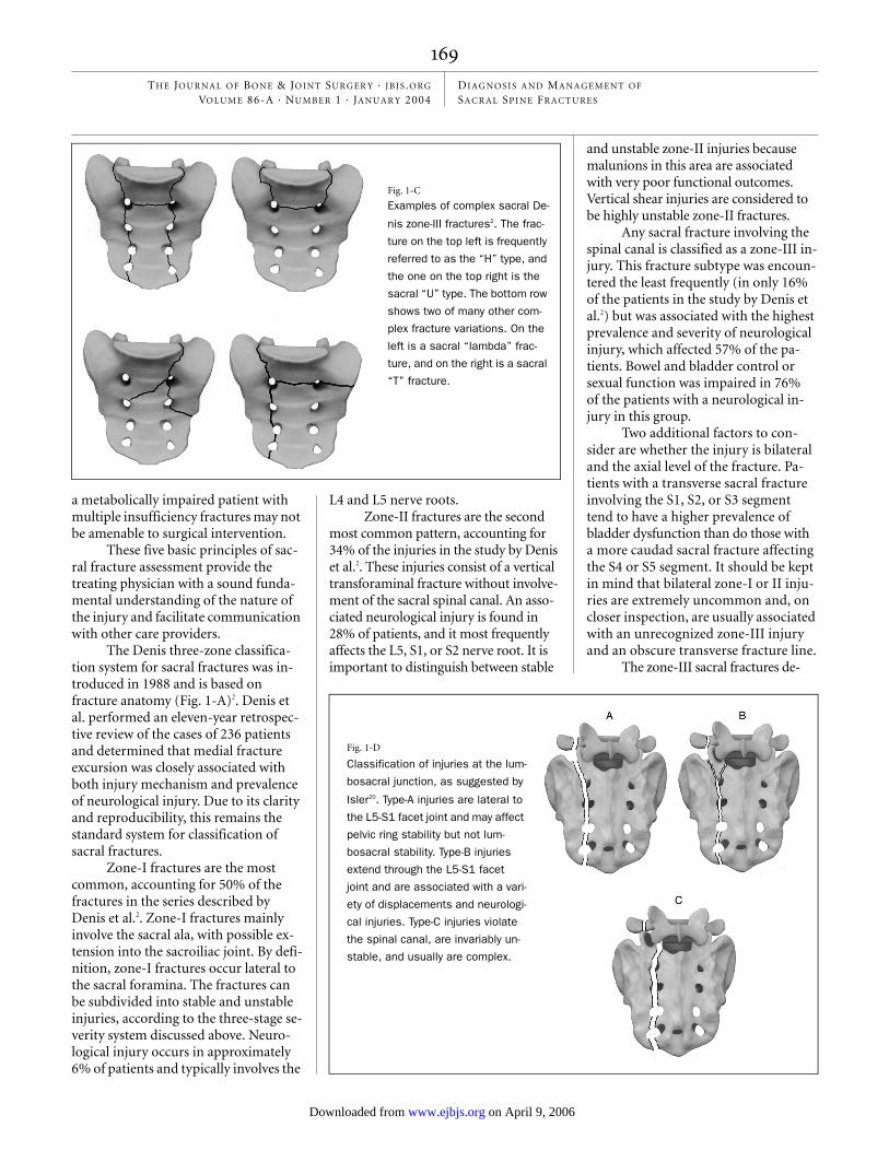
Fig. 1-D
Classification of injuries at the lum-
bosacral junction, as suggested by
Isler20. Type-A injuries are lateral to
the L5-S1 facet joint and may affect
pelvic ring stability but not lum-
bosacral stability. Type-B injuries
extend through the L5-S1 facet
joint and are associated with a vari-
ety of displacements and neurologi-
cal injuries. Type-C injuries violate
the spinal canal, are invariably un-
stable, and usually are complex.
Fig. 1-C
Examples of complex sacral De-
nis zone-III fractures2. The frac-
ture on the top left is frequently
referred to as the “H” type, and
the one on the top right is the
sacral “U” type. The bottom row
shows two of many other com-
plex fracture variations. On the
left is a sacral “lambda” frac-
ture, and on the right is a sacral
“T” fracture.
on April 9, 2006 www.ejbjs.orgDownloaded from

170
TH E JO U R NA L OF BONE & JOINT SURGER Y · JBJS .ORG
VO LU M E 86-A · NUMB ER 1 · JA NU A R Y 2004DI A G N O S I S AND MANAGEM ENT OF SA CR A L SP I N E FR A C TURE S
scribed by Denis et al. have been subclas-sified by Roy-Camille et al.18, with further modification by Strange-Vognsen and Lebech19. With use of that system, injury severity, likelihood of neurological injury, and therapeutic implications are directly related to increasingly severe injury types (1 through 4) (Fig. 1-B). Type-1 frac-
tures are the least severe and demonstrate a simple flexion deformity of the sacrum. Type-2 injuries are partially translated as well as hyperkyphotic. Type-3 injuries display complete translation with no fracture overlap, and type-4 injuries, as described by Strange-Vognsen and Lebech, consist of segmental comminu-
tion of the S1 vertebral body caused by axial loading of the lumbar spine into the cephalad part of the sacrum.
Injury at the Lumbosacral JunctionInjury at the lumbosacral junction is an important, albeit incompletely un-derstood, category of sacral injury. The lumbosacral ligaments are quite strong, so patients presenting with an injury in this transitional zone have usually sus-tained very high-energy trauma.
Like cervical spine injuries, lum-bosacral injuries can be viewed conceptu-ally as unilateral or bilateral dislocations, with or without accompanying fractures. Displacement can vary from lumbosac-ral subluxation to complete lumbopelvic dissociation. Isler proposed a system for assessing lumbosacral injury on the basis of the location of the pelvic ring fracture relative to the L5-S1 facet joint (Fig. 1-D)20. A vertical sacral fracture lateral to the L5-S1 facet joint is unlikely to have an impact on lumbosacral stability but may affect pelvic ring stability. Fractures crossing through the L5-S1 facet joint can be differentiated as extra-articular frac-tures of the lumbosacral junction and ar-ticular dislocations with various stages of displacement of the L5 and S1 articu-lar processes. Fractures crossing into the neural arch medial to the L5-S1 joint are
Fig. 2-B
Fig. 2-B An attempt was made to achieve an indirect foraminal decompression in addition to accomplishing posterior stabilization of the pelvic ring
with early closed reduction and a percutaneously placed sacroiliac screw. There was no improvement of the S1 radiculopathy. Fig. 2-C After there
was no neurological improvement in the first forty-eight hours postoperatively, the decision was made to perform an early decompression of the S1
foramen. This was done under fluoroscopic guidance with a focal hemilaminotomy. Satisfactory decompression was confirmed on the postoperative
computed tomography scan.
Fig. 2-A
Figs. 2-A, 2-B, and 2-C Sacroiliac fixation and foraminotomy. Fig. 2-A A pel-
vic computed tomography scan demonstrating a Denis zone-II transforami-
nal sacral fracture in a twenty-four-year-old woman who was injured in a
motor-vehicle collision. The patient was found to have a dense S1 radiculop-
athy on the side of the fractured ala. The computed tomography scan shows
a large cortical fragment obstructing the S1 foramen.
Fig. 2-C
on April 9, 2006 www.ejbjs.orgDownloaded from

171
TH E JO U R NA L OF BONE & JOINT SURGER Y · JBJS .ORG
VO LU M E 86-A · NUMB ER 1 · JA NU A R Y 2004DI A G N O S I S AND MANAGEM ENT OF SA CR A L SP I N E FR A C TURE S
usually complex and inherently unstable, necessitating stabilization.
Spinal cord injuries have been classifed in a methodical fashion by the American Spinal Injury Association partly on the basis of the original work of Frankel5. This system, however, incom-pletely addresses sacral injuries and the greater variability of neural deficits aris-ing from root injuries. Gibbons et al. designed a useful four-stage system spe-cifically to grade sacral neurological in-juries21, but unfortunately this system has not come into common usage. The stages of the system consist of 1 (no injury), 2 (paresthesias only), 3 (motor loss but bowel and bladder control intact), and 4 (impaired bowel and/or bladder control).
TreatmentEarly ManagementEarly treatment of substantial unstable sacral injuries may include temporary reduction of a displaced pelvic ring fracture and interventional radiologi-cal techniques such as angiographic embolization of bleeding pelvic vessels.
Options for pelvic reduction include temporary skeletal traction, applica-tion of an anterior external fixator, placement of a pelvic clamp, or use of a wrap-around sheet. In the acute posttraumatic setting, the goal is to achieve a noninvasive form of pelvic reduction and volume reduction and to minimize additional blood loss22.
Nonoperative ManagementNonoperative care consists mainly of ac-tivity modification aimed at preventing further fracture displacement. This may consist of prolonged bed rest in skeletal traction, bed rest in a brace or cast with a unilateral or bilateral hip spica (i.e., pantaloon spica), brace immobilization (with a thoracolumbar spinal orthosis with a hip spica) with protected weight-bearing, or early mobilization with pro-tected weight-bearing.
The typical time frame for heal-ing of a posterior pelvic ring fracture is two to four months. This allows for a transitional period of protected weight-bearing for one to two months8. When-
ever treatment involves prolonged recumbency, it is necessary to address the potential dangers of thromboembo-lism, pulmonary complications, and skin breakdown. Countermeasures may include prophylactic anticoagulation and pneumatic compression boots as well as utilization of a spinal injury bed such as the Roto Rest bed (Kinetic Con-cepts, San Antonio, Texas). Vigorous pulmonary toilet to prevent atelectasis and pneumonia should also be insti-tuted. Repeat imaging studies should be performed to verify that fracture-healing is proceeding with satisfactory alignment. Progressive fracture dis-placement, deterioration of neurologi-cal function, or persistent pain with attempts at mobilization may indicate failure of conservative treatment. As a result of the high cost of labor-intensive care necessary for nonoperative man-agement, these strategies have largely fallen out of favor for the treatment of patients with unstable injuries.
Indications for nonoperative management are vague and historically
Fig. 3-A
Figs. 3-A and 3-B Technique of iliolumbar fixation. Fig. 3-A Following lumbar pedicle screw fixation, iliac screws are placed
in a perpendicular trajectory parallel to the inclination angle of the outer table of the ilium. A suitable starting point is pre-
dictably located over the inferomedial aspect of the posterior superior iliac spinous process, with a trajectory leading 1 cm
above the iliac notch. The anterior target area lies in the region of the anterior inferior iliac spinous process. Visualization of
the outer iliac table and the sciatic notch as well as fluoroscopic guidance with a true lateral projection can be helpful tech-
nical aids. Fig. 3-B The final assembly, shown here with two iliac screws on either side, provides unparalleled stability.
Fig. 3-B
on April 9, 2006 www.ejbjs.orgDownloaded from

172
TH E JO U R NA L OF BONE & JOINT SURGER Y · JBJS .ORG
VO LU M E 86-A · NUMB ER 1 · JA NU A R Y 2004DI A G N O S I S AND MANAGEM ENT OF SA CR A L SP I N E FR A C TURE S
have included nearly all sacral fracture patterns. Contraindications to nonop-erative care are relative but include frac-tures with soft-tissue compromise, an incomplete neurological deficit with
objective evidence of neural compres-sion, and extensive disruption of the posterior lumbosacral ligaments. Pa-tients with multiple injuries often bene-fit from timely surgical intervention in
order to facilitate rehabilitation22. How-ever, we are not aware of any meaning-ful studies comparing the results of operative and nonoperative treatment.
Surgical Decision-MakingSurgical intervention for patients with a sacral fracture should incorporate clear and realistically attainable goals, includ-ing fracture stabilization and lumbosac-ral realignment, optimization of the chances for neurological recovery, ade-quate débridement of open injuries and compromised soft tissues, and minimi-zation of additional morbidity.
Surgical options range from min-imally invasive techniques to formal open reduction and internal fixation. Techniques for neural decompression include laminotomy and foraminot-omy, anterior bone disimpaction, and lumbosacral plexus neurolysis. Ante-rior sacral and pelvic stabilization tech-niques involve various methods of anterior stabilization of the pelvic ring (e.g., application of a sacroiliac plate). Posterior stabilization techniques in-clude percutaneous sacroiliac screw fix-ation, bilateral sacroiliac screw fixation with posterior tension-band plate fixa-tion, posterior alar plate fixation, and lumbopelvic segmental fixation.
The timing of any surgical inter-
Fig. 4-A
Figs. 4-A through 4-D Lumbosacral fracture fixation in a forty-eight-year-old
woman who sustained multiple traumatic injuries. Fig. 4-A The patient was in-
jured in a hang gliding accident. The multiple injuries included a closed head
injury, blunt torso and abdominal injuries, an open tibial fracture, and the De-
nis zone-III, Roy-Camille type-2 sacral fracture shown here. The patient was
found to have absent anal sphincter tone and to be areflexic. A computed axial
tomography scan confirmed severe posterior disruption of the pelvic ring with
foraminal compression of the S2 and S3 segments. Pudendal somatosensory
evoked potentials confirmed the presence of a severe sacral plexus injury.
Fig. 4-B
After initial stabilization, posterior decompression and stabilization with lumbopelvic instrumentation was performed on the third day following the
injury. At the time of writing, the patient was able to walk without pain and had recovered normal voluntary bowel and bladder control, but she re-
ported diminished sexual function.
Fig. 4-C Fig. 4-D
on April 9, 2006 www.ejbjs.orgDownloaded from

173
TH E JO U R NA L OF BONE & JOINT SURGER Y · JBJS .ORG
VO LU M E 86-A · NUMB ER 1 · JA NU A R Y 2004DI A G N O S I S AND MANAGEM ENT OF SA CR A L SP I N E FR A C TURE S
vention should be chosen on the basis of treatment goals, the patient’s general medical status, and the invasiveness of the surgical procedure. Overly aggres-sive early surgery can lead to unaccept-able intraoperative blood loss, soft-tissue breakdown, and infection7. On the other hand, delayed decompression of neural elements beyond two weeks may ad-versely affect chances for neurological recovery2. Most minimally invasive pro-cedures require early closed reduction and are limited in terms of the amount of reduction that is attainable and the overall biomechanical stiffness of the construct. Ultimately, when the treat-ment is being chosen, the advantages and drawbacks of each approach should be carefully weighed; a stereotyped ap-proach to all injuries should be avoided.
Decompression TechniquesNeurological injuries from sacral frac-tures range from incomplete monoradic-ulopathies to a complete cauda equina syndrome3. Sacral roots subjected to con-tusion, compression, or traction caused by angulation, translation, or direct com-pression have a theoretical chance of re-covery. Neural recovery of transected or avulsed sacral nerve roots is unlikely.
Given an overall rate of neurologi-cal improvement of approximately 80% regardless of treatment, the indications for and timing of surgical decompres-sion in patients with neurological injuries are somewhat controversial. From a neu-rophysiological standpoint, decompres-sion of compromised neural elements is preferably performed early, within the first twenty-four to seventy-two hours following injury5. This can be accom-plished indirectly with fracture reduc-tion or directly with a laminectomy. Early surgical decompression may be associ-ated with an increased risk of hemor-rhage and wound-healing complications due to soft-tissue contusion and possibly to cerebrospinal fluid leak. Surgical de-compression as an isolated procedure—i.e., without stabilization—is rarely in-dicated. Surgical decompression may be less useful in patients with transected sacral roots. Huittinen found a 35% prev-alence of root transection in a postmor-tem study of transverse sacral fractures23.
Similarly, reconstruction of nerve roots with avulsion injuries is currently impos-sible. Traumatically transected roots are commonly associated with Denis zone-III injuries with Roy-Camille type-3 dis-placement. Avulsions of the lumbopelvic plexus are associated with severely dis-placed zone-II injuries, such as the so-called vertical shear fracture. Surgery should be considered if there is a reason-able chance of restoring even unilateral lower sacral root function because such function is sufficient for voluntary bowel and bladder control24.
An acceptable approach to early management of sacral injuries is an at-tempt at minimal reduction and stabili-zation. The adequacy of reduction is then assessed with computed tomography combined with repeat neurological and possibly electrodiagnostic examination to characterize persistent neurological deficits. In the presence of satisfactory skeletal stabilization but persistent neu-roforaminal or spinal canal compromise, a focal limited decompression may be performed within the first two weeks af-ter injury, with use of a limited midline exposure and fluoroscopy-guided focal laminectomy6.
An attempt should be made to re-pair any dural tears that are encountered to minimize the chances of a pseudo-meningocele developing. Patients pre-senting with a severely displaced fracture that is unsuitable for closed reduction and percutaneous stabilization should be considered for a comprehensive poste-rior decompression and stabilization procedure with use of the most appro-priate stabilization methods available.
Surgical Stabilization TechniquesStabilization of sacral fractures has evolved from largely improvised use of plates and hooks to the use of specifi-cally designed implant systems incorpo-rating cannulated long large-fragment screws or segmental lumboiliac rod-and-screw fixation systems1. A major goal of surgical intervention is to re-store the stability of the lumbosacral articulation. Anterior approaches to the sacrum for decompression or inter-nal fixation have substantial approach-related morbidity and provide limited
surgical exposure. The vast majority of sacral injuries can be effectively treated with posterior percutaneously based approaches. The role of external fixa-tion, once a popular form of treatment for a variety of pelvic fractures, is now limited to the emergent management of pelvic ring disruptions and to use as supplemental treatment devices for an-terior pelvic ring instability.
The need for anterior stabiliza-tion of the pelvic ring should be consid-ered before embarking on any posterior lumbosacral procedure. Frequently, the anterior pelvic ring injury can be re-aligned and stabilized through limited measures such as anterior plate fixation, external fixation, or the use of retro-grade pubic screws. This can provide protection for the pelvic ring during a procedure performed with the patient prone and can aid in reduction of the posterior part of the pelvic ring.
Posterior fixation ideally offers a high degree of mechanical construct stiffness while producing a low implant profile that minimizes the risk of poste-rior soft-tissue breakdown. Sacroiliac screws, initially described for injuries of the sacroiliac joint, can be used for stabilization of a variety of sacral frac-tures as well (Figs. 2-A, 2-B, and 2-C). They can be placed, with the patient ei-ther supine or prone, with use of con-ventional c-arm imaging and through a percutaneous approach. The inser-tion of sacroiliac screws with the guid-ance of computed tomography imaging is of limited use: it is helpful only for the treatment of displaced fractures in a multiply injured patient. The safety of percutaneously placed sacroiliac screws has been established in several large clinical series and has gained considerable acceptance within the traumatology community6,9,12. Potential drawbacks of this technique include limited biomechanical strength, reli-ance on closed reduction techniques that may be inadequate, and lack of availability of a suitable image intensi-fier. Injury to neural, vascular, and in-testinal structures as a result of drill or screw penetration has been described as a rare complication. The risks of this surgical technique primarily consist of
on April 9, 2006 www.ejbjs.orgDownloaded from
174
TH E JO U R NA L OF BONE & JOINT SURGER Y · JBJS .ORG
VO LU M E 86-A · NUMB ER 1 · JA NU A R Y 2004DI A G N O S I S AND MANAGEM ENT OF SA CR A L SP I N E FR A C TURE S
loss of fracture reduction and fixation in a malreduced position. Percutane-ous placement of sacroiliac screws may be contraindicated in patients with anomalous transitional lumbosacral anatomy or when closed fracture re-duction cannot be accomplished9. Po-tential indications for percutaneous placement of sacroiliac screws include a Denis zone-I, II, or III sacral fracture, which can be adequately reduced in a closed fashion. Denis zone-III, Roy-Camille type-2, 3, or 4 injuries are less amenable to this form of fixation as a stand-alone device because of the in-ability to reduce these injuries by closed means. Similarly, fixation of highly dis-placed zone-II fractures (vertical shear injuries) with this method is very chal-lenging. Zone-II fractures with seg-mental comminution are susceptible to overcompression and secondary fo-raminal entrapment when an iliosacral compression screw is used. Such inju-ries may be considered for fixation with two static sacroiliac screws or for ili-olumbar segmental fixation12.
Open reduction of the posterior aspect of the pelvic ring with plate fixa-tion and screw insertion into the sacral ala, as described by Roy-Camille et al.18, is an infrequently used strategy. The ap-plication of vertically aligned plates on the posterior aspect of the sacral ala with anteroposterior small-fragment screw fixation is also of limited value because of the frequent presence of comminu-tion and osteopenia at the fracture site1,18. Use of a posterior iliac tension-band plate as a supplemental internal fixation method with sacroiliac screw fixation can facilitate open fracture reduction and enhance biomechanical stiffness25. However, it requires a posterior two-incision approach, which has been associated with an increased rate of wound-healing complications.
From a biomechanical perspec-tive, the most stable method of lum-bosacral fixation involves the use of lower lumbar pedicle screw fixation and iliac screw fixation with longitudi-nal and transverse rod connections to facilitate fracture reduction (Figs. 3-A through 4-D). The technique of iliac screw placement follows the basic con-
cept of the Galveston technique but enhances it by allowing placement of multiple large bicortical screws11.
Lumboiliac fixation allows com-plete neurological decompression as needed and can enhance the surgeon’s ability to perform an open reduction of a displaced sacral vertebral body. Supplemental internal fixation can be achieved with sacroiliac screws to main-tain fracture reduction while the lum-boiliac fixation is applied. Because of the immediate stability conferred by lumboiliac fixation, most patients can walk with weight-bearing as tolerated without the use of a brace.
Results of TreatmentThe results of the treatment of sacral fractures have been infrequently re-ported and often poorly documented. Aside from the retrospective multicenter study by Denis et al.2, most studies have been of small cohorts and have had con-siderable selection bias. The severity of the neurological injury frequently is not quantified or differentiated. Surgical techniques and timing of intervention have been highly variable or not re-ported. Investigators assessing the effi-cacy of neurological decompression in patients with sacral fractures usually have not reported the severity or type of preoperative and postoperative neu-rological injury. Outcomes measures such as persistent pain and pelvic insta-bility rarely have been evaluated in a sys-tematic fashion.
Decompression SurgeryEstablishing the benefits of decompres-sion over a nonoperative approach in neurologically impaired patients is dif-ficult. Neurological improvement rates of up to 80% are frequently quoted, re-gardless of the type of operative or non-operative management.
In a retrospective study of forty-four patients, Gibbons et al. reported neurological improvement in eleven of fifteen patients treated nonoperatively compared with seven of eight patients managed surgically21. Four of six pa-tients with loss of bowel and bladder control had improvement after nonop-erative treatment compared with two
of two patients treated surgically. Simi-larly, lower-extremity motor improve-ment was found in four of six patients treated nonoperatively compared with three of four treated surgically.
Denis et al. reported no improve-ment of bowel or bladder control in three patients in whom a transverse sacral frac-ture had been treated nonoperatively2. In contrast, all of five patients treated surgi-cally had complete return of sphincter control. Fountain et al. noted improve-ment of bowel and bladder control in five patients treated surgically, whereas the one patient treated nonoperatively had spontaneous improvement26. Sabiston and Wing generally recommended non-operative care in a series of thirty-five patients with a sacral fracture, and they found no improvement of bowel and bladder control in only one patient with a complete cauda equina syndrome who was treated nonsurgically15.
Instrumentation ProceduresNork et al. reported successful results of percutaneous sacroiliac screw fixation in thirteen patients with a Denis zone-III, Roy-Camille subtype-1 or 2 fracture and no substantial neurological deficit12. No deterioration of the sacral kyphosis angle was found despite the fact that the posttraumatic deformity was stabilized without aggressive attempts at reduc-tion. In one patient, it was necessary to revise the hardware because of disen-gagement of a single iliosacral screw. Six patients presenting with L5 or S1 incom-plete radiculopathy had a decrease in the symptoms without the need for neural decompression. On the basis of their ex-perience, the authors recommended in-sertion of bilateral midline-crossing sacroiliac screws when the technique is used to treat a zone-III “H” or “U” frac-ture configuration.
Using a cadaveric model, Schild-hauer et al. demonstrated that segmen-tal lumbopelvic fixation provided substantially better stiffness than did a dual sacroiliac screw construct11. They reported clinically successful results of lumbopelvic fixation in their series of thirty-four patients with a vertically un-stable zone-I or II fracture. Ninety-one percent of the patients were found to
on April 9, 2006 www.ejbjs.orgDownloaded from

175
TH E JO U R NA L OF BONE & JOINT SURGER Y · JBJS .ORG
VO LU M E 86-A · NUMB ER 1 · JA NU A R Y 2004DI A G N O S I S AND MANAGEM ENT OF SA CR A L SP I N E FR A C TURE S
have fulfilled the authors’ standards for a stable fracture union. They reported a 9% rate of complications, which con-sisted of wound-healing problems and a 3% prevalence of iatrogenic radicu-lopathy. With use of the same concept but a different implant configuration, Abumi et al. treated seven patients with a vertically and rotationally displaced zone-I or II pelvic ring injury with bi-lateral S1 screw fixation and a transverse rod connection attached to a Galveston-type rod extension into the ilium on the injured side27. Satisfactory healing was reported in six of the seven patients. Complications included one deep wound infection and one unresolved neurological deficit.
OverviewAssessment and treatment of thora-columbar and sacral fractures has im-proved considerably as a result of advances in general trauma management and diagnostic modalities. Surgical tech-niques have evolved substantially over the past ten years as well. However, several basic issues, such as the appropriate roles of operative and nonoperative care, have
not been resolved conclusively. The tim-ing of intervention and the optimal sur-gical techniques need to be determined on an individual basis with the potential benefits of early neural decompression, skeletal stabilization, and patient mobili-zation weighed against the risks of sur-gery, such as blood loss, infection, and anesthesia-related complications.
Alexander R. Vaccaro, MDRothman Institute, 925 Chestnut Street, Philadelphia, PA 19107. E-mail address: [email protected]
David H. Kim, MDThe Boston Spine Group, 125 Parker Hill Avenue, Boston, MA 02120
Darrel S. Brodke, MDUniversity of Utah, 30 North 1900 East, 3B165, Salt Lake City, UT 84132
Mitchel Harris, MDDepartment of Orthopaedic Surgery, Wake Forest University Baptist Medical Center, Medical Center Boulevard, Winston-Salem, NC 27157-1070
Jens Chapman, MDM.L. Chip Routt, MD
Orthopaedic Services, Harborview Medical Center, 325 North Avenue, Seattle, WA 98104
Thomas Schildhauer, MDChirurgische Klinik und Poliklïnïk, BG-Kliniken Bergmannsheil, Ruhr-Universität Bochum, Bürkle-de-la-Camp-Platz 1, Bo-chum D-47789, Germany
Rick C. Sasso, MDIndiana Spine Group, 8402 Harcourt Road, Suite 400, Indianapolis, IN 46260-2074
The authors did not receive grants or outside funding in support of their research or prepa-ration of this manuscript. They did not receive payments or other benefits or a commitment or agreement to provide such benefits from a commercial entity. No commercial entity paid or directed, or agreed to pay or direct, any benefits to any research fund, foundation, educational institution, or other charitable or nonprofit organization with which the authors are affiliated or associated.
Printed with permission of the American Academy of Orthopaedic Surgeons. This article, as well as other lectures presented at the Academy’s Annual Meeting, will be available in March 2004 in Instructional Course Lectures, Volume 53. The complete volume can be or-dered online at www.aaos.org, or by calling 800-626-6726 (8 A.M.-5 P.M., Central time).
References
1. Pohlemann T, Angst M, Schneider E, Ganz R, Tscherne H. Fixation of transforaminal sacrum fractures: a biomechanical study. J Orthop Trauma. 1993;7:107-17.
2. Denis F, Davis S, Comfort T. Sacral fractures: an important problem. Retrospective analysis of 236 cases. Clin Orthop. 1988;227:67-81.
3. Schnaid E, Eisenstein SM, Drummond-Webb J. Delayed post-traumatic cauda equina compres-sion syndrome. J Trauma. 1985;25:1099-101.
4. Browner BD, Cole JD, Graham JM, Bondurant FJ, Nunchuck-Burns SK, Colter HB. Delayed posterior internal fixation of unstable pelvic frac-tures. J Trauma. 1987;27:998-1006.
5. Schmidek HH, Smith DA, Kristiansen TK. Sac-ral fractures. Neurosurgery. 1984;15:735-46.
6. Routt ML Jr, Simonian PT, Swiontkowski MF. Stabilization of pelvic ring disruptions. Orthop Clin North Am. 1997;28:369-88.
7. Kellam JF, McMurtry RY, Paley D, Tile M. The unstable pelvic fracture. Operative treatment. Orthop Clin North Am. 1987;18:25-41.
8. Tile M. Pelvic ring fractures: should they be fixed? J Bone Joint Surg Br. 1988;70:1-22.
9. Routt ML Jr, Nork SE, Mills WJ. Percutaneous fixation of pelvic ring disruptions. Clin Orthop. 2000;375:15-29.
10. Rommens PM, Vanderschot PM, Broos PL. Conventional radiography and CT examination of pelvic ring fractures. A comparative study of 90 patients. Unfallchirurg. 1992;95:387-92.
11. Schildhauer TA, Ledoux WR, Chapman JR, Hen-ley MB, Tencer AF, Routt ML Jr. Triangular os-teosynthesis and iliosacral screw fixation for unstable sacral fractures. A cadaveric and bio-mechanical evaluation under cyclic loads. J Or-thop Trauma. 2003;17:22-31.
12. Nork SE, Jones CB, Harding SP, Mirza SK, Routt ML Jr. Percutaneous stabilization of U-shaped sacral fractures using iliosacral screws: technique and early results. J Orthop Trauma. 2001;15:238-46.
13. Letournel E. [Surgical fixation of displaced pel-vic fractures and dislocations of the symphysis pubis (excluding acetabular fractures) (author’s transl)]. Rev Chir Orthop Reparatrice Appar Mot. 1981;67:771-82. French.
14. Müller ME. Manual of internal fixation: tech-niques recommended by the AO-ASIF Group. 3rd ed. New York: Springer; 1991.
15. Sabiston CP, Wing PC. Sacral fractures: classi-fication and neurologic implications. J Trauma. 1986;26:1113-5.
16. Gustilo RB, Anderson JT. Prevention of infection in the treatment of one thousand and twenty-five open fractures of long-bones: retrospective and prospective analyses. J Bone Joint Surg Am. 1976;58:453-8.
17. Tscherne H, Gotzen L, editors. Fractures with soft tissue injuries. Berlin: Springer; 1984. p 1-58.
18. Roy-Camille R, Saillant G, Gagna G, Mazel C. Transverse fracture of the upper sacrum. Sui-cidal jumper’s fracture. Spine. 1985;10:838-45.
19. Strange-Vognsen HH, Lebech A. An unusual type of fracture in the upper sacrum. J Orthop Trauma. 1991;5:200-3.
20. Isler B. Lumbosacral lesions associated with pel-vic ring injuries. J Orthop Trauma. 1990;4:1-6.
21. Gibbons KJ, Soloniuk DS, Razack N. Neurologi-cal injury and patterns of sacral fractures. J Neurosurg. 1990;72:889-93.
22. Latenser BA, Gentilello LM, Tarver AA, Thal-gott JS, Batdorf JW. Improved outcome with early fixation of skeletally unstable pelvic frac-tures. J Trauma. 1991;31:28-31.
23. Huittinen VM. Lumbosacral nerve injury in frac-ture of the pelvis. A postmortem radiographic and patho-anatomical study. Acta Chir Scand Suppl. 1972;429:3-43.
24. Gunterberg B. Effects of major resection of the sacrum. Clinical studies on urogenital and anorectal function and a biomechanical study on pelvic strength. Acta Orthop Scand Suppl. 1976;162:1-38.
25. Simonian PT, Routt ML Jr. Biomechanics of pelvic fixation. Orthop Clin North Am. 1997;28:351-67.
26. Fountain SS, Hamilton RD, Jameson RM. Trans-verse fractures of the sacrum. A report of six cases. J Bone Joint Surg Am. 1977;59:486-9.
27. Abumi K, Saita M, Iida T, Kaneda K. Reduction and fixation of sacroiliac joint dislocation by the combined use of S1 pedicle screws and the Galveston technique. Spine. 2000;25:1977-83.
on April 9, 2006 www.ejbjs.orgDownloaded from
Top Related