


Languages
Pages
Legal


Derangements of Potassium
Laura Medford-Davis, MDa,b,*, Zubaid Rafique, MDa,b
KEYWORDS
� Potassium � Hypokalemia � Hyperkalemia � Peaked T waves
KEY POINTS
� Potassium balance regulates the excitability of cardiac cells, and both hypokalemia andhyperkalemia can cause cardiac arrest when severe.
� Treatment of hyperkalemia includes cardiac membrane stabilization, transcellular shifting,and total body potassium elimination. Sodium bicarbonate and Kayexalate are not recom-mended for management.
� Treatment of symptomatic hypokalemia consists of repletion with potassium chloride,which is available in liquid, pill, and intravenously (IV) administrable forms. Magnesiumshould be repleted simultaneously to potentiate potassium absorption and avoid furtherpotassium losses.
� Determine and treat the underlying cause of potassium derangement to preventrecurrence.
� Avoid potentiating medications.
� Consider the dietary potassium contribution.
� Consider problems with potassium excretion from the gastrointestinal (GI) tract orkidneys.
� Consider transcellular potassium shifts across cell membranes.
INTRODUCTION AND PATHOPHYSIOLOGY
About 98% of total body potassium (K1) is intracellular,1,2 and 75% of the intracellularpotassium is contained in skeletal muscle cells.3,4 The body maintains the remaining2% extracellular component within a tight range of 3.5 to 5.0 mEq/L (1 mmol equals1 mEq K1).3 The main mechanism for maintaining this transcellular ratio is thesodium-potassium (Na-K) adenosine triphosphatase (ATPase) pump, which usesenergy in the form of adenosine triphosphate to drive K1 into cells in exchange forsodium (Na). The resulting K1 gradient creates a resting membrane potential thatdetermines cardiac and neuromuscular cell excitability and signal conduction.
a Division of Emergency Medicine, Baylor College of Medicine, 1 Baylor Plaza, Houston, TX77030, USA; b Department of Emergency Medicine, Ben Taub General Hospital, Ben TaubGeneral Hospital Emergency Center, 1504 Taub Loop, Houston, TX 77030, USA* Corresponding author. Department of Emergency Medicine, Ben Taub General HospitalEmergency Center, 1504 Taub Loop, Houston, TX 77030.E-mail address: [email protected]
Emerg Med Clin N Am 32 (2014) 329–347http://dx.doi.org/10.1016/j.emc.2013.12.005 emed.theclinics.com0733-8627/14/$ – see front matter � 2014 Elsevier Inc. All rights reserved.
Downloaded from ClinicalKey.com at Inova Fairfax Hospital - JCon June 07, 2016.For personal use only. No other uses without permission. Copyright ©2016. Elsevier Inc. All rights reserved.

Medford-Davis & Rafique330
Because the extracellular K1 level is proportionally so much less than the intracel-lular level, even a small change in the extracellular level significantly alters the restingmembrane potential. Hyperkalemia is less tolerated by the body and causes moresignificant extracellular shifts than hypokalemia. A 100 mEq excess of total bodyK1 increases extracellular levels by 0.5 mEq, whereas a 100 mEq deficit decreasesextracellular levels by just 0.3 mEq.3 Three different mechanisms alter the extracellularK1 concentration: K1 intake, K1 excretion, and transcellular shift of K1 into or out ofcells.3 Many commonmedications affect one of these 3 mechanisms and can provokea potassium imbalance (Boxes 1 and 2).Usually the body’s regulatory mechanisms can resist large fluctuations in daily po-
tassium intake. However, over time or in those persons predisposed to K1 disorders,diet can affect the extracellular K1 level. Patients with altered total-body potassiumstores, who chronically take medications that alter K1 balance, or who have a diseasepredisposing them to K1 imbalance may need to either increase or avoid intake ofpotassium-rich foods (Box 3).Excretion of K1 from the body is primarily managed by the kidneys, which are
responsible for 90% of excretion in normal physiology.2,4 The other 10% is excretedmostly by the intestine into stool, with a small contribution from sweat. In cases ofsevere burns or extreme exercise, sweat and skin losses increase. Similarly, in end-stage renal disease when the kidneys no longer function, the gut upregulates toperform 25% of excretion.2
Box 1
Medications causing hyperkalemia
Inhibit excretion
Decrease aldosterone
Angiotensin converting enzyme inhibitors
Angiotensin receptor blockers
Potassium-sparing diuretics (spironolactone)
Nonsteroidal antiinflammatory drugs (NSAIDs)
Heparin
Nonselective b-blockers
Block sodium channels
Potassium-sparing diuretics (amiloride, triamterene)
Antibiotics (trimethoprim, pentamidine)
Transcellular shift
Inhibit Na-K ATPase pump
Digoxin (dose dependent)
NSAIDs
Nonselective b-blockers
Anesthetics (succinylcholine, suxamethonium)
Data from Alfonzo AV, Isles C, Geddes C, et al. Potassium disorders—clinical spectrum andemergencymanagement. Resuscitation 2006;70:10–25; and Pepin J, Shields S. Advances in diag-nosis and management of hypokalemic and hyperkalemic emergencies. Emerg Med Pract2012;14(2):1–17.
Downloaded from ClinicalKey.com at Inova Fairfax Hospital - JCon June 07, 2016.For personal use only. No other uses without permission. Copyright ©2016. Elsevier Inc. All rights reserved.

Box 2
Medications causing hypokalemia
Transcellular Shift
Insulin
b-Agonists
Bronchodilators
Tocolytics
Synthroid
Phosphodiesterase inhibitors
Caffeine
Theophylline
Overdose
Verapamil
Barium
Decongestants
Increased K1 Excretion
Thiazide diuretics
Loop diuretics (furosemide and bumetanide)
Mineralocorticoids (fludrocortisone)
High-dose glucocorticoids (prednisone)
High-dose penicillins
Aminoglycosides
Cisplatin
Amphotericin
Data from Refs.2–4,13
Derangements of Potassium 331
Na-K ATPase exchangers are present both in the intestine and in the distal renalnephron to excrete K1. In the kidney, high amounts of urine and sodium arriving tothe distal nephron stimulate the Na-K ATPase to excrete more K1,4 as does aldoste-rone. Low renal perfusion, hypovolemia, low sodium levels, or high potassium levelstrigger renal renin release, which leads to aldosterone release by the adrenal glands,3
increasing renal excretion of K1.Finally, certain physiologic states and medications affect the transport of K1 across
the cell membrane, leading to transcellular shifts that can alter extracellular K1 levels.These shifts can be caused by alterations in the Na-K ATPase pump, pH and acid-base status in the body, and tonicity of the serum.4 Hyperglycemia and hypernatremiaare 2 examples of hypertonic states that drive potassium out of cells.3
The symptoms of potassium alterations are typically vague, so a careful history isimportant to raise clinical suspicion of the diagnosis. Symptoms of both hypokalemiaand hyperkalemia affect the cardiac, muscular, and GI systems. The rate of change inextracellular K1 levels is more important than the absolute K1 level in determiningseverity of symptoms and risk for deterioration, so symptoms are unreliable predictorsof absolute K1 values. Workup of any patient whose history suggests potassium
Downloaded from ClinicalKey.com at Inova Fairfax Hospital - JCon June 07, 2016.For personal use only. No other uses without permission. Copyright ©2016. Elsevier Inc. All rights reserved.

Box 3
Foods rich in potassium
Legumes and grains
Whole-grain bread
Wheat bran
Granola
Beans (kidney, pinto, black, navy, and lima)
Nuts, nut butter
Vegetables
Potato
Yam, sweet potato
Tomato
Spinach
Beets
Broccoli
Brussel sprouts
Bamboo shoots
Winter squash (acorn, butternut, pumpkin)
Cabbage
Carrots (raw)
Spinach
Canned mushrooms
Pickles
Fruits
Banana, plantain
Fig
Prune
Raisin
Date
Apricot (especially dried)
Avocado
Melon (cantaloupe and honey dew)
Kiwi
Mango
Citrus (nectarine and orange)
Pear
Dairy
Milk (soy and regular)
Yogurt
Medford-Davis & Rafique332
Downloaded from ClinicalKey.com at Inova Fairfax Hospital - JCon June 07, 2016.For personal use only. No other uses without permission. Copyright ©2016. Elsevier Inc. All rights reserved.
Meats
Beef
Clams
Sardines
Scallops
Lobster
Salmon
White fish
Other
Sports beverages and supplements
Imitation salt (low-sodium products)
Molasses
Chocolate
Data from Bakris GL, Olendzki B. Patient information: low potassium diet (beyond the basics).Available at: www.uptodate.com/contents/low-potassium-diet-beyond-the-basics. AccessedSeptember 26, 2012.
Derangements of Potassium 333
imbalance should include electrocardiography (ECG), basic metabolic panel (withelectrolyte and creatinine levels), complete blood count, and urinalysis. If symptomsare severe, blood gas analysis to determine pH and urine electrolyte levels to differen-tiate the causes are also suggested (Box 4).4 Magnesium levels need to be checked incases of hypokalemia. The ECG may show specific abnormalities that can help makethe diagnosis and suggest risk of cardiac deterioration, but it is not sensitive andabsence of ECG changes does not rule out a significant potassium abnormality.
HYPERKALEMIAEtiology
Hyperkalemia, extracellular K1 levels greater than 5.0 mEq/L or 5.5 mEq/L dependingon the laboratory assay,1–4 can be caused by failure to excrete enough K1, leading tototal body excess, transcellular shifts, or measurement error (Box 5). Many commonmedications inhibit K1 excretion or shift it out of cells and into the extracellular space(see Box 1). Patients with underlying disorders predisposing them to hyperkalemia
Box 4
Diagnostic workup
ECG
BMP/Chem7
Magnesiuma
CBC
UA
Blood gas, Urine electrolytesb
a In hypokalemiab In severe cases
Downloaded from ClinicalKey.com at Inova Fairfax Hospital - JCon June 07, 2016.For personal use only. No other uses without permission. Copyright ©2016. Elsevier Inc. All rights reserved.

Box 5
Disorders causing hyperkalemia
Failure of excretion
Decreased glomerular filtration rate
Renal insufficiency
Renal failure
Heart failure
Obstructive uropathy
Low aldosterone level
Adrenal insufficiency (Addison disease)
Low renin level
Type 4 renal tubular acidosis
Medications that inhibit Na-K ATPase in the distal nephron (see Box 1)
Transcellular shifts (Na-K ATPase pump)
Hemolysis
Rhabdomyolysis
Tumor lysis syndrome
Hematoma reabsorption
Medications that inhibit Na-K ATPase pump (see Box 1)
Insulin deficiency
Diabetes mellitus
Prolonged fasting
Hypertonicity
Hyperglycemia
Hypernatremia
Acidosis
Hyperkalemic periodic paralysis (mutation of skeletal muscle Na-K pump)
Fasting
Intense exercise
High-K1 meal
Measurement error (pseudohyperkalemia)
Hemolysis during blood draw
Prolonged tourniquet use
Small needle caliber
Excessive fist clenching
Excessive plunger force to pull blood into a syringe
Hemolysis before laboratory analysis
Delay between blood draw and analysis
Aggressive sample shaking
Medford-Davis & Rafique334
Downloaded from ClinicalKey.com at Inova Fairfax Hospital - JCon June 07, 2016.For personal use only. No other uses without permission. Copyright ©2016. Elsevier Inc. All rights reserved.

Hyperviscosity
Extreme leukocytosis
Extreme thrombocytosis
Polycythemia vera
Patient hyperventilation during blood draw (respiratory alkalosis causing transient K1 shift)
Large, rapid potassium load
Massive blood transfusion protocol
High-dose potassium penicillin
Poisoning/ingestion
Data from Refs.1–4
Derangements of Potassium 335
are much more sensitive to initiation of these medications. Ingesting too much K1rarely causes hyperkalemia except in patients with preexisting K1 homeostasisabnormalities.Renal disorders are the most common cause of hyperkalemia, followed by cell lysis,
which releases large intracellular K1 stores.2 High K1 levels stimulate aldosteronesecretion, which acts on the renal tubules to increase K1 excretion into the urine,but in primary mineralocorticoid deficiency this homeostasis mechanism fails to acti-vate, whereas in renal failure the kidney is unable to respond to aldosterone.4 Highplasma K1 levels also stimulate insulin secretion from the pancreas, which drivesK1 intracellularly,4 although patients with diabetes mellitus may not have sufficient in-sulin production capacity to counteract high K1 levels.
Clinical Presentation
Patients with hyperkalemia may be completely asymptomatic or may have cardiac,muscular, or GI complaints. Hyperkalemia depolarizes the cardiac membrane, slowingconduction.5 Patients may experience palpitations or generalized fatigue and malaise.Muscle cramps,4 paresthesias, and weakness are common.2 Weakness can progressto a flaccid paralysis.2 Nausea, vomiting, and diarrhea can also occur.4
Physical examination findings include bradycardia and/or irregular cardiac rhythmwith frequent premature ventricular contractions.2 Neurologic examination may revealdecreased deep tendon reflexes and decreased strength with intact sensation.2,3,5
Diagnostic Testing
An ECG is the first step in the workup of a patient with hyperkalemia. Hyperkalemiaincreases myocyte sensitivity in different areas of the heart progressively as K1 levelsrise:2
1. Atria2. Ventricles3. Bundle of His4. Sinoatrial node5. Interatrial tracts
Therefore, the ECG progresses through several phases that are loosely correlatedwith absolute serum K1 levels and with the rate of increase in serum K1 levels.2,6
The first and most common sign is tall, “peaked” T waves with a narrow base(Fig. 1). These occur most frequently in the precordial leads V2-V4. A sensitive sign
Downloaded from ClinicalKey.com at Inova Fairfax Hospital - JCon June 07, 2016.For personal use only. No other uses without permission. Copyright ©2016. Elsevier Inc. All rights reserved.

Fig. 1. ECG with peaked T waves in hyperkalemia.
Medford-D
avis
&Rafiq
ue
336
Dow
nloaded from C
linicalKey.com
at Inova Fairfax Hospital - JC
on June 07, 2016.For personal use only. N
o other uses without perm
ission. Copyright ©
2016. Elsevier Inc. A
ll rights reserved.

Derangements of Potassium 337
is if the amplitude of the T exceeds the amplitude of the R.7 As the atria are affected thePR interval lengthens, and as the ventricles are affected the QRSwidens. When hyper-kalemia affects the conduction system, the P waves decrease in amplitude until theECG develops a “nodal” rhythm with absent P waves (Fig. 2). The QRS continuesto widen until the S and T waves merge into a “sine wave” pattern (Fig. 3). The sinewave pattern usually shortly precedes ventricular fibrillation (VFib) and cardiac arrest.However, research has shown poor sensitivity for these changes, with only 32% of
ECGs in known hyperkalemia having peaked T waves and only 52% exhibiting anyECG change. If strict measurement criteria were used, just 18% of ECGs displayeda change despite hyperkalemia.8 When limited to hyperkalemia greater than6.0 mEq/L rather than 5 mEq/L, 46% to 64% had changes, suggesting a nonsignifi-cant trend with degree of hyperkalemia.1
Treatment
Most experts agree that ECG changes and/or symptoms should be treated expedi-tiously, but because these can occur at varying absolute K1 levels and some patientscan have K1 levels greater than 7.5 mEq/L without ECG changes or symptoms, it isstill undecided whether an absolute K1 threshold exists whereby risk of arrhythmianecessitates treatment in the asymptomatic patient.In the setting of ECG changes, cardiac membrane stabilization is the first priority
to prevent arrhythmias and cardiac arrest. Calcium is the mainstay of cardiacstabilization to restore resting membrane potential, either as gluconate or chloridecompounds.4 Calcium chloride has approximately 3 times more elemental calciumthan calcium gluconate (6.8 mEq/10 mL vs 2.2 mEq/10 mL),2 and it has greaterbioavailability because calcium gluconate has to be metabolized into an activeform.4 However, calcium gluconate has less risk of tissue necrosis, which allows fastertransfusion.4 There is no randomized evidence supporting one or the other,9 anddecision on which agent to use remains provider and facility dependent.Next, potassium levels can be lowered either temporarily by medications that
cause an intracellular shift of K1 or by therapies that eliminate K1 from the bodyto decrease total body stores (Table 1). Insulin and b-agonists stimulate the Na-KATPase pump to pull more K1 into cells4 and are the mainstays of evidence-basedtherapy for transcellular shift. The 2 medications are synergistic, so using both incombination results in a larger reduction of extracellular K1 than using eitheralone.5,9,10 There is no difference in inhaled or IV forms of b-agonists, although IVforms are not currently available in the United States, nor is there a difference be-tween albuterol and levalbuterol.9 Both metered-dose inhalers and nebulizers arealso equally effective, although it may be easier to deliver consistent dosing withthe nebulized form.4
Sodium bicarbonate has been used to promote intracellular shift of K1, but no goodevidence supports its routine use in hyperkalemia. It is significantly less effective thaninsulin or b-agonists, with a maximum effect of 0.4 mEq/L,10 and it has only been help-ful in patients with a nongap metabolic acidosis.2,4 Furthermore, several studies havefound no effect compared with placebo9 and no additional effect compared withinsulin or b-agonists alone.1
Reduction of total bodyK1 canbe accomplishedby enhancing the body’s renal orGIelimination. Loop diuretics are useful in patients who still produce urine.3,5 Sodiumpolystyrene, or Kayexalate, has been commonly used in practice since studiessuggesting its efficacy were published in 1961, with the presumed mechanismof increased Na-K exchange across the bowel wall.2,11 Onset of action is delayed by2 to 6 hours, decreasing its utility in the management of acute hyperkalemia in the
Downloaded from ClinicalKey.com at Inova Fairfax Hospital - JCon June 07, 2016.For personal use only. No other uses without permission. Copyright ©2016. Elsevier Inc. All rights reserved.
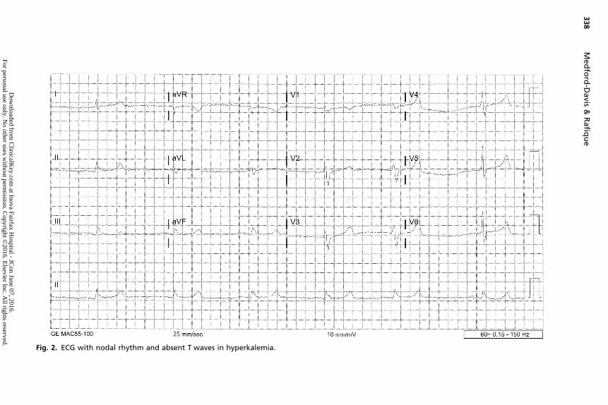
Fig. 2. ECG with nodal rhythm and absent T waves in hyperkalemia.
Medford-D
avis
&Rafiq
ue
338
Dow
nloaded from C
linicalKey.com
at Inova Fairfax Hospital - JC
on June 07, 2016.For personal use only. N
o other uses without perm
ission. Copyright ©
2016. Elsevier Inc. A
ll rights reserved.
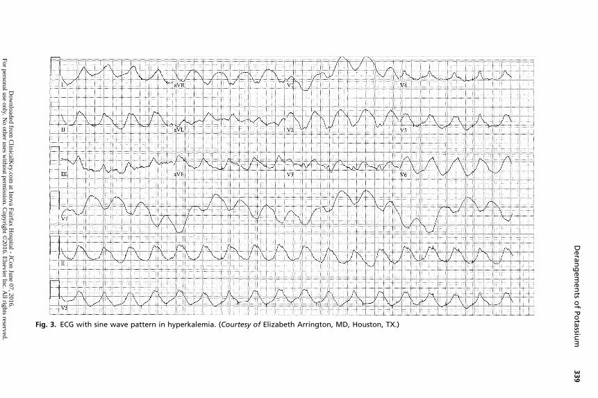
Fig. 3. ECG with sine wave pattern in hyperkalemia. (Courtesy of Elizabeth Arrington, MD, Houston, TX.)
Derangements
ofPotassiu
m339
Dow
nloaded from C
linicalKey.com
at Inova Fairfax Hospital - JC
on June 07, 2016.For personal use only. N
o other uses without perm
ission. Copyright ©
2016. Elsevier Inc. A
ll rights reserved.

Table 1Treatment of hyperkalemia
Medication DoseTime to Onset(min)
Magnitudeof KDReduction(mEq/L)
Durationof Effect(h) Caveats
Calcium 1–2 g(10 mL)
Immediate No effecton K1
0.5–12,4,5 � May potentiatearrhythmias indigoxin toxicity
� Extravasationcan cause tissuedamage andphlebitis
b-Agonists(albuterol)
20 mg Within302–5,9,10
0.5–1.51,2,4,5,9 2–61–5 � Patients takingnonselectiveb-blockers maybe resistant tothis effect2,5
� Tremors, anxiety
Insulin 10 units 15–302,4,5,9,10 0.6–1.22–5,10 2–62,5,10 � Give withDextrose 50%;monitor glucoseclosely forhypoglycemia
� Patients withESRD are moresusceptible tohypoglycemiabecause ofdecreasedexcretion ofinsulin
Loopdiuretics(Lasix,Bumex)
155 1–33,5 Not effective inpatients withESRD who nolonger makeurine
Dialysis N/A Immediate 1–22,5 2–62,5
Abbreviations: ESRD, end-stage renal disease; N/A, not applicable.
Medford-Davis & Rafique340
emergency department.4,5 It also causes significant constipation and so is usuallyadministered in combination with a stool softener. However, subsequent researchhas not replicated the 1961 results, instead showing that Kayexalate makes no differ-ence in serum K1 levels9,10,12 and that stool softeners alone are equally effective.5 Adangerous side effect of Kayexalate is intestinal necrosis, and the US Food and DrugAdministration has issued a warning against administering it in combination with thestool softener sorbitol, which is thought to increase the incidence of intestinal necro-sis.4,5 In light of its delayed onset of action, uncertain efficacy, and high-risk side-effectprofile, Kayexalate is not recommended in the treatment of hyperkalemia.9
Finally, dialysis is the definitive treatment for removing potassium from the body.Most of the K1 elimination occurs during the first 2 hours of dialysis.1 However,dialysis only filters the extracellular compartment, minimizing its effect on totalbody K1 stores, and in cases of elevated total body K1 stores, the body quickly
Downloaded from ClinicalKey.com at Inova Fairfax Hospital - JCon June 07, 2016.For personal use only. No other uses without permission. Copyright ©2016. Elsevier Inc. All rights reserved.

Derangements of Potassium 341
reequilibrates to a hyperkalemic state. Within 6 hours, extracellular K1 returns to70% of its predialysis levels.2 In situations of resuscitation from cardiac arrest,case reports have demonstrated successful return of spontaneous circulation andgood neurologic outcomes despite prolonged arrest when dialysis is initiated duringcardiopulmonary resuscitation (CPR) to correct hyperkalemia.2
Key Points
� Treatment of hyperkalemia includes
� Cardiac membrane stabilization� Transcellular shifts� Total body potassium elimination� Changes in practice� Sodium bicarbonate is not an indicated therapy; can consider its use inacidotic patients with hyperkalemia
� Do not give Kayexalate� In cases of cardiac arrest due to hyperkalemia, perform prolonged CPR untilK1 level is corrected; the patient should not be pronounced dead until theirK1 level is normalized.
� Next steps� Repeat serial K1 measurements to monitor for rebound hyperkalemia� Prevent recurrence
F
- Avoid potentiating medications (see Box 1)- Determine and treat underlying cause (see Box 5)
� Failure to excrete enough K1 to maintain a balance� Transcellular shifts� Suspect measurement error in otherwise normal patients and repeatlaboratory analysis before initiating treatment
HYPOKALEMIAEtiology
Hypokalemia, extracellular K1 concentration less than 3.5 mEq/L, can be causedby increased K1 loss from the body, transcellular shifts, decreased K1 intake,and sometimes by magnesium depletion (Box 6).2 K1 is excreted into the stoolvia Na-K ATPase exchange pumps in the GI tract, and any disease state (eg,diarrhea) or medication (eg, laxatives) that increases stool output increases GIK1 losses.The kidneys are primarily responsible for maintaining K1 balance via the Na-K
ATPase exchange pumps in the distal nephron. Anything that activates these pumpsor increases the flow of Na or urine through the distal nephron increases renal K1 los-ses.4 Low magnesium (Mg) levels stimulate Na-K ATPase activity. Some drugs suchas amphotericin, aminoglycosides, and cisplatin lower Mg, which secondarily lowersK1 (see Box 2).13 If Mg remains low, any K1 intake is quickly excreted by the kidneysdespite a total body K1 deficit.Aldosterone via the renin-angiotensin-aldosterone system is a primary activator of
the Na-K ATPase pump in the distal nephron.3 High aldosterone levels occur duringdehydration in reaction to hypovolemia and in cases of high cortisol and adrenocorti-cotropic hormone or aldosterone-secreting tumors (Cushing syndrome; Connsyndrome).4
High-urine-flow states increase renal excretion of K1. Diuresis, whether druginduced or driven by the body’s elimination of other substances such as excessglucose or water, therefore increases K1 losses. A high-sodium diet and drugs
Downloaded from ClinicalKey.com at Inova Fairfax Hospital - JCon June 07, 2016.or personal use only. No other uses without permission. Copyright ©2016. Elsevier Inc. All rights reserved.

Box 6
Disorders causing hypokalemia
Excessive loss
GI tract
Diarrhea
Laxative abuse
Fistula
Ileostomy
Renal
Increased Na1 delivery to the distal nephron
High-sodium diet
Drugs (eg, penicillins)
Increased urinary flow
Hyperglycemia
Mannitol
High-volume IV normal saline
Activation of Na-K ATPase
Hypomagnesemia
Renal tubular acidosis types 1 and 2
Liddle syndrome
High aldosterone levels
Primary
Conn syndrome
Cushing syndrome
Secondary
Hypovolemia (eg, vomiting)
Bartter syndrome
Gitelman syndrome
Excessive licorice intake (with glycyrrhizic acid, not sold in the United States)
Cutaneous
Excessive sweating
Extensive burns
Transcellular shift
Alkalosis
Hypernatremic hypokalemic paralysis
Thyrotoxic periodic paralysis
Other
Low dietary intake (when chronic)
Data from Refs.2–4,13
Medford-Davis & Rafique342
Downloaded from ClinicalKey.com at Inova Fairfax Hospital - JCon June 07, 2016.For personal use only. No other uses without permission. Copyright ©2016. Elsevier Inc. All rights reserved.

Derangements of Potassium 343
such as penicillins also increase renal K1 excretion by delivering more Na to the distalnephron where the Na gradient drives the Na-K ATPase exchange.3 Cutaneous lossesare usually negligible except in cases of excessive sweating as happens duringextreme exercise or heat exhaustion or when large-surface-area burns break downthe skin barrier.4
Several commonmedications can shift K1 intracellularly, causing an apparent hypo-kalemia despite normal total bodyK1 stores (seeBox2). Alkalosis, respiratory ormeta-bolic, also drives K1 intracellularly. In addition to stimulating renal losses, a highaldosterone level leads to a metabolic alkalosis, which can perpetuate hypokalemiaby simultaneously shifting K1 intracellularly, further reducing extracellular levels.4
Renal tubular acidosis types 1 and 2 are the only cases in which hypokalemia occursin the setting of an acidosis rather than an alkalosis.4 The body can compensate for alow-K1 diet by severely limiting renal excretion of potassium, but if a low-potassiumdiet continues chronically, it can eventually lead to clinically significant hypokalemia.
Clinical Presentation
Like in hyperkalemia, most hypokalemic patients are asymptomatic, particularly at K1levels greater than 3.0 mEq/L. Patients with heart failure are more likely to be symp-tomatic, and it is recommended that their levels should remain 4.0 mEq/L or moreto prevent arrhythmias.2 Muscular symptoms include generalized malaise, fatigue,lethargy, and weakness.2 Fasciculations and tetany also occur.4 Severe cases maypresent as an ascending paralysis that can include respiratory muscles.2 GI com-plaints include ileus due to impaired smooth muscle activity, which can cause nausea,vomiting, and constipation.2,4 Inability to concentrate the urine can also create a neph-rogenic diabetes insipidus presentation with polyuria and polydipsia.3
On examination, weakness may be evident, with decreased strength typically morepronounced in proximal muscles and lower extremities.3 Mental status and orientationremain intact. Abdominal examination may reveal distention in cases of ileus.4
Diagnostic Testing
An ECG is an initial diagnostic test with immediate results that can be used to guidefurther treatment. Hypokalemia predisposes to several arrhythmias, including first-degree heart block with a prolonged PR interval, second-degree heart block, and atrialfibrillation.4 These rhythms can deteriorate into ventricular tachycardia (VTach), VFib,torsades de pointes, and cardiac arrest. Patients taking digitalis are more susceptibleto arrhythmias in the setting of hypokalemia.2,4
In addition to arrhythmias such as first-degree atrioventricular block with anincreased P wave amplitude,3 there are several more specific signs of hypokalemiathat can appear on ECG. The first sign of hypokalemia is usually decreased T waveamplitude, followed by ST depressions or T wave inversions.3 U waves are the clas-sically taught abnormality and are best seen in V2 and V3 (Fig. 4). As they enlarge,they can merge with and mimic T waves (which are simultaneously decreasing inamplitude), giving the false impression of a prolonged QT. Sometimes U wavesgrow so tall that they appear similar to the peaked T waves of hyperkalemia, but Uwaves usually have a broader base.Additional testing should include a blood gas and pH analysis. In hypokalemia, the
kidney cannot excrete bicarbonate and instead excretes protons and chloride,causing a metabolic alkalosis (or perpetuating an existing alkalosis, which may havecaused the hypokalemia via transcellular shift initially).3 All patients with hypokalemiashould have a magnesium level checked because of the strong correlation betweenhypomagnesemia and hypokalemia.3 It is helpful to determine total body K1 stores
Downloaded from ClinicalKey.com at Inova Fairfax Hospital - JCon June 07, 2016.For personal use only. No other uses without permission. Copyright ©2016. Elsevier Inc. All rights reserved.

Fig. 4. ECG with U waves in hypokalemia. (From Dangodara A. ECG interpretation. In: Glasheen JJ, editor. Hospital medicine secrets. Philadelphia:Mosby/Elsevier, 2007; with permission.)
Medford-D
avis
&Rafiq
ue
344
Dow
nloaded from C
linicalKey.com
at Inova Fairfax Hospital - JC
on June 07, 2016.For personal use only. N
o other uses without perm
ission. Copyright ©
2016. Elsevier Inc. A
ll rights reserved.

Derangements of Potassium 345
to differentiate between transcellular shifts and total K1 loss, but measuring total bodyK1 requires a 24-hour urine collection.13
Treatment
Treatment of hypokalemia includes both potassium and magnesium repletion. Hypo-magnesemia is frequently found in patients with hypokalemia because hypomagnese-mia causes hypokalemia via renal wasting, and other common causes of hypokalemiasuch as diarrhea and diuretic use also cause a simultaneous hypomagnesemia.3,4,7
However, in severe cases of symptomatic hypokalemia, concurrent Mg repletion isrecommended even with normal Mg levels because Mg activates the Na-K ATPasepump to promote cellular uptake of dosed K1.4
Although asymptomatic hypokalemia greater than 3.0 mEq/L can be safely dis-charged with a high-K1 diet (see Box 3) and close follow-up after addressingthe cause, any symptomatic hypokalemia should be repleted in the emergencydepartment. Patients with recent myocardial infarction or with heart failure shouldbe repleted to levels of at least 4.0 mEq/L.3,4 As a guide, a 0.3-mEq/L drop in extra-cellular K1 levels represents a 100-mEq/L total body deficit.4 When K1 is recheckedafter repletion but before transcellular equilibration, 10 mEq of K1 is expected to in-crease extracellular levels by 0.1 mEq/L.The maximum recommended dose of potassium chloride in low-risk hypokalemia
with minimal symptoms and without arrhythmia is 20 to 80 mEq/d.4 It is importantto consider the likely cause of the hypokalemia, as aggressive repletion in cases oftranscellular shift can lead to overcorrection of total body K1 and rebound hyperka-lemia once the cause of the shift is resolved. K1 levels should be rechecked period-ically during and after repletion to prevent overcorrection.4
Patients with minimal symptoms who are tolerating intake by mouth can receive pillor liquid potassium chloride, which has the advantage of a single large dose. If patientspresent with nausea or vomiting and are unable to tolerate intake by mouth, IV correc-tion is recommended at a rate of 10 to 20 mEq/h. Patients should be monitored on atelemetry monitor during IV infusion for the development of dysrhythmias. In patientswith significant dysrhythmias (VTach) as a presenting symptom, 20 mEq can bepushed over 3 to 10 minutes to prevent deterioration into cardiac arrest.2,4 IV potas-sium chloride causes burning and discomfort at the IV site and can cause phlebitis.4 Acentral line is recommended for rates greater than 10 mEq/h.
Key Points
Hypokalemia is a common disorder, but is well tolerated by the body relative tohyperkalemia. However, severe cases of hypokalemia can cause dysrhythmias andcardiac arrest.
� Treatment of symptomatic hypokalemia consists of repletion with potassiumchloride
� Intake by mouth in pill or liquid forms� IV- Max rate 10 to 20 mEq/h- Push rapidly over 5 to 10 minutes in cases of cardiac arrest or impending
arrest (VFib, VTach)� Next steps
� Repeat serial K1 measurements to monitor for recurrence and preventovercorrection
� Remember to replete Mg along with K1
Downloaded from ClinicalKey.com at Inova Fairfax Hospital - JCon June 07, 2016.For personal use only. No other uses without permission. Copyright ©2016. Elsevier Inc. All rights reserved.

Medford-Davis & Rafique346
For
� Prevent recurrence- Avoid potentiating medications (see Box 2)- Determine and treat underlying cause (see Box 6)
person
� Increased K1 losses from the body� GI� Renal� Cutaneous
� Transcellular shifts� Decreased K1 intake
SUMMARY
Changes in potassium elimination, primarily due to the renal and GI systems; shiftingpotassium between the intracellular and extracellular spaces; and dietary potassiumintake are the 3 major causes of potassium derangements. Several common medica-tions can contribute to either hyperkalemia or hypokalemia. False laboratory resultsregarding elevations in potassium value are also a common cause for hyperkalemia,and the test should be repeated if suspicion for pseudohyperkalemia exists. Symp-toms of potassium derangement are vague but can be cardiac, musculoskeletal, orGI. There are no absolute guidelines for when to initiate treatment of potassiumderangement, but it is generally recommended when the patient is symptomaticand/or has changes on the ECG attributable to potassium derangement. Treatmentof hyperkalemia includes cardiac membrane stabilization with IV calcium, pushing po-tassium intracellularly using insulin (which should be given in combination with glucoseto avoid hypoglycemia) and b-antagonists, and removing potassium from the bodyentirely with dialysis. Neither sodium bicarbonate nor Kayexalate are recommendedas evidence-based therapies. Treatment of symptomatic hypokalemia consists ofrepletion with potassium chloride either by mouth or IV. Magnesium should berepleted when repleting potassium. Repeat potassium levels should be checked aftertreatment of potassium derangement to monitor effect and guide further therapy.Medications and diet should be adjusted to prevent recurrence, especially in patientspredisposed by renal insufficiency.
REFERENCES
1. Putcha N, Allon M. Management of hyperkalemia in dialysis patients. Semin Dial2007;20(5):431–9.
2. Alfonzo AV, Isles C, Geddes C, et al. Potassium disorders—clinical spectrum andemergency management. Resuscitation 2006;70:10–25.
3. Schaefer TJ, Wolford RW. Disorders of potassium. Emerg Med Clin North Am2005;23:723–47.
4. Pepin J, Shields S. Advances in diagnosis and management of hypokalemic andhyperkalemic emergencies. Emerg Med Pract 2012;14(2):1–17.
5. Weisberg LS. Management of severe hyperkalemia. Crit Care Med 2008;36(12):3246–51.
6. Webster A, Brady W, Morris F. Recognising signs of danger: ECG changes result-ing from an abnormal serum potassium concentration. Emerg Med J 2002;19:74–7.
7. Slovis C, Jenkns R. ABC of clinical electrocardiography: conditions not primarilyaffecting the heart. BMJ 2002;324:1320–3.
8. Montague B, Ouellette JR, Buller GK. Retrospective review of the frequency ofECG changes in hyperkalemia. Clin J Am Soc Nephrol 2008;3:324–30.
Downloaded from ClinicalKey.com at Inova Fairfax Hospital - JCon June 07, 2016.al use only. No other uses without permission. Copyright ©2016. Elsevier Inc. All rights reserved.

Derangements of Potassium 347
9. Mahoney BA, Smith WA, Lo D, et al. Emergency interventions for hyperkalemia[review]. Cochrane Database Syst Rev 2005;(2):CD003235.
10. Elliott MJ, Ronksley PE, Clase CM, et al. Management of patients with acutehyperkalemia. CMAJ 2010;182(15):1631–5.
11. Scherr L, Ogden DA, Mead AW, et al. Management of hyperkalemia with a cation-exchange resin. N Engl J Med 1961;264:115–9.
12. Gruy-Kapral C, Emmett M, Santa Ana CA, et al. Effect of single dose resin-cathartic therapy on serum potassium concentration in patients with end-stagerenal disease. J Am Soc Nephrol 1998;9:1924–30.
13. Cohn JN, Kowey PR, Whelton PJ, et al. New guidelines for potassium replace-ment in clinical practice. Arch Intern Med 2000;160:2429–36.
Downloaded from ClinicalKey.com at Inova Fairfax Hospital - JCon June 07, 2016.For personal use only. No other uses without permission. Copyright ©2016. Elsevier Inc. All rights reserved.
Top Related