


Languages
Pages
Legal


RM6.50
KDN : PP 18701/03/2015 (03419)
Dental Treatmentduring Puasa Month
SEDATION in Dentistry
Root CanalTreatment
Gum Disease:Do you have it?
Energy inMachu PiccuThe DentalAcademy Malaysia
Energy inMachu Piccu
Issue #5, March 2017a dental health magazine

2 Issue #5, March 2017

3Issue #5, March 2017
EditorDr. R. Rajenthiran B.D.S. (S’pore),
P.H.F, FICD, FADI
Editorial TeamDr. R. Rajenthiran B.D.S. (S’pore),
P.H.F, FICD, FADI
Dr. Hj Firdaus B. Hanapiah BDS (N.Z.) MSc (Lon), FDSRCS (Eng.), FICOI, AMM
Editorial AssistantsA Theivanayaki
Creative TeamMotioFixo Sdn Bhd
Art DirectorFariz Hanapiah
DTP ArtistEllina Amin
Advisory PanelDr. Hj Firdaus B. Hanapiah BDS (N.Z.) MSc (Lon), FDSRCS
(Eng.), FICOI, AMM
Dato’ Dr How Kim Chuan B.D.S (S’pore), MSc Orthodontics, FDS RCS (Eng. & Edin), FICOL (USA), FWCLD, FWFLD Laser
(USA), FICCDE, FICD,FAID.
Dr Maniarasu Poonjolai BDS (Malaya), MDS (Malaya), AM Malaya, FRACDS (Syd), DGDP (UK), RCS (Eng), FICD, Oral
Maxillofacial Surgeon.
Dr Chow Kai Foo BDS (S’pore) FDSRCS (Eng), FICD, AM (Malaysia)
Assoc. Prof Dr. Rathna Devi Vaithilingam BDS (Malaya), MClinDent (Perio) (Malaya), Faculty of Dentistry, Uni.
Malaya
Dr Sharon Lee (BDS (Adel) MDSc (Melb),Prosthodontist
Mej (Dr) Faiz Khaleed, Malaysian Astronaut, National Space Agency ANGKASA.
Dr. Ezani Farhana binti Mohd Monoto - Dental office at the University Health Centre, UPM Serdang
Marketing & DistributionParamsothy S
#05
DisclaimerUniversal MEdiDent Sdn Bhd (UMSB) (454315-H)
Editorial material herein is provided for information only. Readers are cautioned not to use this information as a substitute for regular professional health care and consultation. Although great care has been taken in compiling and checking the information given in the publication, the publisher, authors, advisors and agents shall not be responsible or in any way liable for the continued currency of the information or for any errors, omissions or inaccuracies in this publication whether arising from negligence or otherwise howsoever, or for any consequences arising there from. The inclusion or exclusion of any product does not mean that the publisher advocates
or rejects its use either generally or in any particular field or fields.
Advertisement are subject to editorial acceptance and have no influence on editorial content or presentation. The publisher, authors, advisors, and agents do not guarantee, directly or indirectly, the quality or efficacy of any product or service described in the advertisements or other material which is commercial in nature.
Copyright: © 2017 No portion of this publication may be reproduced in any language, stored in or introduced into a retrieval system, or transmitted, resold, redistributed, in any form by any means electronic, electrostatic, without prior written permission
of the publisher.
Now Everyone Can Smile

Dear Readers.
Welcome to the 5th issue of our Dental Health magazine. This will be our second year and we are growing from strength to strength.
From this year on we will be going digital and online.
Our main emphasis will be on the digital magazine and the print versions will be limited.
This will be important and make our magazine viable. We are confident too that this is the right step and we will be able to make the magazine accessible to a wider part of the population.
The profession i.e the Dentists have enough periodicals, professional
and academic journals to cater for their needs. This dental health magazine is meant as a means for us to educate our patients on the latest trends in dentistry and all about the new forms of treatments available to the public at large.
We have had encouraging response from the profession. That is the reason for us to go digital this year to reach out to the good people in one of the fast growing regions of the world we live in that is Asean.
Welcome to the first digital version of the dental health magazine.
DR. R. RAJENTHIRAN
Regards, The Editor
Editor’s Letter
6 Issue #5, March 2017

7Issue #5, March 2017
DATO’ DR NOOR ALIYAH BINTI ISMAIL
8 Issue #5, March 2017
18
24
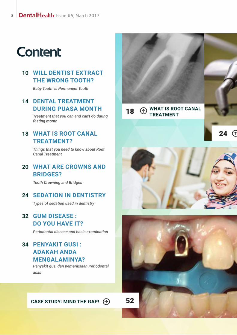
10 WILL DENTIST EXTRACT THE WRONG TOOTH?
Baby Tooth vs Permanent Tooth
14 DENTAL TREATMENT DURING PUASA MONTH
Treatment that you can and can’t do during fasting month
18 WHAT IS ROOT CANAL TREATMENT?
Things that you need to know about Root Canal Treatment
20 WHAT ARE CROWNS AND BRIDGES?
Tooth Crowning and Bridges
24 SEDATION IN DENTISTRYTypes of sedation used in dentistry
32 GUM DISEASE : DO YOU HAVE IT?
Periodontal disease and basic examination
34 PENYAKIT GUSI : ADAKAH ANDA MENGALAMINYA?
Penyakit gusi dan pemeriksaan Periodontal asas
Content
52CASE STUDY: MIND THE GAP!
WHAT IS ROOT CANAL TREATMENT

9Issue #5, March 2017
14
24
36 MINIMALLY INVASIVE TECHNIQUE
For Removal of Impacted Wisdom Tooth
39 KENAPA GIGI SAYA MENJADI JARANG?
Pergerakan gigi yang tidak diingini
39 WHY ARE MY TEETH MOVING APART?
Unintentional Teeth Movement
46 DR EZANI’S RECIPESuper Pink Salad
48 ENERGY IN MACHU PICCU
Journal by Dr.
52 CASE STUDY: MIND THE GAP!
Case study on missing or an unsightly tooth
54 PRACTISE MANAGEMENT TIPS
Building Patient Trust
56 THE DENTAL ACADEMY MALAYSIA
A Clinical Competency training centre.
56 THE DENTAL ACADEMY
DENTAL TREATMENT DURING PUASA MONTH
SEDATION IN DENTISTRY

10 Issue #5, March 2017
Will dentist extract the wrong tooth?Article written by Dr. Tiew Soon Tat
Will dentist extract the wrong tooth?
Children normally shed their baby
teeth at the age of 6 to 12. At the same
time,permanent teeth will be emerging,
forming a mixed dentition. How to
differentiate baby tooth and permanent
tooth? Generally, baby teeth are fairer
and whiter, while permanent teeth are
yellowish in colour. This is due to a
higher calcium composition in enamel
of permanent teeth compared to baby
teeth, causing higher transparency,
revealing the yellowish essence of
permanent teeth.
Recently, a cute mum asked me:
”Dr Tiew, how can you tell which of my
child’s tooth is either baby tooth or
permanent tooth? He lost a tooth before
but I could not remember which one. Is it
possible that you may extract the wrong
tooth?” I was surprised by this question
because dentist should not make this
mistake. Therefore a common sense to
me may be unfamiliar to the parents.
After reassuring the parent, she was
relieved to let her child proceed with
the treatment. This incident had inspired
me to write this article, so that parents
could learn how to differentiate baby
tooth and permanent tooth.
4 differences of baby tooth and permanent tooth
Children normally lose their baby tooth
at the age of 6 to 12, at the same time
permanent tooth emerges, forming a
mixture of baby teeth and permanent
teeth. How to differentiate baby tooth
Generally, baby teeth are fairer and whiter, while permanent teeth are yellowish in colour.

11Issue #5, March 2017
and permanent tooth? Below are some
main aspects:
1. Generally, baby teeth appear to be
fairer and whiter while permanent
teeth will give a more yellowish
hue. This is due to higher enamel’s
calcium composition in permanent
teeth compared to baby teeth,
resulting higher transparency,
revealing the yellowish essence of
permanent teeth.
2. The crown of baby tooth is smaller
than permanent tooth. Generally,
when we grow up, the size of
maxilla and mandible will increase,
leading to increasedcapacity of
oral cavity.Therefore it is common
for the crown of permanent tooth
to be bigger.
3. The neck of baby tooth will be
smaller and getting thinner compare
to permanent tooth. Therefore the
demarcation of crown and root is
clearer in baby tooth.
4. Baby tooth gets attrited easily
compared to permanent teeth
in mixed dentition as they exist
longer in this mixed dentition and
generally permanent teeth have
higher calcium concentration and
harder enamel. Baby tooth also has
larger pulp chamber, higher pulp
horns and larger bifurcated roots.
Teeth aid in digestive system
After explaining the differences between
baby tooth and permanent tooth, let’s
find out how each and every type of
teeth help us to taste delicious food.
Let’s look at how we can classify them.
I believed everyone realizes that our
teeth are a tool that will help us tear,
crush and grind the food that we eat.
Without proper chewing, unrefined food
will cause indigestion, thus burdening
the digestive system.
A research shown, a toothless person
has higher risk of getting stomach
cancer. Due to the difficulty in eating,
a toothless person can only eat certain
food. Depriving of certain vitamins and
minerals would cause malnutrition.
Therefore, food can only be easily
digested and absorbed for body use if
the food is chewed properly.
Baby tooth vs Permanent Tooth
Depriving of certain vitamins and minerals would cause malnutrition. Therefore, food can only be easily digested and absorbed for body use if the food is chewed properly.
Diagram of Baby Tooth andPermanent Tooth

12 Issue #5, March 2017
3 types of teeth and their functions
Each type of tooth has its own role in either baby tooth or permanent tooth. Normally
they are classified into 3 groups:
Incisor
Incisor helps to tear food into pieces. With the help of masticatory muscle, incisor uses
its edge to tear the food into pieces. There are 4central incisorsand 4 lateral incisors.
The 2 lowercentral incisors usually erupt first, quickly followed by the 2 uppercentral.
Once these teeth have erupted, you can expect to see the teeth on either side of
the central incisors coming through next, often 2 at a time. These are lateral incisors.
Canine
Canines are sharper with longer roots embedded in the jaw. Therefore, they are able
to withstand higher force.There are four canines, 2at the top and 2 at the bottom.
Molar
When food is torn by incisors and canines, they are being transferred by the tongue to
molar region for grinding. Molars have lots of pits and fissures that aid in grinding and
resulting in good and effective digestion. The premolars and molars arecollectively
known for grinding. We have eight premolars and 8 to 12 molars.
Hopefully through this article, parents now have a better idea of their children’s teeth.
This article is written and contributed by Dr. Tiew Soon Tat
Source: dentistsnearby.com

13Issue #5, March 2017

14 Issue #5, March 2017
Dental Treatmentduring Puasa MonthA lot of patients would ask the dentist, what treatment can we do when we are fasting and what can’t we do. We would like to try our best to clear your doubt for this matter. From what we know, as long as water or liquid doesn’t enter the throat and stomach, then fasting won’t be cancelled for the day.
Although water will be used to cool the
instruments when in use, for example,
from the high-speed hand-pieces or
scaling. Your dentist will be using a high-
volume saliva and fluid suction to ensure
that fluid doesn’t pool in your mouth.
Your dentist will also let you know in
advance about the possibility of you
accidentally swallowing fluid during a
procedure.
Treatments that involves fluids pooling in the mouth:
� Scaling and prophylaxis
(Pembersihan karang gigi
dan polish gigi)
� Filling (Tampalan gigi)
� Extraction (Cabutan gigi)
� Any other procedures
that involves the dentist’s
handpiece. (Procedur
lain yang menggunakan
handpiece)
Procedures that doesn’t involve fluids:
� Consultation
� Taking an X-rays (OPG or
Intra-oral PA)

15Issue #5, March 2017
✔ Scaling (Mencuci Karang Gigi)
✔ Dental fillings (Menggerudi,
tampalan)
✔ Crown and bridge (Sarung gigi,
Jambatan Gigi)
✔ Extraction (Cabutan Gigi) -
Although it is worth noting that
painkillers will be prescribed by
your dentist after or before the
procedure and that might batalkan
your puasa.
✔ Local Anaesthetic Injections prior
to dental treatments (injeksi bius)
In a nutshell, the following procedures are alright to do during fasting month and won’t be needed to be replaced if done.(Prosedur yang tidak perlu batalkan puasa)
✔ Topical Anesthetic gel
(Ubat Bius untuk gusi)
✔ Root canal treatments
(Rawatan Akar)
✔ Consultation and check-up
✔ Tooth Brushing, Flossing and using
a mouth-wash (Berus gigi, Flos,
Kumur)

Spray mulut dengan syarat ia tidak ditelan tidak membatalkan puasa.
According to sources from IOMS (Islamic Organization for Medical Sciences), The Akademi figh di Jeddah dan WHO, Figh As-Sunnah (Sayyid as-Sabiq), and Fatwa from Yusuf Al-Qaradhawi with Majlis Fatwa Eropah :
Injeksi (cucuk) melalui kulit, kedalam otot
(intramuscular), sendi (intraarticular),
kedalam pembuluh darah (intravenous)
kecuali bahan yang dicucuk mempunyai
zat tidak membatalkan puasa
Menerima Oksigen atau gas untuk bius
tidak membatalkan puasa
Menerima semua bahan yang diserap
kedalam badan melalui kulit, seperti
krim sapuan, minyak dan sebagainya
tidak membatalkan puasa.
According to Garis Panduan Berpuasa bagi pesakit, E-Fatwa.gov.my:
Melakukan prosedure tampalan gigi,
cabutan atau pembersihan gigi dan
memberus gigi dengan siwak (Tooth
cleanser) dengan syarat individu yang
berpuasa tidak menelan apa-apa bahan
semasa prodesure rawatan tersebut
tidak membatalkan puasa.
Penggunaan ubat-ubatan dalam bentuk
sapuan, balutan dan plaster pada
permukaan kulit tidak membatalkan
puasa.
According to Fikah Perubatan:
Mencabut gigi, menampal gigi, mencuci
gigi atau menggosok gigi dengan
siwak ataupun memberus gigi tidak
membatalkan puasa selagi mana tidak
masuk ke dalam halkum (ditelan)
SPRAY MULUT
GAS BIUS
Menerima Oksigen atau gas untuk bius tidak membatalkan puasa.
Mencuci mulut, kumur-kumur, spray
mulut, dengan syarat ia tidak ditelan
tidak membatalkan puasa.
Special Acknowledgement to Aimi Athirah
from International Islamic University
Malaysia (IIUM) for her kind guidance
on this article.
16 Issue #5, March 2017

Take Care of YourTeeth and They’ll Take Care of You
Bad dental hygiene isnt just bad for your teeth; it can harm your entire body. Most people already have chronic gingivitis by the time they’re children, so keep flossing and brushing to prevent the onset of these 14-bad-dental-hygiene-induced ailments.
1. HEART DISEASEIncludes conditions such as coronary heart disease, congenital heart disease, heart attack, and congestive heart failure
2. CLOGGED ARTERIESA build-up of plaque on the-inner walls of the arteries. Arterial plaque reduces blood flow or can block it altogether.
3. STROKEA stoppage of blood flow to the brain, causing brain tissue to die.
4. TOOTH LOSSThe loss of teeth due to gum disease and tooth decay
5. RESPIRATORY DISEASEIncludes disoders affecting the lungs, such as asthma, chronic obstructive pulmonary disease, infections, lung cancer, and many other breathing problems
6. SEVERE GUM DISEASEAn infection of the tissues and bones that surround and support the teeth
7. LUDWIG’S ANGINAA bacterial infection of the floor of the mouth.
8. ENDOCARDITISAn inflammation of your heart’s inner lining.
9. BRAIN ABSCESSA collection of immune cells, pus, and other material in the brain, usually from a bacterial or fungal infection
10. MEDIASTINITISAn inflammation of the mediastinum. This is the area that contains the heart, large blood vessels, windpipe, esophagus, thymus gland, lymph nodes, and connective tissues.
11. OSTEOMYELITIS OF THE JAWAn acute or chronic infection of the jaw bone
12. FACIAL CELLULITISA bacterial skin infection on the face.
13. PNEUMONIAAn inflammation of the lung that involves fluid filling the air sacs
14. SEPSISAn overactive immune response to an infection that results in the formation of blood clots that block the flow of blood to vital organs
POSSIBLE AFFECTS OF:
Gum Disease Gingivitis Tooth Abscesses
SOURCE: GOOGLE HEALTH; MEDICINE PLUS; WEBMO
17Issue #5, March 2017

18 Issue #5, March 2017
WHAT IS A
What is root canal treatment and why do we need it?
Your dentist may have suggested to
you that Root Canal Therapy (otherwise
known as Endodontics) was needed
for a particular tooth. Although you
may have been briefed about some of
the facts concerning the procedures
involved in root canal therapy, you
would like some more information.
Earlier, a badly infected tooth, or one
that just had significant decay, was
doomed to be extracted. Today, the
majority of these teeth can be salvaged
by the Root Canal Specialist.
What are the benefits of endodontic treatment?
Endodontic treatment saves teeth that
would otherwise need to be extracted.
Saving your natural teeth, if possible, is
the best option.
An extraction is truly the last resort!
Indications of root canal treatment
Spontaneous pain or throbbing pain
while biting
➠ Severe Sensitivity to hot and cold
foods
➠ Severe decay or an injury that
creates an abscess (infection) in
the bone
Root Canal Treatment consists of:
The removal of the infected or irritated
nerve tissue that lies within the root of
the tooth. It is this infected pulp tissue
that causes an eventual abscess.
The first step in a root canal is to
obtain access to the nerve. This is
accomplished by establishing a small
access opening in the top of the tooth.
The length of the root canal is determined
and the infected pulp is removed.
At the same visit, the canal where
the nerve is located will be
reshaped and prepared to accept
a special root canal filling material.
The X-Ray shows a root canal treated molar
This article is written and contributed exclusively to dentistsnearby.com by:
Dr.Shekhar Bhatia - BDS(Manipal), MDS(RGUHS), Endodontist
Source: dentistsnearby.com
ROOT CANAL TREATMENT?

19Issue #5, March 2017
The final step in your root canal
will be the sealing of the root canal
with a sterile, plastic material called
gutta percha. This is done in order
to prevent possible future infection.
Is the procedure painful?
Endodontic treatment does not cause
pain; in fact it relieves it. When you
have a severe toothache, the toothache
most likely is due to damaged tissues
in the tooth. Endodontic treatment
removes this damaged tissue from the
tooth, thereby relieving the pain you feel.
If before the procedure or during
procedure patient feels pain this
procedure is done under local
anesthetics.
Will there be pain after procedure?
Cleaning the root canals may cause some
slight tenderness but the discomfort can
be relieved by taking some pain killers. If
pain persists or if you experience severe
pain, call your dentist.
Sometimes when there has been long
standing infection or abscess, there may
be some soreness associated with the
root canal visit. If this should turn out
to be true, you will be given specific
instructions to follow to minimize the
discomfort. When an infection is present,
it may be necessary to take an antibiotic.
How many appointments are necessary?
Usually two or three visits are necessary.
Sometimes it can be completed in one
single visit.
How long will the tooth last?
With proper restoration and dental care
such as proper brushing and flossing,
regular dental check-up, it may last
a lifetime. After the completion of
endodontic treatment, your dentists
will usually advise on a restoration,
for instance a crown that protects the
tooth from future fracture. The teeth
at the back of the mouth, a crown is
commonly required to prevent the
tooth from fracturing; heavy chewing
on a root canal treated tooth should be
avoided until a crown is fitted. A proper
restoration after root canal treatment
prevents leaks of bacteria into the
tooth again and reduces the chances of
needing retreatment of the root canals.
Can the endodontic treatment fail?
Endodontic treatment can have success
rate of up to 95% in general. However
failure can occur if:
h The affected tooth develops decay;
h The restoration on the tooth fails;
h The tooth cracks.Dental Root Canal Process
ROOT CANAL TREATMENT?

20 Issue #5, March 2017
CrownsWhen a tooth are considered too
decayed for comventional chomposite
(tooth-coloured) fillings or amalgams to
be done, your dentist may opt for a more
resilient “restoration” which is what we
term as crowning of the tooth. A crown
can be made completely of metal, which
is normally indicated for your back teeth.
A laminated crown, which is layered
with metal and porcelain can be done
for your front teeth where appearance
is of importance. Or better still, a
crown made completely of porcelain is
usually done now-a-days with the use of
Zirconia. (From wiki, Zirconia : Zirconia
is a very hard ceramic that is used as
a strong base material in some full
ceramic restorations. The zirconia used
in dentistry is zirconium oxide which
has been stabilized with the addition of
yttrium oxide. The full name of zirconia
used in dentistry is yttria-stabilized
zirconia or YSZ.)
While crowning of a tooth costs more
than conventional fillings done normally,
they are definitely more long lasting and
will provide a better sense of security
both to the patient and the dentist, If
done properly, crowning of a tooth are
proven over time to last a very long time.
Dental crowns are “glued” to your teeth
by dental cement which are made of
specially formulated materials which
are loosely similar to those of composite
fillings.
In Malaysia, based on the MDA guidelines
of pricing for dental crowns 2010, they
will cost around RM650 and above for
non precious metal full metal crowns
(which means crowns that are made of
metals that are not expensive) while
metal crowns that contains precious
metal will cost you about RM1000
and above. Ceramic crowns, which
requires a certain amount of expertise
and technique to fabricate will cost
you around RM1100 and above. While
roughly translates that crowning for your
front teeth will usually cost more. Those
porcelain fused metal crowns (layered
crowns with a metal base and porcelain
outermost layer will range from RM700 -
RM 1200 depending on the type of metal
used. These prices were drafted quite
some time ago, therefore it is better to
bet on the safe side and consult your
dentist about the prices he is charging
before moving on.
Dental crowns can also be done on
a child when his or her teeth are
unrestorable by composite fillings,
we call them Stainless steel crowns,
althought they aren’t made of stainless
steel but are mostly Nickel based. These
crowns that are placed on children
are meant to maintain the integrity of
the tooth until they change to their
permanent teeth.
What are Crowns &Bridges?

21Issue #5, March 2017
BridgesDental bridges are done when there is a
tooth missing in between two “healthy”
tooth to replace the missing tooth in
between. With a larger missing teeth
area missing in an area, the probability
of a bridge to fail will increase too. That
is because the bridge itself will be only
supported by less “foundation” and
will experience more force from biting.
Therefore, It is of utmost importance
to discuss the treatment plan with your
dentist thoroughly and you might be
better off with a pair of dentures.
Bridges are made just like crowns are
made with the difference of bridges
consists of a few units of “attached
crowns”.
Based on the Malaysian Dental
Association guidelines of 2010, Bridges
are priced based on the number of
“units” of the bridge. Which means, if
your bridge is a 3 unit bridge using
non precious metal, it will be RM650
x 3 = RM1950. Some dentist will not
be calculating like this and may offer a
cheaper price. Please do take note that,
Crowns and bridges are complicated
pieces of mini prosthesis for your teeth
and our fellow dentists are required
to send them to a dental laboratory
for fabrication, which means prices
of crowns and bridges are actually
determined by the type of metal and the
workmanship from the dental lab’s side.
Source: dentistsnearby.com
Bridges are made just like
crowns are made with
the difference of bridges
consists of a few units
of “attached crowns”
A crown can be made completely of metal, which is normally indicated for your back teeth

22 Issue #5, March 2017

23Issue #5, March 2017

24 Issue #5, March 2017
The following types of sedation are used in dentistry:Inhaled (minimal sedation). You breathe
nitrous oxide -- otherwise known
as “laughing gas” -- combined with
oxygen through a mask that’s placed
over your nose. The gas helps you relax.
Your dentist can control the amount of
sedation you receive, and the gas tends
to wear off quickly. This is the only form
of sedation where you may be able to
drive yourself home after the procedure.
Oral sedation. Depending on the total
dose given, oral sedation can range
from minimal to moderate. For minimal
sedation, you take a pill. The pill will
make you drowsy, although you’ll still
be awake. A larger dose may be given
to produce moderate sedation. This is
the type of anesthesia most commonly
associated with sedation dentistry.
Some people become groggy enough
from moderate oral sedation to actually
fall asleep during the procedure. They
usually can, though, be awakened with
a gentle shake. Examples of Drugs for
oral sedation:
■ Diazepam - It has been around
since the 1960s and is a well
known and time-tested sedative
with amnesic properties. Valium is
particularly useful for appointments
where extensive dentistry is being
performed.
■ Halcion is most well known for the
treatment of insomnia.
■ Zaleplon is commonly used for the
treatment of insomnia.
■ Lorazepam is commonly prescribed
for the treatment of anxiety and
has amnesic properties. It is an
effective sedative and is useful for
appointments <2 hours.
■ Hydroxyzine has anti-anxiety effects
with no amnesic properties.
■ Midazolam is ideal for short
appointments or simple procedures.
There is always risk anesthesia. It is usually safe, though, when given by
experienced dentists.
SEDAT
ION
IN D
ENTI
STRY

25Issue #5, March 2017
IV moderate sedation. You receive the
sedative drug through a vein, so it goes
to work more quickly. This method
allows the dentist to continually adjust
the level of sedation.
Deep sedation and general anesthesia.
You will get medications that will make
you either almost unconscious or totally
unconscious -- deeply asleep -- during
the procedure. While you are under
general anesthesia, you cannot easily
be awakened until the effects of the
anesthesia wear off or are reversed with
medication.
Regardless of which type of sedation you
receive, you’ll also typically need a local
anesthetic -- numbing medication at the
site where the dentist is working in the
mouth -- to relieve pain if the procedure
causes any discomfort.
Sedation dentistry may also be appropriate for people who:
■ have a low pain threshold
■ can’t sit still in the dentist’s chair
■ have very sensitive teeth
■ have a bad gag reflex
■ need a large amount of dental work
completed
IV Moderate Sedation

26 Issue #5, March 2017
HOW SAFE ISSEDATION DENTISTRY?There is always risk anesthesia. It is
usually safe, though, when given by
experienced dentists. However, certain
people, such as those who are obese
or who have obstructive sleep apnea,
should talk to their doctor before having
sedation. That’s because they are more
likely to develop complications from the
anesthesia. Other conditions such as
heart and lungs conditions should also
be disclosed to your dentist and medical
doctor before proceeding.
It’s important to make sure that your
dentist is trained and qualified to
administer the type of sedation you will
be receiving.
1. Before the procedure, your dentist should go over your medical history. Your dentist should also determine whether you are an appropriate candidate for sedation and ask about any medications you’re currently taking.
2. You should ask what dose of the sedative is appropriate for your age and health. You should also ask whether it is within the dose recommended by the FDA.
3. You should receive a form detailing the risks of the procedure. Go over it carefully with your dentist. Ask questions if you’re unclear on any of the wording.
4. The dentist should continuously monitor your pulse rate and oxygen saturation and regular monitoring of depth of sedation and blood pressure throughout the procedure
5. The dentist should also have oxygen -- artificial ventilation -- and drugs that reverse the effects of sedation on hand in case you need them.
To be a smart patient, you should make sure the following things are done:

27Issue #5, March 2017
Documentation on of the following
should be done for records.
■ Names of staffs involved in the
procedure
■ History, examination and
investigative findings
■ Dosages of drugs and their
timings
■ Vital signs: Pulse rate, oxygen
saturation and blood pressure:
before, during and after the
procedure.
SOURCES:
Rai, K, Hegde, A, and Goel, K. Journal of
Clinical Pediatric Dentistry, 2007; vol 32:
pp 1-4.
American Dental Association: “Policy
Statement: The Use of Sedation and General
Anesthesia by Dentists.”
Joel M. Weaver, DDS, PhD, dentist
anesthesiologist; emeritus professor, College
Certain people, such as those who are obese or who have obstructive sleep apnea, should talk to their doctor before having sedation.
Importance of Documentations (Paper work)
of Dentistry, The Ohio State University;
spokesman, American Dental Association.
American Dental Association: “Guidelines
for the Use of Sedation and Anesthesia by
Dentists.” Source: dentistsnearby.com
Recommended read for Dentists:
RECOMMENDATIONS FOR SEDATION
AND ANALGESIA BY NON-
ANAESTHESIOLOGISTS
http://www.dentistsnearby.com/
images/Ministry-Of-Health-Malaysia-
Recommedation-for-sedation-and-
analgesia-2012.pdf
References:
Guidelines for the Use of Sedation
and General Anesthesia by Dentists
(Amedican Dental Association) -
https://www.ada.org/~/media/
ADA/About%20the%20ADA/Files/
anesthesia_use_guidelines.ashx
RECOMMENDATIONS FOR SEDATION
AND ANALGESIA BY NON-
ANAESTHESIOLOGISTS (Ministry of
Health Malaysia) -
http://www.moh.gov.my/
images/gallery/Rujukan/
Recommendations%20for%20
Sedation%20and%20Analgesia%20
-%2016.pdf
WebMD Educational Article -
http://www.webmd.com/oral-health/
sedation-dentistry-can-you-really-
relax-in-the-dentists-chair
28 Issue #5, March 2017

29Issue #5, March 2017

30 Issue #5, March 2017

31Issue #5, March 2017
Infographics from Mortenson Family Dental

PERIODONTITIS
GINGIVITIS
32 Issue #5, March 2017
What is periodontal disease?
How is periodontal disease detected?
Periodontal disease is an umbrella term for diseases of the gums. The two most common types of periodontal disease are gingivitis and periodontitis. Gingivitis is a milder form of gum disease, caused by lack of dental hygiene. The primary sign of gingivitis is bleeding and swollen gums. It may be a precursor to periodontitis which results in not just bleeding and swollen gums but also the breakdown of tissue and bone under the gums and appears in the mouths as shaky teeth with receding gums.
Periodontal disease accounts for approximately 75% of all tooth loss. The cause of periodontal disease is dental plaque which is an accumulation of bacteria on the tooth surface near the gums. Harmful bacteria from the dental plaque and the mediators which results from the body’s response to these harmful bacteria then enter into the bloodstream and has been associated with a number of other diseases, including diabetes, heart disease, preterm and low birthweight babies, respiratory disease and rheumatoid arthritis.
Symptoms of periodontal disease include persistent bad breath, swollen or bleeding gums, pain when chewing, and receding gums. “Pockets” may also be present, indicating a space between the tooth and gums, where more plaque may accumulate. During dental check-ups, routine examinations of gums and teeth will be done. For periodontal disease to be identified, a specific examination called Basic Periodontal Examination (BPE) will be performed by your dentist. BPE can also categorise the severity of the disease.
GUM DISEASEDO YOU HAVE IT?
by Dharani Jayadeva Assoc Prof Dr Rathna Devi Vaithilingam
GINGIVITIS

33Issue #5, March 2017
Preventing periodontal disease
Basic Periodontal Examination
Method
The BPE method was first introduced in 1986 by the British Society of Periodontology. It is a simple but rapid
“screening tool”, used to obtain a basic outline of the severity of periodontal disease in patients to determine route of treatment. It is, however, insufficient in providing a specific diagnosis. Thus for severe cases, further investigations need to be carried out for a proper diagnosis.
When doing a BPE, your dentist classifies your teeth into six sections, namely:
I. the upper right second molar to the upper right first premolar
II. the upper right canine to the upper left canine
III. the upper left first premolar to the upper left second molar
IV. the lower right second molar to the lower right first premolar
V. the lower right canine to the lower left canine
VI. the lower left first premolar to the lower left second molar
Or, as shown below;
Wisdom teeth are excluded in carrying out this examination.
Using a special probe called a CPITN or WHO probe, your dentist will then insert the probe into the space between the gums and the tooth and walk the
It is important to practice good oral hygiene to ensure a healthy lifestyle. Proper oral hygiene, including regular brushing and performing interdental cleaning such as flossing or interdental brushing is usually sufficient in prevention of periodontal disease. However, it is also important to be aware of risk factors such as smoking and uncontrolled diabetes which may worsen periodontal disease progression. Most importantly going for regular dental check-ups and asking your dentist for a BPE will allow for early detection and treatment of periodontal disease.
probe along the gum margin of all the teeth in each section mentioned above. Your dentist will then give a score for each section in the mouth based on the findings following the probing. This procedure is generally painless as only light pressure is used for this procedure.
SCORE ACTION
0 No action is required
1Oral hygiene instruction is given to teach you how to remove plaque which has induced gingivitis
2Oral hygiene instruction, removal of plaque retentive factors (e.g. removal of calculus or tartar on tooth surface by performing scaling)
3Oral hygiene instruction, removal of plaque retentive factors and root surface debridement (cleaning of tartar on the root surface below the gums).
4Similar to (3). The need for further treatment will be assessed and you may be referred to a periodontist(gum specialist).
Your scores and what they mean
*Source: Basic periodontal examination (http://www.bsperio.org.uk/publications/
downloads/94_154250_bpe-2016-po-v5-fi-nal-002.pdf)
GUM DISEASEDO YOU HAVE IT?
by Dharani Jayadeva Assoc Prof Dr Rathna Devi Vaithilingam

34 Issue #5, March 2017
KERADANGANPERIODONTIK
GINGIVITIS
Apakah maksud penyakit gusi?
Bagaimanakah penyakit gusi dikenal pasti?
Penyakit gusi adalah istilah umum yang merangkumi pelbagai jenis penyakit gusi. Dua jenis penyakit gusi paling lazim adalah keradangan gusi (atau gingivitis) dan keradangan peridontik (atau periodontitis). Keradangan gusi adalah jenis yang kurang serius, yang disebabkan oleh penjagaan gigi yang kurang memuaskan. Simptom terawal bagi keradangan gusi adalah pendarahan dan kebengkakan gusi. Kadangkala, keradangan gusi boleh mengakibatkan keradangan periodontik, yang turut melibatkan tisu lembut gusi dan kerosakan tulang di bawah gusi. Ini menyebabkan gigi menjadi longgar dan gusi menyusut.
Penyakit gusi bertanggungjawab untuk hampir 75 peratus semua kehilangan gigi. Sebab utama penyakit gusi adalah plak gigi, iaitu akumulasi bakteria pada permukaan gigi bersebelahan gusi. Kemasukan bakteria berbahaya dari plak gigi ke saluran darah boleh dikaitkan dengan pelbagai jenis penyakit lain, termasuk kencing manis, panyakit jantung, kelahiran bayi yang pramatang dan kurang berat badan, penyakit respiratori serta penyakit rheumatoid arthritis.
Simptom penyakit gusi termasuk nafas berbau, gusi bengkak dan berdarah, kesakitan semasa mengunyah, serta gusi menyusut. Selain itu, mungkin terdapat kehadiran “poket”, yang memberi indikasi ruang antara gigi dan gusi di mana plak gigi boleh mengumpul. Semasa pemeriksaan gigi di klinik gigi, pemeriksaan rutin gigi dan gusi akan dilakukan. Walau bagaimanapun, untuk mengenal pasti penyakit gusi, pemeriksaan khusus untuk gusi iaitu Pemeriksaan Periodontik Asas (PPA) akan dilakukan oleh doktor gigi anda. Pemeriksaan ini juga dapat mengenal pasti tahap keseriusan penyakit gusi.
oleh Dharani Jayadeva Assoc Prof Dr Rathna Devi Vaithilingam
PENYAKIT GUSIADAKAH ANDA MENGALAMINYA?
KERADANGAN GUSI

35Issue #5, March 2017
Pencegahan penyakit gusi
Pemeriksaan Periodontal Asas (Basic Periodontal Examination-BPE)
Cara
Pemeriksaan ini pertama diperkenalkan oleh Persatuan Periodontologi British pada tahun 1986. Pemeriksaan ini adalah cara mudah dan pantas untuk mendapati rangka asas tahap keradangan gusi dalam pesakit, demi mengenal pasti cara perawatan terbaik. Namun demikian, pemeriksaan ini kurang sesuai untuk membuat diagnosis tertentu.
Semasa melakukan BPE, doktor gigi anda akan mengklasifikasi gigi anda kepada enam bahagian, iaitu:
I. gigi molar kedua atas sebelah kanan sehingga gigi pramolar pertama atas sebelah kanan
II. gigi taring atas sebelah kanan sehingga gigi taring atas sebelah kiri
III. gigi pramolar pertama atas sebelah kiri sehingga gigi molar kedua atas sebelah kiri
IV. gigi molar kedua bawah sebelah kanan sehingga gigi pramolar pertama bawah sebelah kanan
V. gigi taring bawah sebelah kanan sehingga gigi taring bawah sebelah kiri
VI. gigi pramolar pertama bawah sebelah kiri sehingga gigi molar kedua bawah sebelah kiri
Atau, seperti dinyatakan dalam gambar rajah di bawah:
Gigi bongsu dikecualikan dalam melakukan pemeriksaan ini.
Dengan menggunakan peralatan prob gigi istimewa yang dinamakan prob gigi CPITN, atau prob gigi WHO, doktor gigi anda akan memasuki prob gigi ke ruang di antara gigi dan gusi.
Dalam pembudayaan cara hidup sihat, penjagaan higin mulut yang baik perlu dipraktikkan. Antara langkah-langkah penjagaan higin mulut yang baik ini, pembersihan permukaan gigi dengan berus gigi serta pembersihan antara celah gigi dengan flos gigi atau berus interdental biasanya cukup untuk mencegah penyakit gusi. Namun, adalah penting untuk menyedari bahawa risik-risiko penyakit gusi seperti tabiat merokok dan penyakit diabetes yang tidak terkawal akan menambah teruk perkembangan penyakit gusi. Disamping itu, pemeriksaan gigi kerap serta permintaan pemeriksaan BPE daripada doktor gigi anda akan membolehkan pengenalpastian serta intervensi awal untuk penyakit gusi.
Kemudian, prob gigi akan digerakkan di sempadan gusi, dalam setiap bahagian seperti ditunjukkan dalam ilustrasi di atas. Doktor gigi anda kemudian akan memberikan skor berdasarkan pendapatan selepas pemeriksaan. Prosedur tersebut biasanya tidak menyakitkan, kerana hanya tekanan ringan digunakan.
SKOR TINDAKAN
0 Tiada tindakan yang diperlukan
1Tunjukajar pembersihan gigi diberikan untuk mengajar anda cara menbersihkan plak gigi yang telah menyebabkan keradangan gusi.
2
Tunjukajar pembersihan gigi, penyingkiran faktor-faktor yang mengekalkan plak (contohnya, pembersihan kalkulus atau karang gigi pada permukaan gigi dengan melakukan penskaleran)
3
Tunjukajar pembersihan gigi, penyingkiran faktor-faktor yang mengekalkan plak dan pembersihan permukaan akar (pembersihan karang gigi pada permukaan akar gigi di bawah gusi).
4Serupa dengan (3). Keperluan pemeriksaan terperinci dikenal pasti dan anda akan dirujuk kepada pakar periodontik (pakar gusi).
Skor anda dan tindakan yang diperlukan
*Sumber: Basic periodontal examination (http://www.bsperio.org.uk/publications/
downloads/94_154250_bpe-2016-po-v5-fi-nal-002.pdf /)

MINIMALLY INVASIVE TECHNIQUE
FOR REMOVAL OF IMPACTED WISDOM TEETH
ARABIC TWO LARGE FLAP UNNECESSARY IN VERTICAL IMPACTION
IMPACTED WISDOM TEETH
Everyone is struck with fear whenever they hear the words “impacted wisdom teeth”. It has been ingrained into the general populations’ mind that removing wisdom teeth is a horrifying procedure that leads to pain, swelling and bleeding. However, there is no longer a need for fear as advances in technology and formation of new techniques have reduced the chances of those complications from occurring.
Introducing, “Minimally Invasive Technique” for removing impacted wisdom teeth – a new technique developed to reduce the 3 main concerns: pain, swelling and bleeding.
1 DESIGN
In conventional surgeries, a soft tissue flap is elevated in order to obtain adequate access to the tooth. As necessary as this is, it causes significant pain, swelling and bleeding as your dentist will be manipulating (and indirectly damaging) a large portion of soft tissues. Whereas in
“Minimally Invasive Technique” we will minimally elevate a flap to gain access to the tooth; meaning absence of vertical releasing incision. Either that, or a surgeon may choose to completely forego the need of a flap using the flapless surgery technique. These will help as the swelling and bleeding is directly proportional to amount of soft tissue flap raised.
36 Issue #5, March 2017
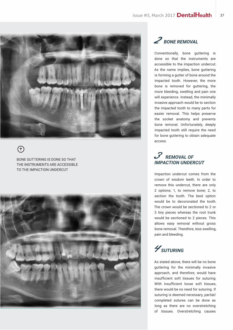
2 BONE REMOVAL
Conventionally, bone guttering is done so that the instruments are accessible to the impaction undercut. As the name implies, bone guttering is forming a gutter of bone around the impacted tooth. However, the more bone is removed for guttering, the more bleeding, swelling and pain one will experience. Instead, the minimally invasive approach would be to section the impacted tooth to many parts for easier removal. This helps preserve the socket anatomy and prevents bone removal. Unfortunately, deeply impacted tooth still require the need for bone guttering to obtain adequate access.
3 REMOVAL OF IMPACTION UNDERCUT
Impaction undercut comes from the crown of wisdom teeth. In order to remove this undercut, there are only 2 options; 1, to remove bone; 2, to section the tooth. The best option would be to decoronated the tooth. The crown would be sectioned to 2 or 3 tiny pieces whereas the root trunk would be sectioned to 2 pieces. This allows easy removal without gross bone removal. Therefore, less swelling, pain and bleeding.
4 SUTURING
As stated above, there will be no bone guttering for the minimally invasive approach, and therefore, would have insufficient soft tissues for suturing. With insufficient loose soft tissues, there would be no need for suturing. If suturing is deemed necessary, partial/completed sutures can be done as long as there are no overstretching of tissues. Overstretching causes
BONE GUTTERING IS DONE SO THAT THE INSTRUMENTS ARE ACCESSIBLE TO THE IMPACTION UNDERCUT
37Issue #5, March 2017

ischemia of tissues which leads to tissue necrosis which is painful and causes swelling.
5 MEDICATIONS
Medications are important for swelling and pain control. Medications given before and after a minor surgical procedure such as removing impacted wisdoms are antibiotics, pain killers and antiswelling. Emphasis is given on medications which have high initial dose and short duration. High initial dose means the duration of onset is quick and duration of taking medication is short e.g. 3 days.
6 ORAL HYGIENE MAINTENANCE
Maintenance of oral hygiene is an area where it is typically neglected by
patients. Surgeons should place great emphasis on maintaining a patient’s oral hygiene to ensure that an extraction socket heals uneventfully. As long as oral hygiene is kept in check, one may not even need to be put on a liquid diet nor bite on a single side for initial days after surgery. A patient may have normal diet, eat and bite as normal as long as they gargle thoroughly after meal times to prevent food from lodging into the socket. When the socket’s clean, there will be no problems.
FIGURE 5 CBCT SCAN SHOWS TRANSVERSELY IMPACTED TEETH FACING LINGUALLY INSTEAD OF THE MORE COMMON MESIOANGLE.
Article by : Dato’ Dr How Kim Chuan & Dr Cheong Jian Haw
38 Issue #5, March 2017

KENAPA GIGI SAYAMENJADI JARANG?
Fungsi pengunyahan yang tidak efisien
boleh menyebabkan gangguan nutrisi dan
masalah kesihatan
Senyuman yang menawan merupakan
satu ciri estetik yang menjadi idaman
ramai. Dengan peredaran masa, semakin
banyak penekanan diberikan kepada
kesihatan mulut dan kepentingannya
sebagai satu komponen utama kesihatan
umum. Gigi yang tersusun cantik adalah
matlamat yang cuba dicapai oleh
kebanyakan anggota masyarakat. Oleh
yang demikian, apabila pergerakan gigi
yang tidak diingini berlaku, ia menjadi
satu isu yang membawa impak yang
negatif kepada diri seseorang itu.
Pergerakan gigi yang tidak diingini
ini, boleh menyebabkan perubahan
orientasi gigi dan hasilnya adalah gigi
yang terputar, condong atau jarang.
Perubahan-perubahan ini mempunyai
impak negatif keatas paras rupa dan
fungsi pengunyahan seseorang individu.
Fungsi pengunyahan yang tidak efisien
boleh menyebabkan gangguan nutrisi
dan masalah kesihatan, manakala kesan
keatas rupa paras boleh menjejaskan
keyakinan diri seseorang. Impak
kesihatan mulut keatas kehidupan
memang jelas dan ianya tidak dapat
diasingkan daripada kesihatan tubuh.
Dalam keadaan mulut yang sihat, gigi kita
kekal ditempat yang sepatutnya melalui
kesinambungan daya yang bertindak ke
atas gigi. Gigi kita sentiasa menerima
daya tolakan dari pelbagai arah.
Contohnya, lidah kita mengenakan daya
tolakan yang akan mencuba menolak gigi
keluar, tetapi daya tolakan ini dilawan
dan diseimbangkan oleh daya daripada
otot bibir yang cuba menolak gigi ke
dalam. Hasilnya, gigi kita kekal ditempat
asal. Tambahan kepada ini, setiap batang
gigi kita akan membantu gigi yang
bersebelahan untuk kekal ditempat
asal dengan cara tidak memberikan
ruang untuk bergerak. Selain itu,
gigi kita juga mempunyai ligamen-
ligamen kecil (ligamen periodontal)
yang berfungsi sebagai alat pautan
gigi kepada tulang rahang. Ligamen
ini turut membantu mengelakkan
pergerakan gigi yang tidak normal.
Sebab
Pergerakan gigi yang tidak normal
berlaku bila terdapat gangguan kepada
Artikel:
Dr Renukanth RamanBDS(Malaya) MClinDent(Perio)(Malaya)
Periodontal Clinical Specialist
Gambar dari http://www.buccasana.es
39Issue #5, March 2017

keseimbangan daya di dalam mulut. Ini
boleh dibahagikan kepada dua kategori,
iaitu:
1. Pergerakan yang berkaitan dengan
penyakit
2. P e r g e r a k a n y a n g t i d a k
berkaitan dengan penyakit
Pergerakan Yang Berkaitan Dengan Penyakit
Punca utama pergerakan jenis ini
adalah Penyakit Gusi juga dikenali
sebagai Penyakit Periodontal. Penyakit
gusi adalah penyakit yang berupaya
merosakkan tisu penyokong gigi. Secara
amnya, tisu penyokong gigi kita terdiri
daripada gusi dan tulang dan kerosakan
kepada struktur penyokong ini boleh
menyebabkan kehilangan gigi. Penyakit
gusi ini disebabkan oleh kuman yang
hidup dalam mulut kita. Kuman ini
jika dibiarkan membiak dengan terlalu
banyak akan mencetuskan proses
keradangan gusi dan seterusnya
kemusnahan tulang penyokong gigi.
Amalan pembersihan gigi yang kurang
efisien akan menyebabkan penyakit gusi
awal atau dikenali sebagai Gingivitis.
Simptom yang lazimnya dialami oleh
seseorang yang menghidap penyakit
awal ini adalah pendarahan gusi bila
memberus gigi. Jika tidak dirawat,
penyakit ini akan parah dan merebak
ketahap Penyakit Periodontitis. Pada
peringkat ini, tulang penyokong gigi
akan musnah akibat daripada kesan
tindakan toksin dari bakteria dan gigi
akan menjadi longgar. Lama-kelamaan
gigi yang longgar ini akan menjadi jarang.
Keadaan ini, jika terus tidak dirawat, akan
menyebabkan kehilangan gigi.
Penyakit gusi merupakan penyakit
yang semakin berleluasa di kalangan
masyarakat Malaysia. Statistik daripada
‘’National Oral Health Survey in Adults’’
pada tahun 2010 menunjukkan bahawa
sebanyak 94% daripada populasi
Malaysia menghidap penyakit gusi.
Angka ini amat merisaukan kerana ia
merupakan kenaikan sebanyak 4.0%
daripada kajian yang serupa yang
dijalankan pada tahun 2000. Apa lagi jika
diambilkira faktor-faktor penyumbang
seperti penyakit kencing manis yang
tidak terkawal, amalan kehidupan
yang tidak sihat, tabiat merokok, serta
paras stres yang tinggi, penyakit gusi
ini boleh mencapai tahap epidemik.
Ilustrasi dari http://www.rydedentalcare.com.au
Pergerakan Yang Tidak Berkaitan Dengan Penyakit
Punca utama pergerakan gigi jenis ini
adalah ketiadaan gigi gantian yang
sesuai selepas gigi dicabut. Walaupun
ianya tidak berlaku kepada semua orang,
namun peratus kejadian pergerakan
jenis ini adalah agak tinggi di kalangan
masyarakat.
Apabila salah satu daripada gigi kita
dicabut, maka wujud satu ruang kosong
dalam susunan gigi. Jika gigi yang dicabut
itu tiada penggantinya, lama-kelamaan
gigi yang bersebelahan mungkin akan
bergerak kedalam ruang kosong tersebut.
Lebih banyak gigi yang dicabut, lebih
banyak ruang akan terbentuk dan ini
mungkin menyebabkan pergerakan
lebih banyak gigi. Akhirnya, gigi menjadi
jarang dan menjejaskan paras rupa.
Sebab-sebab lain yang mungkin boleh
menyebabkan pergerakan sebegini
adalah rawatan ortodontik yang tidak
sempurna dan kehausan tampalan
gigi. Individu yang memakai pendakap
sebagai rawatan ortodontik boleh
mengalami risiko pergerakan gigi yang
tidak diingini sekiranya mereka tidak
patuh kepada arahan pakar ortodontik.
Kejadian pergerakan gigi selepas
rawatan ortodontik selesai boleh berlaku
bila pesakit tidak mematuhi arahan
pemakaian pendakap boleh tanggal
yang bertujuan untuk menstabilkan
kedudukan gigi pada posisi yang baru.
Tampalan gigi yang berada di mulut
untuk masa yang lama akan jadi haus.
Kehausan tampalan gigi ini boleh
mengakibatkan ruang kecil terbentuk dan
sedikit sebanyak boleh menyebabkan
pergerakan gigi, terutamanya pada
tampalan di antara gigi. Walaupun
pergerakan ini adalah kecil, ia mampu
menjejaskan penampilan pesakit.
40 Issue #5, March 2017

Rawatan
Untuk merawat kejadian gigi menjadi
jarang, punca pergerakan gigi perlu
dikenal pasti dan dirawat dengan
sewajarnya.
Sekiranya, pergerakan gigi disebabkan
oleh penyakit gusi, individu tersebut
perlu dirawat oleh Pakar Pergigian
Periodontik. Penyakit gusi perlu
dirawat dan dikawal. Setelah rawatan
gusi selesai, cara-cara untuk merawat
kejarangan gigi boleh dirancang. Salah
satu cara yang efektif adalah rawatan
ortodontik untuk menyusun semula gigi
jarang dan menggantikan gigi yang telah
hilang sekiranya perlu.
Pilihan rawatan ini juga disyorkan untuk
merawat pergerakan gigi yang tidak
berkait dengan penyakit. Setelah ruang
antara gigi ditutup melalui rawatan
ortodontik, gigi yang telah dicabut
perlu digantikan. Antara pilihan rawatan
panggantian gigi yang hilang adalah gigi
palsu, jambatan pergigian dan implan gigi.
Pencegahan
Cara terbaik untuk mengelakkan gigi kita
daripada menjadi jarang adalah melalui
penjagaan kesihatan mulut yang optima.
Dengan mencegah penyakit gusi dan
kehilangan gigi, pergerakan gigi yang
tidak diingini boleh dielakkan. Penjagaan
higin mulut adalah tanggungjawab diri
kita sendiri. Seseorang individu itu harus
kenal tanda-tanda awal penyakit gusi
dan karies gigi. Sekiranya tanda-tanda
awal penyakit ini dapat dikesan, rawatan
awal boleh dilakukan untuk memulihkan
keadaan mulut tanpa berlakunya
kerosakan kekal.
Selain penjagaan higin mulut yang
optimum, lawatan berkala ke klinik
pergigian adalah penting untuk
mengekalkan mulut yang sihat. Sekiranya
anda mempunyai sebarang kemusykilan,
doktor pergigian anda sedia membantu.
Rujukan
1. Eduardo Y.S, Bonsiva S. Treatment Of
A Periodontally Compromised Patient
With Mini-Implant Anchorage. World J
Orthod 2009;10:350–360.
2. Lang N.P, L indhe J . C l inical
P e r i o d o n t o l o g y a n d I m p l a n t
Dentistry. Fifth edition 2008.
3. Storey E. The nature of tooth
movement. American Journal of
Orthodontics 1973;3:292314.
Gambar dari World J Orthod 2009;10:350–360
Gigi yang hilang digantikan dengan sebatang implan gigi
41Issue #5, March 2017

Why are my teethmoving apart?
if the movement of teeth is severe enough to affect
function, this will result in the
inefficient chewing of food which may ultimately lead to
poorer nutrition and subsequent health
problems.
A pleasing smile is a very desirable trait
among many individuals. An increasing
number of people are placing more
emphasis on their oral health as part of
their overall well-being. Well aligned
teeth with no spaces in between has
become the goal that many people are
trying to achieve and maintain. Therefore
if teeth start to move out of position
unintentionally, it becomes a nightmare
for some.
Unintentional movement results in
spaces developing between teeth. It
also may result in the change of the
orientation of teeth ie. teeth start
to tilt or rotate. These changes have
negative effects on the appearance
of the individual and not to mention
function as well. When appearance is
affected, it may result in deterioration
of the individual’s self-esteem and
confidence. Whereas if the movement of
teeth is severe enough to affect function,
this will result in the inefficient chewing
of food which may ultimately lead to
poorer nutrition and subsequent health
problems.
Our teeth are essentially kept in place
by the balance of forces acting upon
them. For example, the tongue will tend
to push the teeth out but this force is
balanced by the muscles of the upper
and lower lip. Each tooth in the mouth
helps the neighbouring tooth stay
in place by not providing space for
movement. Additionally, tiny ligaments
that connect the tooth to the bone (ie.
periodontal ligaments) also play an
important role in ensuring that our teeth
stay in place.
Causes
The undesired movement of teeth
happens when there is an upset to the
balance of forces in the mouth. These
causes can be divided into 2 categories,
which are;
1. Movement of teeth not caused by
diseases of the oral cavity
2. Movement of teeth caused by
diseases of the oral cavity
Movement of teeth not caused by diseases of the oral cavity
The main cause of non-disease related
movement of teeth is tooth extraction
without subsequent replacement.
Written by:
Dr Renukanth RamanBDS(Malaya) MClinDent(Perio)(Malaya)
Periodontal Clinical Specialist
42 Issue #5, March 2017

Although it does not happen to everyone,
the incidence of teeth moving apart
because of extractions of neighboring
teeth is nevertheless relatively high.
When a tooth is removed, this results
in a space. Overtime, the opposing
tooth or the adjacent teeth may start
moving into the empty space. If more
than 1 tooth is extracted, this will result
in more movement. The movement
of teeth immediately adjacent to the
empty space will eventually result in
movement of the other teeth as well. In
the end unsightly spaces will develop
between teeth.
Other causes of non-disease related
movement are such as incomplete
orthodontic treatment and wear and
tear of dental fillings. Individuals who
wear braces as part of their orthodontic
treatment are at risk of having their teeth
moving apart if they do not follow the
instructions of the orthodontist. After
the braces have been removed, quite
often the patient will need to wear an
appliance known as a retainer to make
sure the teeth stay in position. If they
do not wear the retainer according
to instructions, their teeth may start
moving apart.
Dental fillings which have been in the
mouth for many years will experience
wear and tear. As the fillings are worn
down, small spaces will develop which
can allow for small movements of teeth.
This is especially true for fillings located
between teeth. Although the movement
is very slight, it may lead to situations
such as enlarged spaces between
teeth and food impaction problems.
Movement of teeth caused by diseases of the oral cavity
Gum disease or Periodontal disease is
the most common and prevalent cause
of disease related movement of teeth.
Gum disease is basically a disease that
affects the supporting structures of our
Illustration from http://www.rydedentalcare.com.au
Picture From: http://www.buccasana.es
teeth, which comprises of the gums and
bone. This disease is caused by bacteria
that live in the mouth. The bacteria lives
in a soft white layer that forms on our
teeth called Plaque. When our brushing
habits are not efficient enough to
remove the majority of bacterial plaque,
gum disease develops. Initial stages
of gum disease usually manifest as
bleeding when brushing with or without
soreness of the gums. This condition is
called Gingivitis. A bad mouth odor may
also be present. Advanced gum disease,
also known as Periodontitis, happens
when gingivitis is not treated and the
disease is allowed to progress. At this
stage the bone that holds the tooth is
slowly destroyed by toxins released by
the bacteria. As the disease progresses,
more bone is lost and teeth start to
shake. Loose teeth will start to move
apart and spaces will develop between
teeth. Eventually, the affected teeth will
become extremely loose and will have to
be extracted. Gum disease has become
a very prevalent disease in Malaysia.
The last National Oral Health Survey in
Adults done on 2010 revealed that 94%
of the Malaysian adult population have
some form of gum disease. This figure
is very worrying as it represents an
increase of 4% from the previous survey
done in the year 2000. Together with
contributing factors such as uncontrolled
diabetes, smoking, stress and
unhealthy lifestyle habits, gum disease
can grow to epidemic proportions.
Treatment
To correct the problem of spaces which
have developed between teeth, the
cause of the problem must be identified
and treated accordingly.
For example, if the movement of teeth
is cause by gum disease, treating and
controlling the disease is the key. Once
the disease has been treated, options to
close the spaces can be explored. The
most effective method would be closure
43Issue #5, March 2017

Picture from World J Orthod 2009;10:350–360
Single tooth replacement with a dental implant
of the open spaces with braces, followed
by replacement of the missing teeth, if
any.
The same line of treatment is
also recommended to treat tooth
movement not caused by disease.
Once the spaces have been closed,
the missing tooth or teeth needs to
be replaced. The options to replace
missing teeth could be dentures,
dental bridges or dental implants.
Prevention
The best way to prevent unwanted
spacing of teeth is to ensure an
overall healthy oral environment. By
preventing tooth loss and gum disease,
the undesirable movement of teeth can
be avoided. Always be mindful of the
early signs of gum disease. Consistent
bleeding when brushing is an indication
of gingivitis. If detected early, it can be
successfully treated with no lasting ill
effects.
Optimal oral hygiene and regular check
ups with the dentist is the best way to
maintain good oral health. Remember,
dental cavities and gum disease
always starts from poor oral hygiene. If
toothbrushing is done the right way with
the right instruments, these problems
can be avoided. Always ask your dentist
for advice.
References :
1. Lang N.P, L indhe J . C l inical
Periodontology and Implant Dentistry.
Fifth edition 2008.
2. Eduardo Y.S, Bonsiva S. Treatment Of A
Periodontally Compromised Patient With
Mini-Implant Anchorage. World J Orthod
2009;10:350–360.
3. Storey E. The nature of tooth movement.
American Journal of Orthodontics
1973;3:292314.
44 Issue #5, March 2017

45Issue #5, March 2017

46 Issue #5, March 2017
LIFESTYLE : Nutrition
INGREDIENTS:
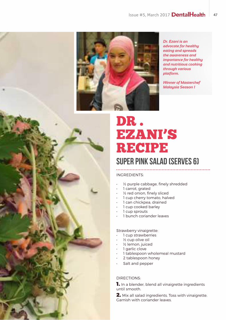
• ½ purple cabbage, finely shredded• 1 carrot, grated• ½ red onion, finely sliced• 1 cup cherry tomato, halved• 1 can chickpea, drained• 1 cup cooked barley• 1 cup sprouts• 1 bunch coriander leaves
Strawberry vinaigrette:• 1 cup strawberries• ½ cup olive oil• ½ lemon, juiced• 1 garlic clove• 1 tablespoon wholemeal mustard• 2 tablespoon honey• Salt and pepper
DIRECTIONS:
1. In a blender, blend all vinaigrette ingredients until smooth.
2. Mix all salad ingredients. Toss with vinaigrette. Garnish with coriander leaves.
SUPER PINK SALAD (SERVES 6)
DR .EZANI’SRECIPE
Dr. Ezani is an advocate for healthy eating and spreads the awareness and importance for healthy and nutritious cooking through various platform.
Winner of Masterchef Malaysia Season 1
47Issue #5, March 2017

48 Issue #5, March 2017
LIFESTYLE : Travelling

I felt a tremendous feeling of awe on my first real sight of this incredible ancient citadel in it’s ruinous splendour. For centuries Machu Picchu had been buried in jungle until July 1911 when Hiram Bingham stumbled upon it. From then on Yale University sent an archaeological expedition to explore it.
A stunning archaeological find, Machu Picchu was the only Inca site to escape 400 years of looting and destruction and it was remarkably preserved. Nor was it an ordinary Inca settlement. Located in an inaccessible location above the Urubamba gorge it contained so many fine buildings that people have been puzzling over it’s meaning since discovery.
I have been fortunate to visit Machu Picchu twice. The first was the usual touristy visit, taking a rickety
ENERGY INMACHU PICCU
designated bus from Aquas Calientes. The speedy uphill drive was rather suicidal I thought with the driver oblivious to the hair-pin curves and driving as if there was no tomorrow. I was so preoccupied with the thought of another suicidal drive down later, it rather took away the excitement of being in Machu Picchu. Fortunately another driver took over our bus and it was quite a relief.
My second visit was with two friends and we decided to treat ourselves and stay up in Machu Picchu where the only accommodation was then was the 36-room Machu Picchu Sanctuary Lodge with an expensive room rate to boot. We were lucky as the rooms were very much in demand and difficult to secure. We can now claim to be among the few people who had actually slept up there in Machu Picchu.
Article by
Dato’ Mohd Yusof Ahmad
- a former diplomat,
having served the Malaysian
Diplomatic Service for 35
years.
49Issue #5, March 2017

The ruins had a completely different feel once the hordes of day tourists had left . I remember asking my friends to join me to explore the place (we had free access to the ruins as guests at the lodge) after the last day tourists had been herded off and before the sun goes down. It was a rude and memorable shock to me when they told me to go ahead first as they wanted to finish watching ‘All About Eve’ from a video they found in the lodge’s library! God, did they come all the way to Machu Picchu to watch ‘ All About Eve’?
So I went on my own. And contrary to what most people expected of the place when deserted or at twilight (there was no sunset anyway, it just slowly and gradually got dark), I felt nothing spiritual nor frightening. What I felt was a tremendous amount of peace, calm and ENERGY! I found a solitary orchid flower growing out from a crack on a wall. And I had the whole place to myself, save for a few solitary people wondering about at a distance, and one man meditating on one of the higher points.
Next morning the three of us were up early to catch the place in a different mood before the tourist crowd arrive from Aguas Calientes. We encountered a whole group of people who had been hiking for 3-4 days from the Sacred Valley on the Inca Trail, and had finally reached their destination in the same tradition the Incas did hundreds of years ago.....
50 Issue #5, March 2017

51Issue #5, March 2017
KEE
PIN
G A
CLE
AN
TO
OTH
BRU
SH R
OU
TIN
E

52 Issue #5, March 2017
CASE STUDY:
Often enough we see lots of patient who come to our clinics wanting to replace a missing or an unsightly tooth in the anterior part of the mouth. Usually the tooth missing is the first or second upper incisors.
Traditionally the dentist can offer a bridge to fill this gap. That means preparing the adjacent teeth which would often be completely healthy teeth to support a bridge. Not only the bridge preparation would mean an unwarranted destruction of the adjacent teeth, but as in our case report below, it would mean unacceptable aesthetics.
CASE REPORT
INTRODUCTION Mary (Not her real name) came to see us 9 months ago for a consultation on a missing tooth. Upon examination we found that she was wearing an upper denture after a loss of an upper right central incisor. She had seen another dentist from elsewhere and had replaced the lost tooth with an extraordinarily big tooth to cover the gap. She complained that her plastic tooth looked odd as it was bigger than her other teeth. Upon further questioning we discovered that she always had a gap between her front incisor teeth, also known technically as a median diastema. She was clearly unhappy with the appearance and
Mind The Gap!
53Issue #5, March 2017
Dr Firdaus Hanapiah BDS (Otago) MSc (Lon.) FDSRCS (England) FICOI
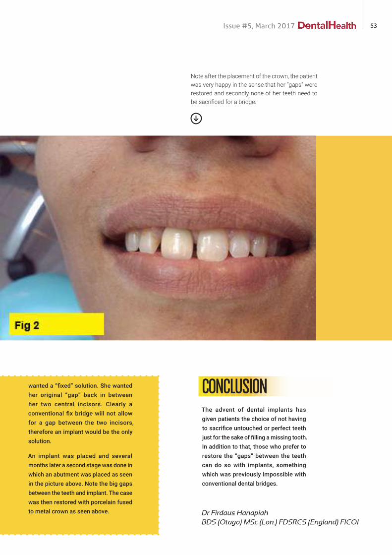
wanted a “fixed” solution. She wanted her original “gap” back in between her two central incisors. Clearly a conventional fix bridge will not allow for a gap between the two incisors, therefore an implant would be the only solution.
An implant was placed and several months later a second stage was done in which an abutment was placed as seen in the picture above. Note the big gaps between the teeth and implant. The case was then restored with porcelain fused to metal crown as seen above.
Note after the placement of the crown, the patient was very happy in the sense that her “gaps” were restored and secondly none of her teeth need to be sacrificed for a bridge.
The advent of dental implants has given patients the choice of not having to sacrifice untouched or perfect teeth just for the sake of filling a missing tooth. In addition to that, those who prefer to restore the “gaps” between the teeth can do so with implants, something which was previously impossible with conventional dental bridges.
CONCLUSION

Dental School being a teacher of the Art & Science of Procedures unfortunately does not teach us the fundamental key of People-to–People business - which is Human Relationship and the bond created.
I always say to my Dr’s- There is a very fine line between Professionalism and a sales man. We are both of them in a scrub.
The business of dentistry is highly complicated that in just of 30 minutes of the first appointment- We meet someone for the first time that comes to us for help- a stranger. In 30 minutes his got to leave our chair being our best friend- someone who feels we care- a
buddy who won’t let us go and we make them pay us for it. How do we do these in just 30 minutes?
As we get very busy in our practices daily and very efficient with our procedures with the latest technologies and scanners, it’s easy for us to bounce from patient to patient and do the procedures very well, but we truly forget that we are in the business of People. The business of human. The business of emotions and feelings.
We are in the business of Humans and Relationships and Connections. Sustaining this part of it and becoming a Super Star in Dentistry and having successful practice is “never how good
your Class II fillings look” . It’s about How when your Class II fillings come out, the Patient insist of Only seeing you and only you even though you failed round 1. This is Success. This is the key that will make you a super star dentist and you’re Practice to succeed.
Here’s one Small tip that I use in every single visit- that I believe has been very beneficial for me. There is a whole day of lecture just on Human Business in Dentistry, but allow me to share you apart of what I do, that has contributed to success.
I believe the most important Five minutes that I have is at the end of the appointment. The last minutes
I read an important quote one time – “that patient don’t care about how much you know until they know how much you care”
PRACTICE MANAGEMENT TIPS: THE LAST MINUTES THAT COUNTS
54 Issue #5, March 2017

PRACTICE MANAGEMENT TIPS:
A key to this is “Eye to eye” contact and the bigger key “is the Human Touch”. The touch that shows you care.
DR BALA SARAVANAN
DR BALA SARAVANAN(BDS, MBA)
before my Patient that entered for Consultation 30mins ago leaves as my Best friend.
This bond is done soon after the procedure is done, the handpiece has been put away, the patient’s face has been cleaned up, and we sit the patient up, and I basically get sort of knee-to-knee with them, and we’ll have a little discussion about the procedure, and then I just chat with them.
This is done as humbly and as considerate as possible depending to the extend of the procedure. I apologize at this point if there should be any
discomfort during the procedure, and explain why was there the discomfort. A key to this is “Eye to eye” contact and the bigger key “is the Human Touch”. The touch that shows you care. The touch that says “That I treated you like my mother, like my father, like my brother”. The touch that’s shows your genuine thoughts of helping. (This touch is done very appropriately- shoulder / hands). I assure you when this is done rightfully; there is a bond that is created. A connection / a chemistry that has been formed.
When this bond is created, is then when I speak about the future treatment options. Not selling it, but just throwing the possibility. Always always allow the Patients to make the concern decision and always make them feel superior.
Hence, no hard selling at this point. Throw them the treatment options. All the treatment options from the least expensive to the most expensive and keep to the one that you want the patient to do to the last option, and tell them if it was me, I would do…… and leave the ball in that court.
You have to make it feel that “You are NOT Financially Driven, and you’re offering the options Cause you care” Dentist have to sell without actually selling and that’s the tricky part. Be genuine to them. Be humble. Tell them what you feel would be the best. I assure you with the trust, the bond created and showing that you are not financially driven, you will get the best out of this patient and I assure you 80% of the time- You’re treatment option will be accepted.
"
"What do you do to build patient trust?
Please Do let me know your thoughts:[email protected]
55Issue #5, March 2017

56 Issue #5, March 2017
“Perfecting the Art of Dentistry”–The Dental Academy MalaysiaRealizing the surplus of Dentist being produced in the Country
& Realizing that the only way to Upgrade the level of Malaysian
Dentist to world class standards of Dentistry is via education- is
how The Dental Academy Malaysia was formed- with the sole
purpose of Upgrading the Dental Landscape. Its focus- To be a
training Platform for every dentist in Perfecting their art & Skills
– or simply said “The Tuition Centre for Dentist”

57Issue #5, March 2017

The Dental Academy Malaysia is a
Purpose built Dental Clinical Training
Academy focusing on Clinical
Competency Training for General Dental
Practioners. Their courses are recognized
& designed for those driven by
excellence & have a thirst for continuous
pursuit of specialized knowledge in
the domain of dentistry. They strive to
upgrade the skill levels of dentist.
They focus their teaching on practical
clinical dentistry. They sharpen and
increase the Clinical skills of Dentist
to be competent & confident in all
domain of dentistry via Simulation / Life
Patient / Discussion / Lecture / Hands-
On–Training as well as being a Personal
Specialist Clinical Mentor & Advisor to
all their participants.
Renowned Clinical Specialist in their
subspecialty will conduct the trainings
and teachings organized by The Dental
Academy/ Dental Associations/ Principle
or Supply Company / Fellowships/
Universities and Other Organization that
require dental teaching and simulation
infrastructure.
Equipped with a State of art Dental
Teaching Facilities and a futuristic
25-40sitter lecture theater, the Teaching
is done with the aid of many State of
the Art Dental Education Technology to
provide the best conductive teaching
possible for a Good “Take Back after
Each Class”
The State of the Art Teaching Facility is
inclusive of 26 Units of Fully Equipped
Dental Simulation/ Dental Microscopes,
High and low-speed headpieces,
phantom head and attached jaw modal.
The Simulations are equipped with
suction and power suction, Light Cure
Machines for effective Restoration
Training & a 23-inch screen to record
simulation exercises, project lectures,
streaming live surgeries. The Academy
also provides all Dental instrumentation
for examination, filling etc.
The Dental Academy Malaysia offers
a Comprehensive roster of Dental
Education Training that will Accelerate
your Learning Experience in gaining
Practical Expertise in Dentistry.
Each course is designed with the dentist
in mind. To ensure minimal disruption to
the Dentist professional commitments,
they have designed each course as a
two or four-day intensive session. To
ensure they extract maximum value, they
keep the enrollment limited to ensure
you receive the necessary personalized
support & attention.
The Dental Academy Malaysia advocates
Experiential Learning as opposed to the
58 Issue #5, March 2017

To ensure minimal disruption to the Dentist professional commitments, they have designed each course as a two or four-day intensive session.
Conventional prescriptive Approach by
converging both knowledge and hands-
on simulated scenarios, we aim to hone
your analytical skill-sets needed when
addressing real-life cases.
In The Dental Academy Malaysia,
Demonstration of a new skill in
Practical Dental Science will be done
using various communication methods
in teaching especially dental modal
demonstration with simulation prior to
real life or Live demonstration.
The mentioned methods are the most
recommended for teaching advanced
technical or neuromuscular skill, hence
the use of dental modal in all aspects
of teaching is cooperated in the Dental
Academy Malaysia Training programs.
In The Dental Academy, video is a
common tool for communication,
documentation and education. Video
provides many advantages including
live-stream demonstration, the creation
of case studies for students, as well as
the post-evaluation of work performed
by students.
The Dental Academy Malaysia Strives to
Make the Dental Learning Environment
as Conductive as possible. Their state of
the art lecture theatre houses up to 40
participants; complete with AV setup and
ample plug points for effective teaching
and learning. Their interactive approach
ensures that the Dentist remain engaged
throughout the sessions.
The Dental Academy Malaysia advocates
Experiential Learning as opposed to the
Conventional prescriptive Approach by
converging both knowledge and hands-
on simulated scenarios, we aim to hone
your analytical skill-sets needed when
addressing real-life cases.
In The Dental Academy Malaysia,
Demonstration of a new skill in
Practical Dental Science will be done
using various communication methods
in teaching especially dental modal
demonstration with simulation prior to
real life or Live demonstration.
The mentioned methods are the most
recommended for teaching advanced
technical or neuromuscular skill, hence
the use of dental modal in all aspects
of teaching is cooperated in the Dental
Academy Malaysia Training programs.
In The Dental Academy, video is a
common tool for communication,
documentation and education. Video
provides many advantages including
live-stream demonstration, the creation
of case studies for students, as well as
the post-evaluation of work performed
by students.
The Dental Academy Malaysia Dental
Life Patient Demonstration Program has
been Developed to Provide Practitioners
with the Confidence, Training and
Experience required to Successfully
undergo all types of dental training
into practical experience. The Dental
Academy Malaysia has a strong focus on
live patient surgery and the surgical and
restorative elements of training where
59Issue #5, March 2017

60 Issue #5, March 2017
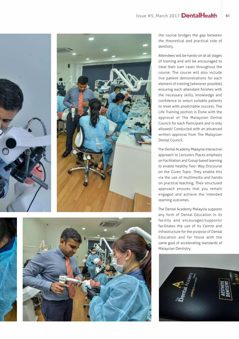
the course bridges the gap between
the theoretical and practical side of
dentistry.
Attendees will be hands-on at all stages
of training and will be encouraged to
treat their own cases throughout the
course. The course will also include
live patient demonstrations for each
element of training (wherever possible),
ensuring each attendant finishes with
the necessary skills, knowledge and
confidence to select suitable patients
to treat with predictable success. The
Life Training portion is Done with the
approval of The Malaysian Dental
Council for each Participant and is only
allowed/ Conducted with an advanced
written approval from The Malaysian
Dental Council.
The Dental Academy Malaysia interactive
approach to Lecturers Places emphasis
on Facilitation and Group based learning
to enable healthy Two- Way Discourse
on the Given Topic. They enable this
via the use of multimedia and hands-
on practical teaching. Their structured
approach ensures that you remain
engaged and achieve the intended
learning outcomes.
The Dental Academy Malaysia supports
any form of Dental Education in its
facility and encourages/supports/
facilitates the use of its Centre and
Infrastructure for the purpose of Dental
Education and for those with the
same goal of accelerating standards of
Malaysian Dentistry.
61Issue #5, March 2017

A: The Dental Academy Malaysia, 6-4 & 8-4, Jalan USJ 9/5R, 47620 Subang Jayahv, Selangor
P: +6012 6665210, +60176331737, +6012293398
W: www.thedentalacademy.com.my
62 Issue #5, March 2017

63Issue #5, March 2017

64 Issue #5, March 2017
Top Related