


Languages
Pages
Legal

Defective biliary secretion of bile acid 3-0- glucuronides in rats with hereditary conjugated hyperbitiru binemial
Folkert Kuipers,‘ Anna Radominska, * Piotr Zimniak, * Joanna M. Little, * Rick Havinga, Roe1 J. Vonk, and Roger Lester*
Department of Pediatrics, University of Groningen, The Netherlands, and Gastroenterology Unit, Department of Medicine, * University of Rochester School of Medicine, Rochester, NY 14642
Abstract Biliary secretion of bile acid glucuronides was stud- ied in control rats and in rats with a congenital defect in hepato- biliary transport of organic anions (GY rats). In control animals, hepatobiliary transport of [3H]lithocholic acid 3-0-glucuronide and [3H]cholic acid 3-0-glucuronide was efficient (> 95 % in 1 h) and comparable to that of [“C]taurocholic acid. Secretion of both glucuronides was impaired in GY rats (24% and 71 % at 1 h), whereas that of taurocholate was similar to control values. However, recovery of the glucuronides in bile was nearly com- plete within 24 h; virtually no radioactivity was found in urine. In control rats, biliary secretion of lithocholic acid 3 - 0 - glucuronide, but not that of cholic acid 3-0-glucuronide or taurocholate, could be delayed by simultaneous infusion of dibromosulphthalein. In mutant rats, dibromosulphthalein infu- sion was also able to inhibit secretion of cholic acid 3-0- glucuronide. [3H]Hydroxyetianic acid, a C20 short-chain bile acid, was secreted by control rats as a mixture of 20% carboxyl- linked and 80 % hydroxyl-linked (3-O-)glucuronide; secretion was very efficient (99% in 1 h). In GY rats, secretion was drastically impaired (16 % at 1 h and 74% over a 24-h period). Initially, the mutant secreted more carboxyl- than hydroxyl- linked glucuronide, but the ratio reached that of control animals after 24 h. The rates of formation of both types of hydroxyetianic acid glucuronide by hepatic microsomes from mutant rats were similar or even slightly higher than those of control micro- somes. These findings indicate that bile acid 3-0-glucuron- ides, but probably not carboxyl-linked glucuronides, are secreted into bile by a transport system shared with organic anions such as conjugated bilirubin and dibromosulphthalein, but different from that for amino acid-conjugated bile acids. -Kuipers, E, A. Radominska, P. Zimniak, J. M. Little, R. Havinga, R. J. Vonk, and R. Lester. Defective biliary secretion of bile acid 3-0-glucuronides in rats with hereditary conjugated hyperbilirubinemia. J. Lipid Res. 1989. 30: 1835-1845.
Supplementary key words taurocholic acid lithocholic acid cholic acid hydroxyetianic acid etianic acid transport UDP- glucuronosyltransferase
Glucuronidation is a well established but quantitatively minor pathway in normal bile acid metabolism. Glu- curonides of all common bile acids have been identified in human serum, bile, and urine (1 -9). Relatively increased
amounts of these bile acid metabolites, in which the glu- curonic acid is linked to the steroid moiety of the mole- cule, are found in urine and serum of patients with cholestatic liver disease (2, 4-9). In this case, glucuronida- tion may occur in addition to conjugation of the bile acid with glycine or taurine. It has recently been shown that, in the rat, glucuronidation is the only mode of conjuga- tion for monohydroxylated short-chain bile acids, a group of acidic steroids abundant in the developing organism (10-15). Short-chain bile acids can be glucuronidated at the hydroxyl group of the steroid ring, the carboxyl group of the side chain, or both (14, 16-18).
It has been assumed that glucuronidation of bile acids prevents possible hepatotoxic effects and promotes urin- ary secretion. From recent findings, however, it is clear that these assumptions are an oversimplification. First, it was recently demonstrated that lithocholic acid 3-0-0- glucuronide (LCG) possesses cholestatic properties ex- ceeding those of lithocholic acid itself (19). Second, it was found that the 3-0-/3-glucuronide of lithocholic acid is secreted in bile, but is minimally secreted in urine even in the presence of complete biliary obstruction (19).
Abbreviations: LCG, lithocholic acid 3-0-glucuronide; CG, cholic acid 3-0-glucuronide; TC, taurocholic acid; hydroxyetianic acid, 3cu-hydroxy-5&androstane-l7&carboxylic acid; GY, “Groningen Yellow” (mutants of Wistar rats with conjugated hyperbilirubinemia); HA, “high androsterone” glucuronidation capacity (Wistar rats with normal levels of 3-hydroxysteroid UDP -glucuronosyltransferase activity); LA, “low androsterone” glucuronidation capacity (mutants of Wistar rats with low levels of 3-hydroxysteroid UDP-glucuronosyltransferase activity); DBSP, dibromosulphthalein; TLC, thin-layer chromatography; UDPGT, UDP -glucuronosyltransferase.
‘Preliminary accounts of this work were presented at the 10th Intema- tional Bile Acid Meeting, Freiburg, FRG, June 1988, and at the Annual Meeting of the American Gastroenterological Association, New Orleans, May 1988 (57).
*To whom reprint requests should be addressed at: Department of Pediatrics, University of Groningen, Bloemsingel 10, 9712 KZ Gronin- gen, The Netherlands.
Journal of Lipid Research Volume 30, 1989 1835
by guest, on Septem
ber 1, 2018w
ww
.jlr.orgD
ownloaded from
Similarly, the glucuronide of cholic acid (20), as well as the glucuronide(s) of the short-chain bile acid hydroxy- etianic acid (14), are rapidly secreted into bile but not into urine upon intravenous administration to bile fistula rats. In agreement with these findings, Stiehl and co-workers (21) found a bi1e:urine excretion ratio for glucuronidated bile acids of 226:l in patients with alcoholic cirrhosis with moderate cholestasis.
Biliary secretion of cholephilic compounds involves up- take from sinusoidal blood by hepatocytes, intracellular transport, and secretion into bile at the canalicular pole of the cell. These processes have been studied in some detail for amino acid-conjugated bile acids, in particular for taurocholic acid (E), and it is generally accepted that carrier-mediated transport processes are involved (22- 28). It is not known whether glucuronidated bile acids are transported by the same systems that are operative for amino acid-conjugated bile acids. The present study was aimed at an initial characterization of the process of hepa- tobiliary transport of glucuronidated bile acids. For this purpose we used a Wistar rat strain (GY = Groningen Yellow) with a hereditary conjugated hyperbilirubinemia that is due to a defective hepatobiliary transport of or- ganic anions such as conjugated bilirubin. We have re- cently demonstrated that the biliary secretion, but not the hepatic uptake, of 3a-sulfated bile acids is impaired in GY rats, and that this inhibition becomes more pronounced with a decreasing number of hydroxyl groups on the bile acid's steroidal nucleus (29). In the present work, we stud- ied the biliary secretion of lithocholic acid 3-0-glucuron- ide (LCG) and cholic acid 3-0-glucuronide (CG), as well as the short-chain bile acid hydroxyetianic acid, and com- pared each with the secretion of E. Since some of the above compounds are not secreted as such, but undergo metabolic conversions during their passage through the liver, the form (or forms) that are actually transported from the hepatocyte into bile have been analyzed and identified.
MATERIALS AND METHODS
Experimental design
The objective of the studies was to examine the biliary secretion of [3H]CG, [3H]LCG, and [ " C ] E in GY and control rats. The biliary secretion of [3H]hydroxyetianic acid, which gives rise to carboxyl- and hydroxyl-linked glucuronides in the rat, was also studied. Additionally, the effects of co-infusion of dibromosulphthalein (DBSP) on biliary secretion of glucuronidated lithocholic and cholic acid was investigated. For this purpose, unlabeled and labeled compounds were prepared and administered in- travenously in trace amounts to unanesthetized control and GY rats prepared with a permanent external biliary fistula. The biliary and urinary secretion of radioactivity
was measured for 24 h, and the radioactive steroidal pro- ducts in bile were characterized (virtually no radioactivity was found in urine).
The specific activities of bile acid-dependent UDP- glucuronosyltransferases were also determined using hepatic microsomes from control and GY rats.
Chemicals
Standard CZ4 bile acids were obtained from Calbiochem-Behring (La Jolla, CA). Hydroxyetianic acid (3a - hydroxy - 56- androstane-l7~-carboxylic acid) and 3- oxo-5~-androstane-17~-carboxylic acid were purchased from Steraloids (Wilton, NH). DBSP was from SERB (Societe &Etudes et de Recherche Biologique, Paris, France). [24-'4C]Taurocholic acid, sp act 56 Ci/mol, [2,4-3H]cholic acid, sp act 25 Ci/mmol, and tritium- labeled water, sp act 1.8 Ci/mol, were obtained from New England Nuclear (Boston, MA). [36-3HJ-3a-Hydroxy- 56-cholanoic acid (tritium-labeled lithocholic acid) was synthesized as described previously (30).
The hydroxyl-linked glucuronides of unlabeled hy- droxyetianic, lithocholic, and cholic acids, as well as hydroxyl-linked glucuronides of tritium-labeled lithocho- lic and cholic acids were chemically synthesized as de- scribed previously (14, 17, 19, 20).
[ 2,2,4,4-3H4]-3a-hydroxy-5~-Androstane-l7~-carboxy- lic acid ([3H]hydroxyetianic acid) was prepared by eno- lization exchange as follows: 3 mg 3-oxo-5P-androstane- 17/3-carboxylic acid acid in 2 ml diethyl ether was mixed with 10 pl (150 pCi) [~arboxyl-~H]acetic acid containing 5 % (w/w) 3HCl and allowed to stand for 40 h. ([Car- b~xyl-~H]acetic acid containing 5 % 3HCl was prepared by mixing 0.362 ml acetic anhydride, 0.05 ml acetyl chloride, and 0.1 ml (10 mCi) 3 H 2 0 for 1 h). The sample was extracted with 4 x 1 ml aliquots of water acidified to pH 2. The pooled aqueous extracts were back-washed with 2 x 2 ml ether. All ether extracts were pooled and dried; the residue was dissolved in 2 ml isopropanol con- taining 2 mg sodium borohydride and the reduction was allowed to proceed for 1 h. The reaction mixture was acidified with 6 N HCl, 1 ml of water was added, and the alcohol was evaporated off under nitrogen. The aqueous solution was extracted with ether, the extract was dried, and the residue was dissolved in ether-methanol (9:l). The sample, a mixture of 3a- and 36-hydroxy acids, was methylated with diazomethane and dried; the methyl es- ters of the 3a- and 36-hydroxy compounds were separated by preparative thin-layer chromatography on LK5 plates (Whatman Chemical Separation Inc., Clifton, NY), de- veloped with benzene-acetone 95:5. Radioactive bands were localized using a Berthold Tracemaster 20 TLC ana- lyzer and visualized by spraying the plates with water (31). The silica gel containing the 3a-hydroxy compound was transferred to a column and the radioactive material was eluted with benzene-acetone 95:5. The recovered
1836 Journal of Lipid Research Volume 30, 1989
by guest, on Septem
ber 1, 2018w
ww
.jlr.orgD
ownloaded from
material was checked by TLC in the same solvent and found to be >99% pure. The methyl ester was hydro- lyzed by dissolving the sample in 2 ml methanol, adding 0.2 ml 5 N NaOH and leaving the mixture at 65OC for 24 h. The methanol was evaporated under NP, water was added, and the sample was extracted with ether as described above. TLC in isooctane-ethyl acetate-acetic acid 10:10:0.2 (v/v), followed by scanning for radioactivity and visualization with Krowicki's reagent (1 % phospho- molybdic acid and 5 % ceric sulfate in 10 % sulfuric acid) (32) demonstrated that the [ 'Hlhydroxyetianic acid was > 99 % chemically and radiochemically pure. The specific radioactivity was 1 Ci/mol.
Animals
Wistar rats with congenital conjugated hyperbiliru- binemia (GY = Groningen Yellow), extensively charac- terized previously (29), were bred at the Central Animal Laboratory, University of Groningen. Biochemically and in the pattern of inheritance, GY rats show great resem- blance to the mutant rat strain described by Jansen et al. (33, 34). Cross-breeding experiments have been recently performed and showed that both strains carry the same genetic defect.
Surgical procedures
Male mutant and normal Wistar rats, 275-325 g, were equipped with permanent silastic catheters in the bile duct and the heart (35). For a number of experiments, an additional, thinner heart catheter was inserted to allow simultaneous blood sampling and continuous intracar- diac infusions (36). The animals were housed in individ- ual Plexiglass cages and kept on a normal laboratory diet. Bile was led outside the cages through a polyethylene tub- ing with a swivel joint, which permitted the animals to move freely. Experiments were performed at least 8 days after surgery, to allow recovery from the operation and the establishment of a new steady state in bile acid synthesis (35). During experiments, the rats were placed in metabol- ic cages for simultaneous collection of urine and bile.
Experimental procedures
Tracer amounts of the bile acids were dissolved in a small volume of 0.1 M sodium hydroxide and added to 2-4 ml of a 4 VJ bovine albumin solution (fraction V, Sig- ma, St. Louis, MO) in saline. The solution was neutral- ized by addition of 0.1 M acetic acid, and passed through a 0.22-pm filter (Schleicher and Schuell, Den Bosch, The Netherlands). Rats received 0.4-1 pCi of bile acid as a bolus injection via the heart catheter. Bile was collected continuously in preweighed test tubes at 10-min intervals, beginning 30 min before injection to establish basal bile flow rates, and continuing for 2 h after the injection. Subsequently, bile was collected as a single sample for 22
h. A single urine sample was collected over the .24-h ex- perimental period. Plasma disappearance of [ 3H]LCG was examined simultaneously with bile secretion in a number of experiments. For this purpose, blood samples of 0.15 ml were taken at 1, 3, 5, 10, 15, 30, 45, 60,90, 120, and 180 min after injection. Blood was immediately transferred to heparinized test tubes and centrifuged to obtain plasma. D-Saccharic acid-1,4-lactone (Sigma) was added to bile and urine samples to prevent hydrolysis of bile acid glucuronides.
In a second set of experiments, mutant and control rats received a bolus injection of 7.7 pmol of DBSP, followed by a constant infusion of 23.1 pmol/h of the dye via the smaller heart catheter for 3 h. This infusion rate equals the estimated transport maximum for biliary secretion of DBSP in normal Wistar rats (29). After 1 h of DBSP infu- sion, 0.5 pCi of [3H]CG or 0.5 pCi of [3H]LCG was in- jected via the larger heart catheter. In all experiments, 0.2 pCi of [ I4C]'IC was injected simultaneously with the glu- curonides, Bile and urine were collected as described above. In addition, blood samples of 0.15 ml were taken at 0, 15, 30, 60, 120, and 180 min after the start of the DBSP infusion, to determine the plasma concentration of the dye.
Bile and urine volumes were determined gravimetrical- ly in tared test tubes. The collection time of bile samples was adjusted to account for the dead space of the tubing system (35). Radioactivity in bile and plasma, in 10-pl and 50-pl aliquots, respectively, was determined in an LKB scintillation counter equipped with external stan- dards to correct for quenching. Bile was decolorized with 50 p1 of hydrogen peroxide prior to counting.
The concentration of DBSP in bile and plasma was de- termined, after appropriate dilution of the samples, as described previously (37).
Isolation and identification of radioactive products in bile
Aliquots of bile from animals that received radiolabeled LCG or CG were diluted with 5 ml of 0.1 M glycine- trichloroacetate buffer, pH 2.8, and the samples were ap- plied to Bond Elut CI8 cartridges (size: 6cc; Analytichem, Harbor City, CA). The cartridges were washed with 5 ml buffer and 2 x 5 ml water, and bile acids were eluted wzh 2 x 5 ml methanol. Methanol eluates were dried, residues were dissolved in methanol, and aliquots were chromato- graphed on Whatman LK5 thin-layer plates in chloro- form-methanol-acetic acid-water 65:25:2:4 (v/v). Ali- quots of bile from animals given hydroxyetianic acid were acidified to pH 2 with 6 N HCl and extracted with 3 x 4 ml ether. Ether extracts were taken to dryness and residues were redissolved in methanol. On several occa- sions, the solid phase extraction method described above for LCG and CG was used with identical results. An ali-
Kuipers et al. Biliary secretion of bile acid glucuronides 1837
by guest, on Septem
ber 1, 2018w
ww
.jlr.orgD
ownloaded from
quot of each sample was chromatographed on TLC plates in ethyl acetate-absolute ethanol-concentrated ammoni- um hydroxide 45:45:15 (v/v).
An additional aliquot of each extract was dried, dis- solved in 1 ml of 0.1 M sodium phosphate buffer, pH 6.8, and treated with 100 units of E. coli /3-glucuronidase (Sig- ma Chemical Co., St. Louis, MO). Samples were in- cubated overnight at 37OC, extracted with CIS cartridges as described above, and subjected to TLC in chloroform- methanol-acetic acid-water 65:25:2:4 (v/v).
Following P-glucuronidase treatment, extracts of bile from animals given LCG were dried, redissolved in 1 ml 0.1 M sodium acetate buffer, pH 5.6, and 25 units of cho- lylglycine hydrolase (Sigma) was added. Samples were in- cubated at 37OC for 18 h, extracted with C I S cartridges, and subjected to TLC analysis in isooctane-ethyl ace- tate-acetic acid 1O:lO:Z (./.).
Selective hydrolysis of the carboxyl-linked glucuronide of hydroxyetianic acid in bile extracts was carried out in 0.1 N NaOH for 3 h at room temperature. For verification of the selectivity of the procedure, a standard mixture of tritium-labeled hydroxyl- and carboxyl-linked glucuron- ides of hydroxyetianic acid was simultaneously subjected to the same hydrolytic treatment. Following hydrolysis, bile acids were extracted with CIS cartridges and analyzed by TLC in ethyl acetate-absolute ethanol-concentrated ammonium hydroxide 45:45:15 (v/v).
Spots on TLC plates were localized by scanning for radioactivity using a Berthold Tracemaster 20, by nonde- structive visualization through spraying with water (for preparative plates) (31), or by visualization through spray- ing with Krowicki’s reagent and heating on a hot plate at 10O-12O0C (32).
In vitro glucuronidation of bile acids
Microsomes of livers from control and mutant Wistar rats were prepared as previously described (17) and stored at - 8OOC. Bile acid:UDP -glucuronosyltransferase (UDP - GT) activity was assayed as previously described (17) ex- cept that the reactions were carried out in 100 mM Na-HEPES buffer, pH 6.5 for carboxyl-linked, and pH 7.5 for hydroxyl-linked glucuronide formation. The glu- curonosyltransferase reaction was linear with protein con- centration up to 1.5 mg/ml and with time for 15 min for both the control and GY rats. The concentration of hydroxyetianic acid was 100 pM in all experiments; this concentration was determined to be saturating but not in- hibitory for both the control and GY rats. Separation of carboxyl- and hydroxyl-linked glucuronides and unre- acted material was performed by TLC (17).
n mutation greatly reducing the 3-hydroxysteroid UDPGT activity is known to occur in Wistar rats with an incidence of about 50 % (38-40). Microsomal prepara- tions derived from all rats were therefore tested with
lithocholic acid (41) in order to classify them as LA (mu- tant) or HA (normal phenotype).
The specific activity of enzymes is expressed as nmol of bile acid glucuronide formed per mg protein and per min, and is reported as mean SD. Protein was measured us- ing the Bio-Rad kit based on the Bradford protein assay (42), with bovine serum albumin as standard.
RESULTS
Biliary secretion of bile acid 3-0-glucuronides
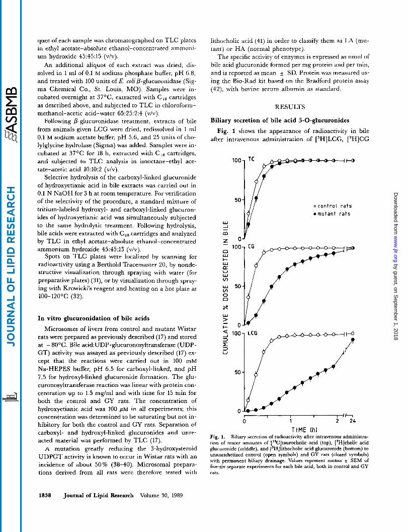
Fig. 1 shows the appearance of radioactivity in bile after intravenous administration of [3H]LCG, [3H]CG
‘“1 TC PP-+--+”
w -J - m
o con t ro l r a t s 0 mutant r a t s
4 100- 3 x 3 V
50 -
I 1 I I I I h 0 1 2 24
TIME Ih) Fig. 1. Biliary secretion of radioactivity after intravenous administra- tion of tracer amounts of [‘*C]taurocholic acid (top), [SH]cholic acid glucuronide (middle), and [3H]lithocholic acid glucuronide (bottom) to unanesthetized control (open symbols) and GY rats (closed symbols) with permanent biliary drainage. Values represent means f SEM of five-six separate experiments for each bile acid, both in control and GY rats.
1838 Journal of Lipid Research Volume 30, 1989
by guest, on Septem
ber 1, 2018w
ww
.jlr.orgD
ownloaded from
and, for comparison, of ["CITE in control and GY rats. Secretion of [14C]TC was slightly delayed in the GY rats when compared to controls, but almost all of the ad- ministered label had been secreted into bile within only 60 min. In contrast, biliary secretion of ['HICG and ['HILCG in GY animals was significantly impaired. The degree of impairment was different for each compound. The recoveries of ['HICG and [3H]LCG in bile of GY rats were 71 * 4 % and 24 * 3 %, respectively, at 60 min after injection, compared to 98 * 1 % and 96 * 2 %, respectively, in controls. However, the administered dose was almost completely recovered from bile collected over a 24-h period; urinary excretion of these compounds was negligible in both controls and GY rats.
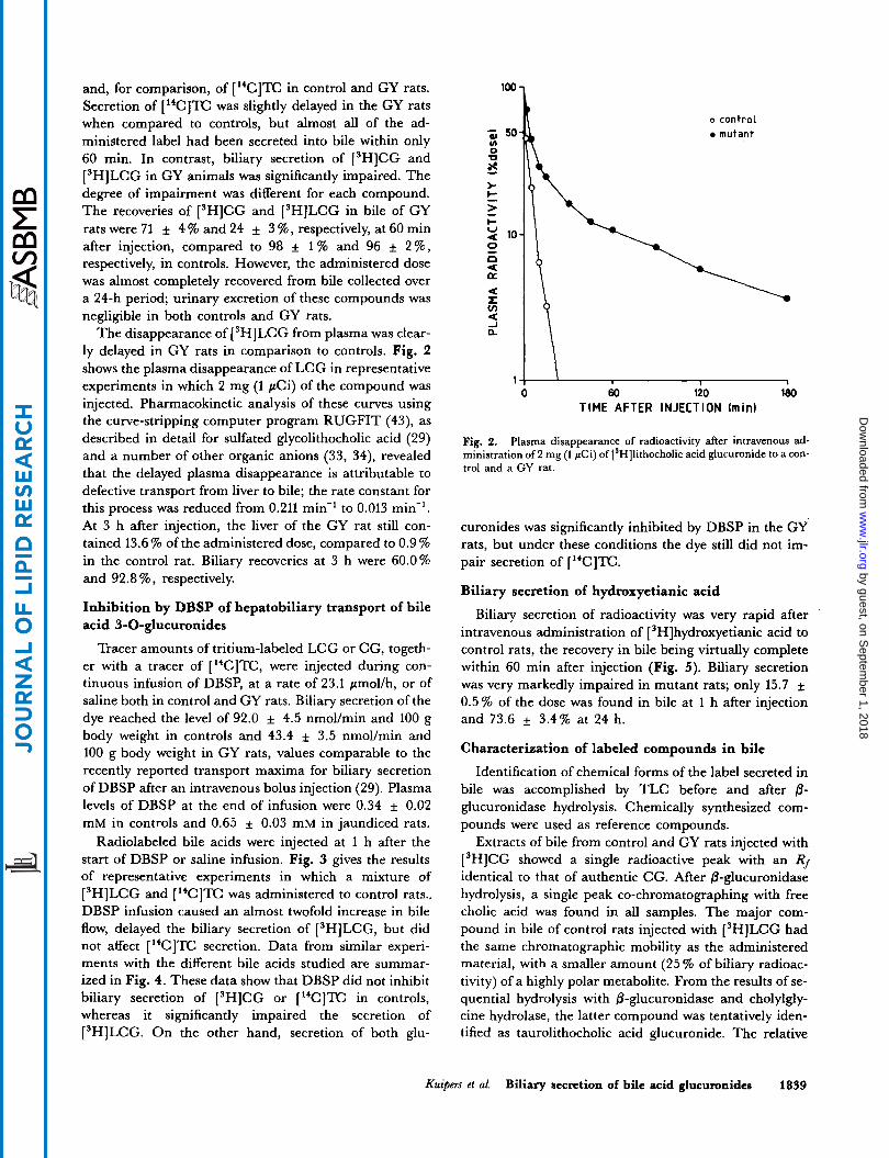
The disappearance of ['HILCG from plasma was clear- ly delayed in GY rats in comparison to controls. Fig. 2 shows the plasma disappearance of LCG in representative experiments in which 2 mg (1 pCi) of the compound was injected. Pharmacokinetic analysis of these curves using the curve-stripping computer program RUGFIT (43), as described in detail for sulfated glycolithocholic acid (29) and a number of other organic anions (33, 34), revealed that the delayed plasma disappearance is attributable to defective transport from liver to bile; the rate constant for this process was reduced from 0.211 min-' to 0.013 min-'. At 3 h after injection, the liver of the GY rat still con- tained 13.6 % of the administered dose, compared to 0.9 % in the control rat. Biliary recoveries at 3 h were 60.0% and 92.8 %, respectively.
Inhibition by DBSP of hepatobiliary transport of bile acid 3-0-glucuronides
Tracer amounts of tritium-labeled LCG or CG, togeth- er with a tracer of ["CIlT, were injected during con- tinuous infusion of DBSP, at a rate of 23.1 pmol/h, or of saline both in control and GY rats. Biliary secretion of the dye reached the level of 92.0 * 4.5 nmol/min and 100 g body weight in controls and 43.4 * 3.5 nmol/min and 100 g body weight in GY rats, values comparable to the recently reported transport maxima for biliary secretion of DBSP after an intravenous bolus injection (29). Plasma levels of DBSP at the end of infusion were 0.34 * 0.02 mM in controls and 0.65 f 0.03 mM in jaundiced rats.
Radiolabeled bile acids were injected at 1 h after the start of DBSP or saline infusion. Fig. 3 gives the results of representative experiments in which a mixture of ['HILCG and ["CIlT was administered to control rats., DBSP infusion caused an almost twofold increase in bile flow, delayed the biliary secretion of ['HILCG, but did not affect ["CI'E secretion. Data from similar experi- ments with the different bile acids studied are summar- ized in Fig. 4. These data show that DBSP did not inhibit biliary secretion of ['HICG or ["CC]TC in controls, whereas it significantly impaired the secretion of ['HILCG. On the other hand, secretion of both glu-
o control mutant
' I
0 60 120 180 TIME AFTER INJECTION (min)
Fig. 2, Plasma disappearance of radioactivity after intravenous ad- ministration of 2 mg (1 pCi) of [SH]lithocholic acid glucuronide to a con- trol and a GY rat.
curonides was significantly inhibited by DBSP in the GY rats, but under these conditions the dye still did not im- pair secretion of ["CITC.
Biliary secretion of hydroxyetianic acid
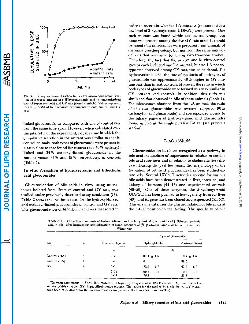
Biliary secretion of radioactivity was very rapid after intravenous administration of [ 'Hlhydroxyetianic acid to control rats, the recovery in bile being virtually complete within 60 min after injection (Fig. 5). Biliary secretion was very markedly impaired in mutant rats; only 15.7 * 0.5% of the dose was found in bile at 1 h after injection and 73.6 * 3.4% at 24 h.
Characterization of labeled compounds in bile
Identification of chemical forms of the label secreted in bile was accomplished by TLC before and after @- glucuronidase hydrolysis. Chemically synthesized com- pounds were used as reference compounds.
Extracts of bile from control and GY rats injected with ['HICG showed a single radioactive peak with an Rf identical to that of authentic CG. After @-glucuronidase hydrolysis, a single peak co-chromatographing with free cholic acid was found in all samples. The major com- pound in bile of control rats injected with ['HILCG had the same chromatographic mobility as the administered material, with a smaller amount (25 % of biliary radioac- tivity) of a highly polar metabolite. From the results of se- quential hydrolysis with @-glucuronidase and cholylgly- cine hydrolase, the latter compound was tentatively iden- tified as taurolithocholic acid glucuronide. The relative
Kuipers et al. Biliary secretion of bile acid glucuronides 1839
by guest, on Septem
ber 1, 2018w
ww
.jlr.orgD
ownloaded from

1 "'- TC
Y I 0
I- W CL U W v)
W i eo
1 o saline
DBSP
0 I
I] 0
- 200 1
1 LL
BILE A C I D m 1
I INFUSION PERIOD I I I I I 1 1
TIME (hl 0 1 2 3
Fig. 3. Biliary secretion of ['*CJtaurocholic acid (top) and (SH]lithocholic acid glucuronide (middle) and bile flow (bottom) in con- trol Wistar rats infused with either dibromosulphthalein (closed sym- bols) or saline (open symbols). Both bile acids were administered simultaneously to unanesthetized rats with permanent biliary drainage at 1 h after initiation of an intravenous infusion of the dye (23.1 pmollh) or its vehicle.
proportion of the tauro-conjugate was higher in bile of GY rats than in that of controls, and increased even fur- ther during the course of the experiment; 59 % of radioac- tivity present in bile collected during the initial 2 h of the experiment and 78 % in samples from 2 to 24 h was in the form of taurolithocholic acid glucuronide.
Bile of three out of the four control rats, collected dur- ing the initial 2 h after injection of [3H]hydroxyetianic acid, contained the hydroxyl-linked glucuronide and the carboxyl-linked glucuronide of [3H]hydroxyetianic acid in a 4:l ratio (Table l), as shown by TLC (see ref. 17 and Methods). The identity of the carboxyl-linked glucuron- ide was further confirmed by selective alkaline hydrolysis under mild conditions (see Methods); this procedure led to the disappearance of the carboxyl-linked glucuronide on the TLC plate with a concomitant appearance of an equivalent amount of free hydroxyetianic acid, but left
the hydroxyl-linked glucuronide unchanged. One control rat secreted radioactivity predominantly as the carboxyl- linked glucuronide of hydroxyetianic acid (88 %), the re- mainder being in the form of a very polar, as yet unidentified, metabolite; no hydroxyl-linked glucuronide was present. This rat is probably a representative of the low 3-hydroxysteroid UDPGT mutation (LA mutation) mentioned in the Methods section; the latter enzyme is responsible for the formation of the hydroxyl-linked (3-0-), but not of the carboxyl-linked glucuronide of hydroxy- etianic acid (41). Bile of GY mutant rats collected within the first 2 h after injection was enriched in the carboxyl-
c E 0 m \ aJ u) 0 0
.-
W 1 m z z 0 I- W
- - E5 W v)
W > I- U .-I 3
-
f U
100 1 CG
0 SALINE
U O B S P
100 LCG 1
CONTROL MUTANT
Fig. 4. Biliary recovery at 30 min after injection ( % dose) of ["C]taurocholic acid (top), [3H]cholic acid glucuronide (middle), and [3H]lithocholic acid glucuronide (bottom) in control and GY rats. The compounds were administered intravenously to unanesthetized rats with permanent biliary drainage at 1 h after initiation of an intravenous infu- sion of dibromosulphthalein (23.1 mmol/h) or its vehicle (saline). Values are means + SEM of four to six separate experiments.
1840 Journal of Lipid Research Volume 30, 1989
by guest, on Septem
ber 1, 2018w
ww
.jlr.orgD
ownloaded from
100 7 y-
ocontroL r a t s mutant r a t s
r I 1 I 1 /h 0 1 2 24
TIME (!-I)
Fig. 5. Biliary secretion of radioactivity after intravenous administra- tion of a tracer amount of ['Hlhydroxyetianic acid to unanesthetized control (open symbols) and CY rats (closed symbols). Values represent means i SEM of four separate experiments in both control and CY rats.
linked glucuronide, as compared with bile of control rats from the same time span. However, when calculated over the total 24 h of the experiment, i.e., the time in which the cumulative secretion in the mutant was similar to that in control animals, both types of glucuronide were present in a ratio close to that found for control rats: 76 % hydroxyl- linked and 24 % carboxyl-linked glucuronide in the mutant versus 81 % and 19%, respectively, in controls (Table 1).
In vitro formation of hydroxyetianic and lithocholic acid glucuronides
Glucuronidation of bile acids in vitro, using micro- somes isolated from livers of control and GY rats, was studied under previously described assay conditions (17). Table 2 shows the synthesis rates for the hydroxyl-linked and carboxyl-linked glucuronides in control and GY rats. The glucuronidation of lithocholic acid was measured in
order to ascertain whether LA mutants (mutants with a low level of 3-hydroxysteroid UDPGT) were present. One such mutant was found within the control group, but none was present among the five GY rats used. It should be noted that microsomes were prepared from animals of the same breeding colony, but not from the same individ- ual rats that were used for the in vivo transport studies. Therefore, the fact that the in vivo and in vitro control groups each included one LA animal, but no LA pheno- type was observed among GY rats, was coincidental. For hydroxyetianic acid, the rate of synthesis of both types of glucuronide was approximately 40 7% higher in GY mu- tant rats than in HA controls. However, the ratio in which both types of glucuronide were formed was very similar in GY mutants and controls. In addition, this ratio was similar to that observed in bile of these animals (Table 1). For microsomes obtained from the LA animal, the ratio of the two glucuronides was reversed (approx. 90% carboxyl-linked glucuronide) and corresponded closely to the biliary pattern of hydroxyetianic acid glucuronides found in vivo in the single putative LA rat (see previous section).
DISCUSSION
Glucuronidation has been recognized as a pathway in bile acid metabolism of importance in relation to specific bile acid substrates and in relation to cholestatic liver dis- ease. During the past few years, the enzymology of the formation of bile acid glucuronides has been studied ex- tensively. Several UDPGT activities specific for various bile acids have been demonstrated in liver, intestine, and kidney of humans (44-47) and experimental animals (48-50). One of these enzymes, the 3-hydroxysteroid UDPGT, has been purified to homogeneity from rat liver (49), and its gene has been cloned and sequenced (51, 52). This enzyme catalyzes the glucuronidation of bile acids at the 3-OH position in the A-ring. The specificity of bile
TABLE 1. The relative amounts of hydroxyl-linked and carboxyl-linked glucuronides of [3H]hydroxyetianic acid in bile, after intravenous administration of tracer amounts of ['Hlhydroxyetianic acid to control and CY
Wistar rats ~~ - ~
Type of Glucuronide
Rat n Time after Iniection Hvdroxvl-Linked Carboxvl-Linked
Control (HA) 3
Control (LA) 1
GY 4
h
0-2
0-2
0-2
2-24 0-24
76
81.1 i 1.6 18.9 f 1.6 0 88.0
32.2 i 4.7 67.8 f 4.7
90.3 * 0.1 10.0 f 0.1 76.4 23.6
The values are means f SEM. HA, mutant with high 3-hydroxysteroid UDPGT activity; LA, mutant with low activity of this enzyme; CY, hyperbilirubinemic mutant. The values for the total 0-24 h bile for the GY mutant have been calculated from the measured values for partial collections (0-2 h and 2-24 h).
Kuipers et al. Biliary secretion of bile acid glucuronides 1841
by guest, on Septem
ber 1, 2018w
ww
.jlr.orgD
ownloaded from

TABLE 2. Specific activities of UDP-glucuronosyltransferase(s) toward hydroxyetianic and lithocholic acid in hepatic microsomes from control Wistar and GY Wistar rats
Hydroxyetianic Acid Glucuronide Lithocholic Acid Glucuronide
Microsomes n Hydroxyl-Linked Carboxyl-Linked H ydroxyl-Linked Carboxyl-Linked
nmol/mg‘ min nmoumg. min
Control (HA) 5 2.39 f 0.49. 0.98 i 0.21’ 1.85 i 0.33’ 0
Control (LA) 1 0.11 0.70 0 0
GY (HA) 5 3.67 i: 0.35* 1.36 f 0.12’ 3.07 i 0.37’ 0
The values are means i: SD; n denotes the number of individual rats; determinations were each done in quad- ruplicate. Reactions were carried out as described in Methods, and products were separated and identified (using appropriate synthetic glucuronide standards) by TLC as hydroxyl- or carboxyl-linked glucuronides ( 1 7). HA, mu- tant with high 3-hydroxysteroid UDPGT activity; LA, mutant with low activity of this enzyme; GY, hyperbilirubi- nemic mutant.
‘Difference between HA control and GY mutant rats is statistically significant by both the Mann-Whitney and the t-test (P < 0.001).
acid UDPGT activities for the site of glucuronidation, the configuration of the bile acid’s hydroxyl groups, and the length of the side chain have recently been studied exten- sively, using microsomes prepared from human (47) and rat liver (17, 41, 50). These studies revealed the existence of three types of bile acid glucuronides: hydroxyl-linked glucuronides at position 3 and at position 6 of the steroid nucleus, and a carboxyl-linked glucuronide in which the glucuronic acid is linked to the side chain. The latter type of glucuronide has been described for short-chain ((320, (223) bile acids (16, 17) as well as for a conventional (CZ4) bile acid
Much less is known about the physiology of bile acid glucuronides. The presence of increased amounts of these compounds in urine of patients with cholestatic liver dis- ease has led to the assumption that bile acids are removed from the body as glucuronides via the kidneys in situa- tions when biliary secretion is impaired or blocked. As already pointed out in the Introduction, experimental data do not bear out this assumption for bile acids. It is now well established that bile is the main excretory route for bile acid 3-0-glucuronides (14, 16, 19-21). Yet, the mechanism(s) for the hepatobiliary transport of these bile acid metabolites have, to our knowledge, not been stud- ied. In the present paper, we used GY rats to study this process. GY rats show a markedly reduced ability for biliary secretion of organic anions, such as conjugated bilirubin and DBSP (29). Jansen and co-workers found that secretion of a neutral steroid (ouabain) (34) and of cysteinyl leukotriene (53) is also impaired. In addition, glutathione is completely absent in bile of these animals, whereas it is present in bile of control Wistar rats in con- centrations of 3-4 mM (54, 55). In contrast, hepato- biliary transport of amino acid-conjugated bile acids is not affected in mutant animals (29, 33, 34). The nature of the secretory defect in GY rats is not yet known.
(50).
In a previous study (29), we demonstrated that biliary secretion of a number of 3a-sulfated bile acids is sig- nificantly impaired in GY mutant rats, whereas that of the unsulfated parent bile acids is not. We were able to demonstrate that the reduced biliary secretion of these sulfates is not due to an impaired hepatic uptake of the compounds from sinusoidal blood. These data were taken to indicate that, in the rat, separate transport systems exist for secretion of sulfated and unsulfated bile acids from hepatocyte into bile. The data presented in this report allow us to extend this conclusion to bile acid 3 - 0 - glucuronides. Bile secretion of LCG, CG, and also of the 3-0-glucuronide of hydroxyetianic acid was very efficient in control rats but markedly impaired in GY rats. Ap- parently, the introduction of a negatively charged group to the part of a bile acid molecule opposite to the side chain turns it into a less favorable substrate for the “bile acid transporting system” and directs it to the “organic anion transporting system,” which is deficient in GY rats. It should be noted that considerable amounts of the taurine conjugate of LCG are formed, especially in the GY strain of rats. However, taurine conjugation does not change the overall distribution of negative charges, i.e., the factor that appears to determine which transporter is used by the molecule, and is therefore of limited relevance to the present considerations. Direct studies on isolated membranes may be best suited for defining how LCG and tauro-LCG differ in transport kinetics or mechanism.
In analogy to bile acid sulfates (29), the presence of ad- ditional hydroxyl groups in CG as compared to LCG allowed for a more efficient hepatobiliary transport of CG in GY rats. In addition, secretion of CG, just as that of E, was not inhibited by co-infusion of DBSP in controls, whereas sqcretion of LCG was. Only in GY rats was DBSP able to inhibit CG secretion under conditions which did not affect TC transport. Whether these obser-
1842 Journal of Lipid Research Volume 30, 1989
by guest, on Septem
ber 1, 2018w
ww
.jlr.orgD
ownloaded from
vations reflect a more efficient transport of CG compared to LCG via the “organic anion” transport system, or addi- tional transport of CG via the “bile acid” transport system, remains to be established.
In addition to experiments performed with bile acid 3-O-glucuronides, we also studied secretion of the short- chain bile acid hydroxyetianic acid. Using this compound, we were able to compare the secretion pattern of endo- genously formed carboxyl- and hydroxyl-linked bile acid glucuronides. By direct TLC comparison of the radioac- tive products in bile it was found that the two classes of glucuronides were formed in both control and GY Wistar rats. In control animals exhibiting high hepatic an- drosterone glucuronidation capacity, Le., the HA pheno- type associated with normal levels of 3-hydroxysteroid UDPGT activity (38-41), the ratio of hydroxyl- to carboxyl-linked glucuronides in bile was 4:l. In an earlier work addressing the hepatic metabolism of hydroxyetianic acid (14), we were able to identify only the major metabo- lite, namely the hydroxyl-linked glucuronide. Improved methodology allowed us now to characterize unequivocal- ly the second metabolite of hydroxyetianic acid. This was achieved not only thrbugh its chromatographic behavior and differential stability toward alkali (this communica- tion), but primarily through isolation from bile and characterization by high-field proton magnetic resonance (not shown; the spectra were identical with those de- scribed previously (17) for metabolites obtained in vitro).
The secretion of hydroxyetianic acid in GY rats was sig- nificady impaired, and the carboxyl-linked glucuronide was identified as the major radioactive component in bile during the first 2 h after injection. Initially it seemed possible that this difference was the result of abnormal bile acid glucuronidation in GY rats. However, a direct comparison of the pattern and of the specific activity of glucuronidation of hydroxyetianic acid by control and GY liver microsomes showed that the enzymatic activities were almost identical, or even slightly higher in GY rats. This suggests that the relative lack of the hydroxyl-linked glucuronide in bile immediately after injection of hy- droxyetianic acid to GY rats is due to altered biliary secre- tion, rather than altered synthesis. While the transport of the hydroxyl-linked glucuronide is impaired in the GY mutant, the secretion of the carboxyl-linked glucuronide remains efficient. A carboxyl-linked glucuronide of a bile acid resembles structurally an amino acid-conjugated bile acid; in both cases, the single negative charge is associated with the side chain. Therefore, our transport studies lend support to the conclusion of Anwer et al. (56) that the pre- sence of one or two charges in the side chain is a prere- quisite for efficient secretion of a bile acid derivative from the hepatocyte into bile via the bile acid transporter. On the other hand, a hydroxyl-linked glucuronide of a bile acid bears one negative charge on the side chain and a second negative charge in the vicinity of ring A, i.e., at the
opposite end of the molecule. This structure makes it similar to bile acid 3-0-sulfates or to dianionic organic dyes, compounds that are poor substrates for the bile acid transporter but are secreted by the organic anion trans- porter. Increased serum levels of bile acid 3-0-glucu- ronides in cholestasis may therefore be, at least partly, the result of less efficient hepatobiliary transport due to com- petition for secretion with bilirubin conjugates. The for- mation of the carboxyl-linked glucuronide (16, 17, this work) would redirect the bile acid to a different transport system and thus facilitate secretion.
This work was supported in part by the National Institute of Child Health and Human Development grant HD-14198 and by National Institutes of Health grant DK-38678. E K. is a Re- search Fellow from the Royal Netherlands Academy of Arts and Sciences. Manuscript received 16 Noucmbn 1988 and in revised form GJune 1989.
REFERENCES
1. Back, P., K. Spaczynski, and W. Gerok. 1974. Bile salt glu- curonides in urine, Hoppe-Scyler’s Z . Physiol. Chem. 355:
2. Frohling, W., A. Stiehl, P. Czygan, M. Liersch, B. Kom- merell, W. Rotthauwe, and M. Becker. 1977. Induction of bile acid glucuronide formation in children with intra- hepatic cholestasis. In Bile Acid Metabolism in Health and Disease. G. Paumgartner and A. Stiehl, editors. University Park Press, Baltimore, MD. 101-104,
3. Almt, B., and J. Sjovall. 1980. Analysis of bile acid glu- curonides in urine. Identification of 3a, 6a, l2a-trihy- droxy-5/3-cholanoic acid. J. Steroid Biochem. 13: 907-916.
4. Almt, B., A. Norden, and J. Sjovall. 1978. Glucuronides of unconjugated 6-hydroxylated bile acids in urine of a patient with malabsorption. Clin. Chim. Acta. 86: 251-259.
5. Frohling, W., and A. Stiehl. 1976. Bile salt glucuronides: identification and quantitative analysis in the urine of pa- tients with cholestasis. Eut: .I. Clin. Invest. 6: 67-74.
6. Stiehl, A., M. Becker, P. Czygan, W. Frohling, B. Kom- merell, H. W. Rotthauwe, and M. Senn. 1980. Bile acids and their sulphated and glucuronidated derivatives in bile, plasma and urine of children with intrahepatic cholestasis: effects of phenobarbital treatment. Eur. J. Clin. Invest. 10:
7. Takikawa, H., H. Otsuka, T. Beppu, Y. Seyama, and T. Yamakawa. 1983. Serum concentrations of bile acid glu- curonides in hepatobiliary diseases. Digestion. 27: 189- 195.
8. Takikawa, H., T. Beppu, Y. Seyama, K. Obinata, and H. Nittono. 1985. Serum concentrations of glucuronidated and sulfated bile acids in children with cholestasis. Biochem. Med. 33: 381-386.
9. Takikawa, H., T. Beppu, Y. Seyama, and T. Sugimoto. 1986. Glucuronidated and sulfated bile acids in serum of patients with acute hepatitis. Dig. Dk. Sci. 31: 487-491.
10. St. Pyrek, J., R. Sterzycki, R. Lester, and E. Adcock. 1982. Constituents of human meconium. 11. Identification of steroidal acids with 21 and 22 carbon atoms. Lipidr. 17: 241-249. St. Pyrek, J., R. Lester, E. Adcock, and A. T. Sanghvi. 1983. Constituents of human meconium. I. Identification of 3-hydroxy-etianic acids. J. Stmid Biochem. 18: 341-351.
749-752.
307-316.
11.
Kuipers et al. Biliary secretion of bile acid glucuronides 1843
by guest, on Septem
ber 1, 2018w
ww
.jlr.orgD
ownloaded from
12.
13.
14.
15.
16.
17.
18.
19.
20.
21.
22.
23.
24.
25.
26.
27.
28.
29.
30.
Little, J. M., J. St. Pyrek, ,R. Sterzycki, and R. Lester. 1982. Hepatic metabolism of short-chain bile acids: epimerization of the 3P-OH group. Gastroenterology. 82: 119 (Abstr.) Lester, R., J. St. Pyrek, and J. M. Little. 1982. Short chain bile acids in cholestatic serum. Gastroenterology, 82: 113 (Abst r.) Little, J. M., J. St. Pyrek, and R. Lester. 1983. Hepatic metabolism of 3a-hydroxy-5P-etianic acid (3a-hydroxy-5P- androstane-17~-carboxylic acid) in the adult rat. J Clin. In- vest. 71: 73-80. St. Pyrek, J., J. M. Little, and R. Lester. 1984. Detection of 3-hydroxy-etianic and 3-hydroxy-bisnorcholanoic acids in human serum. J Lipid Res. 25: 1324-1329. Shattuck, K. E., A. Radominska-Pyrek, P. Zimniak, E. W. Adcock, R. Lester, and J. St. Pyrek. 1986. Metabolism of 24-norlithocholic acid in the rat: formation of hydroxyl- and carboxyl-linked glucuronides and effect on bile flow. Hepatology. 6: 869-873. Radominska-Pyrek, A., P. Zimniak, M. Chari, E. Golun- ski, R. Lester, and J. St. Pyrek. 1986. Glucuronides of monohydroxylated bile acids: specificity of microsomal glu- curonyltransferase for the glucuronidation site, C-3 con- figuration, and side chain length. J. Lipid Res. 27: 89-101. Radominska-Pyrek, A,, K. E. Shattuck, P. Zimniak, R. Lester, and J. St. Pyrek. 1986. Diglucuronide: a novel bile acid metabolite. Fed. Pmc. 45: 1886 (Abstr.) Oelberg, D. G., M. V. Chari, J. M. Little, E. W. Adcock, and R. Lester. 1984. Lithocholate glucuronide is a choles- tatic agent. J. Clin. Inuest. 73: 1507-1514. Little, J. M., M. V. Chari, and R. Lester. 1985. Excretion of cholate glucuronide. J Lipid Res. 26: 583-592. Stiehl, A., R. Raedsch, G. Rudolph, U. Gundert-Remy, and M. Senn. 1985. Biliary and urinary excretion of sulfated, glucuronidated and tetrahydroxylated bile acids in cirrhotic patients. Hepatology. 5: 492-495. Frimmer, M., and K. Ziegler. 1988. The transport of bile acids by liver cells. Biochim. Biophys. Acta. 947: 75-99. Meijer, D. K. F. 1987. Current concepts on hepatic trans- port of drugs. J Hepatol. 4: 259-268. Ruifrok, P. G., and D. K. F. Meijer. 1982. Sodium ion- coupled uptake of taurocholate by rat-liver plasma mem- brane vesicles. Liver. 2: 28-34. von Dippe, P., and D. Levy. 1983. Characterization of the bile acid transport system in normal and transformed hepa- tocytes. J Biol. Chem. 258: 8896-8901. Inoue, M., R. Kinne, T. Tran, and I. M. Arias. 1982. Taurocholate transport by rat liver sinusoidal membrane vesicles: evidence of sodium cotransport. Hepatology. 2:
Duffy, M. C., B. L. Blitzer, and J. L. Boyer. 1983. Direct determination of the driving forces for taurocholate uptake into rat liver plasma membrane vesicles. J. Clin. Invest. 72:
Meier, P. J., A. St. Meier-Abt, C. Barrett, and J. L. Boyer. 1984. Mechanisms of taurocholate transport in canalicular and basolateral rat liver plasma membrane vesicles. J Biol.
Kuipers, E, M. Enserink, R. Havinga, A. B. M. van der Steen, M. J. Hardonk, J. Fevery, and R. J. Vonk. 1988. Separate transport systems for biliary secretion of sulfated and unsulfated bile acids in the rat. J Clin. Invest. 81:
Radominska-Pyrek, A., T. Huynh, R. Lester, and J. St. Pyrek. 1986. Preparation and characterization of 3-mono- hydroxylated bile acids of different side chain length and
572-579.
1470-1481.
Chem. 259: 10614-10622.
1593-1599.
31.
32.
33.
34.
35.
36.
37.
38.
39.
40.
41.
42.
43.
44.
45.
46.
47.
configuration at C-3. Novel approach to the synthesis of 24- norlithocholic acid. J Lipid Res. 27: 102-113. Stahl, E., and H. K. Mangold. 1975. Techniques of thin- layer chromatography. In Chromatography. E. Heftmann, editor. Van Nostrand Reinhold Co., New York. 164-188. Banaszek, A., K. Krowicki, and A. Zamojski. 1968. ’I’hin- layer and column chromatography of erythromycins and some degradation products. J Chmmatogr. 32: 581 -583. Jansen, P. L. M., W. H. M. Peters, and W. H. Lamers. 1985. Hereditary chronic conjugated hyperbilirubinemia in mutant rats caused by defective hepatic anion transport. Hepatology. 5: 5 73-5 79. Jansen, P. L. M., G. M. M. Groothuis, W. H. N. Peters, and D. F. K. Meijer. 1987. Selective hepatobiliary transport defect for organic anions and neutral steroids in mutant rats with hereditary conjugated hyperbilirubinemia. Hepatology. 7: 71-76. Kuipers, E, R. Havinga, H. Bosschieter, G. P. Toorop, F. R. Hindriks, and R. J. Vonk. 1985. Enterohepatic cir- culation in the rat. Gastroenterology. 88: 403-411. Kempen, H. J. M., F. Kuipers, T. J. C. van Berkel, and R. J. Vonk. 1987. Effect of infusion of “tris-galactosyl- cholesterol” on plasma cholesterol, clearance of lipoprotein cholesteryl esters, and biliary secretion in the rat. J Lipid Res. 28: 659-666. Vonk, R. J., E. Scholtens, and J. H. Strubbe. 1978. Biliary excretion of dibromosulphthalein in the freely moving unanaesthetized rat; circadian variations and effects of deprivation of food and pentobarbital anaesthesia. Clin. Sci. Mol. Med. 55: 399-406. Matsui, M., and M. Hakozaki. 1979. Discontinuous varia- tion in hepatic uridine diphosphate glucuronyltransferase toward androsterone in Wistar rats. Biochem. Pharmacol. 28: 411-415. Matsui, M., F. Nagai, and S. Ayoagi. 1979. Strain differ- ences in rat liver UDP -glucuronyltransferase activity towards androsterone. Biochem. J 179: 483-487. Matsui, M., and H. K. Watanabe. 1982. Classification and genetic expression of Wistar rats with high and low hepatic microsomal UDP -glucuronosyltransferase activity towards androsterone. Biochem. J. 202: 171-174. Radominska, A., M. D. Green, P. Zimniak, R. Lester, and T. R. Tephly. 1988. Biosynthesis of hydroxyl-linked glu- curonides of short-chain bile acids by rat liver 3- hydroxysteroid UDP -glucuronosyltransferase. J Lipid Res.
Bradford, M. M. 1976. A rapid and sensitive method for the quantitation of microgram quantities of protein utiliz- ing the principles of protein dye binding. Anal. Biochem. 72: 248-254. Scaf, A. H. J. 1988. Pharmacokinetic analyses with Rugfit: an interactive pharmacokinetic computer program. Biopharm. &Drug Dispos. 9: 415-446. Matern, H., S. Matern,-Ch. Schelzig, and W. Gerok. 1980. Bile acid UDP -glucuronyltransferase from human liver. FEBS Lett. 118: 251-254. Parquet, M., M. Pessah, E. Sacquet, C . Salvat, A. Raiz- man, and R. Infante. 1985. Glucuronidation of bile acids in human liver, intestine, and kidney. FEBS Lett. 189:
Matern, S., H. Matern, E. H. Farthmann, and W. Gerok. 1984. Hepatic and extrahepatic glucuronidation of bile acids in man: characterization of bile acid undine 5’-diphos- phate-glucuronosyltransferase in hepatic, renal, and in- testinal microsomes. J. Clin. Invest. 74: 402-410. Radominska, A., P. Zimniak, Y. M. Irshaid, R. Lester,
29: 501-508.
183-187.
1844 Journal of Lipid Research Volume 30, 1989
by guest, on Septem
ber 1, 2018w
ww
.jlr.orgD
ownloaded from
T. R. Tephly, and J. St. Pyrek. 1987. Glucuronidation of 6a-hydroxy bile acids by human liver microsomes. J. Clin. Invest. 8 0 234-241.
48. Matem, H., S. Matern, and W. Gerok. 1982. Isolation and characterization of rat liver microsomal UDP -glucuronyl- transferase activity toward chenodeoxycholic acid and testosterone as a single form of enzyme. J. Biol. C h . 257
49. Kirkpatrick, R. B., C. N. Falany, and T. R. Tephly. 1984. Glucuronidation of bile acids by rat liver 3-OH androgen UDP -glucuronyltransferase.J. Biol. C h . 259: 6176- 6180.
50. Zimniak, P., A. Radominska, M. Zimniak, and R. Lester. 1988. Formation of three types of glucuronides of 6-hydroxy bile acids by rat liver micros0mes.J. LipidRcs. 29: 183-190. Mackenzie, P. I. 1986. Rat liver UDP -glucuronosyltrans- ferase. cDNA sequence and expression of a form glucu- ronidating 3-hydroxyandrogens. J. Biol. Chm. 3 0 14112- 14117.
52. Jackson, M. R., and B. Burchell. 1986. The full length coding sequence of rat liver androsterone UDP -glucuronyl- transferase cDNA and comparison with other members of this gene family. Nucleic Acidr Res. 14: 779-795.
7422-7429.
51.
53. Huber, M., A. Guhlmann, P. L. M. Jansen, and D. Kep- pler. 1987. Hereditary defect in hepatobiliary cysteinyl leukotriene elimination in mutant rats with defective hepatic anion excretion. Hepatology. 7 : 224-228.
54. Kuipers, E, R. Houwen, E. Smit, M. Dijkstra, J. T. P. Derksen, and R. J. Vonk. 1988. On the role of glutathione in biliary copper secretion in rats. Gastmentemlogy. 94: A557 ( Abs t r. )
55. Oude Elferink, R. P. J., W. G. M. Liefting, J. de Haan, and P. L. M. Jansen. 1987. Absence of glutathione in bile of rats with hereditary conjugated hyperbilirubinemia (TR-rats). Hepatology, 7: 1039 (Abstr.)
56. Anwer, M. S., E. R. L. O'Maille, A. E Hofmann, R. A. DiPietro, and E. Michelotti. 1985. Influence of side-chain charge on hepatic transport of bile acids and bile acid ana- logues. Am. J. Physiol. 249: G479-G488.
57. Kuipers, E, A. Radominska, P. Zimniak, R. Havinga, R. J. Vonk, and R. Lester. 1988. Defective bile secretion of bile acid 3-0-glucuronides in rats with hereditary con- jugated bilirubinemia: implication for the mechanism of lithocholate glucuronide-induced cholestasis. Gastroentml- 00. 94: A558 (Abstr.)
Kuipers et al. Biliary secretion of bile acid glucuronides 1845
by guest, on Septem
ber 1, 2018w
ww
.jlr.orgD
ownloaded from
Top Related