


Languages
Pages
Legal


CroniconO P E N A C C E S S EC DENTAL SCIENCE
Research Article
Ultrasonic Evaluation of Autologous Fat Injection to Face and Neck
Ahmad Fayez Ahmad1*, Hekmat Yacoub2 and Michel Betros3
1Master Student, Department of Maxillofacial Surgery, Tishreen Hospital, Lattakia, Syria 2Assistant Professor, Department of Surgery, Tishreen Hospital, Lattakia, Syria3Professor, Department of Plastic Surgery, Tishreen Hospital, Lattakia, Syria
Citation: Ahmad Fayez Ahmad., et al. “Ultrasonic Evaluation of Autologous Fat Injection to Face and Neck”. EC Dental Science 18.6 (2019): 1142-1149.
*Corresponding Author: Ahmad Fayez Ahmad, Master Student, Department of Maxillofacial Surgery, Tishreen Hospital, Lattakia, Syria.
Received: April 08, 2019; Published: May 14, 2019
Abstract
Minimally-invasive autologous fat injection of the head and neck region can be considered a valid alternative to major invasive surgical procedures both for aesthetic and functional purposes.
Keywords: Autologous Fat; Facial Trauma; Ultrasound Technology; Head and Neck; Autologous Fat Injection; Fat Grafting; Coleman Technology
To evaluate the technique of fat-transfer to renovate the post traumatic tissue defects. as an effective alternative to invasive surgi-cal techniques in the oral and maxillofacial area to achieve an improvement on aesthetical and functional sides. Twenty clinical cases were used (60% males, 40% females).
These twenty patients were having a trauma to the facial area or they have had a physical deficiency in the soft tissues after the maxillofacial surgery that was done at Tishreen University Hospital/Lattakia - Syria.
Ultrasonic waves were used to evaluate our results during a fellow up of at least six months period.
Introduction
The use of fat to improve contour irregularities and correct depressions goes back to the end of the Nineteenth Century, many authors [1-3] described the advantages to be obtained from fat grafting for various clinical applications in functional/reconstructive and cosmetic surgery.
However, the technique fell out of favor because the tendency of the grafts to resorb, form cysts and be almost completely replaced by fibrous tissue, made the results unpredictable, especially in the field of facial aesthetics. The advent of liposuction, in the early 1980s, re-newed interest in autologous fat re-injection [4] but, despite numerous attempts, the injected lipoaspirate continued to disappear almost completely. The problems of reabsorption were finally overcome in the 1990s, when Sidney Coleman developed a new atraumatic tech-nique for fat harvesting and placement [5] that preserved the fragile adipocytes. In his opinion, the keys for success were: 1) harvesting with low negative pressure; 2) purifying the lipoaspirate by centrifugation; and 3) placing minimal amounts of adipocytes in multiple tun-nels in order to maximize contact with the surrounding tissues and increase the survival rate [6]. He formalised the steps of the procedure, christened it Lipostructure® [7] and since then, the outcomes of various applications,
In the last 20 years, with the improvement in harvesting techniques, there has been a renewed interest and more wide spread clinical use of fat grafts in Aesthetic and Reconstructive Surgery in general and in Cranio Maxillofacial Surgery in particular.
Autologous fat possesses many qualities of ideal filler, including its lack of immunogenicity, abundant supply, relatively low cost and potential for durable results. Several investigators have reported short-term and long-term viability of transferred fat.

1143
Ultrasonic Evaluation of Autologous Fat Injection to Face and Neck
Citation: Ahmad Fayez Ahmad., et al. “Ultrasonic Evaluation of Autologous Fat Injection to Face and Neck”. EC Dental Science 18.6 (2019): 1142-1149.
Importance of Study
The importance of this study derived from the increasing need to provide aesthetic and therapeutic methods, which decrease the amount of surgical interventions, specifically in the remaining time, in which we suffer from the increasing of traumatic injuries in maxil-lofacial area. In addition, the increasing need to provide the aesthetic requirements for patients, who suffered from surgical sequelae in the maxillofacial. Whereas autologous fat is the closest to perfect filling and it is easily available, simple to harvest. And with no additional coast to the patient. With no risk of rejection or any other immune responses.
Aims of the Study
1- Evaluating the rate of fat grafting absorption after injecting with continued observing for six months.
2- Determine the appropriate time for the second injecting procedure.
Materials and Methods
Selection and description of the sample
During the years 2017 and 2018 a total of fifty-five patients had consulted our Maxillofacial Clinic at Tishreen University Hospital. With post trauma defects, out of this number a group of twenty of them were selected to undergo our experiment. The other were excluded because of not accepting the idea or they did not fit with our criteria to run this experiment.
These twenty patients (12 males, 8 females; their age run between 20 and 39 years old) were selected according to the following:
1- Patients who suffer from post-traumatic facial deformities resulting from insufficient or not primary treatment.
2- Patients who suffer from post-traumatic tissue defects in only one area at the face.
3- 12 months post the primary treatment.
4- More than Eighteen years old.
Patient who were excluded because of:
1- Intensive wasting weight patients.
2- Injuries that require orthopedic grafting or correction.
3- Tumors patients.
4- Patients with neurological or behavioral injury.
5- Birth deformities.
6- Patients suffering from dermal diseases.
7- Significant changes in patient weight during the period of observation.
Harvesting the fat tissue
Through a 3 mm surgical incision using blade 15, the fat obtained depending on siphon syringe technique for fat harvesting. Cannula used (3 mm diameter, 15 cm length) with a blunt head, which has one side hole and connected to a 20 cc syringe. During the graft har-vesting, a negative pressure applied with its minimum limit. The fat is ended in tubes ready for Centrifugation at 3000\cycles\min for 3 minutes.
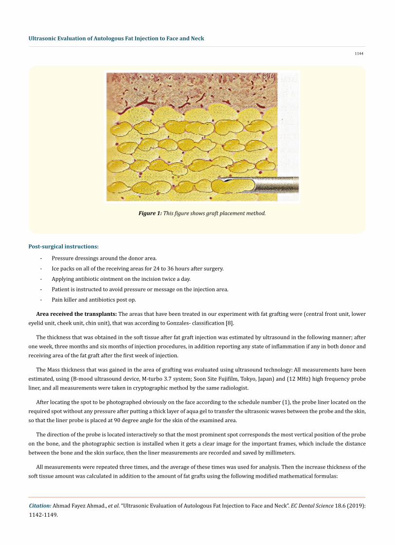
Graft placement method: Several tunnels were created to deposited small fat packs, and only by pulling the cannula off. This method achieves large contacts surface with capillaries which increases the graft viability to the maximum where the fat was injected according to cross and overlapping levels (first subcutaneous in a deep level then superficial subcutaneous) using a 7 - 9 mm length and 16- blunt cannula then the fatwas injected slowly with an over-correction not extremely.
Citation: Ahmad Fayez Ahmad., et al. “Ultrasonic Evaluation of Autologous Fat Injection to Face and Neck”. EC Dental Science 18.6 (2019): 1142-1149.
Ultrasonic Evaluation of Autologous Fat Injection to Face and Neck
1144
Figure 1: This figure shows graft placement method.
Post-surgical instructions:
- Pressure dressings around the donor area.
- Ice packs on all of the receiving areas for 24 to 36 hours after surgery.
- Applying antibiotic ointment on the incision twice a day.
- Patient is instructed to avoid pressure or message on the injection area.
- Pain killer and antibiotics post op.
Area received the transplants: The areas that have been treated in our experiment with fat grafting were (central front unit, lower eyelid unit, cheek unit, chin unit), that was according to Gonzales- classification [8].
The thickness that was obtained in the soft tissue after fat graft injection was estimated by ultrasound in the following manner; after one week, three months and six months of injection procedures, in addition reporting any state of inflammation if any in both donor and receiving area of the fat graft after the first week of injection.
The Mass thickness that was gained in the area of grafting was evaluated using ultrasound technology: All measurements have been estimated, using (B-mood ultrasound device, M-turbo 3.7 system; Soon Site Fujifilm, Tokyo, Japan) and (12 MHz) high frequency probe liner, and all measurements were taken in cryptographic method by the same radiologist.
After locating the spot to be photographed obviously on the face according to the schedule number (1), the probe liner located on the required spot without any pressure after putting a thick layer of aqua gel to transfer the ultrasonic waves between the probe and the skin, so that the liner probe is placed at 90 degree angle for the skin of the examined area.
The direction of the probe is located interactively so that the most prominent spot corresponds the most vertical position of the probe on the bone, and the photographic section is installed when it gets a clear image for the important frames, which include the distance between the bone and the skin surface, then the liner measurements are recorded and saved by millimeters.
All measurements were repeated three times, and the average of these times was used for analysis. Then the increase thickness of the soft tissue amount was calculated in addition to the amount of fat grafts using the following modified mathematical formulas:

Citation: Ahmad Fayez Ahmad., et al. “Ultrasonic Evaluation of Autologous Fat Injection to Face and Neck”. EC Dental Science 18.6 (2019): 1142-1149.
Ultrasonic Evaluation of Autologous Fat Injection to Face and Neck
1145
Increase the thickness of the soft tissue = (the thickness of the soft tissue after injection-the sickness of the soft tissue after injection)/ the thickness of the soft tissue before injection (x100).
The amount of remaining fat graft = (the thickness of the soft tissue in each post-injection period except after one week of injection)/(the thickness of the soft tissue after one week of injection) x 100.
Figure 2: This figure shows the gonzales classification.
Figure 3: An illustration showing the locations of the anatomical points.

Citation: Ahmad Fayez Ahmad., et al. “Ultrasonic Evaluation of Autologous Fat Injection to Face and Neck”. EC Dental Science 18.6 (2019): 1142-1149.
Ultrasonic Evaluation of Autologous Fat Injection to Face and Neck
1146
Fat grafting receiving area Anatomical point Description Forehead zone Glabella (GL) Above the middle point between the eyebrows 10 mm
Palpebra inferior zone Inferior eye orbit (OR) Middle inferior orbital Malar zone Zygomatic (ZY) Down line convergence of the eye lateral to Zygomatic arc
Mental (chin) zone Mental Eminence (ME) The most salience point in the chin
Table 1: Shows the anatomical points approved for ultrasound measurements (Manhein., et al. 2000).
Results
Time periodThe areasIncrease the thickness of the soft tissueThe amount of remaining fat graft
After weekLower eyelid unit208.24%Central front unit553.49%
Chin unit400.00%Cheek unit165.98%
Total331.92%After three monthsLower eyelid unit17.06%37.98%
Central front unit53.49%23.49%Chin unit81.82%36.36%
Cheek unit22.41%46.02%Total43.69%64.75%
After six
months
Lower eyelid unit-34.71%55.78%Central front unit-32.56%43.94%
Chin unit2.60%56.43%Cheek unit-33.20%54.58%
Total-24.47%45.28%
Discussion of results
Graft survival
Although the clinical experience with fat grafts is remarkable positive, yet there is limited quality experimental evidence that proves that the transferred fat survives. Coleman believes that the graft volume stabilizes at 3 - 4 months, although a subtle volumetric decrease may occur up to 1 year; beyond that, he claims that the volume remains constant for 8 - 12 years [9-12].
THE result of our study showed that the absorption rate in the injected fat graft reached 35.25% after three month of injection and 54.72% after six months.
While Hörl’s study showed that a reduction in graft volume of approximately 50% was demonstrated at 3 months, increasing to 55% at 6 months, and remaining stable thereafter until 12 months after re-implantation. But Hörl., et al. 54 used magnetic resonance imaging to document volume changes after autologous fat graft survival for correction of facial defects [13] and he depended on the procedures which Coleman recommended to prepare and inject the fat graft.

Citation: Ahmad Fayez Ahmad., et al. “Ultrasonic Evaluation of Autologous Fat Injection to Face and Neck”. EC Dental Science 18.6 (2019): 1142-1149.
Ultrasonic Evaluation of Autologous Fat Injection to Face and Neck
1147
Denadai’s following method, was the most similar to our method in studying the fat graft, and others [14] to treat Parry-Romberg syn-drome patients, where they depended in their study on observing the injected fat graft using ultrasound and calculate the fat graft volume in the same way followed in our research, to consume with results that after six months of injection the absorption amount of fat graft reaches 32.3%. The low absorption rate can be explained of their most research sample which includes children, where their research that include children supported the low absorption rate of fat graft and this hypothesis is supported by Raposo-Amaral CE., et al. [15] and Guibert M and Fanchi G., et al [16].
Our study agrees with Xie Y’s study and others in terms of injected fat graft absorption rate. To show that absorption rate is 47.92% 6 months later, where fat grafts were applied on facial hemiatrophy patients, and volumetric changes of injected fat graft were observed by holography using laser camera as he depended on low sedimentation speed. It showed high satisfaction rate after three months to decrease after 12 months and the study didn’t show any complications in donor or receiving area [17].
The result s of our research differed from Rieck and Schlaak’s research, which showed 30% of survival rate after 6 months of injection when it hypodermoclysis. But it remains only 6% after six months when it is injected into muscles, and this difference can be explained because their studies were performed in different areas on mice (hypodermic and muscles). They depended on cell-labelling technique for studying the fat graft survival rate [18].
We disagree with Bachhav, M and Agrawal, K and others, which showed that absorption rate is 21.85% of the absorption volume after 12 months of the injection procedure and it is possible that the low absorption rate related to low sedimentation speed, which used in the research (500 rpm). At this study, the researchers relied on MRI and photographs of the face to determine volumetric changes in the autologous fat graft that is used to correct the post-traumatic facial defects [19]. Nelson and Stewart’s study showed absorption rate after six months was 29% and 43% after 12 months, where the study included 46 patients with HIV-associated lipodystrophy for 3 years by autologous fat transfer and artificial filling substance injection and the observation is done by holographic techniques.
In 2016, Jiang and others had proceeded a research about Romberg syndrome treatment using autologous fat transfer technique, the study included 13 patients who underwent autologous fat transfer twice by three to six months. The observation done by holographic techniques to evaluate the injected fat graft volume. Researchers depended on our method in different sedimentation speed 300 rpm. Injected fat survival rate was 75.1% in the second time, while fat graft survival rate was much higher than the first one 43.3%.
Timing of subsequent injection
Additional procedures always deemed necessary to achieve an optimal result since the overall absorption rate of fat grafting reached 54.72% after 6 months of injection according to our study, and ranges from 50 to 90% according to several studies [5,20,21].
We also noticed in our study that patients’ level of satisfaction decreases aesthetically 6 months post-transplantation. So, our research indicates the importance of the subsequent injection after 6 months of the first injection, and so we agree with [22] who indicate the tim-ing of additional fat grafting sessions should be deferred until 6 months postoperatively to diminish the inflammatory response.
However, no scientific study has addressed the timing of subsequent fat grafting and therefore, only expert opinion has been mentioned in the literature regarding this specific issue.
Conclusions
- The autologous fat transfer technique can be considered an effective technique in managing the injuries of Maxillofacial trauma.
- The reliance on Coleman’s recommendations for the harvesting, preparation and injection of lipid taste provides good results, but we support excessive over-correction.
- The next injection after six months of the first injection.

Citation: Ahmad Fayez Ahmad., et al. “Ultrasonic Evaluation of Autologous Fat Injection to Face and Neck”. EC Dental Science 18.6 (2019): 1142-1149.
Ultrasonic Evaluation of Autologous Fat Injection to Face and Neck
1148
Recommendations
The results of this study recommend the following:
- Adopt the autologous fat transfer technique to correct the soft tissues caused by the trauma.
- Dependence on ultrasound technology.
- Work on the study of functional improvement in the injection area.
Bibliography
1. Neuber F. “Fat transplantation”. Chir Kongr Verhandlungen der Deutschen Gesellschaft für Chirurgie 22 (1893): 66.
2. Peer LA. “Loss of weight and volume inhuman fat grafts: with postulation of a “cell survival theory””. Plastic and Reconstructive Surgery 5 (1950): 217.
3. Illouz YG. “The fat cell “graft”. A new technique to fill depressions”. Plastic and Reconstructive Surgery 78.1 (1986): 122-123.
4. Coleman SR. “The technique of periorbital lipoin filtration”. Operative Techniques in Plastic and Reconstructive Surgery 1 (1997): 120-126.
5. Coleman SR. “Structural fat grafts: the ideal filler?” Clinics in Plastic Surgery 28.1 (2001): 111-119.
6. Bergara AR and Itoiz AO. “Present state of the surgical treatment of chronic frontal sinusitis”. Archives of Otolaryngology 61.6 (1955): 616-628.
7. Anderson OA., et al. “Periocular autologous Coleman fat graft survival and histopathology”. Ophthalmic Plastic and Reconstructive Surgery 24.3 (2008): 213-217.
8. Gonzalez-Ulloa M. “Restoration of the face covering by means of selected skin in regional aesthetic units”. British Journal of Plastic Surgery 9.3 (1956): 212-221.
9. Mojallal A. “Historical review of the use of adipose tissue transfer in plastic and reconstructive surgery”. Annales de Chirurgie Plastique Esthétique 49.5 (2004): 419-425.
10. Rohrich RJ., et al. “In search of improved fat transfer viability: A quantitative analysis of the role of centrifugation and harvest site”. Plastic and Reconstructive Surgery 113.1 (2004): 391-395.
11. Coleman SR. “Structural fat grafting: More than permanent filler”. Plastic and Reconstructive Surgery 118.3 (2006): 108-120.
12. Kaufman MR., et al. “Autologous fat transfer for facial recontouring: Is there science behind the art”. Plastic and Reconstructive Surgery 119.7 (2007): 2287-2296.
13. Hörl HW., et al. “Technique for liposuction fat reimplantation and long term volume evaluation by magnetic resonance imaging”. An-nals of Plastic Surgery 26.3 (1991): 248-258.
14. Denadai R., et al. “Predictors of autologous free fat graft retention in the management of craniofacial contour deformities”. Plastic and Reconstructive Surgery 140.1 (2017): 50e-61e.
15. Raposo-Amaral CE., et al. “Parry-Romberg syndrome: Severity of the deformity does not correlate with quality of life”. Aesthetic Plastic Surgery 37.4 (2013): 792-801.

Citation: Ahmad Fayez Ahmad., et al. “Ultrasonic Evaluation of Autologous Fat Injection to Face and Neck”. EC Dental Science 18.6 (2019): 1142-1149.
Ultrasonic Evaluation of Autologous Fat Injection to Face and Neck
1149
16. Guibert M., et al. “Fat graft transfer in children’s facial malformations: A prospective three dimensional evaluation”. Journal of Plastic Reconstructive and Aesthetic Surgery 66.6 (2013): 799-804.
17. Xie Y., et al. “Correction of hemifacial atrophy with autologous fat transplantation”. Annals of Plastic Surgery 59.6 (2007): 645-653.
18. Rieck B and Schlaak S. “Measurement in vivo of the survival rate in autologous adipocyte transplantation”. Plastic and Reconstructive Surgery 111.7 (2003): 2315-2323.
19. Agrawal K., et al. “Autologous Fat Transfer for Esthetic Contouring of Face in Posttraumatic Nonfunctional Maxillofacial Deformities”. Craniomaxillofacial Trauma and Reconstruction 9.2 (2015): 113-120.
20. Ramon Y., et al. “Enhancing the take of injected adipose tissue by a simple method for concentrating fat cells”. Plastic and Reconstruc-tive Surgery 115.1 (2005): 197-201.
21. Toledo LS. “Syringe liposculpture”. Clinics in Plastic Surgery 23.4 (1996): 683-693.
22. Pu LLQ. “Towards more rationalized approach to autologous fat grafting”. Journal of Plastic Reconstructive and Aesthetic Surgery 65.4 (2012): 413-419.
Volume 18 Issue 6 June 2019©All rights reserved by Ahmad Fayez Ahmad., et al.
Top Related