


Languages
Pages
Legal


CroniconO P E N A C C E S S EC PAEDIATRICS
Case Report
Meconium Ileus in a Female Neonate: A Case Report and Review of Literature
Volkan Sarper Erikci* and Merve Dilara ÖneyAssociate Professor, Department of Pediatric Surgery, Tepecik Training Hospital, Sağlık Bilimleri University, Izmir, Turkey
Citation: Volkan Sarper Erikci and Merve Dilara Öney. “Meconium Ileus in a Female Neonate: A Case Report and Review of Literature”. EC Paediatrics 8.4 (2019).
*Corresponding Author: Volkan Sarper Erikci, Associate Professor, Department of Pediatric Surgery, Tepecik Training Hospital, Sağlık Bilimleri University, Izmir, Turkey.
Received: February 26, 2019
AbstractMeconium ileus (MI) is a congenital form of mechanical intestinal obstruction caused by inspissated meconium at the level of
terminal ileum. Although MI was once considered to be closely associated with cystic fibrosis (CF), MI may also occur in the absence of CF as well. A 2-day-old newborn girl with MI who required surgical intervention for the evacuation of thick meconium is presented in this study. It is also aimed in this report to review the presentation, imaging findings and management of MI under the light of relevant literature. The health providers should keep this anomaly in mind and a prompt pediatric surgical consultation is recom-mended and the patient should be treated accordingly.
Keywords: Meconium Ileus (MI); cystic Fibrosis (CF)
IntroductionMeconium ileus (MI) is a congenital form of mechanical intestinal obstruction caused by inspissated meconium at the level of terminal
ileum [1,2]. Although MI was once considered to be closely associated with cystic fibrosis (CF), MI may also occur in the absence of CF as well [3,4]. Here we present a 2-day-old newborn girl with MI who required surgical intervention for the evacuation of thick meconium. It is also aimed in this report to review the presentation, imaging findings and management of MI under the light of relevant literature.
Case Report
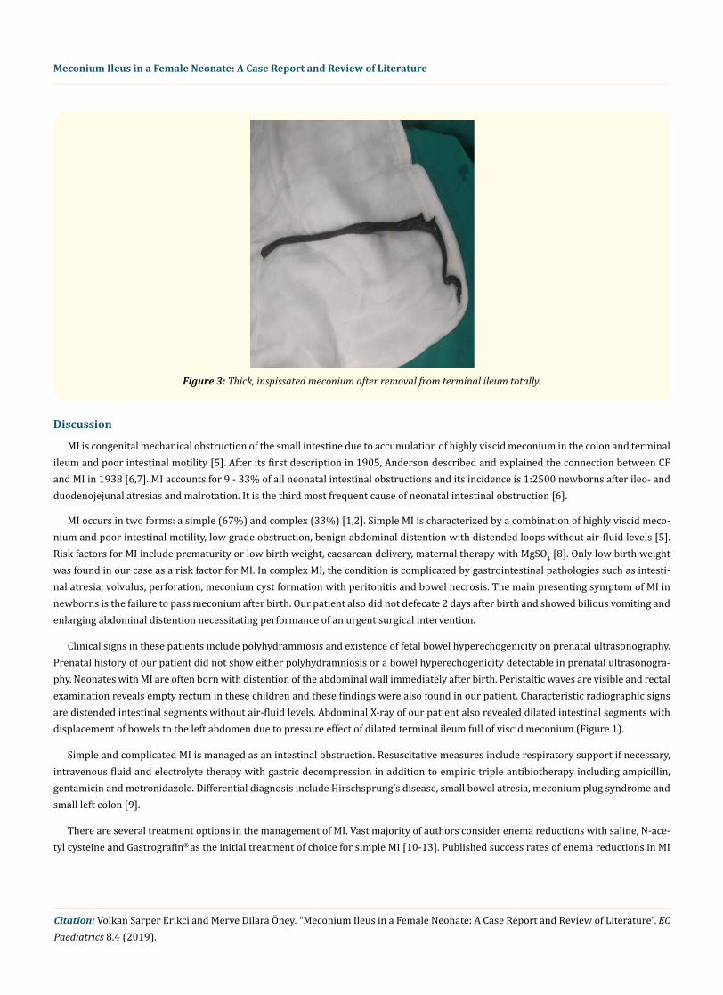
A 2-day-old female weighing 1990 g at 35 weeks gestation was born via caesarean section to a 32-year-old mother (G1P1). The patient was admitted to our department with abdominal distention, bilious emesis and failure to pass meconium since birth. Antenatal history was unremarkable. An erect abdominal X-ray showed dilated loops of small intestine with no passage of intestinal gas beyond large bowel without air-fluid levels (Figure 1). A water soluble enema study showed an unused colon. A nasogastric tube drainage failed to alleviate the obstruction. Initial medical management included restoration of adequate body temperature, hydration and electrolyte balance toge-ther with empiric antibiotic coverage including ampicillin, gentamicin and metronidazole and blood sample was taken for DNA analysis of the CFTR gene including mutations of ΔF508. Exploratory laparotomy was performed with a transverse upper abdominal incision. A dilated terminal ileum full of inspissated meconium with an unused colon was found (Figure 2). Following enterotomy 5 cm proximal to ileocecal valve, proximal irrigation of meconium using warm saline was carried out. Inspissated meconium was totally evacuated from the intestine (Figure 3). A loop ileostomy at the enterotomy site was performed. Bowel activity returned 5 days following surgery. The baby is gaining weight and the stoma closure is planned to be closed at 8 weeks post-surgery.

Citation: Volkan Sarper Erikci and Merve Dilara Öney. “Meconium Ileus in a Female Neonate: A Case Report and Review of Literature”. EC Paediatrics 8.4 (2019).
Meconium Ileus in a Female Neonate: A Case Report and Review of Literature
Figure 1: X-ray showing dilated loops of small intestines with no passage of intestinal gas beyond large bowel (Arrow showing displaced intestine into the left abdominal cavity).
Figure 2: Operative view showing dilated terminal ileum full of inspissated meconium. Note enterotomy was performed and the inspissated meconium is being removed.
Citation: Volkan Sarper Erikci and Merve Dilara Öney. “Meconium Ileus in a Female Neonate: A Case Report and Review of Literature”. EC Paediatrics 8.4 (2019).
Discussion
MI is congenital mechanical obstruction of the small intestine due to accumulation of highly viscid meconium in the colon and terminal ileum and poor intestinal motility [5]. After its first description in 1905, Anderson described and explained the connection between CF and MI in 1938 [6,7]. MI accounts for 9 - 33% of all neonatal intestinal obstructions and its incidence is 1:2500 newborns after ileo- and duodenojejunal atresias and malrotation. It is the third most frequent cause of neonatal intestinal obstruction [6].
MI occurs in two forms: a simple (67%) and complex (33%) [1,2]. Simple MI is characterized by a combination of highly viscid meco-nium and poor intestinal motility, low grade obstruction, benign abdominal distention with distended loops without air-fluid levels [5]. Risk factors for MI include prematurity or low birth weight, caesarean delivery, maternal therapy with MgSO4 [8]. Only low birth weight was found in our case as a risk factor for MI. In complex MI, the condition is complicated by gastrointestinal pathologies such as intesti-nal atresia, volvulus, perforation, meconium cyst formation with peritonitis and bowel necrosis. The main presenting symptom of MI in newborns is the failure to pass meconium after birth. Our patient also did not defecate 2 days after birth and showed bilious vomiting and enlarging abdominal distention necessitating performance of an urgent surgical intervention.
Clinical signs in these patients include polyhydramniosis and existence of fetal bowel hyperechogenicity on prenatal ultrasonography. Prenatal history of our patient did not show either polyhydramniosis or a bowel hyperechogenicity detectable in prenatal ultrasonogra-phy. Neonates with MI are often born with distention of the abdominal wall immediately after birth. Peristaltic waves are visible and rectal examination reveals empty rectum in these children and these findings were also found in our patient. Characteristic radiographic signs are distended intestinal segments without air-fluid levels. Abdominal X-ray of our patient also revealed dilated intestinal segments with displacement of bowels to the left abdomen due to pressure effect of dilated terminal ileum full of viscid meconium (Figure 1).
Simple and complicated MI is managed as an intestinal obstruction. Resuscitative measures include respiratory support if necessary, intravenous fluid and electrolyte therapy with gastric decompression in addition to empiric triple antibiotherapy including ampicillin, gentamicin and metronidazole. Differential diagnosis include Hirschsprung’s disease, small bowel atresia, meconium plug syndrome and small left colon [9].
There are several treatment options in the management of MI. Vast majority of authors consider enema reductions with saline, N-ace-tyl cysteine and Gastrografin® as the initial treatment of choice for simple MI [10-13]. Published success rates of enema reductions in MI
Figure 3: Thick, inspissated meconium after removal from terminal ileum totally.
Meconium Ileus in a Female Neonate: A Case Report and Review of Literature

Citation: Volkan Sarper Erikci and Merve Dilara Öney. “Meconium Ileus in a Female Neonate: A Case Report and Review of Literature”. EC Paediatrics 8.4 (2019).
ranges between 5 - 83% [10-17]. This treatment option was not used in our case due to progressive abdominal distention, clinical deteri-oration and an urgent surgical intervention was performed.
The goal of operative management in MI is to evacuate meconium from the intestine while preserving maximum intestinal length. Vari-ous surgical procedures have been proposed. In 1971 Kalayoğlu., et al. reported the first successful procedure for uncomplicated MI [18]. Resection of the dilated segment of the bowel and creation of a double barrelled Mikulicz type enterostomy followed by postoperative irrigations of the distal limb with pancreatic enzymes have also been suggested by Gross [19]. Bishop and Koop suggested the resection of the dilated intestine and creation of a Roux-en-Y ileostomy with exteriorization of the distal limb which decreases the risk of electrolyte imbalance and may avoid additional surgery [20]. T-tube enterostomy with postoperative irrigation was recommended by O’Neill without bowel resection. Ileotomy through a purse-string suture with irrigations using acetyl cysteine was recommended by Venugopal [21]. In recent years, for simple MI unresponsive to enemas, either primary resection-irrigation and anastomosis or T-tube ileostomy with irrigation have been proposed in the management of these sick children [13,16,22,23]. In our patient due to clinical deterioration and rather unstable clinical parameters, urgent surgical intervention was performed and following enterotomy and evacuation of inspissated meconium, simple loop ileostomy was performed in order to decrease operative time. She is now well and gaining weight and the stomal closure will be performed at the 7 - 8 postoperative week.
Conclusion
In conclusion, surgical intervention in simple uncomplicated MI, unresponsive to enemas or in children with clinical deterioration, is safe. Laparotomy and evacuation of inspissated meconium followed by ileostomy is useful in the management of these children. Evacuati-on of thick and viscid meconium will accelerate intestinal activity and promote early oral feeding. The health providers dealing with such kinds of patients should keep this anomaly in mind and a prompt pediatric surgical consultation is recommended and the patient should be treated accordingly.
Bibliography
1. Wyllie R. “Intestinal atresiai stenosis and malrotation”. In: Kliegmann RM, Behrman RE, Jenson HB, Stanton BF., et al. (eds) Nelson textbook of pediatrics, 18th edition. Saundres Elsevier, Philadelphia (2007): 1559-1562.
2. Ziegler MM. “Meconium ileus”. Current Problems in Surgery 31.9 (1994): 731-777.
3. Gorter RR., et al. “Clinical and genetic characteristics of meconium ileus in newborns with and without cystic fibrosis”. Journal of Pediatric Gastroenterology 50.5 (2010): 569-572.
4. Fakhoury K., et al. “Meconium ileus in the absence of cystic fibrosis”. Archives of Disease in Childhood 67.10 (1992): 1204-1206.
5. Paradiso VF., et al. “Meconium obstruction in absence of cystic fibrosis in low birth weight infants: an emerging challenge from in-creasing survival”. Italian Journal of Pediatrics 37 (2011): 55.
6. Rivosecchi M. “Meconium ileus”. In: Puri P, Höllwart M eds. Pediatric Surgery Diagnosis and Management. Berlin: Springer-Verlag (2009): 415-422.
7. Ziegler MM. “Meconium ileus”. In: O’Neill Ja, Coran AG, Fonkalsrud E, Grosfeld JL eds. Pediatric Surgery 6th edition. New York: Mosby (2006): 1289-1303.
8. Emil S., et al. “Meconium obstruction in extremely low birth weight neonates: guidelines for diagnosis and management”. Journal of Pediatric Surgery 39.5 (2000): 731-737.
9. Kiely E. “Meconium ileus”. In: Puri P editor. Newborn Surgery. USA: Taylor & Francis Group, LLC (2003): 465-471.
10. Noblett HR. “Treatment of uncomplicated meconium ileus by Gastrografin ® enema: a preliminary report”. Journal of Pediatric Sur-gery 4.2 (1969): 190-197.
Meconium Ileus in a Female Neonate: A Case Report and Review of Literature

Citation: Volkan Sarper Erikci and Merve Dilara Öney. “Meconium Ileus in a Female Neonate: A Case Report and Review of Literature”. EC Paediatrics 8.4 (2019).
11. Burke MS., et al. “New strategies in non-operative management of meconium ileus”. Journal of Pediatric Surgery 37.5 (2002): 760-764.
12. Boyd A., et al. “Gastrografin® enema in meconium ileus: the persistent approach”. Pediatric Surgery International 3.2-3 (1988): 139-140.
13. Rescorla FJ and Grosfeld JL. “Contemporary management of meconium ileus”. World Journal of Surgery 17.3 (1993): 318-325.
14. Ein SH., et al. “Bowel perforation with non operative treatment of meconium ileus”. Journal of Pediatric Surgery 22.2 (1987): 146-147.
15. Copeland DR., et al. “Diminishing role of contrast enema in simple meconium ileus”. Journal of Pediatric Surgery 44.11 (2009): 2130-2132.
16. Nguyen LT., et al. “Meconium ileus: is stoma necessary?” Journal of Pediatric Surgery 21.9 (1986): 766-768.
17. D’Agostino S., et al. “Uncomplicated meconium ileus: efficacy of enterotomy and bowel irrigation”. Pediatric Surgery International 10.5-6 (1995): 329-331.
18. Kalayoğlu M., et al. “Meconium ileus: a critical review of treatment and eventual prognosis”. Journal of Pediatric Surgery 6.3 (1971): 290-300.
19. Gross RE. “Intestinal obstruction in the newborn resulting from meconium ileus”. In: The Surgery of Infancy and Childhood. Philadel-phia: WB Saunders Co., (1953): 175-191.
20. Bishop HC and Koop CE. “Management of meconium ileus: resection, Roux-en-Y anastomosis and ileostomy irrigation with pancreatic enzymes”. Annals of Surgery 145.3 (1957): 410-414.
21. Venugopal S and Shandling B. “Meconium ileus: laparotomy without resection, anastomosis, or enterostomy”. Journal of Pediatric Surgery 14.6 (1979): 715-718.
22. Rescorla FJ., et al. “Changing patterns of treatment and survival in neonates with meconium ileus”. Archives of Surgery 124.7 (1989): 837-840.
23. Mak GZ., et al. “T-tube ileostomy for meconium ileus: four decades of experience”. Journal of Pediatric Surgery 35.2 (2000): 349-352.
Volume 8 Issue 4 April 2019©All rights reserved by Volkan Sarper Erikci and Merve Dilara Öney.
Meconium Ileus in a Female Neonate: A Case Report and Review of Literature
Top Related