


Languages
Pages
Legal


HAL Id: hal-00563042https://hal.archives-ouvertes.fr/hal-00563042
Submitted on 4 Feb 2011
HAL is a multi-disciplinary open accessarchive for the deposit and dissemination of sci-entific research documents, whether they are pub-lished or not. The documents may come fromteaching and research institutions in France orabroad, or from public or private research centers.
L’archive ouverte pluridisciplinaire HAL, estdestinée au dépôt et à la diffusion de documentsscientifiques de niveau recherche, publiés ou non,émanant des établissements d’enseignement et derecherche français ou étrangers, des laboratoirespublics ou privés.
Correlations between gastropod shell dissolution andwater chemical properties in a tropical estuary
David J. Marshall, Jose H. Santos, Kenneth M.Y. Leung, Wang H. Chak
To cite this version:David J. Marshall, Jose H. Santos, Kenneth M.Y. Leung, Wang H. Chak. Correlations betweengastropod shell dissolution and water chemical properties in a tropical estuary. Marine EnvironmentalResearch, Elsevier, 2008, 66 (4), pp.422. �10.1016/j.marenvres.2008.07.003�. �hal-00563042�

Accepted Manuscript
Correlations between gastropod shell dissolution and water chemical properties
in a tropical estuary
David J. Marshall, Jose H. Santos, Kenneth M.Y. Leung, Wang H. Chak
PII: S0141-1136(08)00179-7
DOI: 10.1016/j.marenvres.2008.07.003
Reference: MERE 3269
To appear in: Marine Environmental Research
Received Date: 4 December 2007
Revised Date: 3 July 2008
Accepted Date: 5 July 2008
Please cite this article as: Marshall, D.J., Santos, J.H., Leung, K.M.Y., Chak, W.H., Correlations between gastropod
shell dissolution and water chemical properties in a tropical estuary, Marine Environmental Research (2008), doi:
10.1016/j.marenvres.2008.07.003
This is a PDF file of an unedited manuscript that has been accepted for publication. As a service to our customers
we are providing this early version of the manuscript. The manuscript will undergo copyediting, typesetting, and
review of the resulting proof before it is published in its final form. Please note that during the production process
errors may be discovered which could affect the content, and all legal disclaimers that apply to the journal pertain.

ACCEPTED MANUSCRIPT
Correlations between gastropod shell dissolution and water chemical
properties in a tropical estuary
David J. Marshalla*, Jose H. Santosc , Kenneth M. Y. Leungb and Wang H. Chaka
a Department of Biology, Universiti Brunei Darussalam, Tungku Link, Gadong BE1410,
Brunei Darussalam b The Swire Institute of Marine Science and Division of Ecology & Biodiversity, School
of Biological Sciences, The University of Hong Kong, Pokfulam, Hong Kong, P. R.
China c Department of Chemistry, Universiti Brunei Darussalam, Tungku Link, Gadong
BE1410, Brunei Darussalam
*Corresponding author:
Dr. David J. Marshall, Department of Biology, Universiti Brunei Darussalam, Tungku
Link, Gadong BE 1410, Brunei Darussalam. Telephone: +673-2463001; Fax: +673 -
2461502; e-mail: [email protected]

ACCEPTED MANUSCRIPT
2
Abstract
Although poorly reported in the scientific literature, acidic waters characterize many
South East Asian estuaries. The observation of shell dissolution in a typically marine
gastropod whelk (Thais) prompted investigation into determining relationships between
shell properties of this whelk and the water chemistry (including pH) of the Sungai
Brunei estuary (Borneo) in which it occurs. Shell weight, shell length and topographical
shell features were determined for populations of Thais gradata distributed along a
gradient of pH and salinity ranging between 5.78 and 8.3 pH units, and 3.58 and 31.2 psu.
Shell weight varied independently of the co-varying acidity, salinity and calcium levels
experienced. In contrast, shell length and a semi-quantitative variable based on shell
sculpturing (shell erosion rank, SER) were significantly correlated with these water
chemistry variables. This study brings attention to the potential use of estuarine
organisms and systems in investigating current marine acidification questions.
Keywords: Acidification; Calcium carbonate; Estuaries; Gastropods; Thais; pH
ACCEPTED MANUSCRIPT
3
1. Introduction
Acidification of marine systems, notably that relating to elevated atmospheric carbon
dioxide (CO2) and the oceanic carbonate equilibrium, has attracted considerable recent
interest (Broecker, Takahashi, Simpson & Peng, 1979; Feely, Sabine, Lee, Millero,
Lamb, Greeley et al., 2002; Caldeira & Wickett, 2003; Orr, Fabry, Aumont, Bopp,
Doney, Feely et al., 2005). Current prediction of fairly rapid acidification of the ocean
surface waters (decades; Orr et al., 2005) is disturbing in the context of our limited
understanding of how organisms and ecological systems might respond to this (Seibel &
Walsh, 2003; Pörtner, Langenbuch & Reipschläger, 2004; Shirayama & Thornton, 2005).
Predictably, organisms with reduced pH tolerance or with calcium carbonate skeletal
structures (such as, coralline algae, corals, echinoderms, molluscs) will be most affected
(Pörtner et al., 2004; Jokiel, Rodgers, Kuffner, Andersson, Cox & Mackenzie, 2008;
Kuffner, Andersson, Jokiel, Rodgers & Mackenzie, 2008; Hall-Spencer, Rodolfo-
Metalpa, Martin, Ransome, Fine, Turner et al., 2008), though community cascading is
likely to cause local ecological devastation, especially in the case of coral reef systems
(Pelejero, Calvo, McCulloch, Marshall, Gagan, Lough et al., 2005). In the above context,
investigations into environmental circumstances leading to carbonate undersaturation and
consequently to dissolution of shells and skeletons in marine ecosystems are highly
relevant and important.
Although the effects of environmental CO2 and pH change on organismal
functions (growth, physiology and toxicity) are becoming better known (Yamada &
Ikeda, 1999; Hinga, 2002; Seibel & Walsh, 2003; Michaelidis, Ouzounis, Paleras &
Pörtner, 2005; Shirayama & Thornton, 2005; Berge, Bjerkeng, Pettersen, Schaanning &
Øxnevad, 2006) information on in situ shell dissolution of oceanic organisms is limited to
pelagic pteropod (Clios) and heteropod molluscs (Orr et al., 2005). In littoral systems,

ACCEPTED MANUSCRIPT
4
shell dissolution studies have focused on unicellular foraminiferans which are used to
assess current and historical environmental perturbations (Geslin, Debenay, Duleba &
Bonetti, 2002; Hayward, Grenfell, Nicholson, Parker, Wilmhurst, Horrocks et al., 2004).
While the above is not necessarily all embracing from an ecological perspective of shell
dissolution, only a single known study concerns in situ dissolution for gastropods in
coastal systems (Hall-Spencer et al., 2008).
Acidification is generally overlooked in coastal ecological studies, despite its
prevalence in estuarine systems (Howland, Tappin, Uncles & Plummer, 2000; Braga,
Bonetti, Burone & Filho, 2000; Abril, Etcheber, Delille, Frankignoulle & Borges, 2003;
Lin, Wood, Haskins, Ryffel & Lin, 2004; García-Luque, Forja & Gómez-Parra, 2005).
While lowered salinities reduce the buffering capabilities of estuaries, acidification can
derive from high natural and or anthropogenic organic inflows through elevated
microbial activity (Mirlean, Baraj, Niencheski, Baisch & Robinson, 2001; Abril et al.,
2003; Braungardt, Achterberg, Elbaz-Poulichet & Morley, 2003; Lin et al., 2004; Powell
& Martens, 2005, Zai, Dai, Cai, Wang & Wang, 2005). Heterotrophic microbial
metabolism significantly increases acidity through the production of CO2 and carbonic
acid in the muddy substratum of estuaries, and in some cases, bacterioplankton may
account for 60-100% of planktonic oxygen consumption (Jonas, 1997). Acidification is
exacerbated in turbid eutrophic estuaries as a consequence of reduced photosynthetic
activity and biological withdrawal of CO2 (Abril et al., 2003). Although the waters
feeding the Sungai Brunei estuary (Brunei Darussalam, Borneo, South East Asia) are
naturally acidic, biogenic CO2 production is implied by the combination of extraordinary
natural and anthropogenic organic loads, lowered dissolved oxygen levels, elevated
dissolved CO2 levels and highly turbid conditions (Chua, Chou & Sadorra, 1987; DJM,
unpublished).
ACCEPTED MANUSCRIPT
5
This study stemmed from the observation of shell dissolution in a gastropod
whelk (Thais gradata (Jonas)) which inhabits the Sungai Brunei estuarine system. We
aimed to derive relationships between shell dissolution properties of this whelk and water
chemical attributes underlying water carbonate saturation of the estuary (particularly pH,
calcium and salinity), and to determine which measurable aspects of the shell were most
sensitive to dissolution. This study highlights the use of estuarine organisms and estuaries
to address acidification questions relating to a broader marine context.
2. Materials and methods
2.1. Estuary and the snails
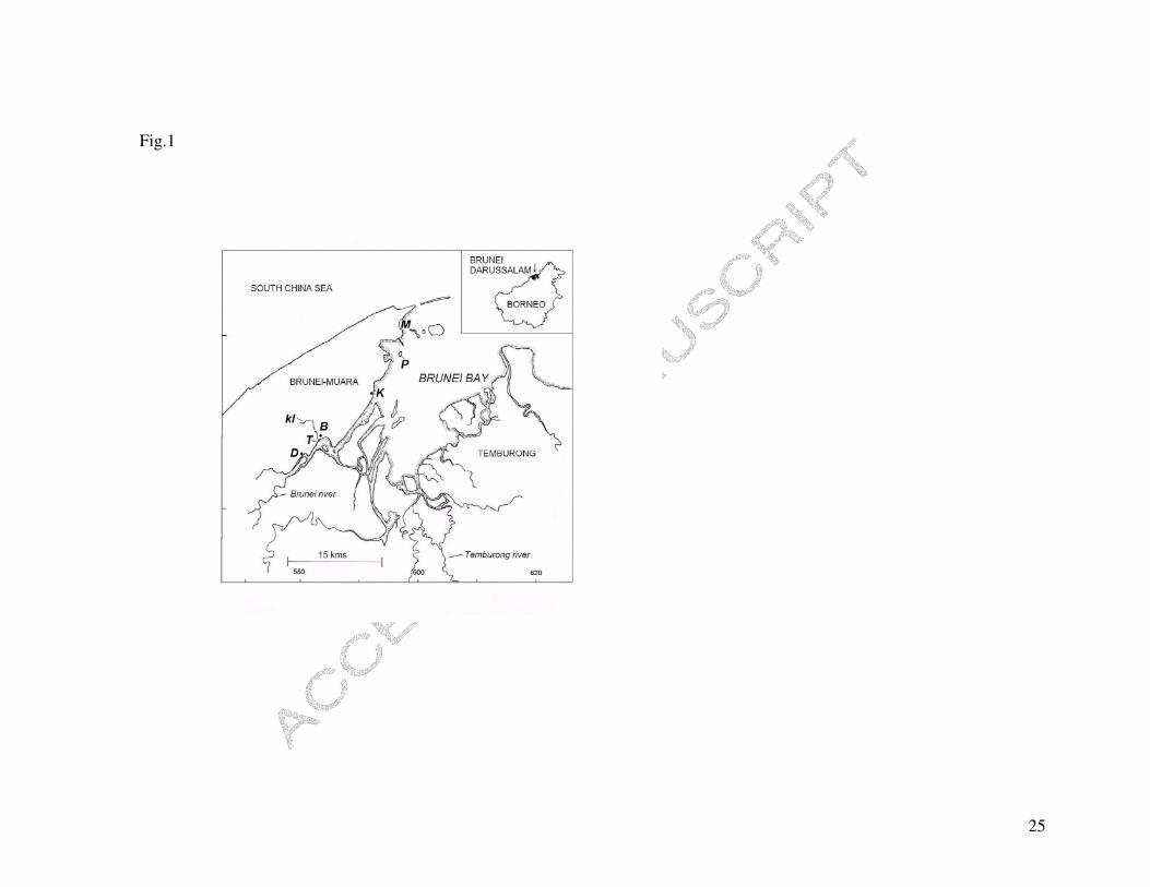
The Sungai Brunei estuarine and bay system covers an area of 1380 km2, with the inner
bay being fed by several large rivers in Brunei Darussalam (Sungai Brunei and
Temburong) and Sarawak, Malaysia (Sungai Limbang and Trusan; Fig. 1; Chua et al.,
1987). Waterways are fringed with extensive mangrove stands. The estuary is distinctly
brown and turbid, and carries a high organic load deriving from the mangroves and urban
centres (Bandar, Kiulap and Gadong; Chua et al., 1987). It has been central to the
lifestyles of the Bruneian people for the past four centuries, and currently supports one of
the oldest water villages in South East Asia (approximately 29 000 people living in stilt
houses over the water).
The gastropod whelk, Thais gradata (Jonas) is endemic to southern China and the
Malay Archipelago (Tan, 1999). Although typically an inhabitant of mangrove trees, in
the Brunei estuary it is highly abundant on artificial embankments and piers, built to
retain waters and to facilitate small-craft commutation. A congeneric estuarine species, T.
malayensis, thought to occur in Sungai Brunei estuary, is distributed across South East
Asia and northern Australia (Tan & Sigurdsson, 1996).

ACCEPTED MANUSCRIPT
6
2.2. Water chemistry
Four recording stations were established along the Sungai Brunei estuary, to cover the
range of physicochemical conditions that characterize it (Damuan, D, low salinity,
04°52’05.6”N, 114°54’37.6”E; Bandar, B, low salinity 04°53’09.3”N, 114°56’53.2”E;
Kota Batu, K, midrange salinity, 04°55’42.2”N, 115°00’54.3”E and Muara, M, high
salinity, 05°01’01.1”N, 115°03’57.8”E; Fig. 1). Water samples were collected from each
station on 24 occasions (13 during November 2005 to March 2006 and 11 during August to
November 2007). Sampling included a range of tidal conditions and, on any day, all
stations were visited in the same order in a 2 h timeframe. Duplicate air-tight samples
(allowing no head space) were collected from the upper 1 m column adjacent to Thais
habitat, using 1000 mL acid-washed, polythene jars. Samples were returned to the
laboratory in 1 h of completing the sampling series, and were equilibrated at 28°C for 1 h in
a Grant constant temperature water-bath. Field water temperature was relatively constant,
varying between 27.9 and 33.6°C (Fluke thermocouple thermometer 54 II). Salinity and pH
were determined simultaneously at 28°C; salinity to ± 0.01 psu (YSI Model 85D multi-
meter or HACH HQ40d meter and IntelliCal probe) and pH to ± 0.01 units using an NBS
scale (Thermo Orion Model 260A or HACH HQ40d calibrated with Mettler Toledo SRM
NIST precision buffers). Laboratory measured salinity and pH were invariable to in situ
field measurements taken on five occasions. For the 2007 samples, 100 mL sub-samples
were treated with concentrated nitric acid for calcium analysis. Calcium concentrations of
these samples were determined using Shimadzu AA-6701F atomic absorption
spectrophotometer in direct injection mode utilising acetylene-air fuel mixture. Total
alkalinity was determined for a separate set of samples collected on a single day (27 June
2008, N = 12). Determination involved using potentiometric titration and double-indicator
titration employing phenolphthalein and methyl orange indicators. A titrant of standard HCl
ACCEPTED MANUSCRIPT
7
solution containing 0.7 mol L-1 NaCl was used to adjust the total ionic strength to
approximate that of the seawater.
2.3. Shell weight, shell length and aperture relationships
Snails (N = 377) were collected from four populations (Tamoi, T; Bandar, B; Kota Batu,
K and Pulau Bedukang, P; Fig. 1) during July to December 2005. Populations B and K
corresponded with the water sampling stations. However, because T and P were difficult
to access rapidly for water sampling, this was undertaken at nearby stations D (where
snails occurred at low abundance) and M (where no snails occurred; Fig. 1), which
showed similar water conditions respectively to those where snails were collected. Snails
were haphazardly collected in a manner to include a broad size range. Shells supporting
epizoic barnacles were avoided, except population T which mostly possessed barnacles.
Shell scrubbing and removal of epibionts preceded all determinations. To avoid
microenvironment effects of mud-bank water, snails were collected from vertical hard
substrata, their preferred habitat which drains rapidly during low tides.
Shell length, shell weight, aperture length, and shell structural features were
determined in the laboratory. Shell length and aperture length (as a proxy for growth and
age) were measured using digital Vernier calipers (precision: 0.01 mm; see Fig. 2).
Qualitative features of the shells were assessed by a method described below. Shells were
then cracked open with a bench vice to separate shell material from the tissue, and the
shell material was dried at 60°C for 3 d to achieve a constant weight, determined to 0.001
g with a Mettler Toledo PB303. We determined how shell weight and shell length were
related to aperture length and, whether and how the populations differed with respect to
these relationships (General Linear Models, Statistica Version 6, Statsoft Inc.). Because
preliminary observations showed that smaller shells elongate more rapidly while

ACCEPTED MANUSCRIPT
8
remaining thin, causing a non-linear shell weight to shell elongation relationship, shells
having an aperture shorter than 15 mm long were excluded from the analyses. Each
model comprised a categorical predictor (snail population with four levels; T, B, K and
P), a dependent variable (shell weight or shell length), and continuous variable (aperture).
All regressions conformed to assumptions of normality, heteroscedasticity, and
homogeneity of variance (Cochran’s test).
2.4. Shell erosion rank (SER)
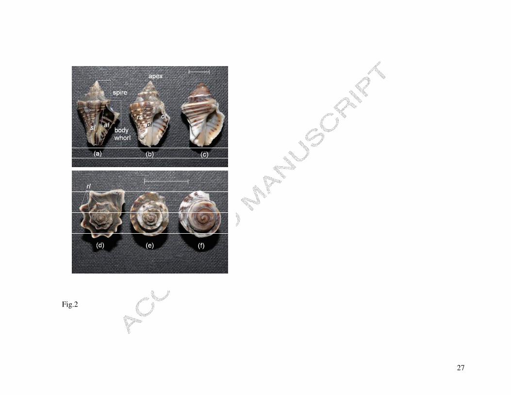
The surface of non-eroded Thais shells is characterized by an outer periostracum growing
over fine axial riblets which interconnect with spiral riblets to form cancellate patterning
(axial riblets and the periostracum are not readily discernible in Fig. 2). Dissolution
causes the sequential loss of these features in the above order. Because the apex (being
the oldest part of the shell) is always eroded and the outer lip (comprising the most
recently deposited shell) is usually intact, these parts were excluded from the shell
assessment. Extreme dissolution leads to smoothing of the shell (loss of periostracum,
axial riblets and spiral riblets) and rounding of the apex, longitudinal ribs (or spines) and
whorl shoulders (Fig. 2c and 2f). Although Tan & Sigurdsson (1996) suggested that the
development of spines is individually variable, spines were conspicuous in all individuals
of population P (Fig. 2).
Each snail was ranked according to the following criteria. Rank 1 (non-erosion):
periostracum and axial riblets distinct on most of the shell, and longitudinal ribs on spire
whorls, pronounced (Fig. 2a and 2d). Rank 2: longitudinal ribs on spire whorls, eroded
and indistinct; spiral and axial riblets on body whorl adjacent to the parietal lip, distinct
(Fig. 2b and 2e). Rank 3: axial and spiral riblets directly adjacent to parietal lip eroded,
but conspicuous elsewhere on the body whorl (especially on the surface opposite the

ACCEPTED MANUSCRIPT
9
aperture, not shown in figure). Rank 4: axial and spiral riblets eroded on entire body
whorl (Fig. 2c and 2f). Aperture length, as an assessment of size, was determined along
with the SER for each shell for N = 33 for each population. Because very small shells had
experienced limited acidic exposure, only shells > 20 mm were used in SER
determinations.
3. Results
3.1. Water chemistry determinations
The stations differed markedly in pH, salinity and calcium regime; mean values showed a
decreasing trend from the seaward to the landward stations (Stn. M > K > B � D; Table
1). pH varied between 5.78 and 8.3 units and salinity between 3.8 and 31.2 psu, with
lowest and highest values for each variable recorded at stations B and M, respectively
(Table 1). The water chemistry was most variable at B and least variable at M (Table 1).
Relationships for the water chemical variables are: pH = 4.10 + 2.62 log10 salinity;
calcium = 21.25 + 12.65 salinity; R2 = 0.89, P < 0.001; Fig. 3). The spatial pattern of
water chemistry was consistent across stations (M > K > B � D) on each recording
session, but varied temporally on different days in relation to tidal flux and rainfall events
(Fig. 3). Preliminary data showed that total alkalinity was related to salinity as follows:
total alkalinity (mmol.L-1) = 0.884 + 0.033 salinity (psu), R2 = 0.94, P < 0.001.
3.2. Shell weight, shell length and aperture relationships
Relationships for shell weight against aperture length differed significantly among
populations; the landward populations generally had heavier shells (Fig. 4a; Table 2).
Shell length as a function of aperture also differed among the populations, with shorter
shells found in the landward populations, for the range of shell sizes examined (Fig. 4b;

ACCEPTED MANUSCRIPT
10
Table 2). The effect of size was removed by scaling data to a standard aperture length
(SAL) using the slope coefficient (b) for the individual regressions (see Fig. 4) as follows
(see Packard & Boardman, 1988):
Standard shell weight (or shell length) = (SAL / aperture length)b × shell weight (or shell
length)
SAL is the grand mean for aperture length, which for shell weight = 19.1 mm and for
shell length = 18.9 mm. Standardized shell weight was not significantly different between
populations B, K and P, but T was significantly greater than the others (Fig. 5). However,
standardized shell length increased progressively across populations, from landward to
seaward (Fig. 5). Population T thus comprises snails possessing not only the heaviest
shells, but also the shortest shells (Fig. 5). Significant positive correlations were found for
means of standard shell length against estuarine pH and calcium (pH = -2.25 + 0.36 shell
length, R2 = 0.96, P = 0.022; calcium = -865.32 + 43.09 shell length, R2 = 0.95, P =
0.027; Fig. 5c).
3.3 Shell erosion rank (SER)
Shell erosion rank (SER) differed strikingly among the populations, with the landward
populations (T and B) scoring much higher than the seaward populations (K and P; Fig.
6a). There was no significant difference between populations T and B (Fig. 6a). Shells
from population P showed little dissolution effect (Fig. 2). Furthermore, SER was
independent of aperture length, for shell lengths > 20 mm (Fig. 6b). SER was negatively
related to estuarine water pH (pH = 8.52 − 0.55 SER; R2 = 0.98, P = 0.008) and calcium
concentration (calcium = 431 – 66.6 SER, R2 = 0.97, P = 0.014; Fig. 6c).

ACCEPTED MANUSCRIPT
11
4. Discussion
Acidification has not received the same consideration in estuarine ecology as have
processes involving other physicochemical variables (temperature, salinity and hypoxia),
despite its implied effect on the structuring and functioning of estuarine systems
(Knutzen, 1981; Diaz & Rosenburg, 1995; Burnett, 1997; Ringwood & Keppler, 2002).
Relevant studies on estuarine acidification mostly concern spatial and temporal patterns
of the water chemistry and not biological effects (Howland et al., 2000; Mirlean et al.,
2001; Braungardt et al., 2003; Jennerjahn, Ittekkot, Klöpper, Adi, Nugroho, Sudiana et
al., 2004; Lin et al., 2004; García-Luque et al., 2005). We recorded the predictable
pattern of a declining landward gradient in pH, salinity and calcium concentration in the
Sungai Brunei estuary (Fig. 3). Although the water chemistry at any station was
temporally variable in relation to tidal flux and local rainstorm events, shell dissolution in
populations of Thais gradata correlated with the average pH and calcium concentration
experienced (Figs. 5 and 6). While these correlations suggest the potential for using shell
dissolution to capture the effects of highly variable estuarine water chemistry, dissolution
is caused by carbonate undersaturation, and determination of the water carbonate
saturation state (�) at the various stations in relation to the measured chemical
parameters would be required for a complete interpretation. Saturation state is however
expected to closely track the observed variation in pH (see Kuffner et al., 2008).
Acidification of the Sungai Brunei estuary seems primarily to arise from the
combination of natural and anthropogenic eutrophication and heterotrophic metabolism.
Although acidic freshwater inflows characterize the system (Chua et al., 1987),
significant biogenic carbonic acid production due to mangrove bacterial decomposition
must affect the landward stations of the estuary (see Braga et al., 2000; Abril et al.,
2003). Similar pH levels at stations D and B, despite the more seaward location of B,

ACCEPTED MANUSCRIPT
12
suggests acidic priming from the major urbanized areas via Sungai Kedayang (Fig. 1).
Raw and treated sewage enters the system near station B from the water village Kampong
Ayer, and downstream of station B from Pintu Malim, the largest sewage treatment plant
in the country. Acidification through heterotrophic metabolism is further implied by
elevated dissolved carbon dioxide and lowered dissolved oxygen levels at the landward
stations (Chua et al, 1987; DJM, unpublished data). Seaward increases in pH results from
buffering and increased hardness of the more saline waters (Mirlean et al., 2001; García-
Luque et al., 2005). Although water chemistry (pH, carbonate saturation state and
alkalinity) commonly varies dielly in coastal systems in relation to net CO2 production by
primary producers (Kuffner et al., 2008), we presume that this effect is reduced by the
highly turbid conditions that characterize this system and is probably masked by rainfall
events and tidal flow patterns.
The three shell variables assessed (shell weight, shell length, and shell erosion
rank) differed in sensitivity to the measured conditions underlying dissolution (pH and
calcium concentration). While shell length and shell surface sculpturing (SER) varied
among populations in a predictable way, shell weight was contrary to the prediction
(Figs. 4, 5 and 6). Snails exposed to higher acidity levels sometimes possessed heavier
shells (populations T > B = K = P; Fig. 5), suggesting that factors other than dissolution
influence shell weight loss or gain in field populations. Theoretically, several intrinsic
factors may affect shell production or shell weight loss under these circumstances. For
example, internal shell dissolution contributes to regulating pH of the extracellular fluids
in many shellfish experiencing acidic conditions (Michaelidis et al., 2005). On the other
hand, gastropod shell production is affected by nutritional state, organismal growth rate,
and even predation pressure. Slower growth rates due to constrained feeding are known
to cause shell thickening in congeners of this species (Palmer, 1981), and the possibility

ACCEPTED MANUSCRIPT
13
exists for reduced food availability at the more acidic localities. Moreover, congeners are
known to induce shell production under predation pressure (Palmer, 1981; Appleton and
Palmer, 1988; Palmer 1992), though it is unknown how populations of predatory crabs
vary spatially in the Sungai Brunei estuary. It is further possible that snails of the heavily-
shelled Tamoi population induce shell production in response to epibiont colonization, or
as a consequence of shell thinning detected through proprioception (but see Bibby,
Cleall-Harding, Rundle, Widdicombe & Spicer, 2007). In contrast, dissolution of external
shell features and erosion of the apex (causing shortening) are largely independent of the
intrinsic factors affecting shell weight. Furthermore, the capacity to increase calcification
under increased acidity has been recently shown in brittlestars (Wood, Spicer &
Widdicombe, 2008).
The absence of previous reports raises the question on how widespread gastropod
shell dissolution in estuaries actually is. Does this represent a low incidence of estuarine
acidification, is it more typical to the lesser studied tropical estuaries, or is it particular of
estuarine-dwelling Thais snails? A few published studies, including from South East Asia
(Jennerjahn et al., 2004; Zai, Dai, Cai, Wang & Wang, 2005), show that common features
of estuarine acidification include high turbidity, eutrophication and heavy rainfall events
(Abril, et al., 2003; Paquay, Mackenzie & Borges, 2006). Nonetheless, the structural and
mineralogical properties of Thais shells suggest a relatively high potential for dissolution.
These shells are typically aragonitic and lack the outer slower-dissolving calcitic layer
found in some gastropods (Cubillas et al., 2005), notwithstanding other influences of
dissolution such as crystal size and the proportion of organic matrix in the shell (Harper,
2000). The global distribution of snails that possess these kinds of shells and live in
estuaries characterized by acidification is unknown. We assume that the visible absence
of shell dissolution in a variety of littorinid and neritid snails which cohabit with T.

ACCEPTED MANUSCRIPT
14
gradata in the Sungai Brunei estuary relates to the combination of different shell
properties and their more upper-shore distributions, exposing them relatively infrequently
to the acidic water.
Aside from their shell properties, Thais snails contrast markedly with other
marine organisms in physiological ability to tolerate and grow under highly variable and
extreme acidic conditions (Hall-Spencer et al., 2008 give recent data on field acidity
conditions supporting other coastal gastropods). Studies examining acidity tolerances of
marine animals have however mainly concerned plankton and bivalve molluscs
(Knutzen, 1981; Bamber, 1990; Yamada & Ikeda, 1999; Hinga, 2002; Ringwood &
Keppler, 2002; Michaelidis et al., 2005; Sato, Watanabe, Toyota & Ishizaka, 2005; Berge
et al., 2006). Growth rates of bivalves are impeded by increasing acidity down to 6.7 pH
units when all growth ceases, and significant mortality occurs at around 6.0 pH units
(Bamber, 1990; Michaeldis et al., 2005; Berge et al., 2006). Oceanic copepods are even
more limited in their tolerance of acidification and die when exposed to a reduction of
only 0.2 pH units (Seibel & Walsh, 2003). In view of the likelihood of their evolutionary
historical association with acidic conditions, Thais gradata could serve as a model
system to understanding mechanisms for survival of elevated CO2 and acidity, including
behavioral isolation, and physiological acclimation and adaptation. This study further
highlights the potential for using estuaries in understanding the consequences of marine
acidification in general.
Acknowledgements
Research grant funding to DJM was provided through the Ministry of Development
(MOD), Brunei Darussalam. Kamal Ariffin and Mohammad Amad are thanked for

ACCEPTED MANUSCRIPT
15
technical support and data provision. The comments of two anonymous referees lead to
an improved version of the manuscript.
References
Abril, G., Etcheber, H., Delille, B., Frankignoulle, M., & Borges, A.V. (2003). Carbonate
dissolution in the turbid and eutrophic Loire estuary. Marine Ecology Progress
Series, 259, 129-138.
Appleton, R.D., & Palmer, A.R. (1988). Water-borne stimuli released by predatory crabs
and damaged prey induce more predator-resistant shells in a marine gastropod.
Proceedings of the National Academy of Sciences (USA), 85, 4387-4391.
Bamber, R.N. (1990). The effects of acidic seawater on three species of lamellibranch
mollusc. Journal of Experimental Marine Biology and Ecology, 143, 181-191.
Berge, J.A., Bjerkeng, B., Pettersen, O., Schaanning, M.T., & Øxnevad, S. (2006).
Effects of increased seawater concentrations of CO2 on growth of the bivalve
Mytilus edulis L. Chemosphere, 62, 681-687.
Bibby, R., Cleall-Harding, P., Rundle, S., Widdicombe, S., & Spicer, J. (2007). Ocean
acidification disrupts induced defences in the intertidal gastropod Littorina
littorea. Biology Letters, doi:10.1098/rsbl.2007.0457
Braga, E.S., Bonetti, C.V.D.H., Burone, L., & Filho, J.B. (2000). Eutrophication and
bacterial pollution caused by industrial and domestic wastes at the Baixada
Santista estuarine system – Brazil. Marine Pollution Bulletin, 40, 165-173.
Braungardt, C.B., Achterberg, E.P, Elbaz-Poulichet, F., & Morley, N.H. (2003). Metal
geochemistry in a mine-polluted estuarine system in Spain. Applied
Geochemistry, 18, 1757-1771.
Broecker, W.S., Takahashi, T., Simpson, H.J. & Peng, T-H. (1979). Fate of fossil fuel
carbon dioxide and the global carbon budget. Science, 206, 409-418.

ACCEPTED MANUSCRIPT
16
Burnett, L.E. (1997). The challenges of living in hypoxic and hypercapnic aquatic
environments. American Zoologist, 37, 633-640.
Caldeira, K. & Wickett, M.E. (2003). Oceanography: Anthropogenic carbon and ocean
pH. Nature, 425, 365.
Chua, T-E., Chou, L.M., & Sadorra, M.S.M. (1987). The coastal environmental profile of
Brunei Darusslam: Resource assessment and management issues. ICLARM
Technical Report 18, Fisheries Department, Ministry of Development, Brunei
Darussalam.
Cubillas, P., Köhler, S., Prieto, M., Chaïrat, C., & Oelkers, E.H. (2005). Experimental
determination of the dissolution rates of calcite, aragonite, and bivalves. Chemical
Geology, 216, 59-77.
Diaz, R.J., & Rosenburg, R. (1995). Marine benthic hypoxia: A review of its ecological
effects and the behavioral responses of benthic macrofauna. Oceanography and
Marine Biology Annual Review, 33, 245-303.
Feely, R.A., Sabine, C.L., Lee, K., Millero, F.J., Lamb, M.F., Greeley, D., Bullister, J.L.,
Key, R.M., Peng, T-H., Kozyr, A., Ono, T., & Wong, C.S. (2002). In situ calcium
carbonate dissolution in the Pacific Ocean. Global Biochemical Cycles, 16, 1144,
doi:10.1029/2002GB001866.
García-Luque, E., Forja, J.M., & Gómez-Parra, A. (2005). Characterization of
atmosphere-water exchange processes of CO2 in estuaries using dynamic
simulation. Journal of Marine Systems, 58, 98-106.
Geslin, E., Debenay, J-P., Duleba, W., & Bonetti, C. (2002). Morphological
abnormalities of foraminiferal tests in Brazilian environments: comparison
between polluted and non-polluted areas. Marine Micropaleontology, 45, 151-
168.

ACCEPTED MANUSCRIPT
17
Hall-Spencer, J.M., Rodolfo-Metalpa, R., Martin, S., Ransome, E., Fine, M., Turner,
S.M., Rowley, S.J., Tedesco, D. & Buia, M-C. (2008). Volcanic carbon dioxide
vents show ecosystem effects of ocean acidification. Nature,
doi:10.1038/nature07051.
Harper, E.M. (2000). Are calcitic layers an effective adaptation against shell dissolution
in the Bivalvia? Journal of Zoology, 251, 179-186.
Hayward, B.W., Grenfell, H.R., Nicholson, K., Parker, R., Wilmhurst, J., Horrocks, M.,
Swales, A., & Sabaa, A.T. (2004). Foraminiferal record of human impact on
intertidal estuarine environments in New Zealand’s largest city. Marine
Micropaleontology, 53, 37-66.
Hinga, K.R. (2002). Effects of pH on coastal marine phytoplankton. Marine Ecology
Progress Series, 238, 218-300.
Howland, R.J.M., Tappin, A.D., Uncles, R.J., & Plummer, D.H. (2000). Distributions and
seasonal variability of pH and alkalinity in the Tweed Estuary, UK. The Science
of the Total Environment, 251, 125-138.
Jennerjahn, T.C., Ittekkot, V., Klöpper, S., Adi, S., Nugroho, S. P., Sudiana, N., Yusmal,
A., Prihartano, & Gaye-Haake, B. (2004). Biogeochemistry of a tropical river
affected by human activities in its catchment: Brantas River estuary and coastal
waters of Mandura Strait, Java, Indonesia. Estuarine, Coastal and Shelf Science,
60, 503-514.
Jokiel, P.L., Rodgers, K.S., Kuffner, I.B., Andersson, A.J., Cox, E.F., & Mackenzie, F.T.
(2008). Ocean acidification and calcifying reef organisms: a mesocosm
investigation. Coral Reefs, doi: 10.1007/s00338-008-0380-9

ACCEPTED MANUSCRIPT
18
Jonas, R.B. (1997). Bacteria, dissolved organics and oxygen consumption in salinity
stratified Chesapeake Bay, an anoxia paradigm. American Zoologist, 37, 612-620.
Knutzen, J. (1981). Effects of decreased pH on marine organisms. Marine Pollution
Bulletin, 12, 25-29.
Kuffner, I.B., Andersson, A.J., Jokiel, P.L., Rodgers, K.S., & Mackenzie, F.T. (2008).
Decreased abundance of crustose coralline algae due to ocean acidification.
Nature Geoscience, doi:10.1038/ngeo100.
Lin, C., Wood, M., Haskins, P., Ryffel, T., & Lin, J. (2004). Controls on water
acidification and deoxygenation in an estuarine waterway, eastern Australia.
Estuarine, Coastal and Shelf Science, 61, 55-63.
Michaelidis, B., Ouzounis, C., Paleras, A., & Pörtner, H.O. (2005). Effects of long-term
moderate hypercapnia on acid-base balance and growth rate in marine mussels
Mytilus galloprovincialis. Marine Ecology Progress Series, 293, 109-118.
Mirlean, N., Baraj, B., Niencheski, L.F., Baisch, P., & Robinson, D. (2001). The effect of
accidental sulphuric acid leaking on metal distributions in estuarine sediment of
Patos Lagoon. Marine Pollution Bulletin, 42, 1114-1117.
Orr, J.C., Fabry, V.J., Aumont, O., Bopp, L., Doney, S.C., Feely, R.A., Gnanadesikan,
A., Gruber, N., Ishida, A., Joos, F., Key, R.M., Lindsay, K., Maier-Reimer, E.,
Matear, R., Monfray, P., Mouchet, A., Najjar, R.G., Plattner, G-K., Rodgers,
K.B., Sabine, C.L., Sarmiento, J.L., Schlitzer, R., Slater, R.D., Totterdell, I.J.,
Weirig, M-F., Yamanaka, Y., & Yool, A. (2005). Anthropogenic ocean
acidification over the twenty-first century and its impact on calcifying organisms.
Nature, 437, 681-686.
Packard, G.C., & Boardman, T.J. (1988). The misuse of ratios, indicies and percentages
in ecophysiological research. Physiological Zoology, 61, 109.

ACCEPTED MANUSCRIPT
19
Palmer, A.R. (1981) Do carbonate skeletons limit the rate of body growth? Nature, 292,
150-152.
Palmer, A.R. (1992). Calcification in marine molluscs: How costly is it? Proceedings of
the National Academy of Sciences (USA), 89, 1379-1382.
Paquay, F.S., Mackenzie, F.T., & Borges, A.V. (2006). Carbon dioxide dynamics in
rivers and coastal waters of the “big island” of Hawaii, USA, during baseline and
heavy rain conditions. Aquatic Geochemistry, doi 10.1007/s10498-006-9005-5.
Pörtner, H.O., Langenbuch, M., & Reipschläger, A. (2004). Biological impact of elevated
ocean CO2 concentrations: lessons from animal physiology and earth history.
Journal of Oceanography, 60, 705-718.
Pörtner, H.O., Langenbuch, M., & Michaelidis, B. (2005). Synergistic effects of
temperature extremes, hypoxia, and increases in CO2 on marine animals: From
Earth history to global change. Journal of Geophysical Research, 110, C09S10,
doi 10.1029/2004JC002561.
Powell, B., & Martens, M. (2005). A review of acid sulfate soil impacts, actions and
policies that impact on water quality in Great Barrier Reef catchments, including
a case study on remediation at East Trinity. Marine Pollution Bulletin, 51, 149-
164.
Pelejero, C., Calvo, E., McCulloch, M.T., Marshall, J.F., Gagan, M.K., Lough, J.M., &
Opdyke, B.N. (2005). Ocean Science: preindustrial to modern interdecadal
variability in coral reef pH. Science, 309, 2204-2207.
Ringwood, A.H., & Keppler, C.J. (2002). Water quality variation and clam growth: Is pH
really a non-issue in estuaries? Estuaries, 25, 901-907.

ACCEPTED MANUSCRIPT
20
Sato, T., Watanabe, Y., Toyota, K., & Ishizaka, J. (2005). Extended probit mortality
model for zooplankton against transient change of PCO2. Marine Pollution
Bulletin, 50, 975-979.
Shirayama, Y., & Thornton, H. (2005). Effect of increased atmospheric CO2 on shallow
water marine benthos. Journal of Geophysical Research C: Oceans, 110, 1-7.
Seibel, B.A., & Walsh, P.J. (2003). Biological impacts of deep-sea carbon dioxide
injection inferred from indices of physiological performance. Journal of
Experimental Biology, 206, 641-650.
Tan, K.S., & Sigurdsson, J.B. (1996). New species of Thais (Neogastropoda, Muricidae)
from Singapore, with a re-description of Thais javanica (Philippi, 1848). Journal
of Molluscan Studies, 62, 517-535.
Tan, K.S. (1999). Imposex in Thais gradata and Chicoreus capucinus (Mollusca,
Neogastropoda, Muricidae) from the Straits of Johor: A case study using penis
length, area, and weight as measures of imposex severity. Marine Pollution
Bulletin, 39, 295-303.
Yamada, Y., & Ikeda, T. (1999). Acute toxicity of lowered pH to some zooplankton.
Plankton Biology and Ecology, 46, 62-67.
Wood, H.L., Spicer, J.I., & Widdicombe, S. (2008). Ocean acidification may increase
calcification rates, but at a cost. Proceedings of the Royal Society B, 275, 1767-
1773.
Zai, W., Dai, M., Cai, W-J., Wang, Y., & Wang, Z. (2005). High partial pressure of CO2
and its maintaining mechanism in a subtropical estuary: the Pearl River estuary,
China. Marine Chemistry, 93, 21-32.

ACCEPTED MANUSCRIPT
21
Figure captions
Fig. 1. Map of Brunei bay and the Sungai Brunei estuary. Sampling stations are D,
Damuan; T, Tamoi; B, Bandar; K, Kota Batu; P, Pulau Bedukang; M, Muara and
kl, Sungai Kedayang.
Fig. 2. Thais gradata shells from the Sungai Brunei estuary, showing different
dissolution effects. Side and apical views of shells from (a, d) Pulau Bedukang
(P), (b, e) Kota Batu (K), and (c, f) Bandar (B), respectively indicating shell
dissolution ranks (SERs) 1, 2 and 4 (see text for further details). Abbreviations are
as follows: sl, shell length; al, aperture length; rs, spiral riblets, rl; longitudinal
ribs; pl, parietal lip and ol, outer lip. Scale bar for (a), (b) and (c) indicates 10 mm,
and that for (d), (e) and (f) indicates 20 mm.
Fig. 3. Relationships between salinity, pH and calcium concentration in the Sungai
Brunei estuary. (a) pH against salinity; Muara (open circles); Kota Batu (closed
circles); Damuan (triangles) and Bandar (squares).
Fig. 4. Relationships between aperture length, shell length and shell weight of the four
populations of Thais gradata from the Sungai Brunei estuary. T, Tamoi; B,
Bandar; K, Kota Batu; P, Pulau Bedakang. Regression equations are indicated in
figure; P < 0.001. (a) Shell weight against aperture length; line B is omitted
for clarity. (b) Shell length against aperture length; line T is omitted for clarity.
Statistical analyses are given in Table 2.
Fig. 5. Means ± 95% C.I. for (a) standardized shell weight (�2 = 13.4, df = 3, P = 0.038)
and (b) standardized shell length (�2 = 57.07, df = 3, P < 0.001). T, Tamoi; B,
Bandar; K, Kota Batu; P, Pulau Bedukang. Number in parenthesis indicates
sample size. (c) Mean standardized shell length against means for pH (circles,
thick line) and calcium (squares, thin line; see Fig. 4 and Tables 1, 2).

ACCEPTED MANUSCRIPT
22
Fig. 6. Means ± 95% C.I. for (a) shell erosion rank (F3,128 = 89.95, P < 0.001) and (b)
aperture length (�2 = 15.87, df = 3, P = 0.0012) using the same snails (N = 33) for
each population. T, Tamoi; B, Bandar; K, Kota Batu; P, Pulau Bedukang.
Significant differences in (a) are indicated by different letters associated with
error bars. (c) Mean shell erosion rank (SER) against mean pH (circles, thick line)
and calcium (squares, thin line; see Table 1).

ACCEPTED MANUSCRIPT
23
Table 1. Descriptive statistics for pH, salinity and Ca for each station Station Mean ± SD Max Min N pH D 6.80 ± 0.23 7.11 6.33 22 B 6.83 ± 0.39 7.51 5.78 24 K 7.44 ± 0.43 8.09 6.5 23 M 8.02 ± 0.15 8.3 7.73 24 Salinity D 12.25 ± 4.27 19.56 5.32 22 (psu) B 13.50 ± 5.49 22.7 3.58 24 K 18.47 ± 4.89 26.9 9.5 23 M 27.16 ± 3.0 31.2 19.6 25 Calcium D 227.9 ± 55.7 296 111 11 (ppm) B 226.2 ± 85.9 333 43 11 C 292.5 ± 50.4 374 209 11 D 374.6 ± 47.1 427 280 11 D = Damuan; B = Bandar; K = Kota Batu; M = Muara

ACCEPTED MANUSCRIPT
24
Table 2. Statistical analyses of General Linear Models for shell weight and shell length against aperture of the four populations of Thais gradata in the Sungai Brunei estuary (see Fig. 4) SS df MS F P Shell weight versus aperture length (Fig. 4a)
Intercept 47.3 1 47.3 337.82 < 0.0001 Aperture length 99.8 1 99.8 712.91 < 0.0001
Population 10.9 3 3.6 26.04 < 0.0001 Error 16.2 116 0.14
Shell length versus aperture length (Fig. 4b)
Intercept 5.03 1 5.03 4.86 0.028 Aperture length 4113.8 1 4113.8 3974.89 < 0.0001
Population 113.4 3 44.46 42.96 < 0.0001 Error 384.99 372 1.04
Comparisons of population relationships for shell weight against aperture length are: T = B but � K and P; B = K but � P; K � P; P < 0.01 and for shell length against aperture length are: T = B and K but � P; B � K and P; K � P; P < 0.01.
ACCEPTED MANUSCRIPT
25
Fig.1
ACCEPTED MANUSCRIPT
27
Fig.2

ACCEPTED MANUSCRIPT
28
Fig.3
Salinity (psu)0 5 10 15 20 25 30 35
Cal
cium
(pp
m)
0
100
200
300
400
500
(a)
(b)
0 5 10 15 20 25 30 35
pH
5.5
6.0
6.5
7.0
7.5
8.0
8.5(a)
pH = 4.10+2.62 log10 Salinity
Calcium = 21.25 + 12.65 SalinityR2 = 0.89; P < 0.001

ACCEPTED MANUSCRIPT
29
Fig.4
14 15 16 17 18 19 20 21 22 23 24 25
She
ll w
eigh
t (g)
1
2
3
4
5
6
7
T
K
P
T: SW = -8.7 + 0.63 AL; R2 = 0.9B: SW = -6.65 + 0.51 AL; R2 = 0.92K: SW = -6.15 + 0.48 AL; R2 = 0.87P: SW = -4.26 + 0.35 AL; R2 = 0.69
(a)
Aperture length (mm)
10 11 12 13 14 15 16 17 18 19 20 21 22 23 24 25
She
ll le
ngth
(mm
)
14
16
18
20
22
24
26
28
30
32
34
36
B
K
P
T: SL = -1.69 + 1.47 AL; R2 = 0.89
B: SL = 0.06 + 1.35 AL; R2 = 0.89K: SL = 1.16 + 1.34 AL; R2 = 0.9P: SL = 2.48 + 1.32 AL; R2 = 0.95
(b)

ACCEPTED MANUSCRIPT
30
Fig.5
T B K P
Sta
ndar
d sh
ell w
eigh
t (g)
1.82.02.22.42.62.83.03.23.43.63.84.04.2
(27)
(20) (53)(21)
Populations T B K P
Sta
ndar
d sh
ell l
engt
h (m
m)
24
25
26
27
28
29
30
(61)
(104)
(142)
(70)
(a)
(b)
Standard shell length (mm)24 25 26 27 28 29
pH
6.6
6.8
7.0
7.2
7.4
7.6
7.8
8.0
8.2
Cal
cium
(ppm
)
200
220
240
260
280
300
320
340
360
380
400(c) pH = -2.25 + 0.36 shell length,
R2 = 0.96, P = 0.022calcium = -865.32 + 43.09 shell length, R2 = 0.95, P = 0.27

ACCEPTED MANUSCRIPT
31
Fig.6
T B K P
She
ll er
osio
n ra
nk
0.60.81.01.21.41.61.82.02.22.42.62.83.03.23.43.6
Populations
T B K P
Ape
rture
leng
th (m
m)
17.0
17.5
18.0
18.5
19.0
19.5
20.0
20.5
21.0
21.5
22.0
22.5
(a)
(b)
aa
bc
Shell erosion rank 0.5 1.0 1.5 2.0 2.5 3.0 3.5
pH
6.6
6.8
7.0
7.2
7.4
7.6
7.8
8.0
8.2
Cal
cium
(ppm
)
200
220
240
260
280
300
320
340
360
380
400(c) pH = 8.52 - 0.55 SER,
R2 = 0.98, P = 0.008calcium = 431 - 66.6 SER, R2 = 0.97, P = 0.014
Top Related