


Languages
Pages
Legal

Copyright © 2010 Pearson Education, Inc.
Figure 7.1 The human skeleton.
Copyright © 2010 Pearson Education, Inc.
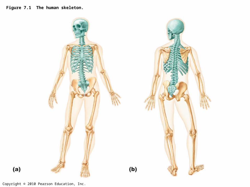
Figure 7.1a The human skeleton.
Copyright © 2010 Pearson Education, Inc.
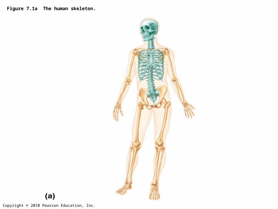
Figure 7.1b The human skeleton.
Copyright © 2010 Pearson Education, Inc.
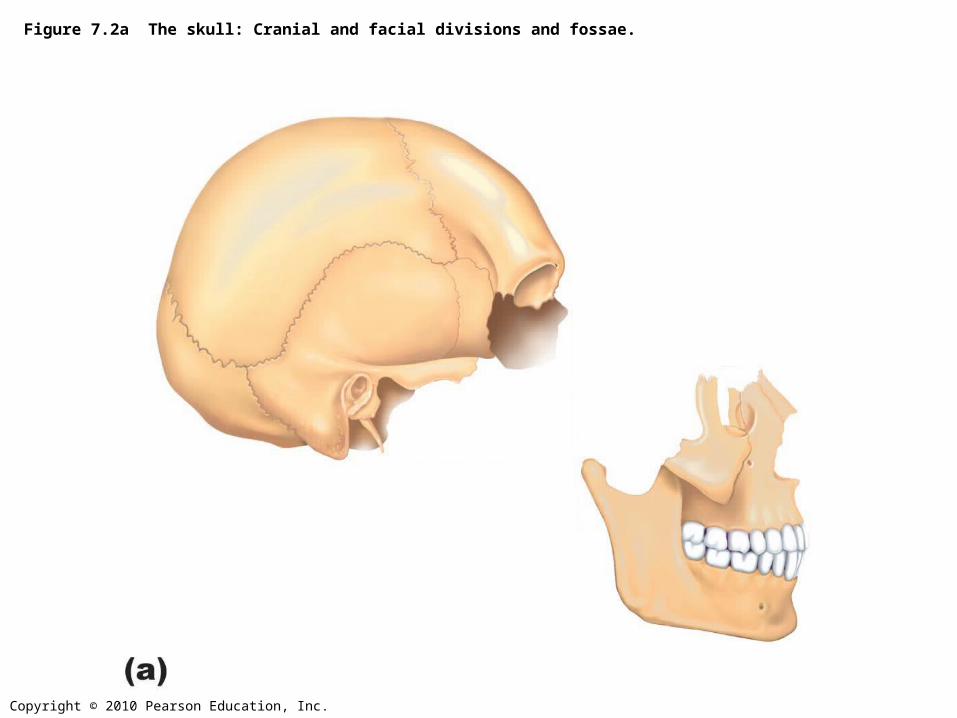
Figure 7.2a The skull: Cranial and facial divisions and fossae.
Copyright © 2010 Pearson Education, Inc.
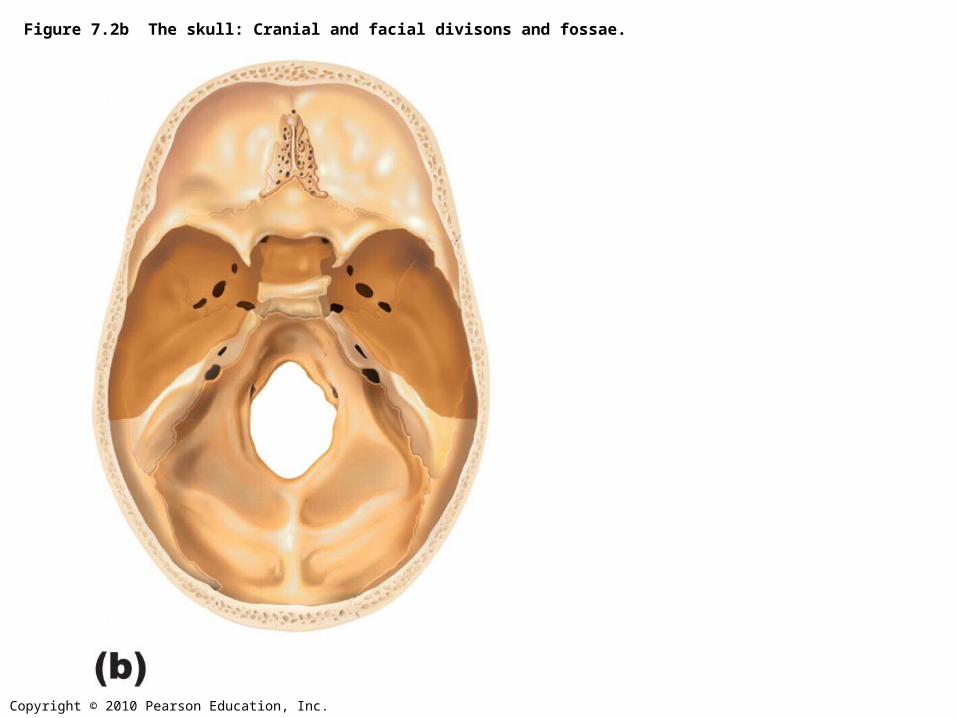
Figure 7.2b The skull: Cranial and facial divisons and fossae.
Copyright © 2010 Pearson Education, Inc.
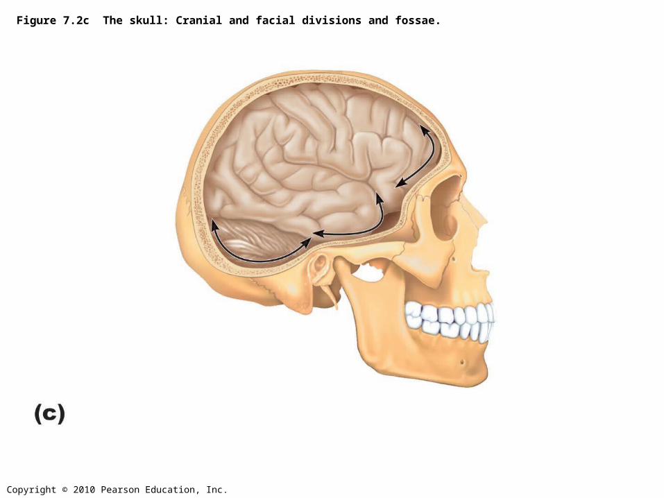
Figure 7.2c The skull: Cranial and facial divisions and fossae.
Copyright © 2010 Pearson Education, Inc.
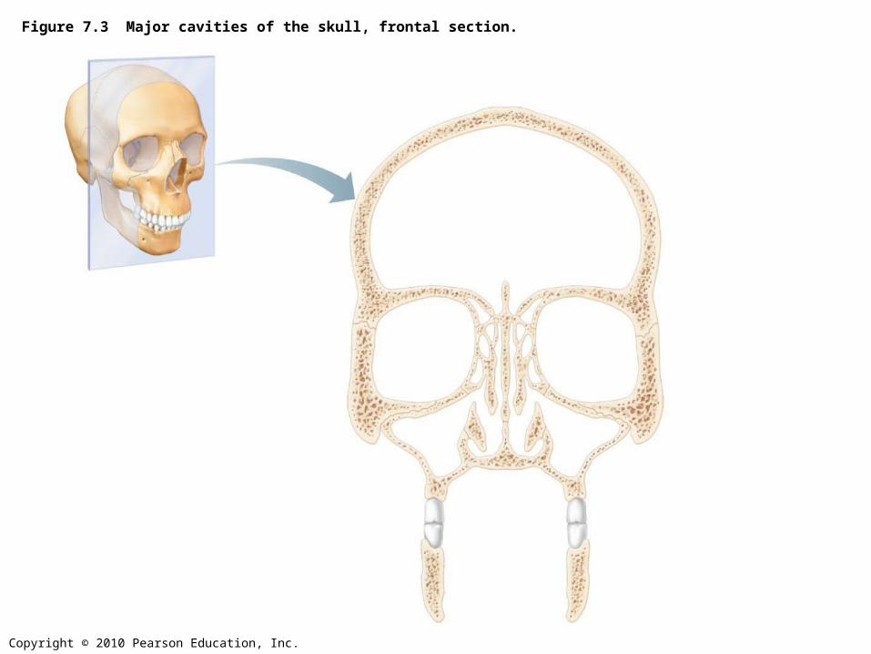
Figure 7.3 Major cavities of the skull, frontal section.
Copyright © 2010 Pearson Education, Inc.
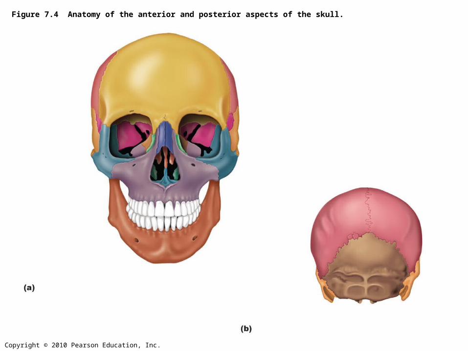
Figure 7.4 Anatomy of the anterior and posterior aspects of the skull.
Copyright © 2010 Pearson Education, Inc.
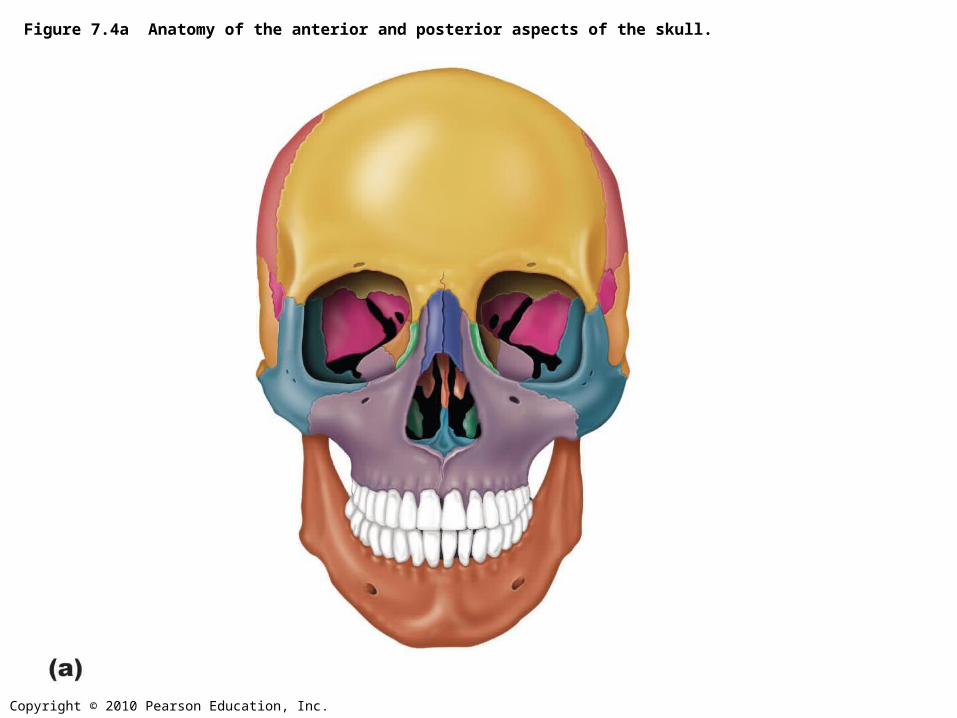
Figure 7.4a Anatomy of the anterior and posterior aspects of the skull.
Copyright © 2010 Pearson Education, Inc.
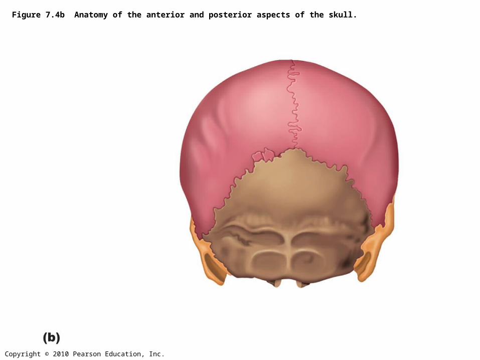
Figure 7.4b Anatomy of the anterior and posterior aspects of the skull.
Copyright © 2010 Pearson Education, Inc.
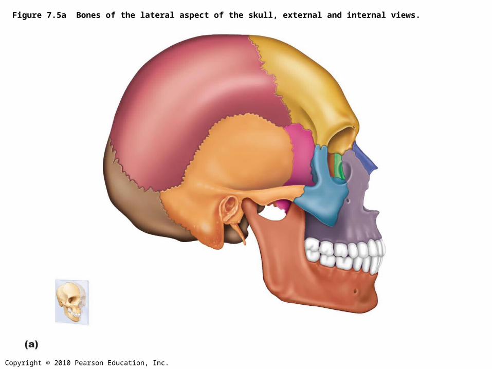
Figure 7.5a Bones of the lateral aspect of the skull, external and internal views.
Copyright © 2010 Pearson Education, Inc.
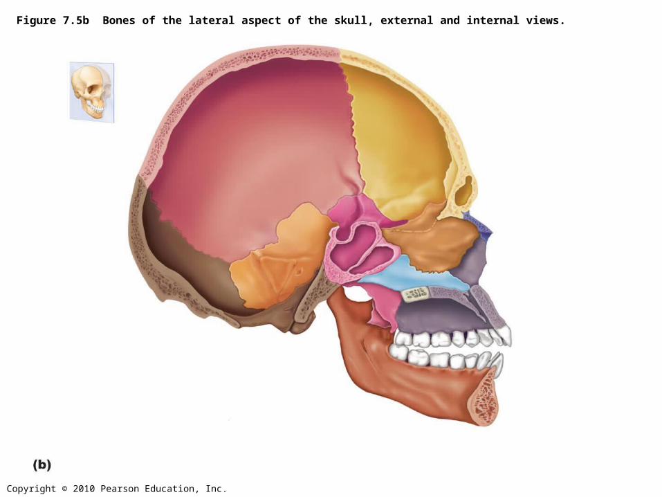
Figure 7.5b Bones of the lateral aspect of the skull, external and internal views.
Copyright © 2010 Pearson Education, Inc.
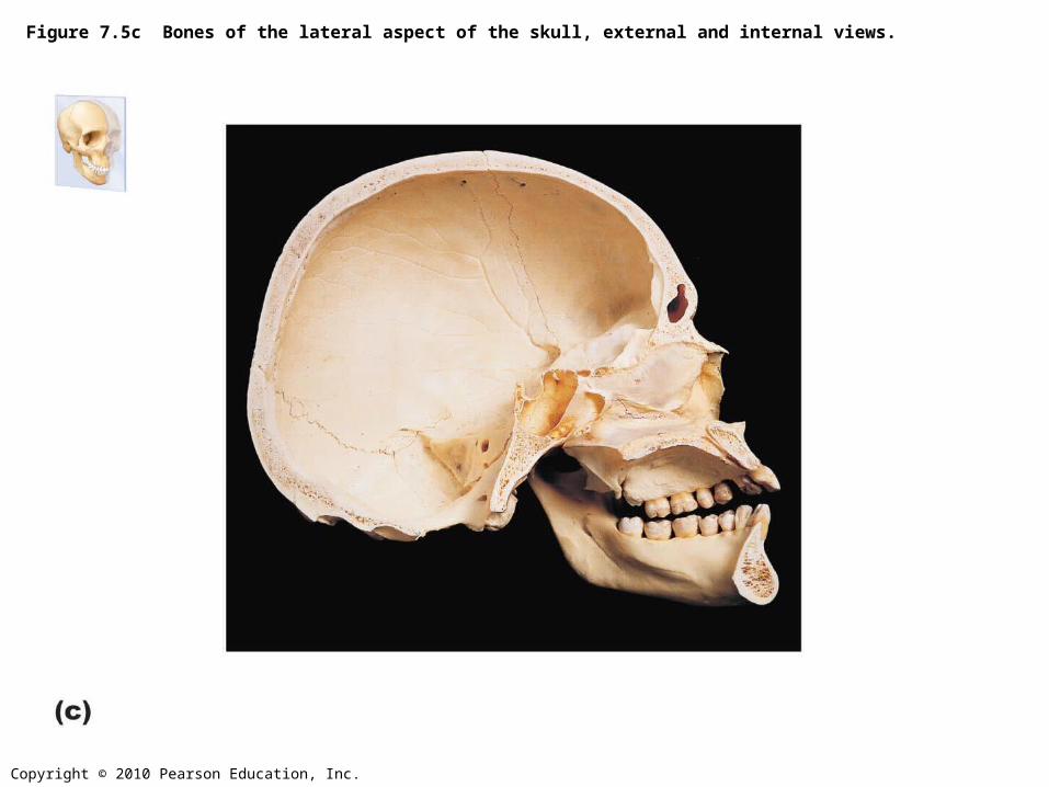
Figure 7.5c Bones of the lateral aspect of the skull, external and internal views.
Copyright © 2010 Pearson Education, Inc.
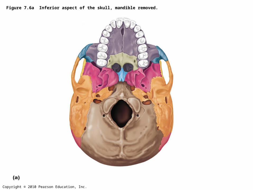
Figure 7.6a Inferior aspect of the skull, mandible removed.
Copyright © 2010 Pearson Education, Inc.
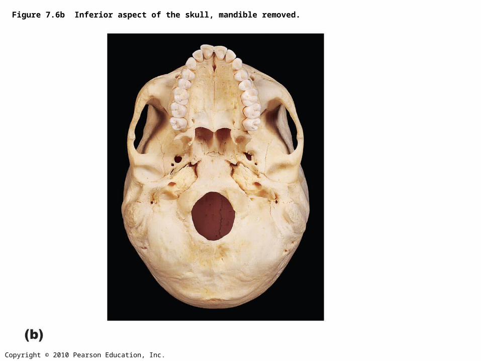
Figure 7.6b Inferior aspect of the skull, mandible removed.
Copyright © 2010 Pearson Education, Inc.
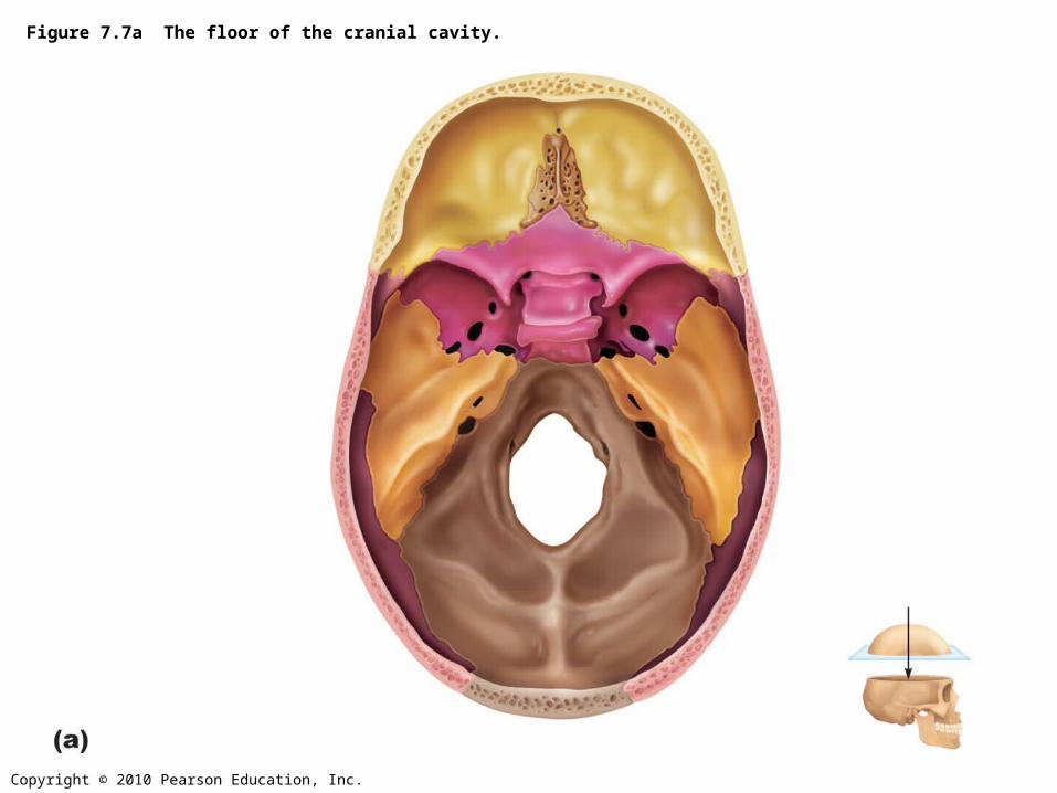
Figure 7.7a The floor of the cranial cavity.
Copyright © 2010 Pearson Education, Inc.
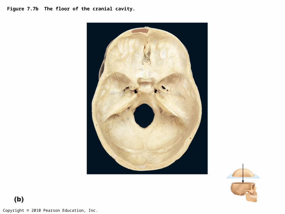
Figure 7.7b The floor of the cranial cavity.
Copyright © 2010 Pearson Education, Inc.
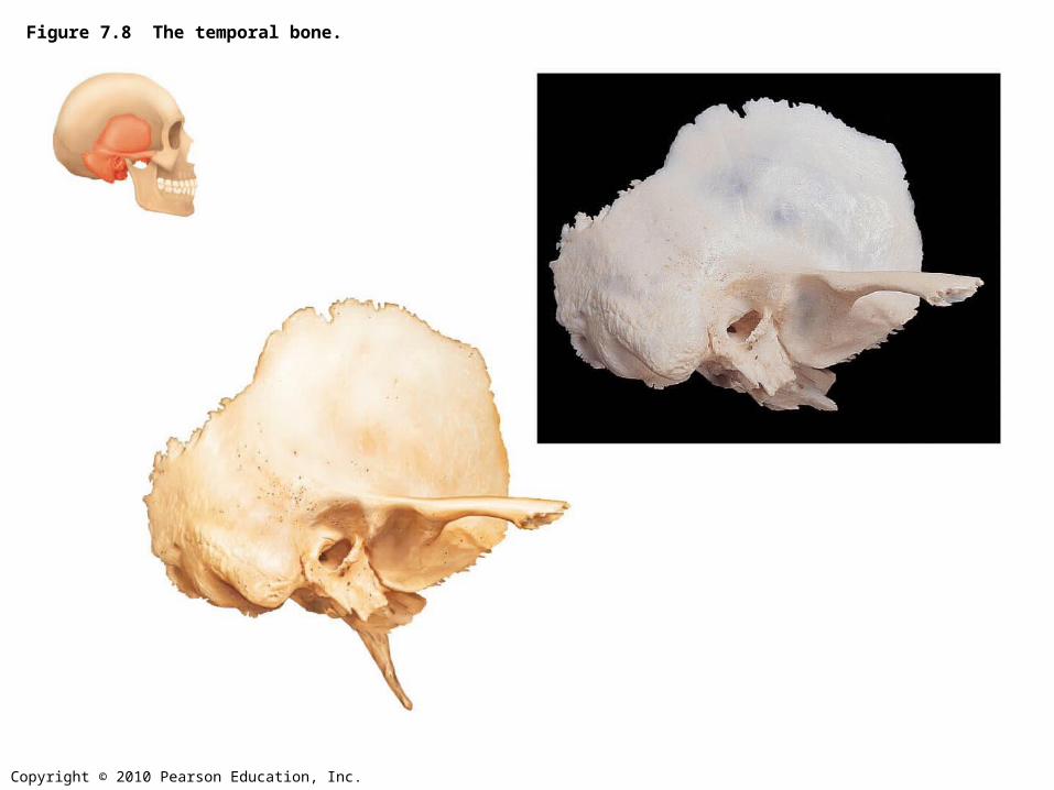
Figure 7.8 The temporal bone.
Copyright © 2010 Pearson Education, Inc.
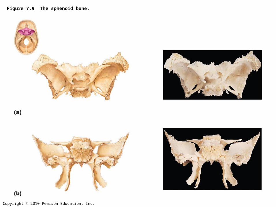
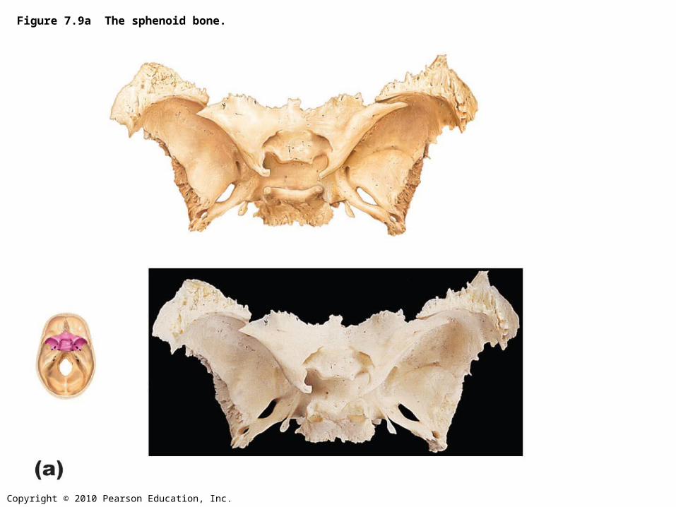
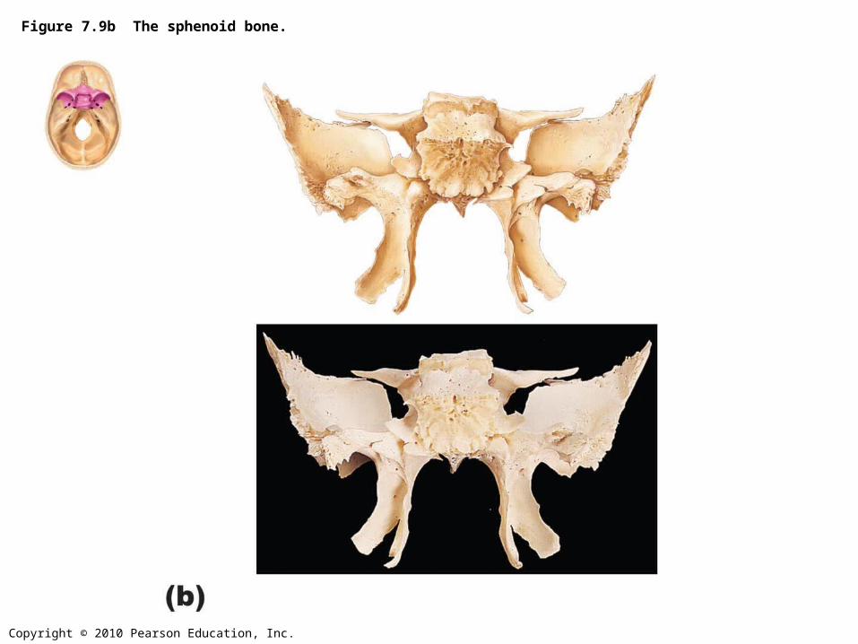
Figure 7.9 The sphenoid bone.
Copyright © 2010 Pearson Education, Inc.
Figure 7.9a The sphenoid bone.
Copyright © 2010 Pearson Education, Inc.
Figure 7.9b The sphenoid bone.
Copyright © 2010 Pearson Education, Inc.
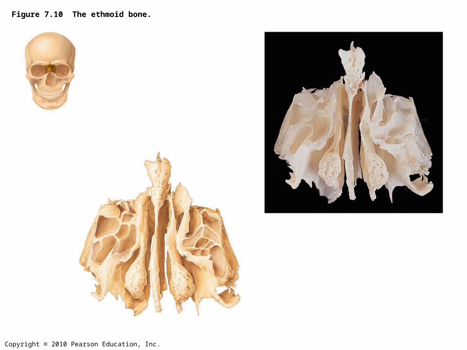
Figure 7.10 The ethmoid bone.
Copyright © 2010 Pearson Education, Inc.
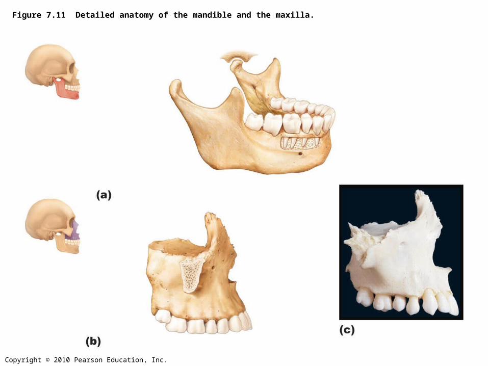
Figure 7.11 Detailed anatomy of the mandible and the maxilla.
Copyright © 2010 Pearson Education, Inc.
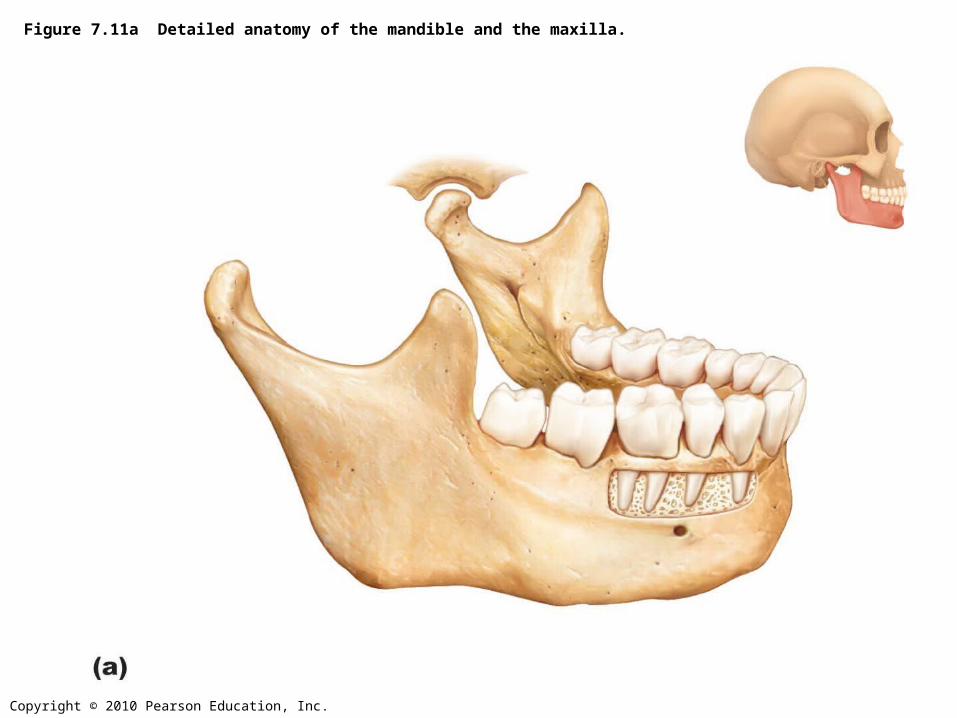
Figure 7.11a Detailed anatomy of the mandible and the maxilla.
Copyright © 2010 Pearson Education, Inc.
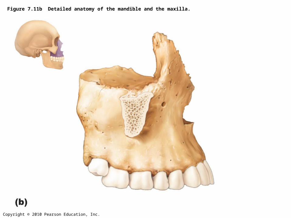
Figure 7.11b Detailed anatomy of the mandible and the maxilla.
Copyright © 2010 Pearson Education, Inc.
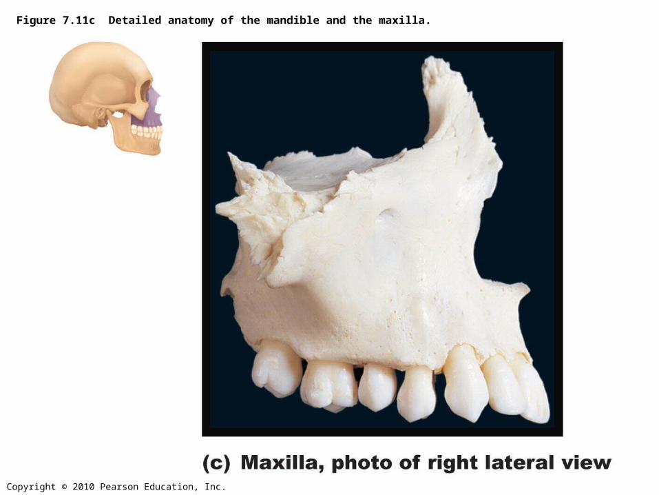
Figure 7.11c Detailed anatomy of the mandible and the maxilla.
Copyright © 2010 Pearson Education, Inc.
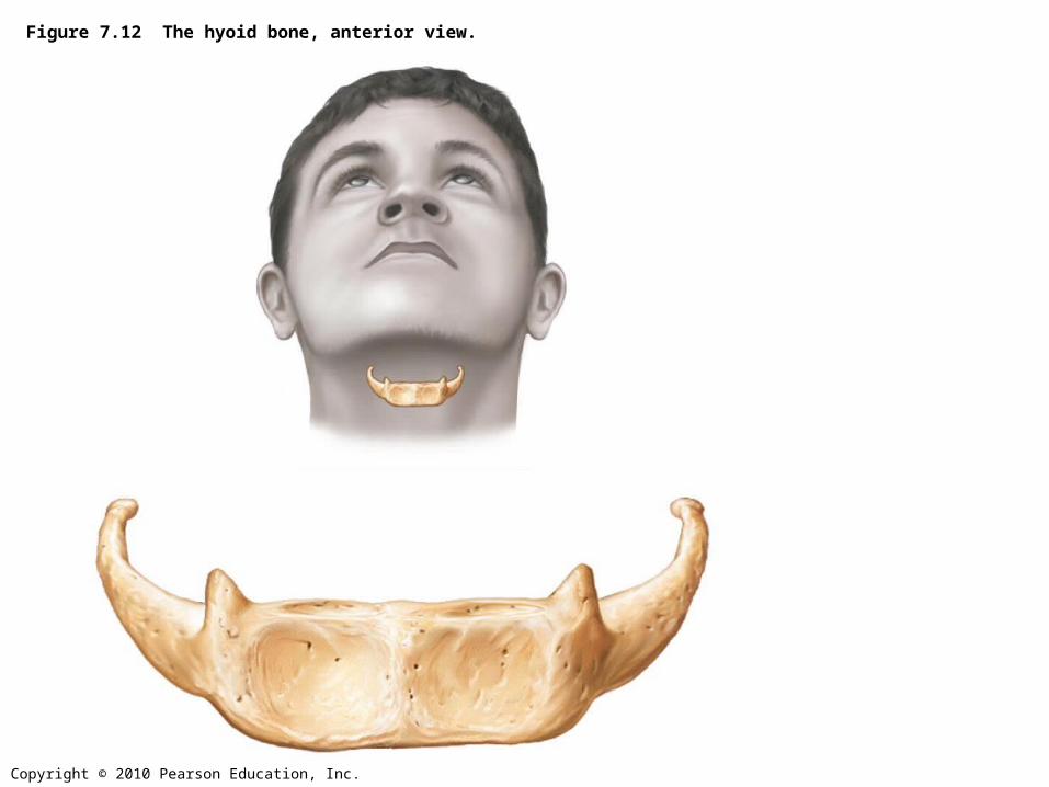
Figure 7.12 The hyoid bone, anterior view.
Copyright © 2010 Pearson Education, Inc.
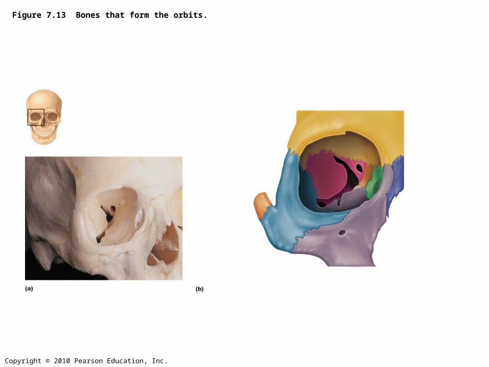
Figure 7.13 Bones that form the orbits.
Copyright © 2010 Pearson Education, Inc.
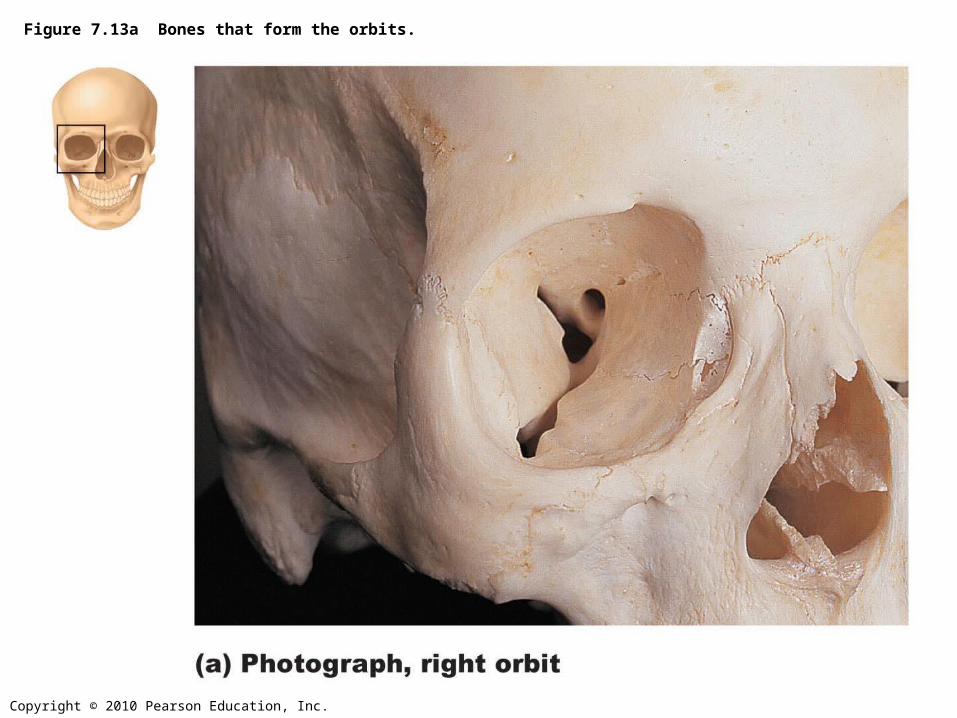
Figure 7.13a Bones that form the orbits.

Copyright © 2010 Pearson Education, Inc.
Figure 7.13b Bones that form the orbits.

Copyright © 2010 Pearson Education, Inc.
Figure 7.14a Bones of the nasal cavity.
Copyright © 2010 Pearson Education, Inc.
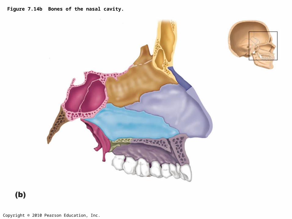
Figure 7.14b Bones of the nasal cavity.
Copyright © 2010 Pearson Education, Inc.
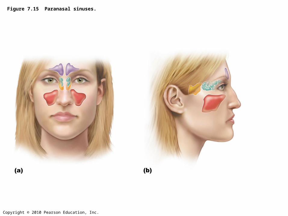
Figure 7.15 Paranasal sinuses.
Copyright © 2010 Pearson Education, Inc.
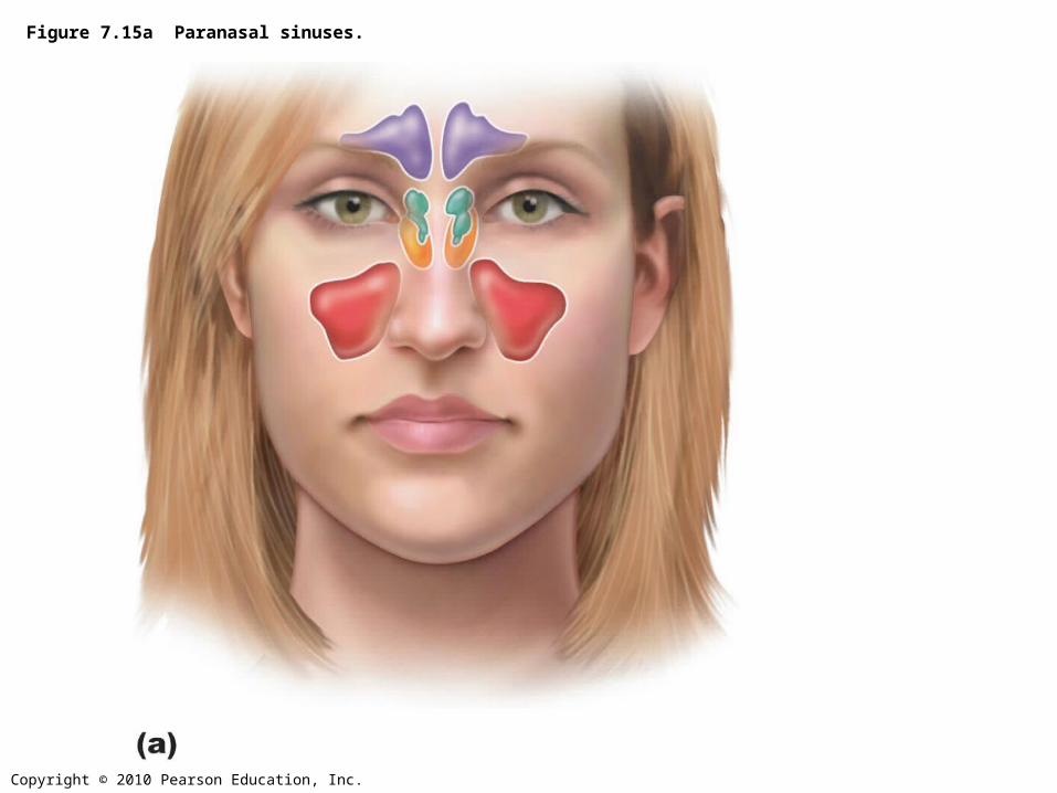
Figure 7.15a Paranasal sinuses.
Copyright © 2010 Pearson Education, Inc.
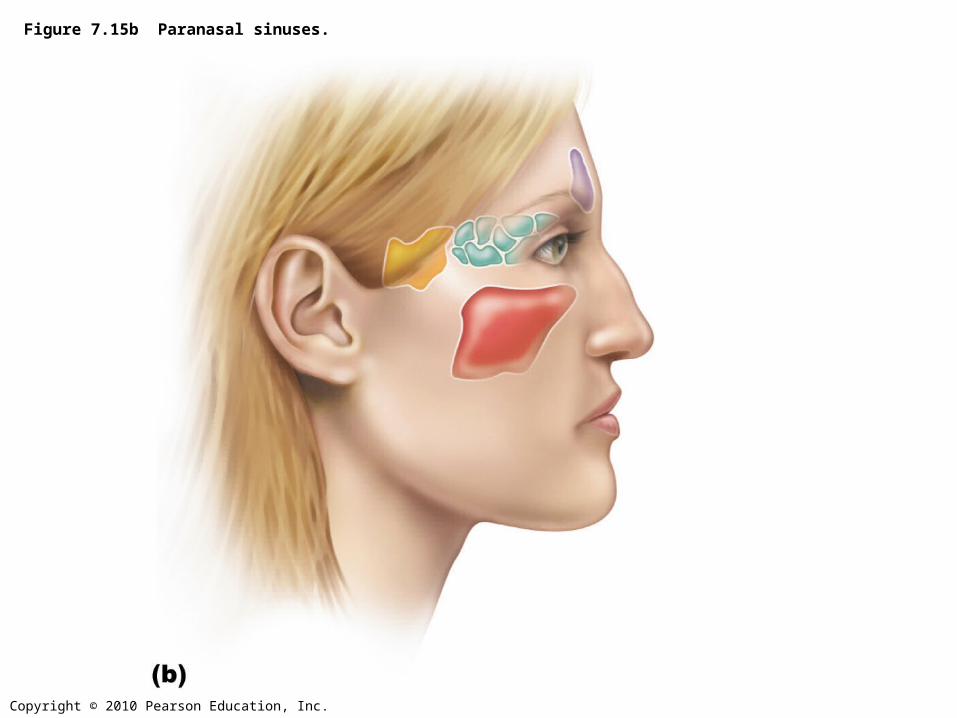
Figure 7.15b Paranasal sinuses.
Copyright © 2010 Pearson Education, Inc.
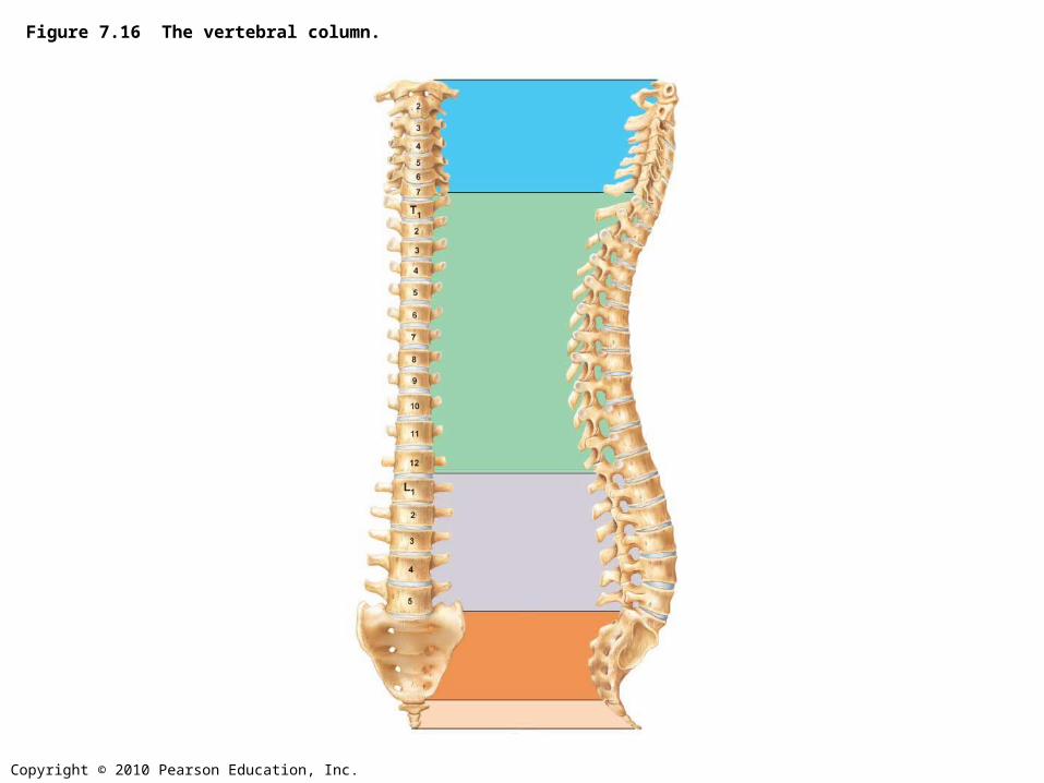
Figure 7.16 The vertebral column.
Copyright © 2010 Pearson Education, Inc.
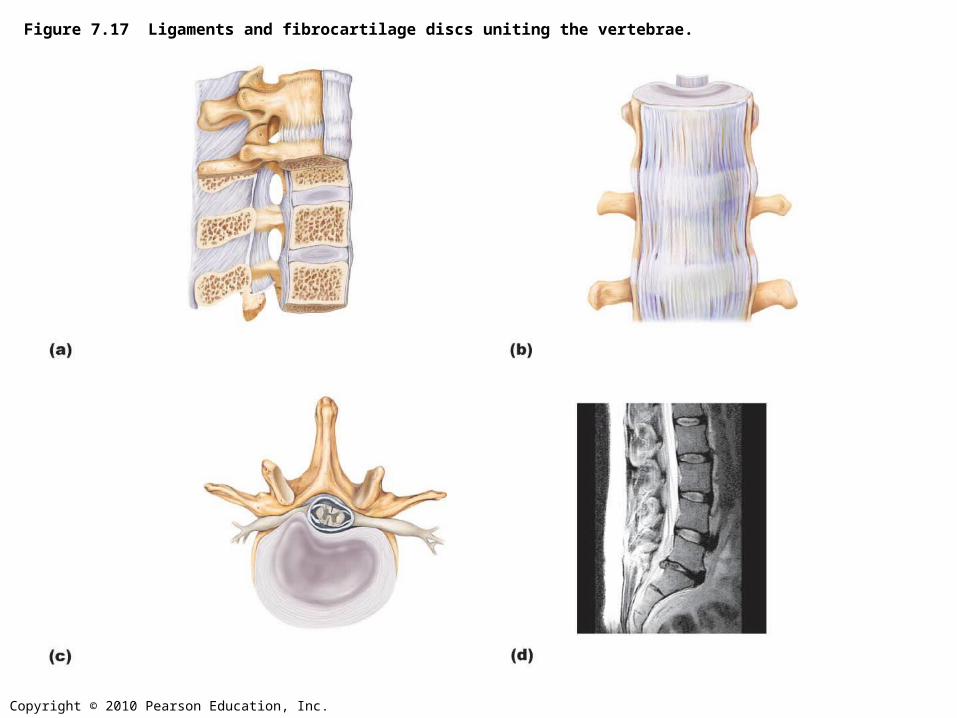
Figure 7.17 Ligaments and fibrocartilage discs uniting the vertebrae.

Copyright © 2010 Pearson Education, Inc.
Figure 7.17a Ligaments and fibrocartilage discs uniting the vertebrae.
Copyright © 2010 Pearson Education, Inc.
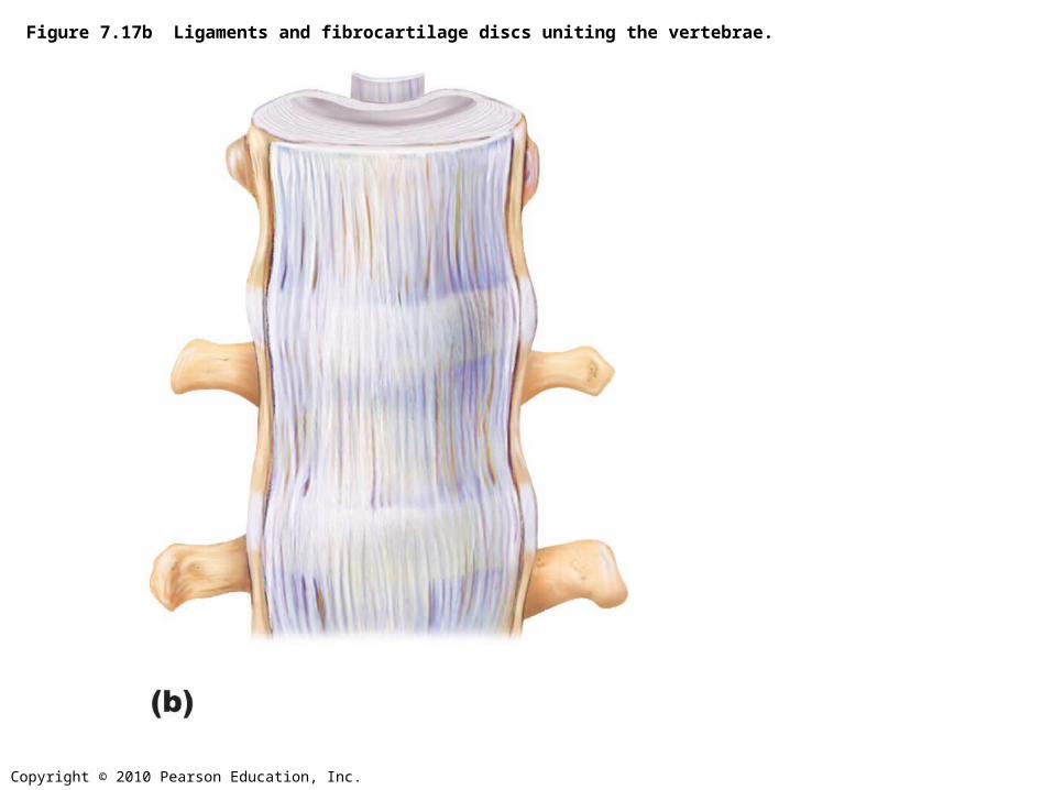
Figure 7.17b Ligaments and fibrocartilage discs uniting the vertebrae.
Copyright © 2010 Pearson Education, Inc.
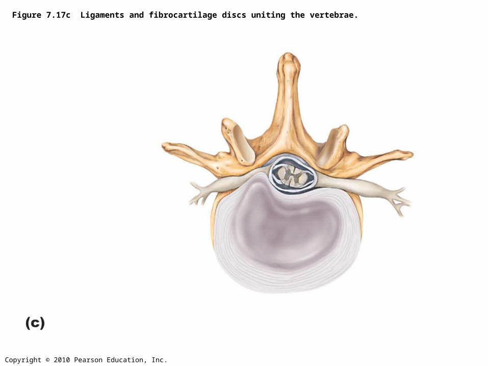
Figure 7.17c Ligaments and fibrocartilage discs uniting the vertebrae.
Copyright © 2010 Pearson Education, Inc.
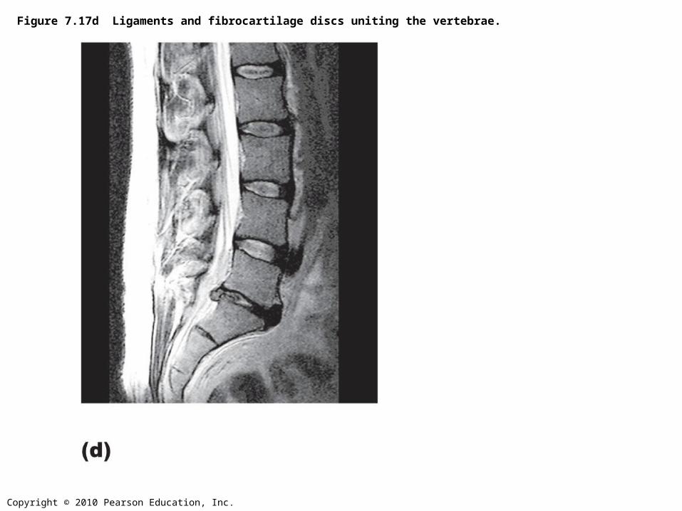
Figure 7.17d Ligaments and fibrocartilage discs uniting the vertebrae.
Copyright © 2010 Pearson Education, Inc.
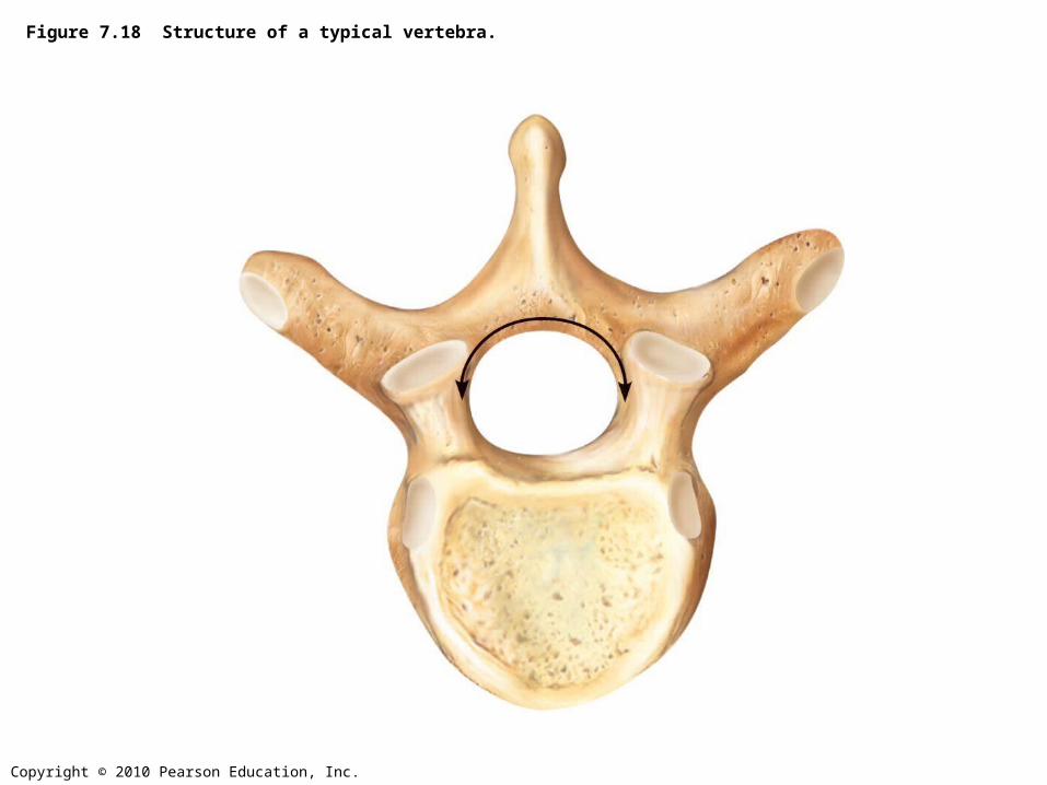
Figure 7.18 Structure of a typical vertebra.
Copyright © 2010 Pearson Education, Inc.
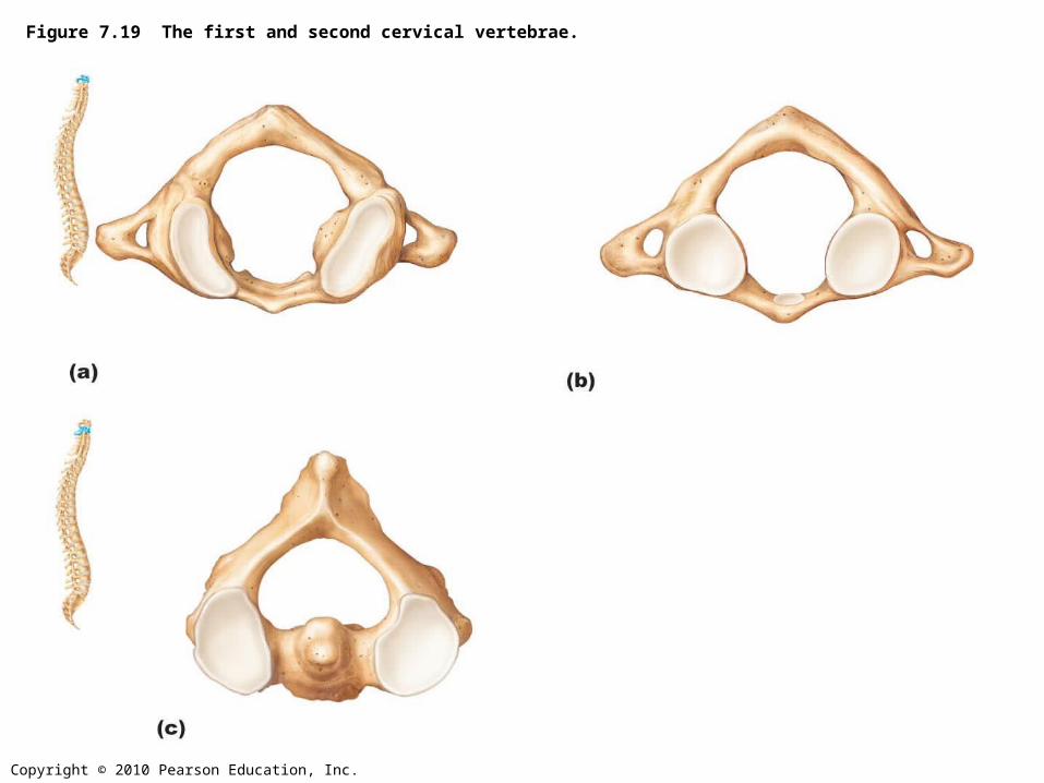
Figure 7.19 The first and second cervical vertebrae.
Copyright © 2010 Pearson Education, Inc.
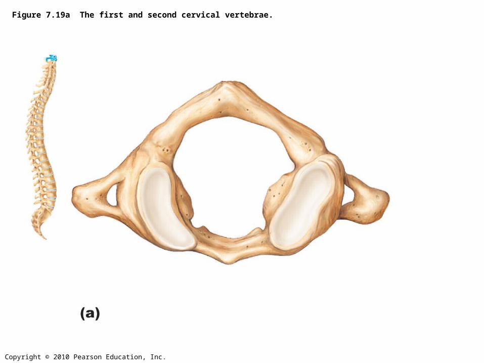
Figure 7.19a The first and second cervical vertebrae.
Copyright © 2010 Pearson Education, Inc.
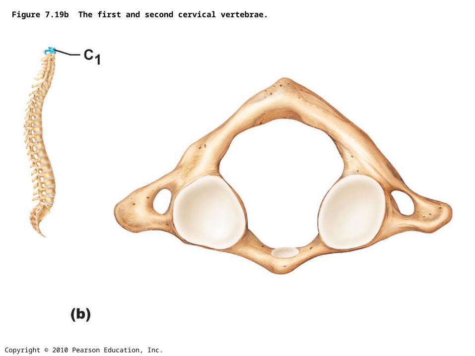
Figure 7.19b The first and second cervical vertebrae.

Copyright © 2010 Pearson Education, Inc.
Figure 7.19c The first and second cervical vertebrae.
Copyright © 2010 Pearson Education, Inc.
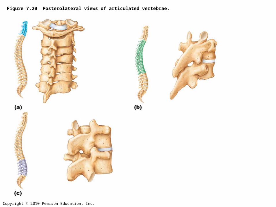
Figure 7.20 Posterolateral views of articulated vertebrae.
Copyright © 2010 Pearson Education, Inc.
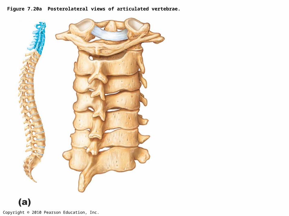
Figure 7.20a Posterolateral views of articulated vertebrae.

Copyright © 2010 Pearson Education, Inc.
Figure 7.20b Posterolateral views of articulated vertebrae.
Copyright © 2010 Pearson Education, Inc.
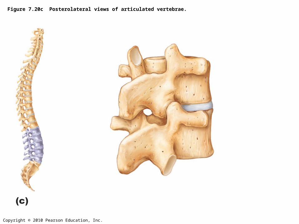
Figure 7.20c Posterolateral views of articulated vertebrae.

Copyright © 2010 Pearson Education, Inc.
Figure 7.21 The sacrum and coccyx.
Copyright © 2010 Pearson Education, Inc.
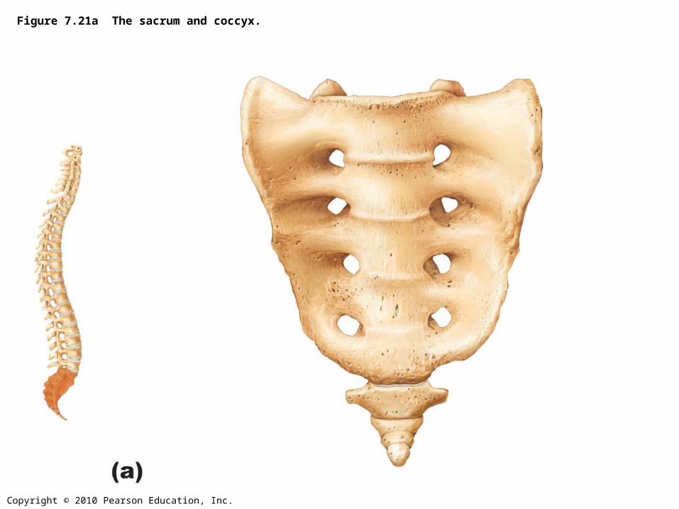
Figure 7.21a The sacrum and coccyx.
Copyright © 2010 Pearson Education, Inc.
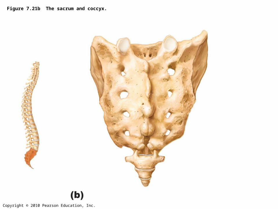
Figure 7.21b The sacrum and coccyx.
Copyright © 2010 Pearson Education, Inc.
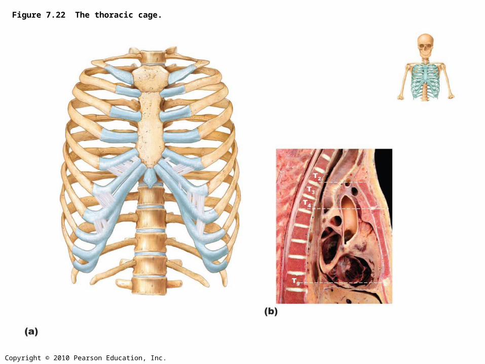
Figure 7.22 The thoracic cage.
Copyright © 2010 Pearson Education, Inc.
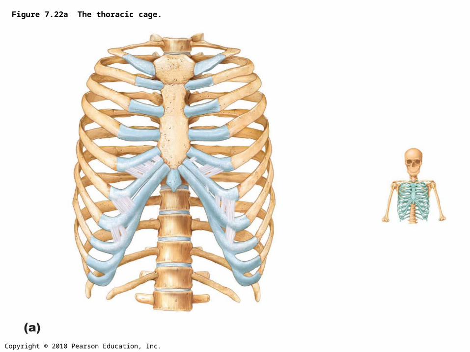
Figure 7.22a The thoracic cage.
Copyright © 2010 Pearson Education, Inc.
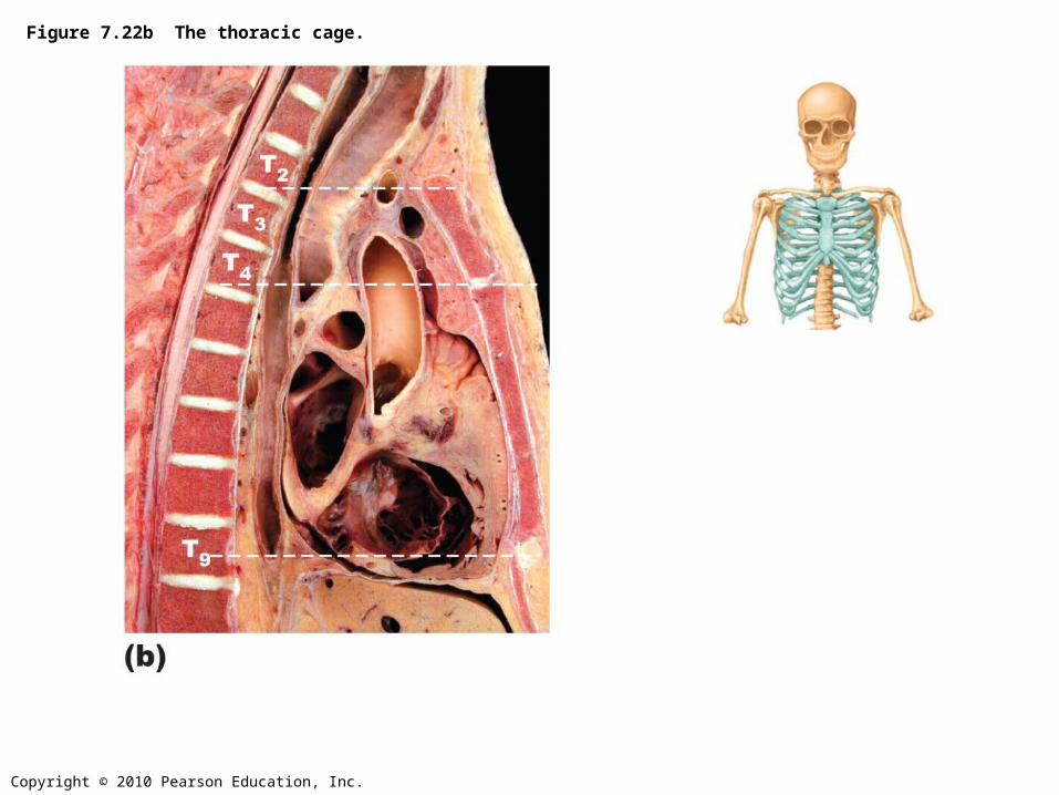
Figure 7.22b The thoracic cage.
Copyright © 2010 Pearson Education, Inc.
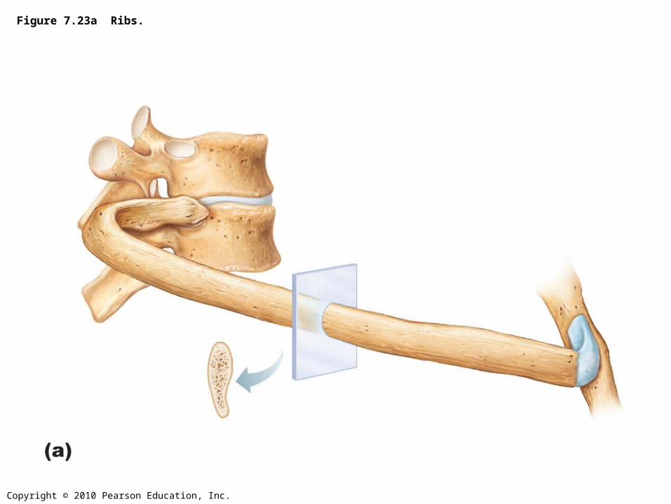
Figure 7.23a Ribs.
Copyright © 2010 Pearson Education, Inc.
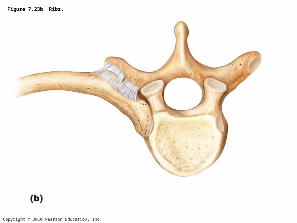
Figure 7.23b Ribs.
Copyright © 2010 Pearson Education, Inc.
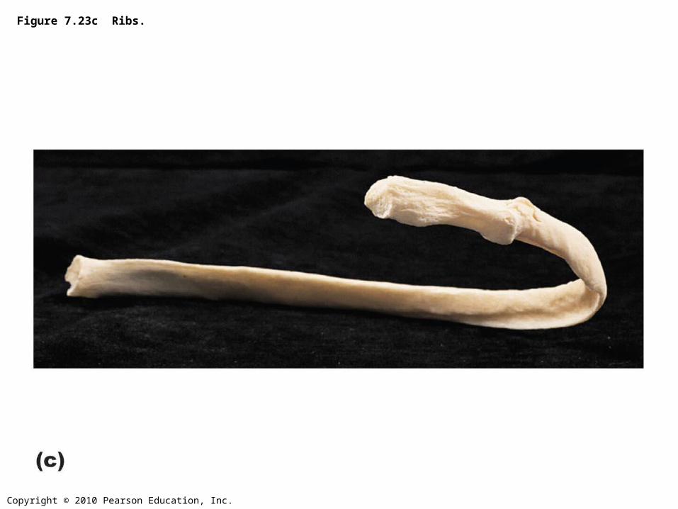
Figure 7.23c Ribs.
Copyright © 2010 Pearson Education, Inc.
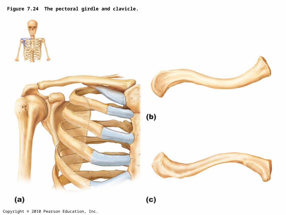
Figure 7.24 The pectoral girdle and clavicle.
Copyright © 2010 Pearson Education, Inc.
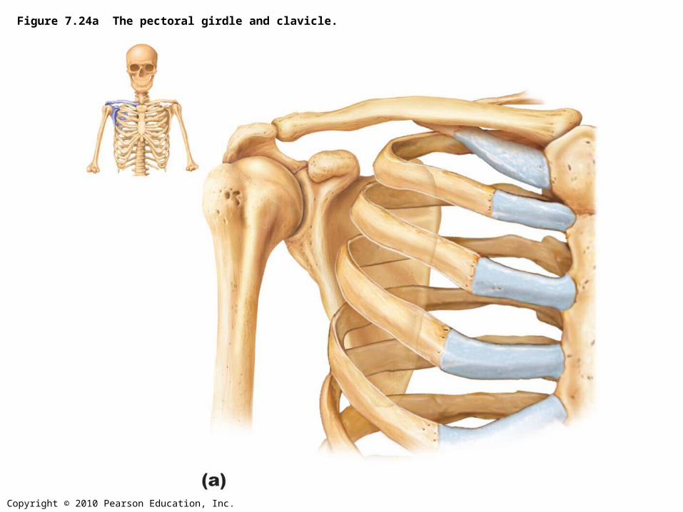
Figure 7.24a The pectoral girdle and clavicle.
Copyright © 2010 Pearson Education, Inc.
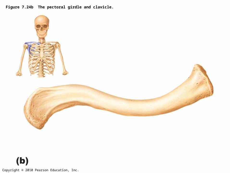
Figure 7.24b The pectoral girdle and clavicle.
Copyright © 2010 Pearson Education, Inc.
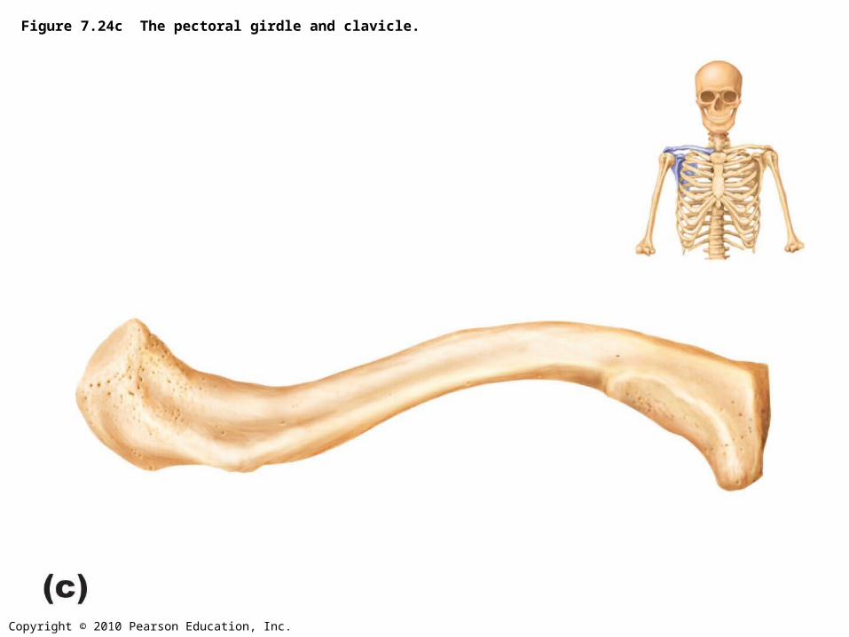
Figure 7.24c The pectoral girdle and clavicle.
Copyright © 2010 Pearson Education, Inc.
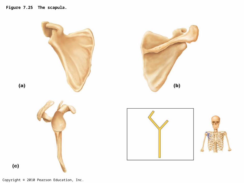
Figure 7.25 The scapula.
Copyright © 2010 Pearson Education, Inc.
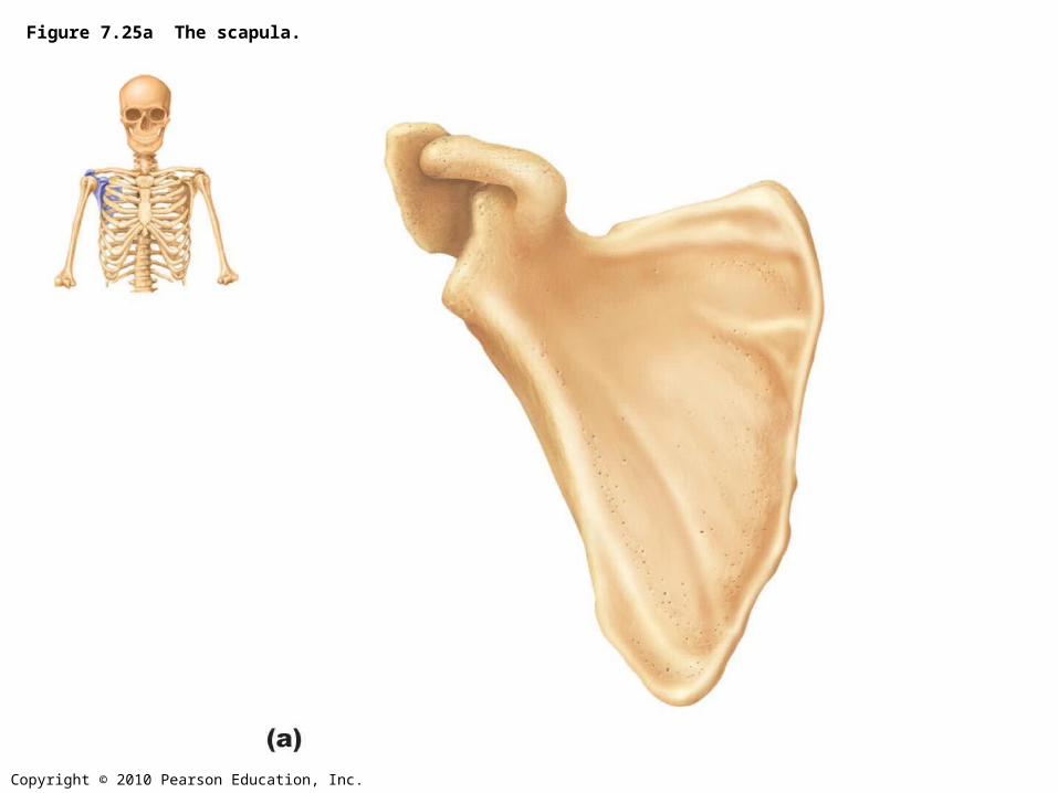
Figure 7.25a The scapula.
Copyright © 2010 Pearson Education, Inc.
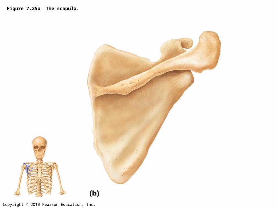
Figure 7.25b The scapula.
Copyright © 2010 Pearson Education, Inc.
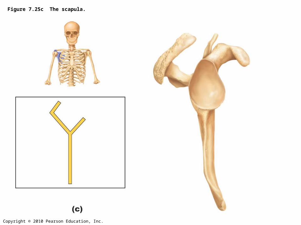
Figure 7.25c The scapula.
Copyright © 2010 Pearson Education, Inc.
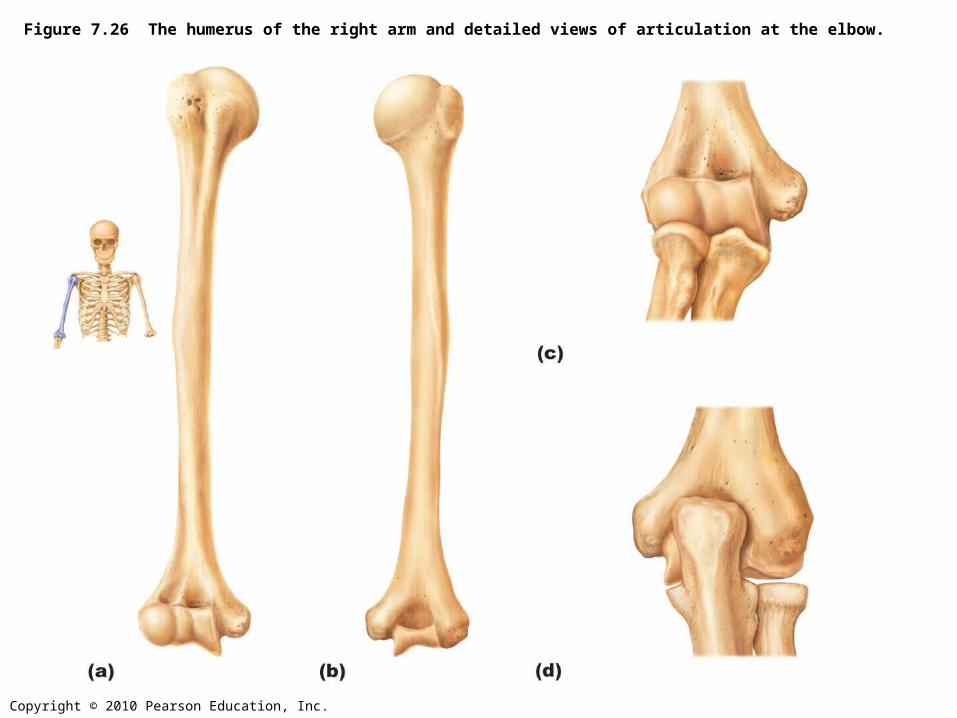
Figure 7.26 The humerus of the right arm and detailed views of articulation at the elbow.
Copyright © 2010 Pearson Education, Inc.
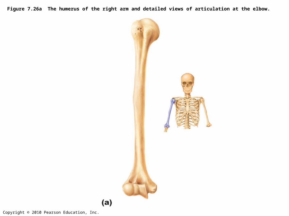
Figure 7.26a The humerus of the right arm and detailed views of articulation at the elbow.
Copyright © 2010 Pearson Education, Inc.
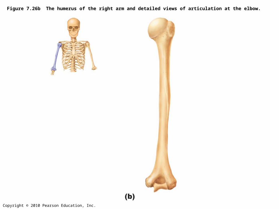
Figure 7.26b The humerus of the right arm and detailed views of articulation at the elbow.
Copyright © 2010 Pearson Education, Inc.
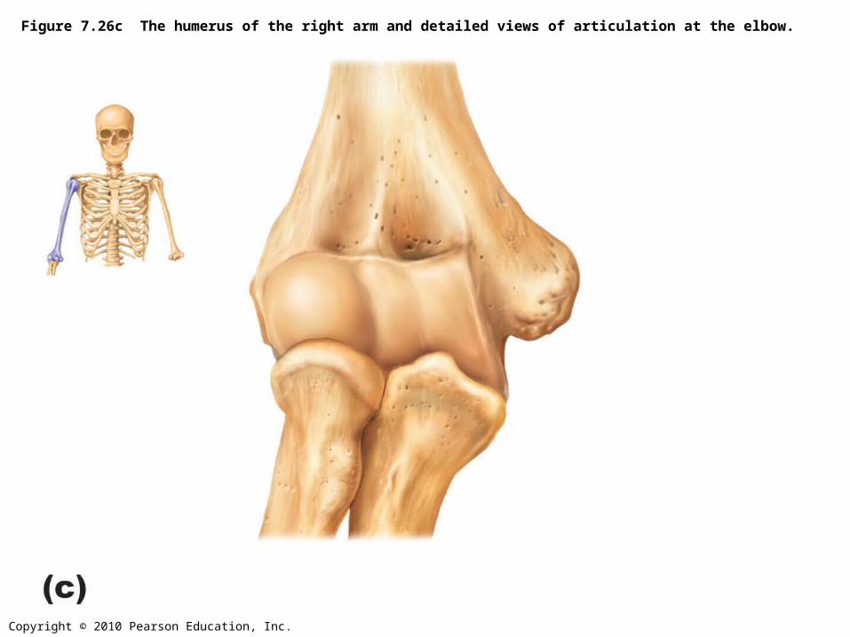
Figure 7.26c The humerus of the right arm and detailed views of articulation at the elbow.
Copyright © 2010 Pearson Education, Inc.
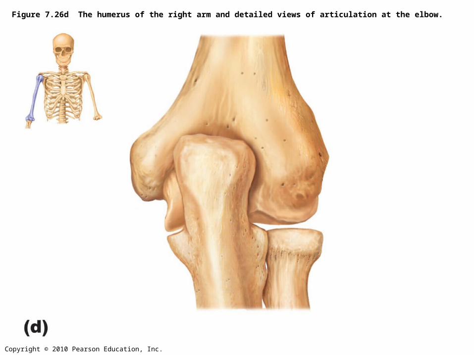
Figure 7.26d The humerus of the right arm and detailed views of articulation at the elbow.

Copyright © 2010 Pearson Education, Inc.
Figure 7.27 Radius and ulna of the right forearm.
Copyright © 2010 Pearson Education, Inc.
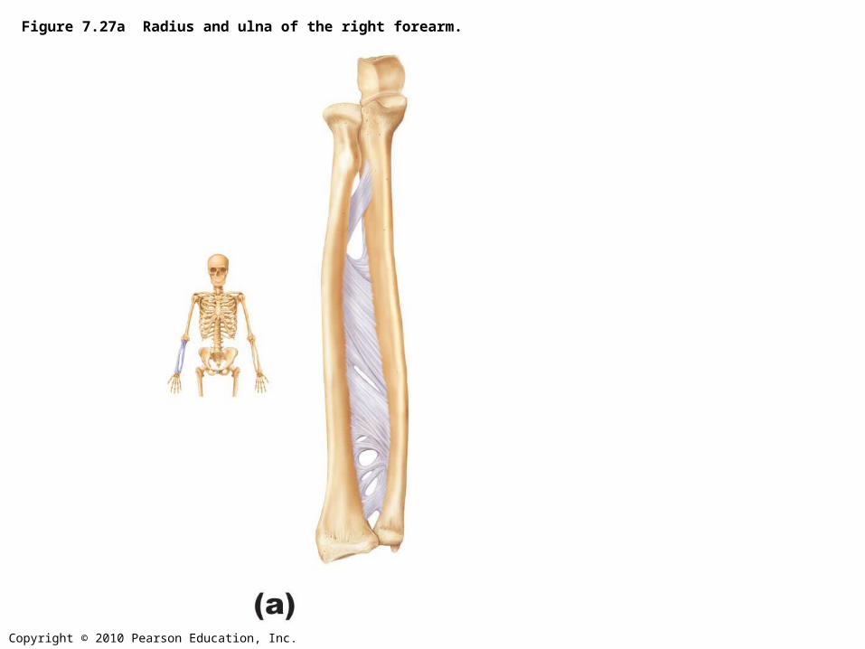
Figure 7.27a Radius and ulna of the right forearm.
Copyright © 2010 Pearson Education, Inc.
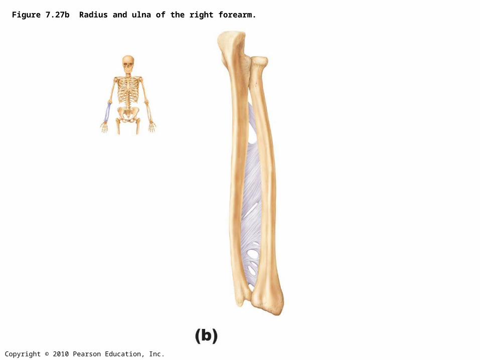
Figure 7.27b Radius and ulna of the right forearm.
Copyright © 2010 Pearson Education, Inc.
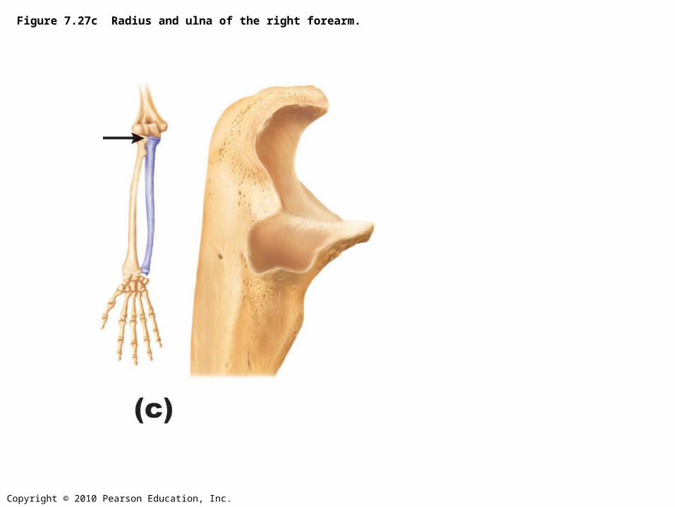
Figure 7.27c Radius and ulna of the right forearm.
Copyright © 2010 Pearson Education, Inc.
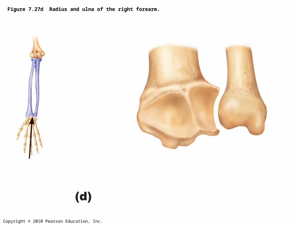
Figure 7.27d Radius and ulna of the right forearm.
Copyright © 2010 Pearson Education, Inc.
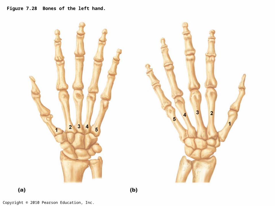
Figure 7.28 Bones of the left hand.
Copyright © 2010 Pearson Education, Inc.
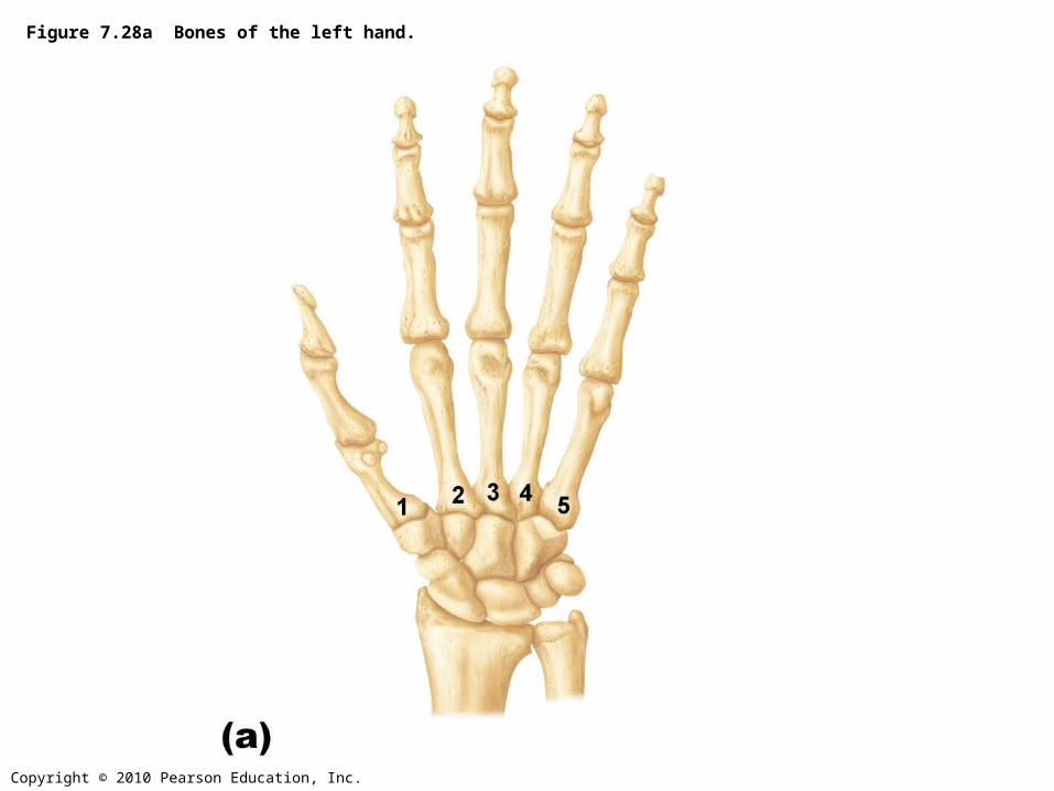
Figure 7.28a Bones of the left hand.
Copyright © 2010 Pearson Education, Inc.
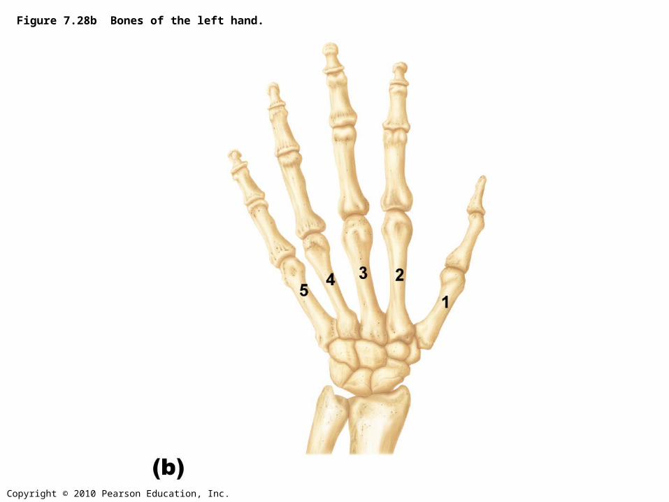
Figure 7.28b Bones of the left hand.
Copyright © 2010 Pearson Education, Inc.
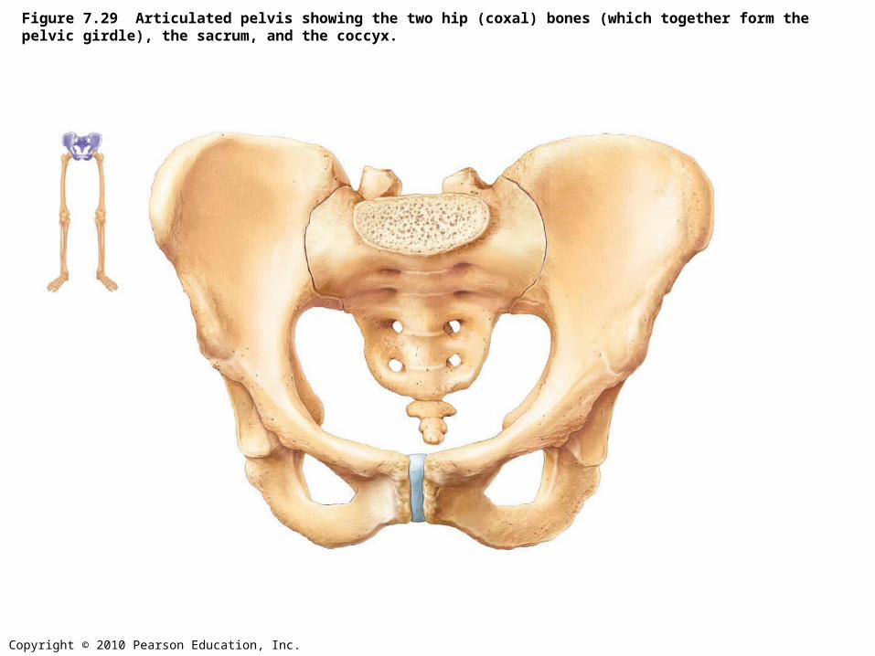
Figure 7.29 Articulated pelvis showing the two hip (coxal) bones (which together form the pelvic girdle), the sacrum, and the coccyx.

Copyright © 2010 Pearson Education, Inc.
Figure 7.30a Bones of the bony pelvis.
Copyright © 2010 Pearson Education, Inc.
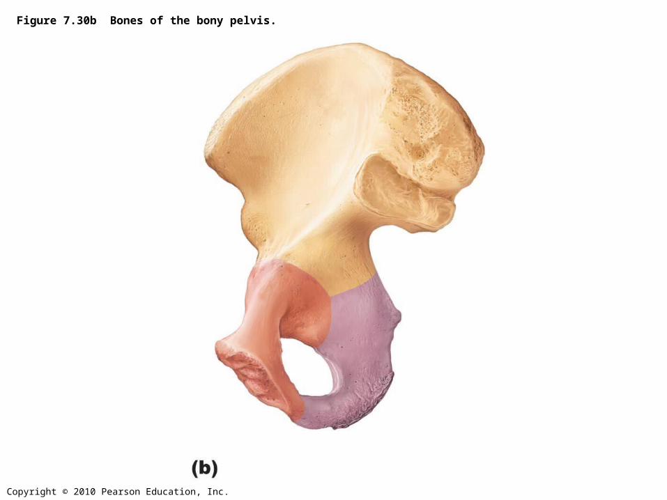
Figure 7.30b Bones of the bony pelvis.
Copyright © 2010 Pearson Education, Inc.
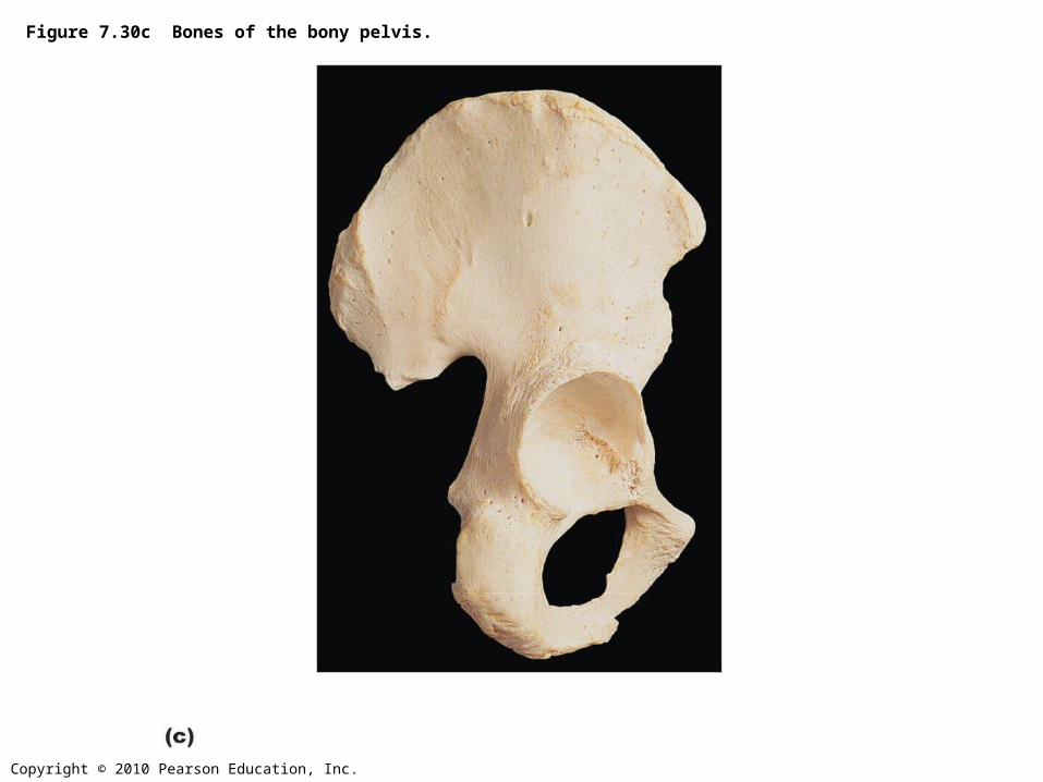
Figure 7.30c Bones of the bony pelvis.
Copyright © 2010 Pearson Education, Inc.
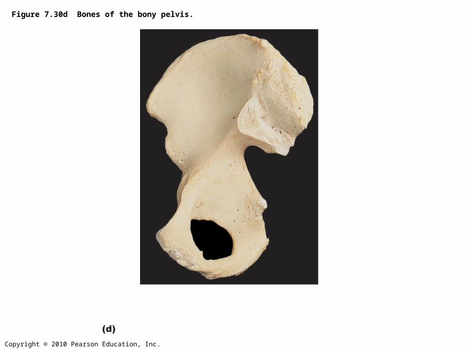
Figure 7.30d Bones of the bony pelvis.
Copyright © 2010 Pearson Education, Inc.
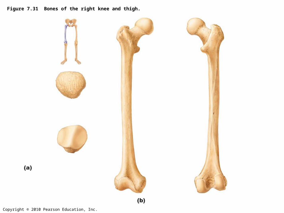
Figure 7.31 Bones of the right knee and thigh.
Copyright © 2010 Pearson Education, Inc.
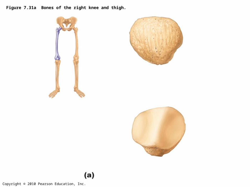
Figure 7.31a Bones of the right knee and thigh.
Copyright © 2010 Pearson Education, Inc.
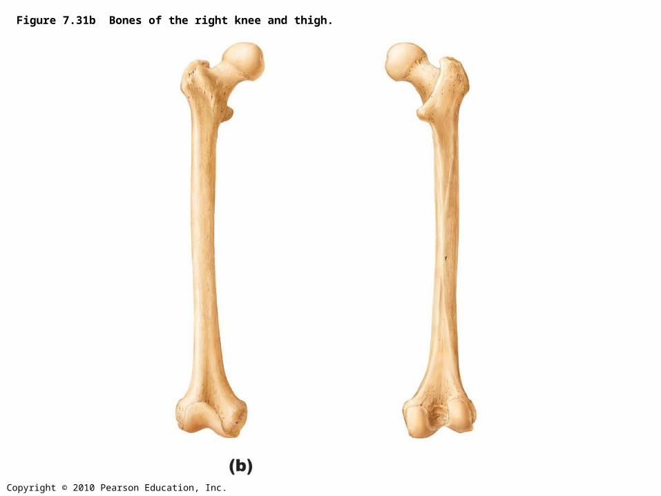
Figure 7.31b Bones of the right knee and thigh.
Copyright © 2010 Pearson Education, Inc.
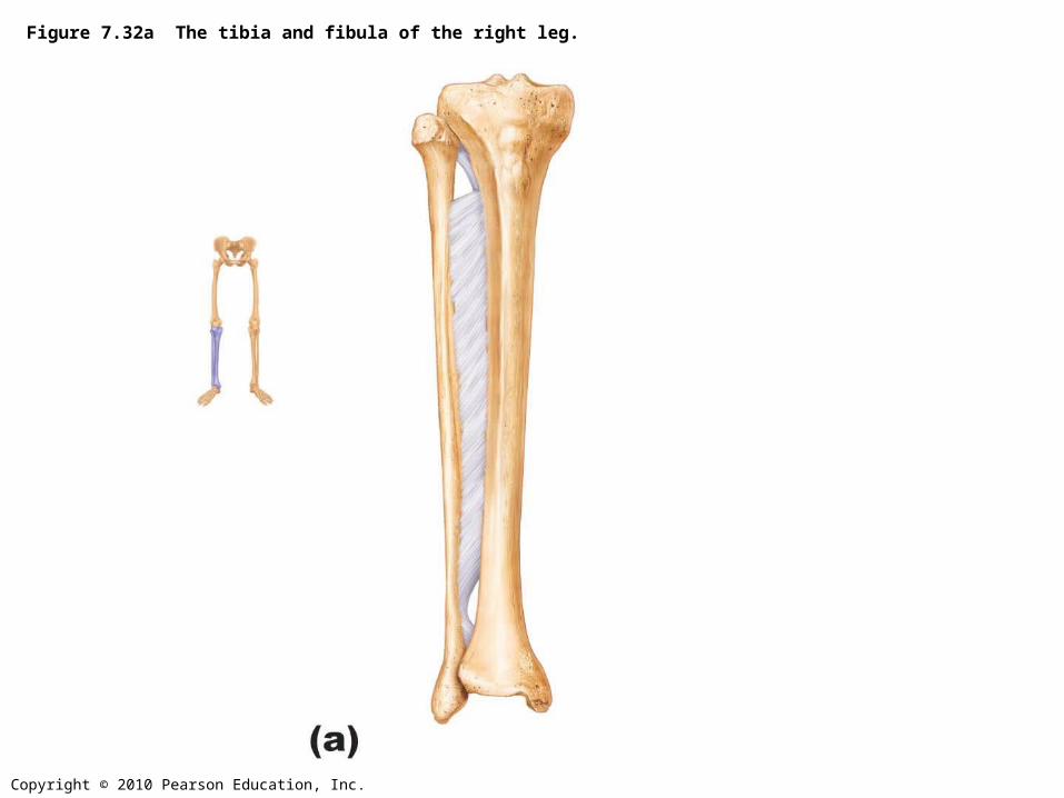
Figure 7.32a The tibia and fibula of the right leg.
Copyright © 2010 Pearson Education, Inc.
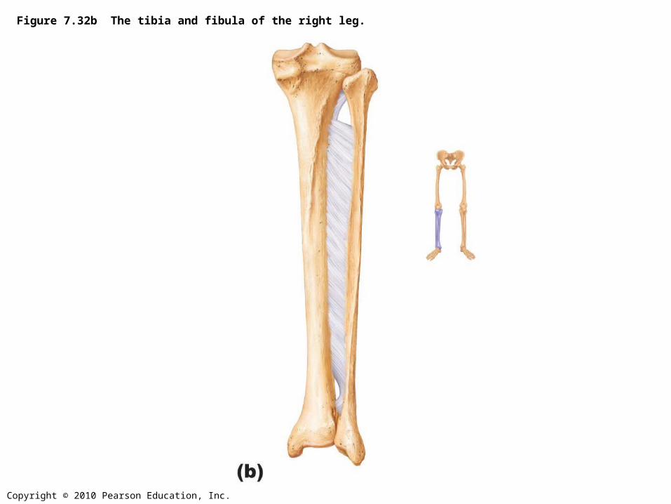
Figure 7.32b The tibia and fibula of the right leg.
Copyright © 2010 Pearson Education, Inc.
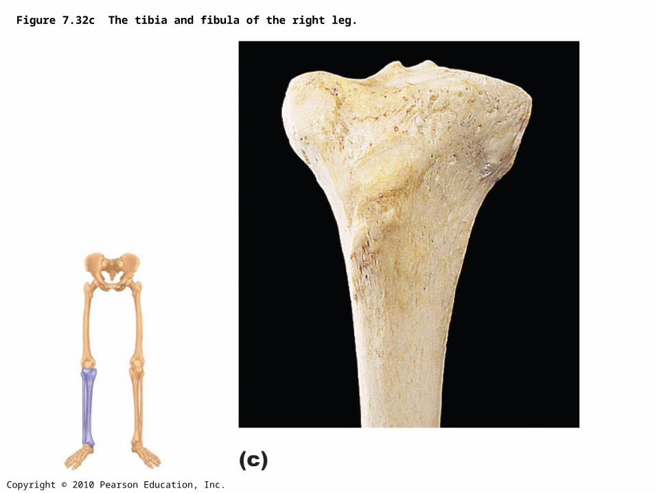
Figure 7.32c The tibia and fibula of the right leg.
Copyright © 2010 Pearson Education, Inc.
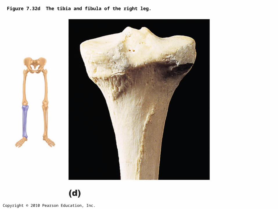
Figure 7.32d The tibia and fibula of the right leg.
Copyright © 2010 Pearson Education, Inc.
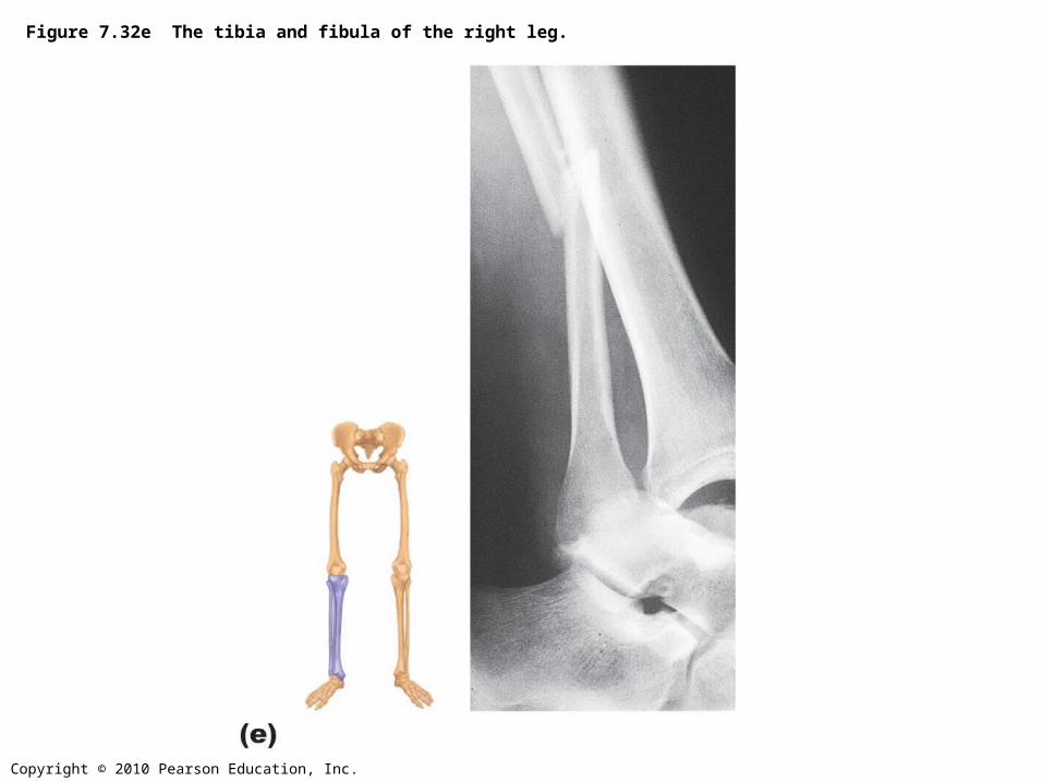
Figure 7.32e The tibia and fibula of the right leg.
Copyright © 2010 Pearson Education, Inc.
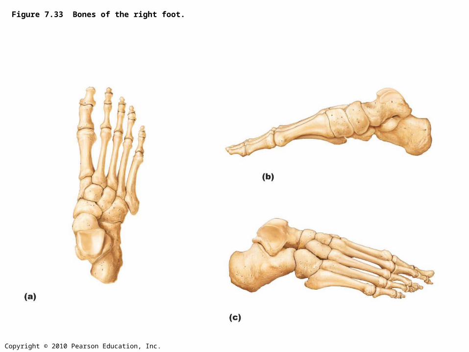
Figure 7.33 Bones of the right foot.
Copyright © 2010 Pearson Education, Inc.
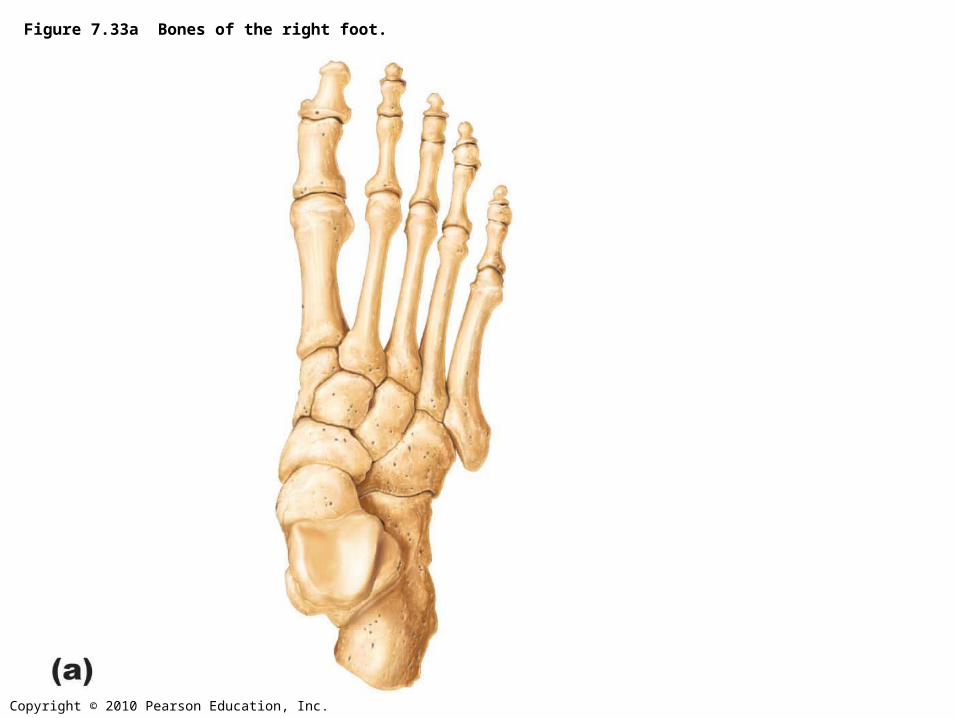
Figure 7.33a Bones of the right foot.
Copyright © 2010 Pearson Education, Inc.
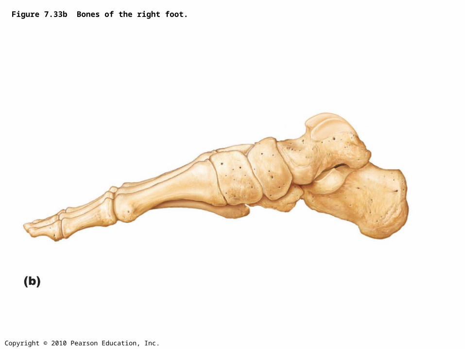
Figure 7.33b Bones of the right foot.
Copyright © 2010 Pearson Education, Inc.
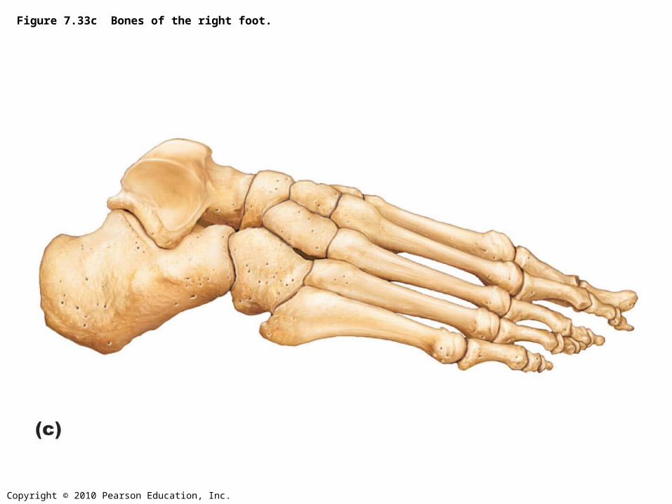
Figure 7.33c Bones of the right foot.

Copyright © 2010 Pearson Education, Inc.
Figure 7.34 Arches of the foot.
Copyright © 2010 Pearson Education, Inc.
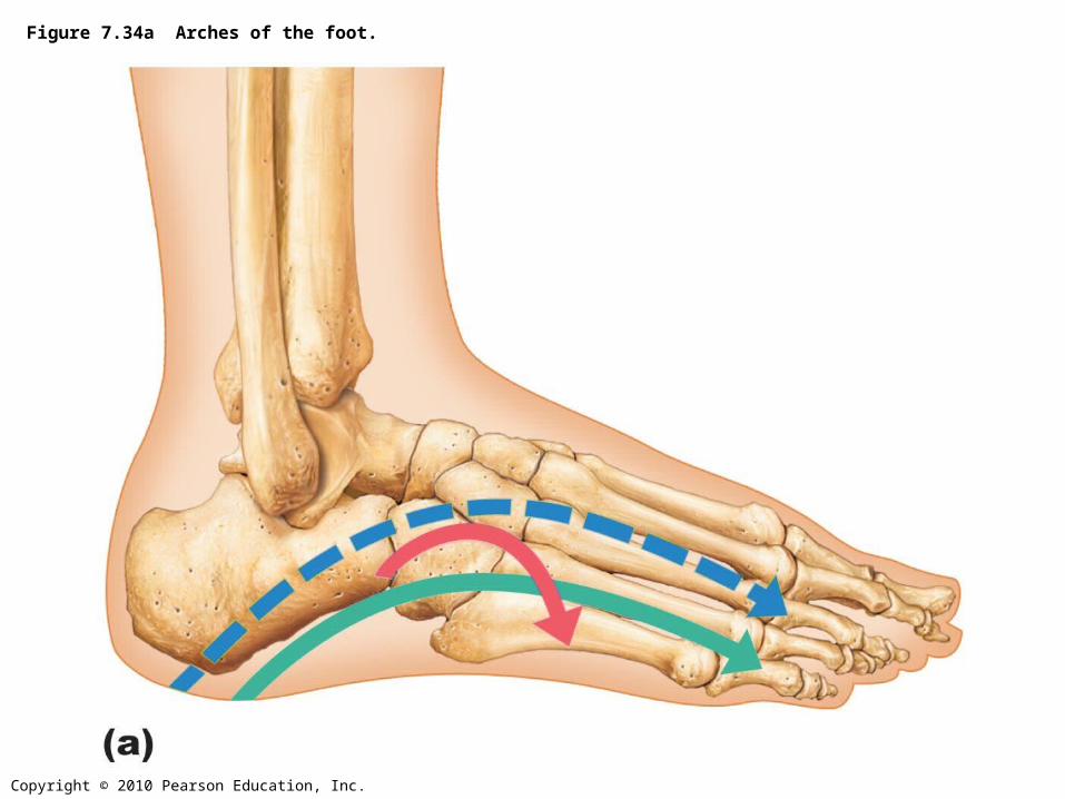
Figure 7.34a Arches of the foot.
Copyright © 2010 Pearson Education, Inc.
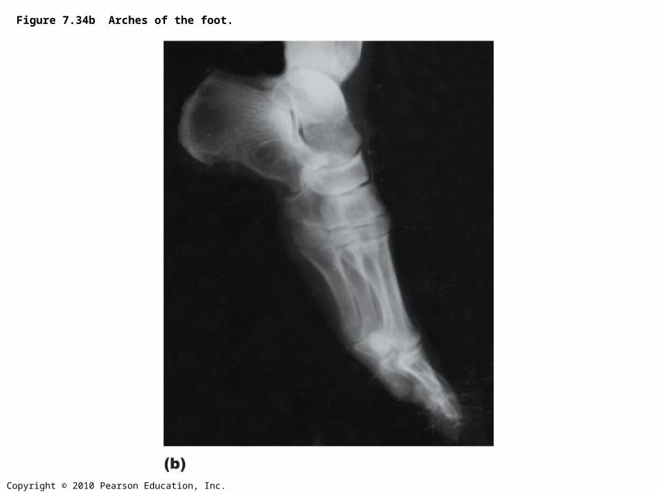
Figure 7.34b Arches of the foot.
Copyright © 2010 Pearson Education, Inc.
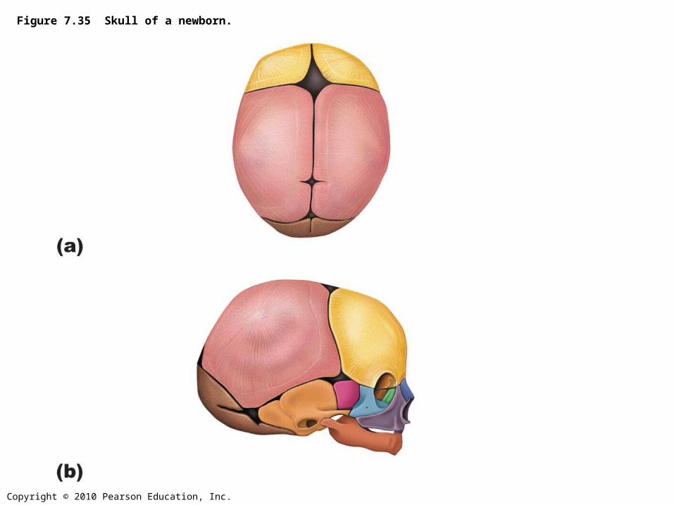
Figure 7.35 Skull of a newborn.
Copyright © 2010 Pearson Education, Inc.
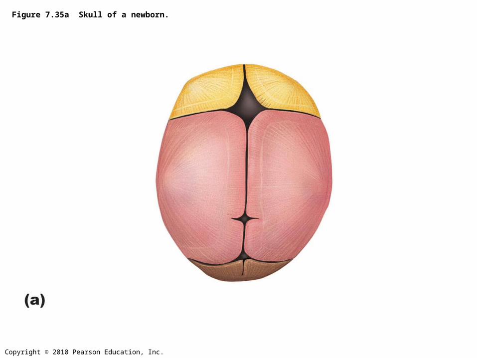
Figure 7.35a Skull of a newborn.
Copyright © 2010 Pearson Education, Inc.
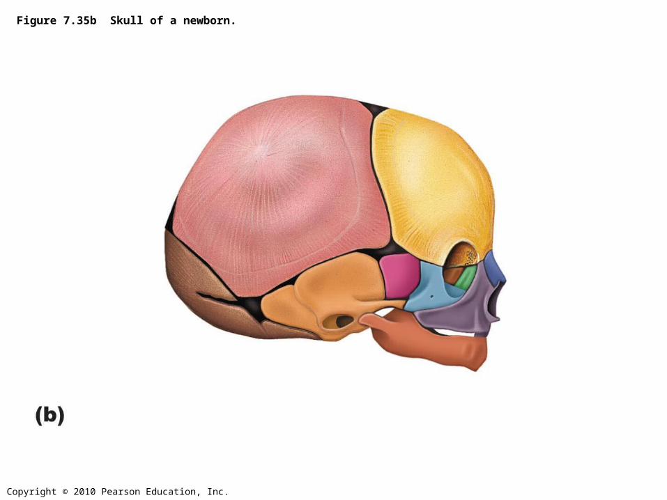
Figure 7.35b Skull of a newborn.
Copyright © 2010 Pearson Education, Inc.
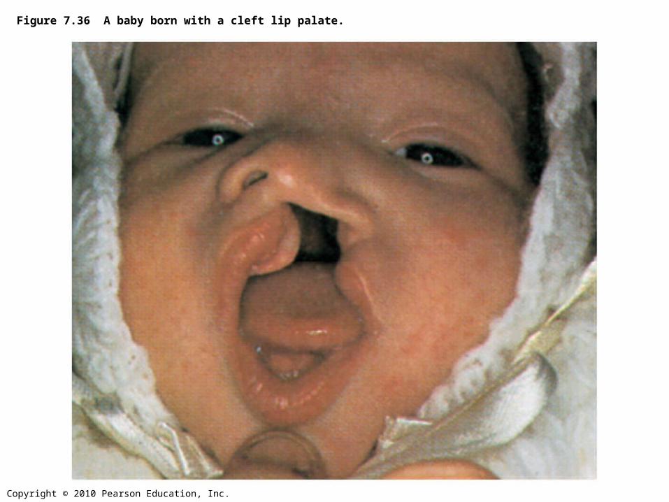
Figure 7.36 A baby born with a cleft lip palate.
Copyright © 2010 Pearson Education, Inc.
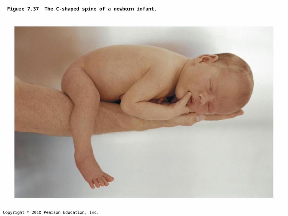
Figure 7.37 The C-shaped spine of a newborn infant.
Copyright © 2010 Pearson Education, Inc.
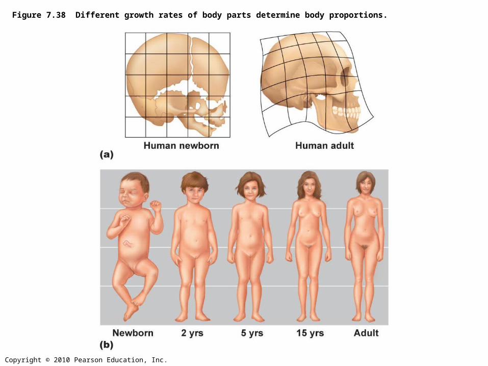
Figure 7.38 Different growth rates of body parts determine body proportions.
Top Related