


Languages
Pages
Legal


Coordination in Plants
You see plants all around you. But, are they of the same size or
height? Of course not! You see big trees, medium-sized shrubs, and
even plant saplings. This tells us that plants exhibit some growth
changes as well as some movements. This coordination in plants is
attributed to the presence of plant hormones. Unlike animals, plants do
not have any muscular system or nervous system. But, they are still
able to show movement and also coordination. These movements are
always controlled and not haphazard. Let us learn more about
coordination in plants.
What is coordination in plants?
Coordination is the ability to use different parts of the plant together,
smoothly and efficiently. In plants, coordination is due to the result of
a chemical system, wherein plant hormones or phytohormones have a
major role.
Movement in plants
Plants exhibit two types of movements.

1. Growth-dependent movements called the Tropic Movements. (
towards or away from a stimulus)
2. Non-growth dependent movements called the Nastic
Movements. ( independent of stimulus)
Tropic movements
These can be classified again into 5 types. They are:
● Phototropism (light)
● Geotropism (gravity)
● Hydrotropism (water )
● Chemotropism (chemicals)
● Thigmotropism (touch)
1. Phototropism – It is the movement of plants in response to
light. The shoot system of a plant exhibits this characteristic.
The shoot moves towards the light.
2. Geotropism – It is the movement of a plant part towards the
soil. This is a characteristic of the root system. The roots
always move in the direction of the earth’s gravity.
3. Hydrotropism– It is the movement of a plant towards the water.
The stimulus here is water.

4. Chemotropism – It is the movement of plants in response to a
chemical stimulus. A classic example of this type of movement
is the growth of the pollen tube towards the ovule, during
fertilization, in a flower.
5. Thigmotropism – It is a directional movement in plants in
response to touch. For e.g. the plant tendrils climb around any
support which they touch.
Nastic Movements
Nastic movements in plants are not directional movements. They are
not dependent on stimulus and are growth independent. For example,
the leaves of a touch me not plant (Mimosa pudica), fold up
immediately when touched. These kinds of changes occur due to the
changes in the amount of water in the leaves. Depending on the
quantity, they either swell up or shrink.

Plant hormones or phytohormones
They are responsible for the control and coordination of plants. There
are different types of hormones, which affect the growth of a plant.
Phytohormones are chemical compounds which are released by
stimulated cells. These hormones are diffused around the plant cells.
They have a role to play in the cell division, cell enlargement, cell
differentiation, fruit growth, falling of leaves, ripening of fruits,
ageing of plants etc.

The different types of phytohormones are:
1. Auxins
2. Gibberellins
3. Cytokinins
4. Abscisic acid
Auxins – They help in the cell growth at the shoot tips. By elongating
the cells, they help in the growth process.
Gibberellins – These hormones are responsible for the cell growth in
the stem, seed germination, and flowering.
Cytokinins – They promote cell division in plants. They also promote
the opening of the stomata and delay ageing in leaves.
Abscisic acid – This hormone inhibits the growth of the plant. And
therefore, it promotes dormancy in seeds and buds. The detachment of
fruits, flowers, and falling of leaves etc. are promoted by this
hormone.
Learn more about Animal Hormones here.

Solved Examples for You
Q: Name a plant hormone that promotes growth? How does it
promote the growth of a tendril around a support?
Solution: Auxins promote growth in plants. They are synthesized in
the shoot tips of the plant. When a plant tendril comes in contact with
a support, the auxins at the tip of the tendril stimulate faster growth of
cells, thus making the tendril grow faster and have a coiled
appearance.

Hormones In Animals
Animals are more complex than plants. They have many
specialised organs that perform specialised functions for their
control and coordination. This coordination in animals occurs due
to the nervous coordination (Nervous System) and the chemical
coordination (Endocrine System). Both these systems act in a
coordinated manner in animals, so as to regulate the various body
activities. Let’s learn more about the hormones in animals.
Endocrine System
The endocrine system comprises of different endocrine glands and
hormones. These endocrine glands in animals help in the chemical
coordination. They secrete chemicals called hormones. They are
special messengers that control many body functions, including
hunger, body temperature, mood, growth and development,
metabolism, reproductive processes etc. The endocrine glands are
ductless and hence are also called as ductless glands.
What are Hormones?
Hormones are chemicals secreted by the endocrine glands directly
into the bloodstream. Through the blood, these hormones in

animals reach their target organs to stimulate or inhibit specific
physiological processes. The site of production of the hormones is
different and the site of action is different. Even though there are
different hormones in the bloodstream, each will act only on the
specific target organ. There are around 20 major hormones in
animals that are released by the endocrine glands into the blood,
playing a major role in many of the physiological processes
happening in the body.
Did you know that the hormone levels in the body can be
influenced by several factors? Stress, infection, minerals in the
blood etc. influence the hormone levels in animals.
Different Endocrine Glands
● Hypothalamus
● Pituitary gland
● Thyroid gland
● Parathyroid gland
● Pineal gland
● Adrenal gland
● Pancreas
● Testes
● Ovaries
(Source – Wikipedia)
Hypothalamus
This gland forms an important link between the nervous system
and the endocrine system, via the pituitary gland.
Some of the important functions are:
● Helps in maintaining the body temperature, controls sleep,
hunger, thirst, emotions and moods.

● It also controls the release of 8 major hormones by the
pituitary gland.
● Controls the sexual behaviour and reproduction.
● It controls the circadian rhythm of the body.
Pituitary Gland
The pituitary gland is very small in size but is called as the Master
Gland, as many endocrine glands are controlled by the hormones
secreted by it. It also stimulates other endocrine glands to produce
hormones. Some of the hormones released by this gland are
growth hormone, thyroid stimulating hormone, MSH, LH, FSH
etc.
Thyroid Gland
It is the largest endocrine gland that is shaped like a butterfly. It
produces the thyroxine hormone, which controls the metabolic
rate in the body. Apart from that, it also plays a role in the bone
growth, development of the brain and nervous system in children.
Iodine is important for the synthesis of thyroxine.
Parathyroid Gland

This gland releases parathormone which helps in regulating the
calcium and phosphorus levels in the bone.
Pineal gland
This produces melatonin hormone that regulates the sleep
patterns.
Adrenal gland
These glands are located on top of the kidneys and produce
hormones such as adrenaline, cortisol, and aldosterone etc. These
hormones control stress, help control blood sugar, burn protein
and fat and also regulate blood pressure.
Pancreas
They secrete two important hormones – insulin and glucagon.
Both work together to maintain the glucose levels in the blood.
Testes
These glands are present in males and produce testosterone
hormone.
Ovaries

These glands are present in females. The hormones produced by
ovaries are oestrogen and progesterone.
Solved Examples For You
1. Why is the use of iodised salt recommended?
Ans. The hormone thyroxine that is produced in the thyroid gland
is stimulated by iodine. Hence it is advisable to have sufficient
intake of iodine, so that thyroid glands function normally.
Deficiency of thyroid hormone leads to goitre.
2. The gland that produces insulin – Pancreas
Human Brain
Weighing at just 2% of the total body weight, the human brain is
probably the most complex structure. It is the centre of control of
all the body functions and senses. The average weight of the
human brain is around 1.5 kg and it contains about 86 billion
nerve cells (neurons) that control every action you take. Read
more…

The human brain along with the spinal cord, make up the Central
Nervous System in humans. It is the command centre for the
Nervous system. The brain controls even the smallest of our
actions such as laughing; crying out in pain, walking, breathing,
etc. It is a communication centre where decision making happens
and is also responsible for the communication and coordination
between different parts of the body. Now, this communication
takes place with the help of neurons or nerve cells that send
information in the form of chemical and electrical signals. The
brain receives input from the sensory organs and then sends out
the output to the muscles.
Parts of the human brain
The human brain is made up different parts and compartments,
for each of the functions. Take a look at the different parts of the
brain.
● Forebrain
● Midbrain
● Hind Brain

(Source – Wikipedia)
Fore Brain
It is the main thinking part of the brain and controls the
voluntary actions. The forebrain processes sensory information
that is collected from the various sense organs such as ears, eyes,
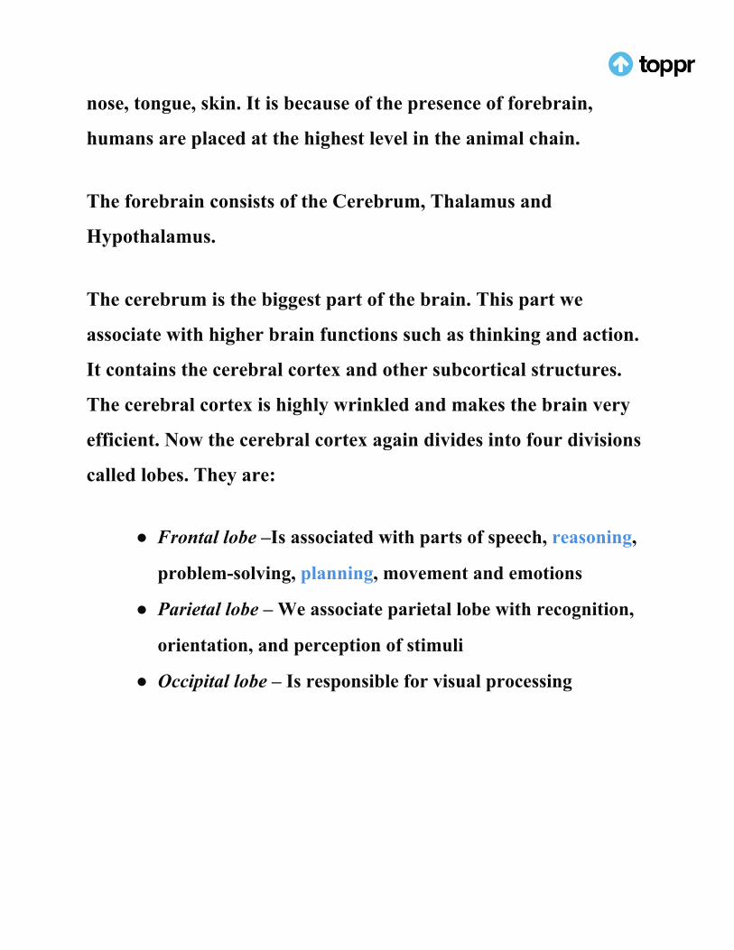
nose, tongue, skin. It is because of the presence of forebrain,
humans are placed at the highest level in the animal chain.
The forebrain consists of the Cerebrum, Thalamus and
Hypothalamus.
The cerebrum is the biggest part of the brain. This part we
associate with higher brain functions such as thinking and action.
It contains the cerebral cortex and other subcortical structures.
The cerebral cortex is highly wrinkled and makes the brain very
efficient. Now the cerebral cortex again divides into four divisions
called lobes. They are:
● Frontal lobe –Is associated with parts of speech, reasoning,
problem-solving, planning, movement and emotions
● Parietal lobe – We associate parietal lobe with recognition,
orientation, and perception of stimuli
● Occipital lobe – Is responsible for visual processing

● Temporal lobe – Finally temporal lobe is associated with
memory, speech perception and recognition of auditory
stimuli
The cerebrum is divided into two halves by a deep furrow. These
halves are the left and right hemispheres. Each side functions
slightly different from the other, even though they are
symmetrical. So the right hemisphere links to creativity whereas
the left hemisphere relates to logic abilities. And Corpus callosum
connects the two hemispheres.
Mid Brain
The midbrain connects the forebrain and the hindbrain. It acts as
a bridge and transmits signals from hindbrain and forebrain. It is
associated with motor control, vision, hearing, temperature
regulation, alertness.
Hind Brain
It is the control centre for visceral function.As a result, this part of
the brain plays a role in controlling the heart rate, breathing,
blood pressure, sleep and waking up functions etc. The hindbrain

has three parts, namely – medulla oblongata, pons and
cerebellum.
The cerebellum is responsible for maintaining equilibrium,
transfer of information, fine adjustments to motor actions,
coordinating eye movements etc. Coordination and body balance,
posture during walking, riding, standing, swimming, running, are
all maintained by the cerebellum.
Learn more about Reflex Action here in detail.
Solved Questions For You
Q: You are walking in a straight line or bending down to pick up
a pencil. Which part of the brain controls these actions?
Ans: Cerebellum
Q: What connects the two hemispheres of the brain?
Ans: Corpus callosum.

Nervous System
Did you know there are more nerve cells in our bodies than there
are stars in the Milky Way? The nervous system is one of the most
important systems in the human body that sends information
from one part of the body to another. It is because of this system
that we are able to control and coordinate our movements and
actions.
Parts of the Nervous System
It consists of two main parts.
● The Central Nervous System ( CNS)
● Peripheral Nervous System (PNS)

(Source – Wikipedia)
Central Nervous System
This system consists of the Brain and Spinal Cord. Find more
about human brain in the related posts.
Peripheral Nervous System
It consists of the cranial nerves coming from the brain and the
spinal nerves coming from the spine. There are 12 pairs of cranial
nerves and 31 pairs of spinal nerves in humans.
The peripheral nervous system is made of up of the Autonomic
nervous system and Somatic Nervous System. The Sympathetic

nervous system and Parasympathetic nervous system fall under
the autonomic nervous system.
The following figure explains the interrelation between the
various nervous systems.
(Source – Wikipedia)
Nerves

A nerve is a thread like structure that comes out of the brain and
the spinal cord. So these nerves branch out to all the parts of the
body and are mainly responsible for carrying information and
messages from part to the other. All the nerves make up the
peripheral system. They carry information between the brain and
spinal cord.
Types of Nerves
There are different types of nerves, according to the action they
perform. They are:
● Sensory Nerves – These send messages to the brain from all
the sensory organs,
● Motor Nerves – They carry messages from the brain to the
muscles in the body.
● Mixed Nerves – They carry the sensory and motor nerves.
They help in conducting the incoming sensory information
and also the outgoing information to the muscle cells.

Based on which part the nerves connect to the Central Nervous
System, they are classified as:
● Cranial Nerves – They start from the brain and carry
messages from the brain to the rest of the body. Certain
nerves are sensory nerves while some are mixed nerves.
● Spinal Nerves – These nerves originate from the Spinal
Cord. They carry messages to and from the central nervous
system. They consist of mixed nerves.
Neurons
Nerves are made up special cells called the nerve cells or neurons.
These neurons are the basic unit of the nervous system.
( Source – Wikipedia)

Three parts make up a neuron – axon, cell body and nerve
endings. The cell body is the main part and has all the components
of the cell such as the nucleus, mitochondria, endoplasmic
reticulum etc. Axons are long cable-like projections that carry the
messages along the length of the cell. These axons are covered by a
protective covering called the myelin sheath. This myelin is made
of fat and has a role in speeding up the transmission of messages
down a long axon. Dendrites are the small branch-like projections
that form connections with other neurons these dendrites can be
present at both ends of the cell.
Synapse is the junction between two nerve cells. It consists of a
minute gap. Impulses or messages pass by diffusion of a
neurotransmitter.
Some Amazing Facts
● Neurons have high energy requirements and are bundled
with blood vessels.
● There are billions of neurons in the body, about 100 billion
in the brain and 13.5 million in the spinal cord.

● Axons can transmit electrical signals at a rate of 2,500 per
second.
Solved Questions for You
Q: What is the gap between two nerve cells called?
Ans. Synapse
Q: How does the nervous system mechanism function?
Ans. Broadly, the mechanism of the nervous system can be
summarised as follows:
● Information is transmitted in the form of electric impulse.
● The flow of information is very quick.
● Axons and dendrites transmit information throughout the
body, in a coordinated effort.

Reflex Action
What will be your reaction, when you accidentally touch a hot,
burning object? Naturally, your reflexes will kick into action and
you withdraw your hand. You will not wait for some time and
then move your hand slowly. Otherwise, your hand will get burnt!
In such situations, your reactions are quick, involuntary and
happen without much thinking. This is nothing but a reflex action.
Reflex Action
Even though it is the brain that gives messages for muscles to act,
in a reflex action, the spinal cord is the one that sends the
messages to the muscles. Control and coordination in reflex
actions are different. Our brain is a complex centre and the
communication between the thinking part of the brain and other

muscles is very intense. But, all this takes some time. This may not
work well in sudden situations because even nanoseconds lost are
crucial when communication happens between the brain and
sense organs. Now, here is when the spinal cord comes into action.
When the body senses danger, the input-output of messages
between the nerves and the corresponding action must be
completed very quickly. The spinal cord sends the required
signals to the muscles and the reflex action is completed. Since the
action is instantaneous, the brain involvement is not much in
reflex action.
Reflex Arc
Reflex actions happen through the reflex arc, which is a neural
pathway that controls the reflexes. It acts on an impulse even
before it reaches the brain. Some stimuli require an automatic,
instantaneous response without the need of conscious thought.
The following diagram shows the reflex arc pathway.

(Source – NCERT)
The receptor here is the sense organ that senses danger. The
sensory neurons pick up signals from the sensory organ and send
them through other neurons which are interconnected. It is then
received by the relay neuron which is present in the spinal cord.
Immediately, the spinal cord sends back signals to the muscle
through the motor neuron. The muscles attached to the sense
organ move the organ away from danger. In reflex actions, the
signals do not travel up to the brain.
A Few Examples of Reflex Action

● When light acts as a stimulus, the pupil of the eye changes
in size.
● Coughing or sneezing, because of irritants in the nasal
passages.
● Knees jerk in response to a blow or someone stamping the
leg.
● The sudden removal of the hand from a sharp
● Sudden blinking when an insect comes very near to the
eyes.
Solved Questions for You
Q: What are receptors?
Ans: Specialised cells which react to a stimulus are called
Receptors.
Q: What is a reflex arc?
Ans: A reflex arc is a path along which the nerve impulse travels
from the receptor cells to the effector muscles.

Q: Differentiate between walking and any reflex action
Ans: Walking is a voluntary action that involves the exchange of
messages between the brain and the leg muscles Reflex action, on
the other hand, is an involuntary action in response to a stimulus
that does not involve thinking.
Top Related