


Languages
Pages
Legal

Confocal & Two-photon Microscopy
Contents
1. Two-Photon Microscopy : Basic principles and Architectures
2. Resolution and Contrast in Confocal and Two-Photon microscopy
3. Example of two-photon images
4. Extensive application: Fluorescence Correlation Microscopy,
Life-time imaging
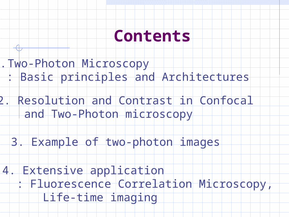
Figure 2, Relevant time scale.
1. Two-Photon Microscopy (vs One-photon)
Figure 1. Jablonski diagram
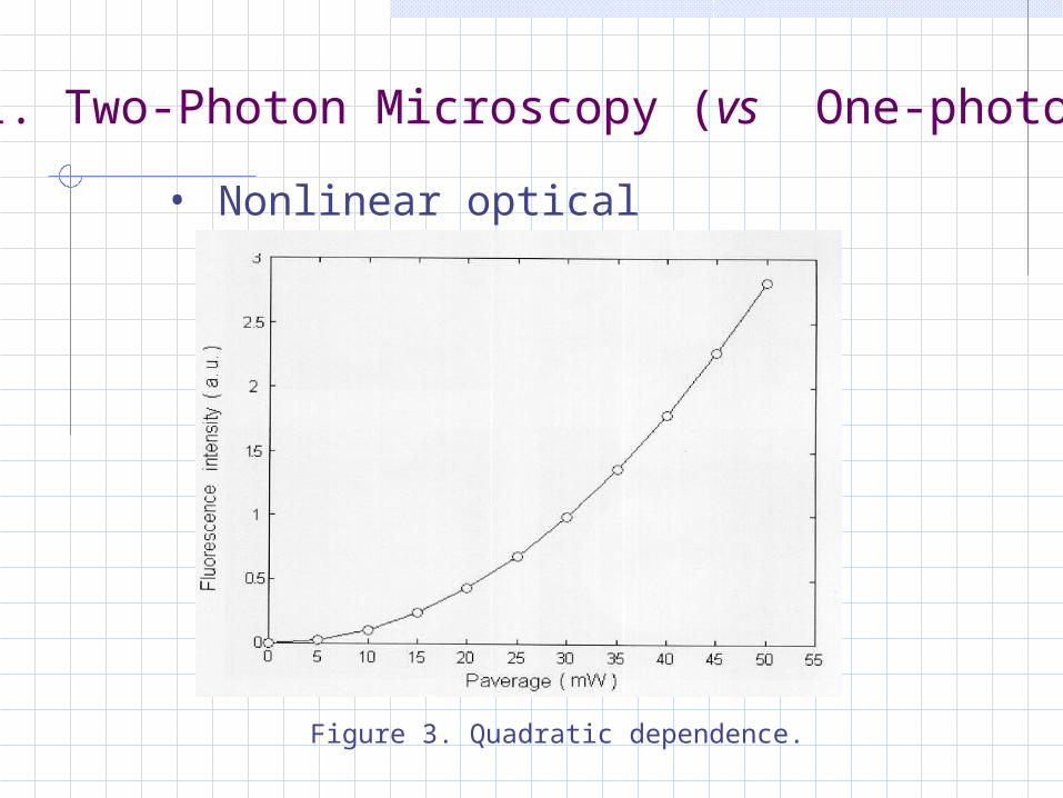
• Nonlinear optical excitation , I2p P2
1. Two-Photon Microscopy (vs One-photon)
Figure 3. Quadratic dependence.
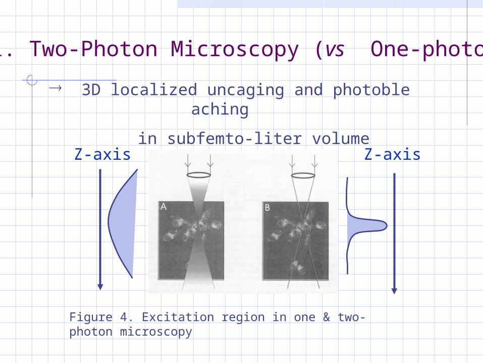
3D localized uncaging and photobleaching
in subfemto-liter volume
1. Two-Photon Microscopy (vs One-photon)
Figure 4. Excitation region in one & two-photon microscopy
Z-axis Z-axis
1. Two-Photon Microscopy (vs One-photon)
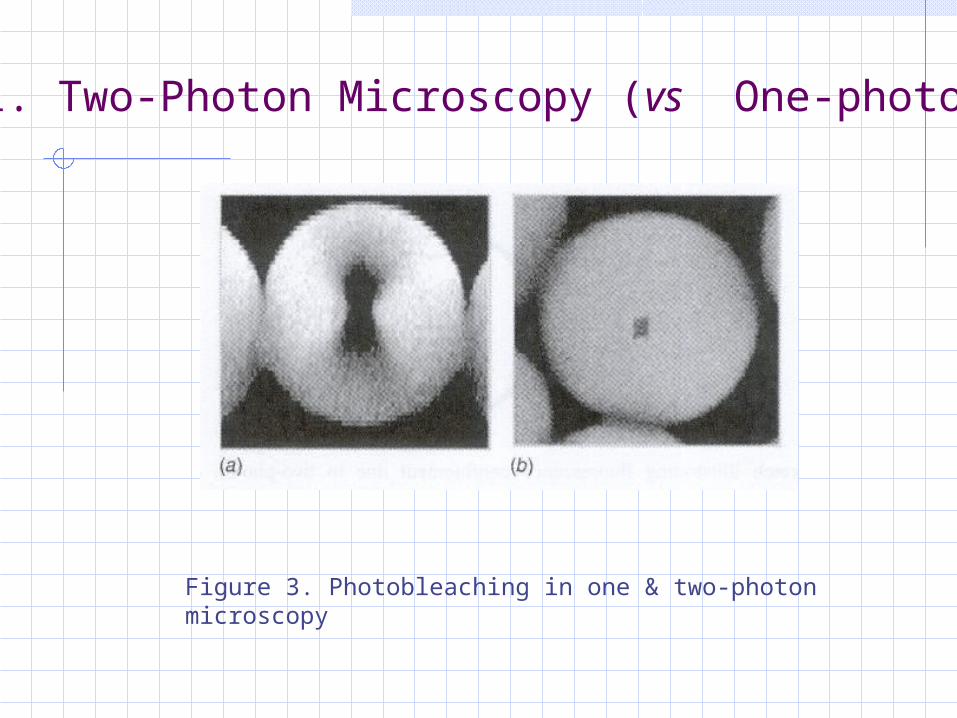
Figure 3. Photobleaching in one & two-photon microscopy
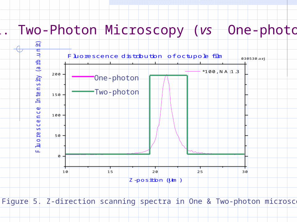
10 15 20 25 30
0
50
100
150
200 *100, NA:1.3
030530.orjFluorescence distribution of octupole film
Flu
ore
sce
nce
In
ten
sity (
arb
.un
it)
Z-position (m)
1. Two-Photon Microscopy (vs One-photon)
One-photon
Two-photon
Figure 5. Z-direction scanning spectra in One & Two-photon microscopy
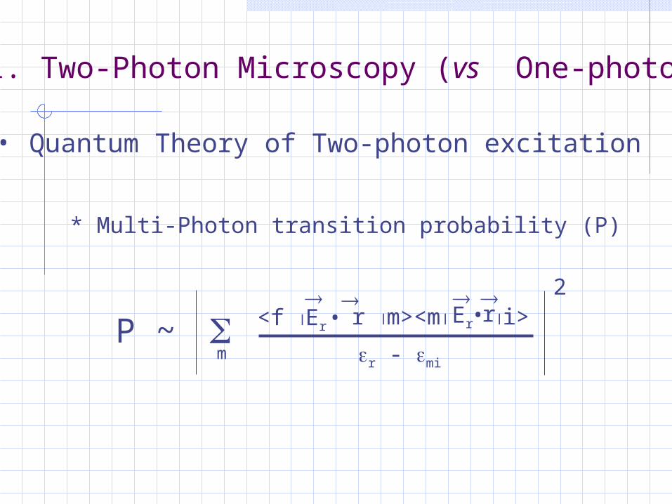
• Quantum Theory of Two-photon excitation
P ~ <f Er •
r m><m Er
•ri>
r - mi
2
m
* Multi-Photon transition probability (P)
1. Two-Photon Microscopy (vs One-photon)
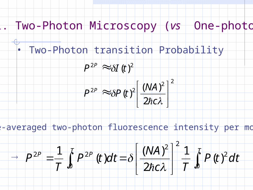
• Two-Photon transition Probability
2222
22
2
)()(
)(
c
NAtPP
tIP
P
P
* Time-averaged two-photon fluorescence intensity per molecule
TT PP dttPTc
NAdttP
TP
0
2
2
0
222 )(
1
2
)()(
1
1. Two-Photon Microscopy (vs One-photon)
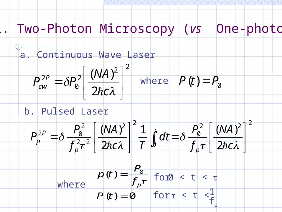
a. Continuous Wave Laser22
20
2
2
)(
c
NAPP P
cw where
0)( PtP
b. Pulsed Laser222
0
0
22
22
202
2
)(1
2
)(
c
NA
f
Pdt
Tc
NA
f
PP
pp
Pp
where0)(
)( 0
tP
f
Ptp
p 0 < t < for
1 < t < fp
for
1. Two-Photon Microscopy (vs One-photon)
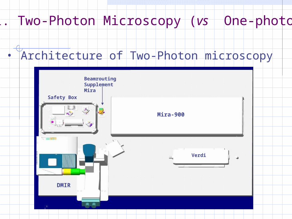
• Architecture of Two-Photon microscopy
1. Two-Photon Microscopy (vs One-photon)
Safety Box
BeamroutingSupplement Mira
Mira-900
DMIR
Verdi
1. Two-Photon Microscopy (vs One-photon)
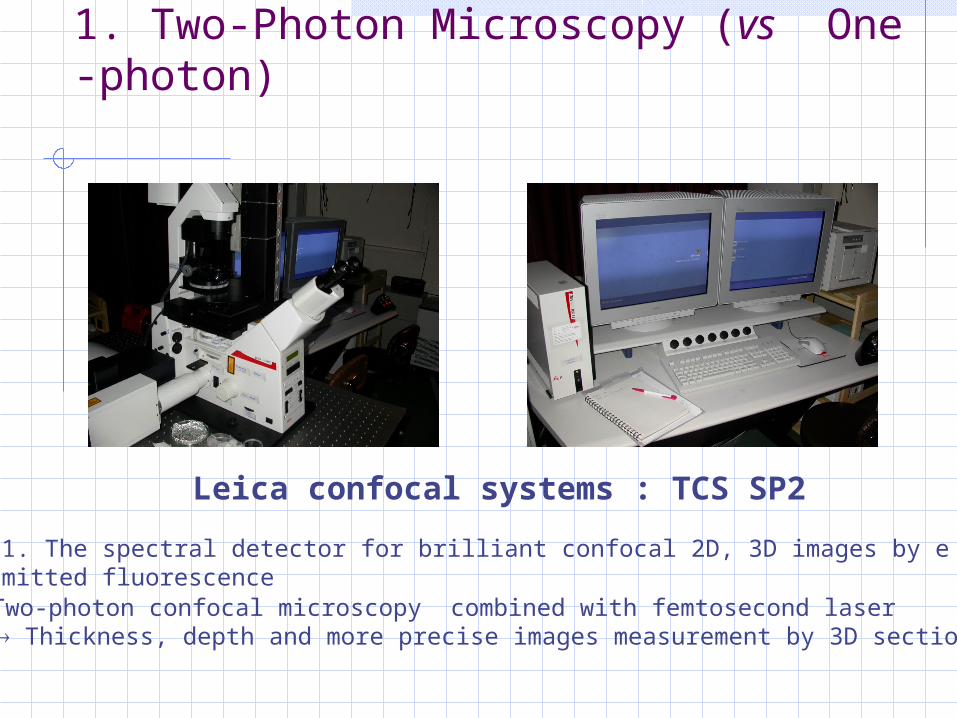
Leica confocal systems : TCS SP2
2. Two-photon confocal microscopy combined with femtosecond laser Thickness, depth and more precise images measurement by 3D sectioning
1. The spectral detector for brilliant confocal 2D, 3D images by emitted fluorescence
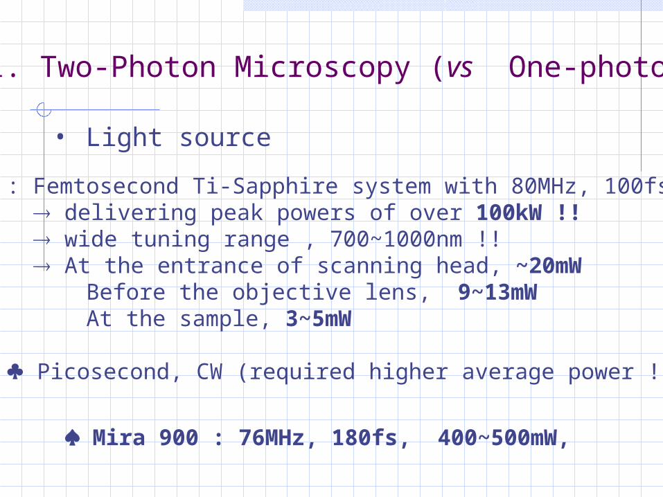
: Femtosecond Ti-Sapphire system with 80MHz, 100fs delivering peak powers of over 100kW !! wide tuning range , 700~1000nm !! At the entrance of scanning head, ~20mW Before the objective lens, 9~13mW At the sample, 3~5mW
Picosecond, CW (required higher average power !!)
• Light source
1. Two-Photon Microscopy (vs One-photon)
Mira 900 : 76MHz, 180fs, 400~500mW,
1. Two-Photon Microscopy (vs One-photon)
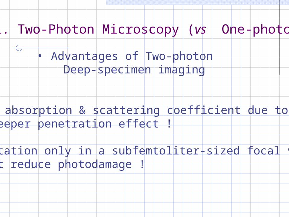
• Advantages of Two-photon Deep-specimen imaging
a. Lower absorption & scattering coefficient due to IR : Deeper penetration effect !
b. Excitation only in a subfemtoliter-sized focal volume : It reduce photodamage !
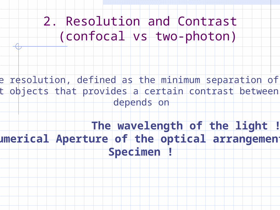
The resolution, defined as the minimum separation of twoPoint objects that provides a certain contrast between them,
depends on
The wavelength of the light !Numerical Aperture of the optical arrangement !
Specimen !
2. Resolution and Contrast (confocal vs two-photon)
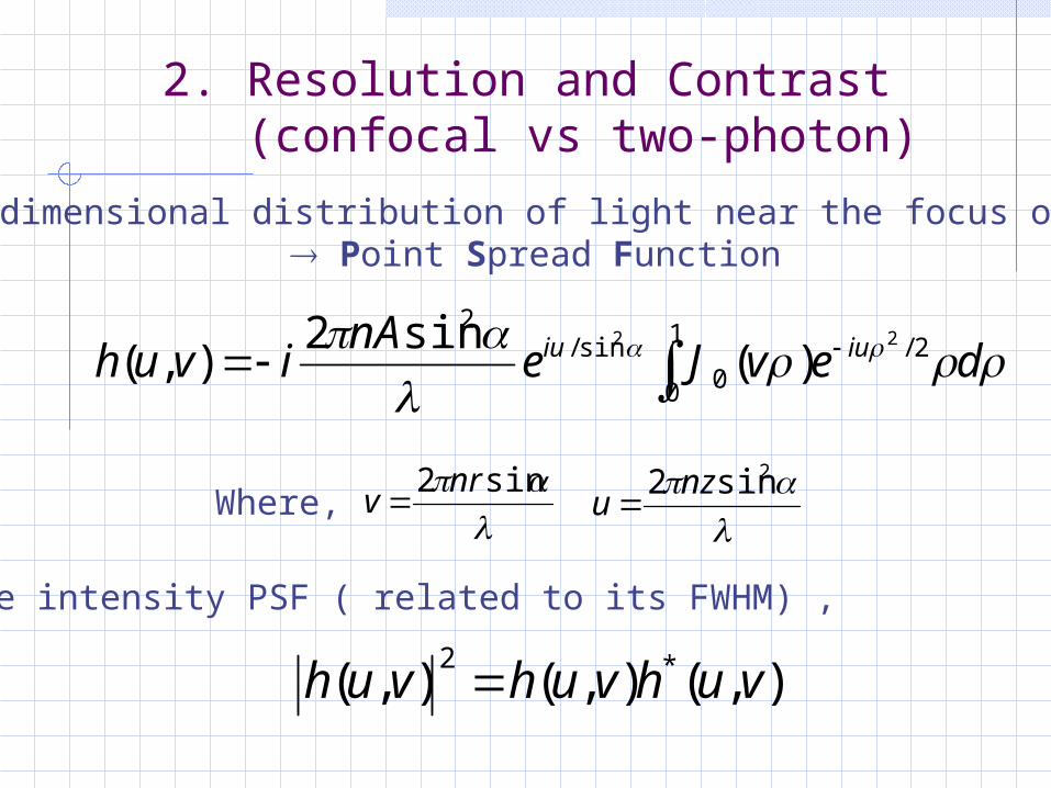
•Three-dimensional distribution of light near the focus of lens Point Spread Function
1
0
2/0
sin/2
22
)(sin2
),(
devJenA
ivuh iuiu
sin2 nr
v
2sin2 nzu Where,
The intensity PSF ( related to its FWHM) ,
),(),(),( *2vuhvuhvuh
2. Resolution and Contrast (confocal vs two-photon)
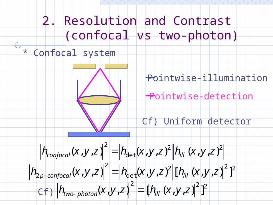
* Confocal system
Pointwise-illumination
Pointwise-detection
22det
2),,(),,(),,( zyxhzyxhzyxh illconfocal
222det
2
2 ]),,([),,(),,( zyxhzyxhzyxh illconfocalp
222]),,([),,( zyxhzyxh illphotontwo Cf)
Cf) Uniform detector
2. Resolution and Contrast (confocal vs two-photon)
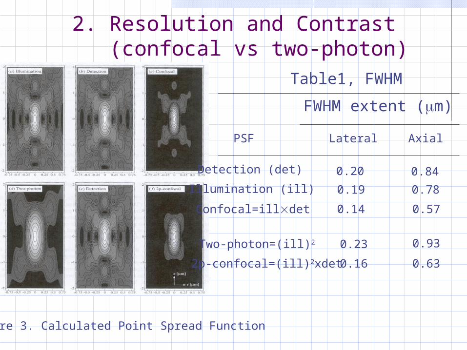
Table1, FWHM
2. Resolution and Contrast (confocal vs two-photon)
Figure 3. Calculated Point Spread Function
FWHM extent (m)
Illumination (ill)
Detection (det)
Lateral AxialPSF
Confocal=illdet
Two-photon=(ill)2
2p-confocal=(ill)2xdet
0.20 0.84
0.19 0.78
0.14 0.57
0.23
0.16
0.93
0.63
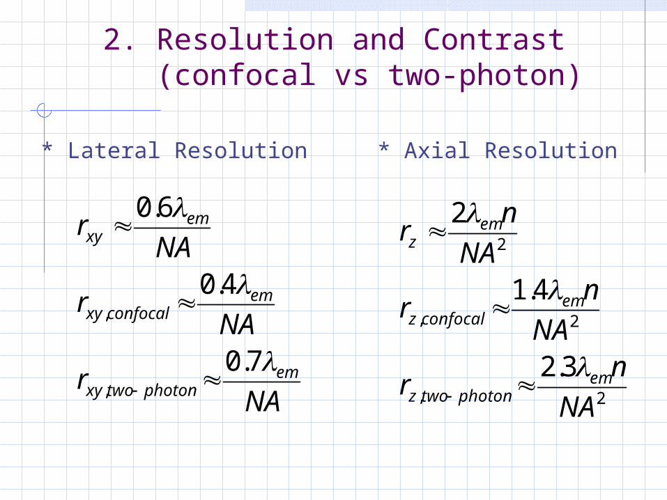
* Lateral Resolution
NAr
NAr
NAr
emphotontwoxy
emconfocalxy
emxy
7.0
4.0
6.0
,
,
2. Resolution and Contrast (confocal vs two-photon)
2,
2,
2
3.2
4.1
2
NA
nr
NA
nr
NA
nr
emphotontwoz
emconfocalz
emz
* Axial Resolution
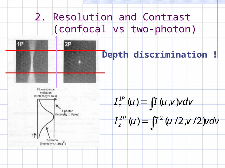
vdvvuIuI
vdvvuIuI
Pz
Pz
)2/,2/()(
),()(
22
1
Depth discrimination !
2. Resolution and Contrast (confocal vs two-photon)
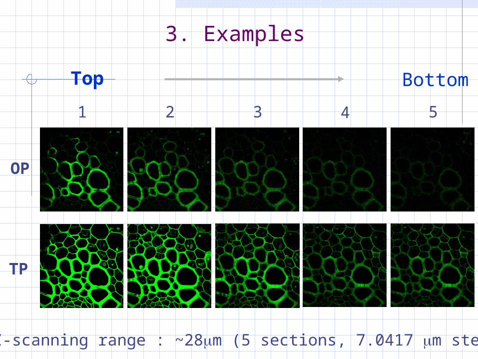
3. Examples
1 43 52
OP
Top Bottom
TP
Z-scanning range : ~28m (5 sections, 7.0417 m step)
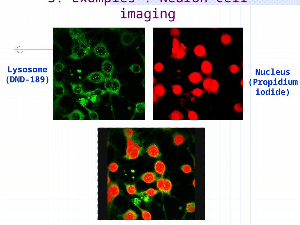
3. Examples : Neuron cell imaging
Lysosome(DND-189)
Nucleus(Propidium
iodide)
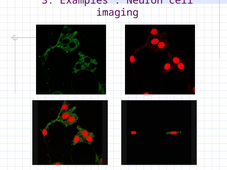
3. Examples : Neuron cell imaging
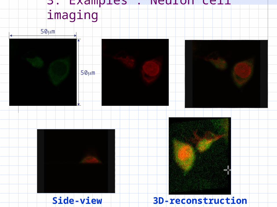
3. Examples : Neuron cell imaging
Side-view 3D-reconstruction
50m
50m
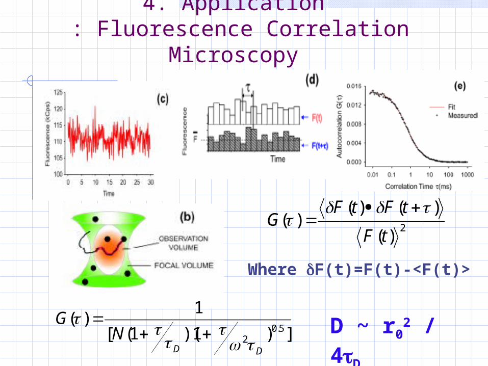
4. Application : Fluorescence Correlation
Microscopy
2)(
)()()(
tF
tFtFG
Where F(t)=F(t)-<F(t)>
])1)(1([
1)(
5.02DD
NG
D ~ r0
2 / 4D
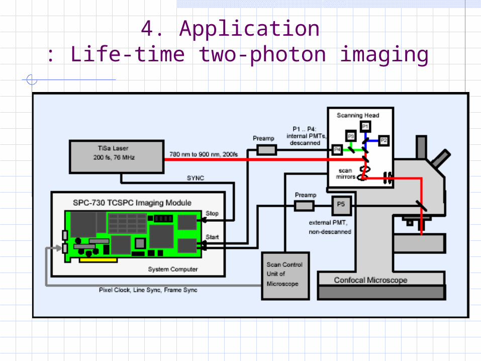
4. Application : Life-time two-photon imaging
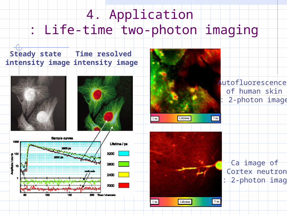
4. Application : Life-time two-photon imaging
Steady state intensity image
Time resolved intensity image
Autofluorescence of human skin
: 2-photon image
Ca image of Cortex neutron
: 2-photon image
Top Related