


Languages
Pages
Legal


CKD-MBD
By Stephen Z. Fadem, M.D., FACP, FASN
Clinical Professor of Medicine
Baylor College of Medicine
May 23, 2007

Goals
• Understand the new definition
that ties vascular calcification,
mineral disorders and bone
abnormalities together - CKD-
MBD
• Understand new concepts on
bone adaptation to CKD
• Be familiar with the therapies
directed toward preventing
extraskeletal calcification and
low bone turnover syndrome
PHOTOS BY STEPHEN FADEM
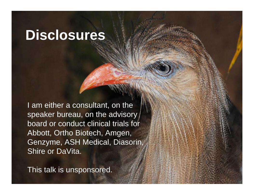
Disclosures
I am either a consultant, on the
speaker bureau, on the advisory
board or conduct clinical trials for
Abbott, Ortho Biotech, Amgen,
Genzyme, ASH Medical, Diasorin,
Shire or DaVita.
This talk is unsponsored.

ESTABLISHED
ASSOCIATION
BETWEEN VASCULAR
CALCIFICATION AND
RENAL FAILURE
1968

Renal Osteodystrophy linkage to
vascular calcification is not new
“Consideration is also given to the
manifestations of soft-tissue
calcification, both of the vascular and
subcutaneous type, and to the effects of
treatment”– Source: Eastwood, JB. Renal osteodystrophy - a radiological view,
CRC Crit Rev Diagn Imaging. 1977 Apr;9(1):77-104.

1985 - Uremic Arterial Disease
• Rabbit model of 9 months renal failure
• All major systemic arteries affected
• Medial degeneration without lipidaccumulation
• No coronary stenosis
• Medial calcification in non-cholesterol-fedrabbits
• Uremic arterial disease different thanatherosclerosis– Acta Pathol Microbiol Immunol Scand [A]. 1985 Mar;93(2):81-
8.Uremic arterial disease in rabbits with special reference tothe coronaryarteries.Tvedegaard E, Falk E, Nielsen M
medial
calcification

1987 Aortic and Mitral valve
Calcification in ESRD• Echocardiography in 87 patients
• 35 to 70 years old
• Maintenance dialysis 7.5 years
• 24 patients: aortic valve calcification
• 31 patients: mitral annular calcification– Source: Lancet. 1987 Oct 17;2(8564):875-7.Aortic and mitral valve calcification inpatients with end-stage renal disease.MaherER, Young G, Smyth-Walsh B, Pugh S, CurtisJR.

1990 - Pulse Wave Velocity -
Aorta and large artery compliance in ESRD
• 90 control and 92 hemodilaysis patients
• Matched for age and MAP
• Aortic calcification - plain films and echo– PWV 1113±319 cm/sec in HD
– 965 ± 216 cm/sec in Control (P=0.0016)
• Pulse Pressure– HD 76.6 ± 23.7 mg Hg
– CS 63.9 ± 22 mg Hg (p=0.007)– Source: Kidney Int. 1990 Jan;37(1):137-42.Aortic and
large artery compliance in end-stage renalfailure.London GM, Marchais SJ, Safar ME, Genest AF,Guerin AP, Metivier F, Chedid K,London AM.CentreHospitalier Manhes, Fleury Merogis, France

2001 - Longitudinal Study linking
calcification with mortality
Source: Hypertension. 2001 Oct;38(4):938-42.Arterial
calcifications, arterial stiffness, and cardiovascular risk
in end-stage renal disease.Blacher J, Guerin AP, Pannier B,
Marchais SJ, London GM.

Linkage of vascular calcification
and bone
• J Am Soc Nephrol. 2003 Jun;14(6):1559-67. BMP-7is an efficacious treatment of vascularcalcification in a murine model ofatherosclerosis and chronic renalfailure.Davies MR, Lund RJ, Hruska KA.
• Kidney Int. 2002 Feb;61(2):638-47. Medialartery calcification in ESRD patients isassociated with deposition of bone matrixproteins.Moe SM, O'Neill KD, Duan D, Ahmed S,Chen NX, Leapman SB, Fineberg N, Kopecky K
• Nephron. 2001 Dec;89(4):455-8.Solubleosteopontin and vascular calcification inhemodialysis patients.Nitta K, Ishizuka T, etal.

LDLR-/- 5/6 Nephrectomized Mice with
Metabolic SyndromeJ Am Soc Nephrol 16: 917-928, 2005
LDLR-/- mice
• High fat, cholesterol diets, and normal kidneys– Decreased bone turnover, vascular calcification and
hyperphosphatemia
• Added 5/6 nephrectomy– LBT worsened
• Treating with BMP-7– Corrected LBT, hyperphosphatemia and vascular
calcification
• Decreased VC by reducing the serum P04 with aphosphate binder
Eur J Clin Invest. 2006 Aug;36 Suppl 2:43-50

BMP-7 and VC in LDLR-/- mice
J Am Soc Nephrol 14: 1559–1567, 2003
Vascular calcium
deposition is blocked
in fat-fed, uremic,
LDLR-/-mice treated
with BMP-7

How the LDLR related to
Bone Formation?
OSTEOBLAST
DIFFERENTIATION

Mechanism of LDLR-/- VSMC
Calcification
J Clin Invest. 2005 May 2; 115(5): 1210 1220.
UPREGULATEDPyrophosphate
-generating
(PPi-generating)
enzyme
Blocks VC

Difference between blood vessel and bone
is the MSX2 in adventitial fibroblasts
BMP2
LRP5/6 + WNT
MSX2
Osteoblastic
vascular
calcification
(WNT signaling does not occur
because there is no LDL Co-
receptor)
WNT
In LDLR -/- mice
skeletonization
decreases
RUNX2(Cbf 1)

COLLAGEN RICH
ECM
Ca-P04
Pi
Pi Na
ALKALINE
PHOSPHATASE
OSTEOBLAST
DIFFERENTIATION
Na Pi co
transporter
NONCOLLAGENOUS
PROTEIN PRODUCTION
Osteocalcin
Osteopontin
Matrix Gla Protein
BMP-2a
Alkaline Phosphatase
APOPTOSIS
Pi Na
CALCIFICATION
Ca++Matrix
Vesicle
Adapted from Shioi, et al. Am J Kidney Dis 38:S47-S49, 2001
Adapted from Giachelli, et al. Am J Kidney Dis 38:S34-S37, 2001
Chen, et al. Kidney International 70:1046-1053, 2006
PDGF
RUNX2(Cbf 1)
RANKL? CLAST-
LIKE
CELLS
OPGFAT/BMP-2 promotes
BMP-7 blocks
Vascular Smooth Muscle CellVascular Smooth Muscle Cell

Bone Morphogenetic Protein
Receptors
SIGMA-
ALDRICH
BMP-7
• TGFß superfamily
• Induces SMAD
• 14 BMPS (BMP-2 inducesvascular calcification)
• 20q13 (long arm, 13th bandof chromosome 20)
• Holt-Oram - BMP-7– Nonapposable thumb, ASD
• BMP-7 downegulated earlyin kidney failure
• Maintains VSMCdifferentiation - blockstransformation to osteoblastRUNX2
MESENCHYME PRECURSOR OSTEOBLAST DIFFERENTIATION

BMP ACTIONS
BMP2/BMP 7 - Needed for Osteoblast Differentiation
BMP7 - Blocks Vascular calcification

BMP-7 has great potential
• Blocks tubular epithelialcell de-differentiation,
• Blocks mesenchymaltransformation andapoptosis
• Preserves glomerularintegrity
• Inhibits injury-mediatedmesangial matrixaccumulation.
• Eliminates peritrabecularfibrosis
• Decreases bone resorptionand restores normal ratesof bone formation
• Increases the skeletaldeposition of ingestedphosphorus and calcium,preventing vascularcalcification in CKDrestoring osteocalcinexpression to normaltissue-restricted sites.
Curr Opin Nephrol Hypertens. 2004 Jul;13(4):417-22.

Circ. Res. 2005;97;105-114


Renal Osteodystrophy
no longer works
• Strong relationship between mineralmetabolism and CKD morbidity
• Osteodystrophy– Implies a bone disorder
– 24 to 37 year old diaysis patients have thecv death rate 70 to 80 year olds
– 99% of patients die of cardiovasculardisease prior to reaching dialysis
• KDIGO establishes new classificationin 2005

Moe S, Drueke T, Cunningham J, et al. Definition, evaluation, and classification of renal
osteodystrophy: A position statement from Kidney Disease: Improving Global Outcomes (KDIGO).
Kidney Int 69:1945-1953, 2006.
KDIGO


CKD-MBD
The broad syndrome that develops as a
systemic disorder of mineral and bone
metabolism caused by CKD
Laboratory
Calcium
Phosphorus
PTH
Vitamin D
Renal Osteodysdrophy
Turnover
Mineralization
Volume
Linear Growth
StrengthCalcification
X-ray
EBCT
Plethysmography

LBC - Evaluation in CKD
• Laboratory
– PTH, Clacium, Phosphorus, alkaline phosphatase(total or bone specific), serum bicarbonate, vitaminD level
• Bone Biopsy Only if
– High PTH and low alkaline phosphatase
– Unexplained bone pain and fractures
• Calcification -
– Soft Tissue Imaging
– Pulse Pressure

ATHEROSCLEROSIS
CKD-MBDOSTEOPOROSIS
Vascular Calcification: Confounding
Disease Processes

Case Study
• 54 year old African American Man onhemodialysis for 4 years. Hypertensive 20years. Diabetes 10 years. L upper arm AVF.– Kt/V 1.3, BP 157/70 mm Hg,
– Serum Phosphorus 6.1 mg/dL
– iPTH 321 pg/L (Bayer Alexis Method)
– Serum Calcium 10.2 mg/dL
– Serum Albumin 4.1 g/dL
– Sevelamer 800 mg, 3 with each meal
– Doxercalciferol 3 mcg/treatment
– Cinacalcet 30 mg each day

Bone Biopsy - TMV 1. OM - Osteo-malacia
2. MUO - Mixeduremicosteo-dystrophy
3. AD -Adynamicbone disease
4. HPT - Hyper-parathyroid-related
5. OF - OsteitisFibrosa

Low Bone Turnover
Photo courtesy Stuart Sprague

High Bone Turnover
Photo courtesy Stuart Sprague

iPTH levels between African
Americans and Caucasians
• 76 ESRD patients (Caucasian = 48, African Americans = 28)
• histomorphometric measurement and iPTH levels
• Age, duration of dialysis, and calcium and phosphorus levels weresimilar between the two groups.
• iPTH levels
– African American group - 534 pg/mL ± 79 vs.
– Caucasian 270 pg/mL ± 46 (P < 0.01).
• iPTH levels with low bone turnover
– African Americans 460 pg/mL ±115 vs
– Caucasians 168 pg/mL ± 41
• Alkaline phosphatase levels
– African American group 162mg/dL ± 31 vs.
– Caucasian 144 mg/dL ± 43, (P < 0.01).
• Correlations between PTH levels and activation frequency
– r = 0.60, P < 0.01 in Caucasians
– r = 0.22, P = NS in African Americans.
Kidney Int. 2003 Aug;64(2):737-42.

How aggressive should we be in
managing PTH levels?

Compare PTH assay to K/DOQI
standard

5/6 Nephrectomized Mice
• Chow fed
– Developed secondary hyperparathyroidism
• Phosphate restricted and treated with
calcitriol
– Adynamic bone disease
– depressions in osteoblast number,
perimeters, bone formation rates, and
mineral apposition rates
J Am Soc Nephrol. 2004 Feb;15(2):359-69

African Americans may be more
resistant to PTH and have higher
levels.
Suppressing the iPTH to accepted
levels could lead to low bone turnover
disease
Kidney International (2003) 64, 737-742

Pathological Fractures
• Dialysis Patients intheir 40s– 80 fold higher risk of
hip fracture
• Hip fracture– Double mortality
• Low or high PTHlevel– a risk factor for hip
fracture

Vascular Calcification
• Associations with dialysis patients– Goodman, W.G., et al., Coronary-artery calcification in young adults with end-
stage renal disease who are undergoing dialysis. N Engl J Med, 2000. 342(20): p.
1478-83.
– Raggi, P., et al., Cardiac calcification in adult hemodialysis patients. A link
between end-stage renal disease and cardiovascular disease? J Am Coll Cardiol,
2002. 39(4): p. 695-701.
• Mortality associated with EBCT– Wayhs, R., A. Zelinger, and P. Raggi, High coronary artery calcium
scores pose an extremely elevated risk for hard events. J Am Coll Cardiol,
2002. 39(2): p. 225-30.
• 65% patients starting HD have vascular calcification.
Patients with zero calcification at onset of HD do not
progress– Block, G.A., et al., Effects of sevelamer and calcium on coronary artery
calcification in patients new to hemodialysis. Kidney Int, 2005. 68(4): p.
1815-1824.

Peripheral
Vascular
Disease• Plain film femoral artery
calcification related to increased allcause mortality
• Increased pulse wave velocity
• Increased pulse pressure
• Inverse relation to bonemineralization
– Bone mineralizes at ages 25 to 25,then decreases,
– accentuated in CKD
• Common in CKDJASN, 2001. 12(12): p. 2838-2847.
AJKD, 2002. 40(3): p. 472-9.
Circulation, 2006. 114(18): p. 1914-1922.
• Low bone turnover - greatestrisk of vascular calcification
• Non calcium binders - mayhave role in decreasingcalcification, increasingtrabeculation
• Some patients never getvascular calcification
KI 2005. 68(4): p. 1815-182

Abdominal Aorta X-ray Score
Kidney International (2006) 70, 1623–1628.
• Plain lateral x-ray of the
lumbar spine– Aortic calcification >7
– CACs on EBT > 1000
– Aortic valve of 75.9
– (p < 0.001)
• CAC Score > 100 (Valve)– Sensitivity 53%
– Specificity 70%
• CAC Score > 100 (Xr >7)– Sensitivity 67%
– Specificity 91%

How aggressive should we be in
managing Phosphorus levels?

Laboratory Evidence
Moe S, Chertow G, CJASN 1:697, 2006

Phosphorus Risks in ESRD
4 AJKD 31:607,1998
7 JASN 15:2208, 2004
8 KI 67:1179, 2005
9 JASN 16:1788, 2005
10 JASN 15:770, 2004
11 KI 70:351, 2006
12 JASN 16:520, 2005

Phosphorus in non dialysis• Association with early atherosclerosis in
patients with presumed normal kidneyfunction (p=0.0003; N=294)
Int J Cardiol, 1997. 60(1): p. 73-79.
• CARE: Normal cr, PO4 3.5 gm/dL -adjusted mortality hazard ration of 1.27 (CI1.02 to 1.59 p=0.03 for trend).
Circulation, 2005. 112(17): p. 2627-33.
• 8 VAMCs: (n=96,619 patients), 7021 nondialysis patients had creatinine levels > 1.2mg/dL.– Serum P04 than 3.5 mg/dL associated with a
significantly increased risk of death
– Mortality rate increased linearly with 0.5 mg/dLserum P04 increments
J Am Soc Nephrol 2005;16:520-8.

Phosphorus• Directly influences the development of
parathyroid hyperplasia and PTH secretion
• Indirectly influences vitamin D resistance.
– Enhances expression of a potent growth
promoter, TGF (transforming growth factor
alpha) and its receptor, EGFR, the
epidermal growth factor receptor.
– TGF /EGFR expression and downstream
signaling lead to severe parathyroid
hyperplasia and vitamin D resistance.
• Lends insight into how vitamin D is less
effective in controlling hyperparathyroidism
when severe hyperphosphatemia is
present.
VIT D EGFR
TGF
NODULAR
HYPERPLASIA
P21
P27P21
P27
PHOS
Kidney International (2002) 62, 1472–1473Nodular parathyroid growth: Role of vitamin D resistance
Adriana S Dusso
VDR

FGF-23 FACTS• What
– In FGF Family
– FIBROBLAST GROWTH FACTOR 23;
– 3 exons, 10 kb of genomic sequence
– 251-amino acids contains an N-
terminal 24-amino acid signal
sequence
• When
– FGF23 gene encodes mutant factor• autosomal dominant hypophosphatemic
rickets
– Tumor induced osteomalacia
– -/- knockout mice• hyperphosphatemia and increased 1 alpha
hydroxylase
• Where
– lies in 54 kb telomeric of FGF6
12p13 (short arm, 13th band)
• Why
– Essential for phosphorusmetabolism
– Essential for adaptation ofhyperphosphatemia induced byCKD
– Present in normal circulation
• How
– Decreased Na dependentphosphate uptake in kidney cells
– Decreases 1 a hydroxylaseactivity
– Binds to Klotho - high affinity -Klotho essential for its function
– Klotho generates receptor fromFGF1
• Breakdown
– cleaved between arg179 andser180,
• Measured
– sandwich ELISA for humanFGF23, using 2 monoclonalantibodies to FGF23.
PHOSPHORUS STIMULATES FGF23 IN CKDSource: Entrez Gene

Elevated Serum Phosphorus
• 30% ingested phosphorus excreted in the gastrointestinal tract,remaining 70% eliminated by the kidney.
• Phosphorus stimulates FGF-23 in CKD
– This increase tubular excretion of P04, maintaining levels.
– Decreases 1, Hydroxylase - which decreases active vitamin D
– Increased PTH synthesis
• J Clin Endocrinol Metab 2006
• Serum P04 would rise sooner in CKD were it not for FGF-23
• Keeps serum phosphorus levels normal during moderate tosevere CKD
• Phosphorus retention begins when these compensatorymechanisms are overcome by the decrease in kidney function(GFR 20-25 mL/min/1.73m2).
• PTH corrects with dietary protein restriction (supplemented 0.3g/kg/bw) in early CKD
– Combe C et al. Nephrol Dial Transplant 1993;8:412-8.

Established relationships between
P04 and PTH
Fadem, SZ and Moe, SM
Adv Chr Kidney Dis 14:44-35, 2006
Dietary P04 control• 800 to 1,000 mg/day limits protein to below requirements
– http://nutrinfo.org – Source: USDA
• Phosphorus - an additive to processed foods
– Restructured meats, spreads, puddings and caramelized colas,
– “Fast foods” and less expensive foods - burdensome to families
– Polyphosphates and pyrophosphates are rapidly absorbed.
• Crossover study of graduate students,
– Diet free of phosphate additives reduced the load by an average of1,154 mg per day,
– Maintained protein content
J Nutr 1977;107:42-50
• Plant foods require phytase for phosphorus breakdown
– Absent in humans
– Phosphate absorption less complete
Semin Dial 2003;16:186-8

A summary of therapy• 1970s - Suppress PTH with oral vitamin D
and control Serum P04 with aluminum
• Aluminum Toxicity lead to Calcium binders
• 1980s iv calcitriol lead to hypercalcemia
• 1990s vitamin D analogs– P04 mortality and Vascular calcification studies
• Late 1990s Sevelamer
• 2000s Lanthanum, CinacalcetAm J Kidney Dis 42:96-107, 2003
Nephrol Dial Transplant 19:1902-1906, 2004
N Eng J Med 294:184-188, 1976
Contrib Nephrol 102:110-124, 1993
Am J Kidney Dis 33:694-701, 1999
Am J Kidney Dis 43:234-243, 2004
N Eng J med 350:1516-1525, 2004

Chertow, GM, Burke, SK, Raggi P. Sevelamer attenuates the
progression of coronary and aortic calcification in hemodialysis patients.
Kidney International 2002;62:245-252

Chertow, GM, Burke, SK, Raggi P. Sevelamer attenuates the
progression of coronary and aortic calcification in hemodialysis patients.
Kidney International 2002;62:245-252
Lower P04 without raising Ca
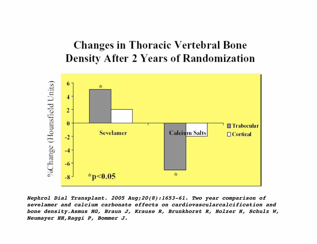
Nephrol Dial Transplant. 2005 Aug;20(8):1653-61. Two year comparison of
sevelamer and calcium carbonate effects on cardiovascularcalcification and
bone density.Asmus HG, Braun J, Krause R, Brunkhorst R, Holzer H, Schulz W,
Neumayer HH,Raggi P, Bommer J.

Sevelamer and Calcium Carbonate
decrease kidney calcification in 5/6
nephrectomy rats
J Am Soc Nephrol 13: 2299–2308, 2002

Binder Studies• CARE Study - 8 week blinded study
– Calcium acetate more efficatious in controllingserum P04 than sevelamer
• Kidney International 65:1914-1926, 2004
• RIND Study 18 month trial– 60 incident patients randomized to calcium binders
- had progressive calcification
– 54 to sevelamer HCL• Kidney International 68:1815-1824, 2005
• LANTHANUM 6 weeks– Significant drop in P04 in one week - 2250 mg/day
• Clinical Nephrology 65:191-202, 2006

Vitamin D
• Vitamin D, especially thenew analogs, confers aprotective effect on patientsurvival
• Reasons– Inflammation
– Renin-angiotensin system
– Myocardium
– Muscle
– Bone (may also decreasebone pain)
Prog Biophys Mol Biol 2006;92:4-8.
Kidney Int 2005;68:1973-81.
Hemodial Int 2005;9 Suppl 1:S25-9.

Vitamin D and Blood Pressure
• NHANES III survey• Representative sample of the US population between
1988 and 1994.
• 12,644 participants with 25OHD levels and BloodPressure measurements,
• Systolic blood pressure was 2.7 mmHg lower (P=0.0005)
– vitamin D levels 85.7 nmole/L compared with the lowestquintile <40.4 nmole/L when adjusted for BMI, age, sexand ethnicity.
• Diastolic blood pressure changes were significant, butnot when adjusted for BMI (p=0.013).
– Scragg R, Sowers M, Bell C. Serum 25-hydroxyvitamin D and blood pressure in the ThirdTational Health and Nutritional Examination Survey. Proc 13th Workshop on Vitamin D:512006.
• Down-regulates renin production

Vitamin D Deficiency
• Calcidiol, 25(OH)D3, low due to– Urban living
– Cultural dress
– Lack of sun explosure
– Lack of physical activity
– CKD population
• Limited studies evaluating theeffects of supplementation withergocalciferol or cholecalciferol
• Measure vitamin D3 in CKD
• Treat with OTC Ergocalciferol

Exercise• Weight bearing on bone mass - Astronauts
– NASA Space Program
– Trabecular bone loss was similar in space travel tothat in prolonged bed rest.
• Weight-bearing exercise in postmenopausalwomen– slow or decrease a decline in bone mineral density
– Increase trochanteric bone mineral content,
– Reducing the risk of falls (15889312)
• Exercise in CKD - need for additional studies

Nocturnal Dialysismmol (P < 0.01)
Kidney International 53:1399-1404, 1998
Conversion from nephron.com
161.6 ± 59.0
(5,010 mg/L)
26.9 ± 9.8
(833.9 mg/L)
Nocturnal
Hemodialysis
75.8 ± 22.5
(1,516 mg/L)
25.3 ± 7.5
(784.3 mg/L)
Conventional
Hemodialysis
100 to 210
mmol/week
(3100 - 6510 mg/wk)
10 and 30
mmol/day
(310-930 mg/day)
Dietary Phosphorus
Absorbed
Weekly intake in ESRD around 1 gm/day
Only 40-80% absorbed
By fourth month patients were on no binders
Dietary Phosphorus intake doubled
Nocturnal hemodialysis may slow vascular calcificationYuen, D., et al., The natural history of coronary calcification progression in a cohort of
nocturnal haemodialysis patients. Nephrol Dial Transplant, 2006. 21(5): p. 1407-12.

PTH caveats• Higher PTH levels and adynamic bone disease in
African Americans
• PTH may be a normal adaptation mechanism inCKD, and we may not want to over treat it
• DOPPS data does not show the strong associationbetween PTH with mortality in K/DOQI range
• The measurement of PTH does not relate to theoriginal Nichols Allegro assay with current BayerCentaur or Roche Elecys assays
• Newer agents such as cinacalcet enable PTHsuppression without hypercalemia
• Therefore, a high serum calcium level in a patient oncinacalcet who has a lowered PTH level could havelow bone turnover disease, particularly if AfricanAmerican

Appendix

Appendix -2

CKD-MBD
• Parathyroid Hormone– Epidemiology
– Accuracy
– Management
• Bone Disease– CKD Adaptation
– Assessment
– Management
• Vascular Calcification– Association with CKD-
MBD
– Assessment (Plain Films)
– Management
• Phosphorus Control
– Consequences
– Management (Diet,
Meds, Dialysis)
• Vitamin D– Vitamin D Deficiency
– Vitamin D and Survival
– Vitamin D in oversuppression
Take Away
• Vascular calcificationplays a key role inCKD mortality
• Vascular calcificationstarts early
• We may be doing adisservice to patientsby not emphasizingearly phosphoruscontrol and oversuppressing PTHlevels
• We should be offeringnocturnal dialysis tomore patients
• Vitamin D has non-skeletal functions thatare ignored not only inCKD but in the generalpopulation


Copyright ©2005 The Endocrine Society
Kobayashi, T. et al. Endocrinology 2005;146:1012-1017
FIG. 1. Differentiation of bone cells of three lineages and its regulation by transcriptionfactors
Top Related