


Languages
Pages
Legal

Screening Programmes
Fetal Anomaly
Information for health professionals
Chorionic villus sampling (CVS) & amniocentesis
ARCHIVED
SEPTEMBER 2017
NHS Fetal Anomaly Screening Programme Chorionic villus sampling (CVS) and amniocentesis – Information for health professionals2
Chorionic villus sampling (CVS) and amniocentesis information for health professionalsThis information sheet is intended to provide a guide for health professionals who are not usually involved in
chorionic villus sampling (CVS) and amniocentesis procedures but who may be asked questions by women
about these procedures. CVS is sometimes also called chorionic villus biopsy or placental biopsy (CVB or PB).
Chorionic villus sampling (CVS) and amniocentesis are invasive diagnostic procedures carried out during
pregnancy. They are usually offered to detect chromosomal disorders such as Down’s syndrome (Trisomy 21),
but are sometimes used for single-gene conditions such as sickle cell disease, thalassaemia major or other rare
conditions.
It is important to remember that women can choose whether or not to undergo these procedures. As health
professionals, you must ensure that they understand the risks that these procedures carry to the fetus.
It is good clinical practice to obtain formal written consent for amniocentesis or CVS before the procedure.
Written or verbal information should include the reason for offering the invasive procedure, an explanation
of the procedure and the cytogenetic results that will become available (Royal College of Obstetricians and
Gynaecologists 2010).
It is important to know the woman’s HIV, rhesus and hepatitis B status before undertaking either of these
invasive procedures.
Both CVS and amniocentesis should be performed under continuous ultrasound guidance. Although the
procedure itself usually takes about 10 minutes to perform, the appointment may need to be longer to allow
for discussion and because the woman may need to rest afterwards.
Summary
ARCHIVED
SEPTEMBER 2017
NHS Fetal Anomaly Screening Programme Chorionic villus sampling (CVS) and amniocentesis – Information for health professionals 3
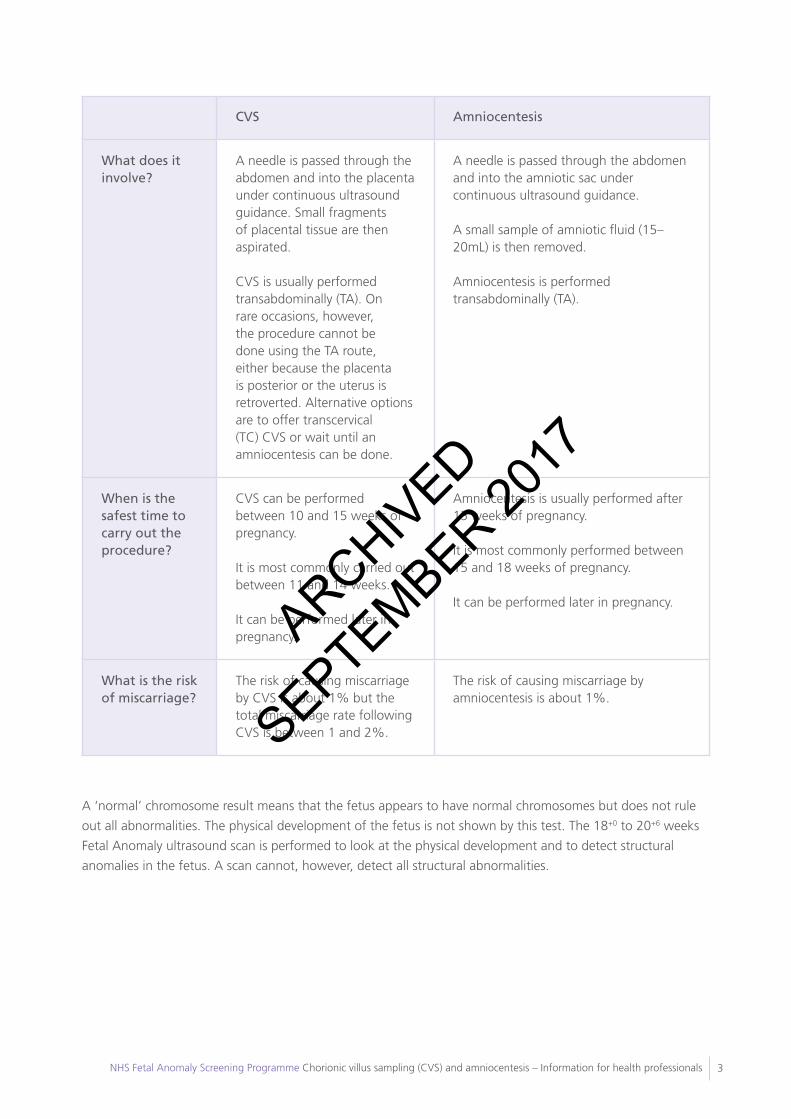
CVS Amniocentesis
What does it involve?
A needle is passed through the abdomen and into the placenta under continuous ultrasound guidance. Small fragments of placental tissue are then aspirated.
CVS is usually performed transabdominally (TA). On rare occasions, however, the procedure cannot be done using the TA route, either because the placenta is posterior or the uterus is retroverted. Alternative options are to offer transcervical (TC) CVS or wait until an amniocentesis can be done.
A needle is passed through the abdomen and into the amniotic sac under continuous ultrasound guidance.
A small sample of amniotic fluid (15–20mL) is then removed.
Amniocentesis is performed transabdominally (TA).
When is the safest time to carry out the procedure?
CVS can be performed between 10 and 15 weeks of pregnancy.
It is most commonly carried out between 11 and 14 weeks.
It can be performed later in pregnancy.
Amniocentesis is usually performed after 15 weeks of pregnancy.
It is most commonly performed between 15 and 18 weeks of pregnancy.
It can be performed later in pregnancy.
What is the risk of miscarriage?
The risk of causing miscarriage by CVS is about 1% but the total miscarriage rate following CVS is between 1 and 2%.
The risk of causing miscarriage by amniocentesis is about 1%.
A ‘normal’ chromosome result means that the fetus appears to have normal chromosomes but does not rule
out all abnormalities. The physical development of the fetus is not shown by this test. The 18+0 to 20+6 weeks
Fetal Anomaly ultrasound scan is performed to look at the physical development and to detect structural
anomalies in the fetus. A scan cannot, however, detect all structural abnormalities.
ARCHIVED
SEPTEMBER 2017
NHS Fetal Anomaly Screening Programme Chorionic villus sampling (CVS) and amniocentesis – Information for health professionals4
1. Introduction............................................................................................................................................................
2. Why is CVS or amniocentesis performed? .............................................................................................................
3. Diagnostic testing in multiple pregnancies...............................................................................................................
4. HIV, rhesus and hepatitis B status...........................................................................................................................
5. Consent..................................................................................................................................................................
6. Results....................................................................................................................................................................
7. Ultrasound scan.....................................................................................................................................................
8. When are the procedures performed?...................................................................................................................
9. What happens during the procedures?..................................................................................................................
10. Discomfort during the procedure.........................................................................................................................
11. Length of the procedure......................................................................................................................................
12. Post procedure care.............................................................................................................................................
13. Barriers to the completion of the procedure........................................................................................................
14. Bringing a partner or friend..................................................................................................................................
15. Eating and drinking..............................................................................................................................................
16. Do the procedures harm the fetus?....................................................................................................................
Miscarriage rate after CVS or amniocentesis ..............................................................................................
Cause of miscarriage after CVS or amniocentesis.......................................................................................
Reducing the risk of miscarriage..................................................................................................................
17. Cytogenetic results..............................................................................................................................................
What is the difference between QF-PCR and karyotyping?........................................................................
QF-PCR result..............................................................................................................................................
Full karyotype result....................................................................................................................................
Does a normal karyotype mean there is nothing ‘wrong’ with the fetus?..................................................
18. Abnormal results.................................................................................................................................................
19. Examples of chromosome abnormalities that can be detected using CVS or amniocentesis..............................
Trisomy 21 (Down’s syndrome)....................................................................................................................
Trisomy 18 (Edwards’ syndrome).................................................................................................................
Trisomy 13 (Patau’s syndrome).....................................................................................................................
Sickle cell and thalassaemia.........................................................................................................................
Other conditions..........................................................................................................................................
20. Further information, charities and support groups..............................................................................................
21. Sources and references.......................................................................................................................................
Contents5
5
5
5
6
6
6
6
7
9
9
9
10
10
10
11
11
11
11
12
12
12
12
13
14
15
15
15
15
16
16
17
19
ARCHIVED
SEPTEMBER 2017
NHS Fetal Anomaly Screening Programme Chorionic villus sampling (CVS) and amniocentesis – Information for health professionals 5
This information sheet is intended to provide a guide for health professionals not usually involved in chorionic
villus sampling (CVS) and amniocentesis procedures but who may be asked questions by women about these
procedures. CVS is sometimes also called chorionic villus biopsy or placental biopsy (CVB or PB).
Parents requesting such a procedure because of their genetic family history should be referred for prenatal
diagnosis (PND) counselling. This counselling is generally carried out by an obstetrician/fetal medicine
specialist, screening midwife or specialist midwife (fetal medicine) or a clinical geneticist.
CVS and amniocentesis are invasive diagnostic procedures carried out during pregnancy. They are usually
offered to detect chromosomal disorders such as Down’s syndrome (Trisomy 21). When used for rarer
indications, such as single-gene conditions (e.g. sickle cell disease, and thalassaemia major), clinical
management policies should be followed, which will usually include establishing both the parents’ genetic
status before the procedure.
A high level of expertise in ultrasound scanning is essential for operators undertaking amniocentesis or CVS in
multiple pregnancies (Royal College of Obstetricians and Gynaecologists 2010).
It is important to know the woman’s HIV, rhesus and hepatitis B status before undertaking either
amniocentesis or CVS.
The woman’s rhesus status should be stated on the referral form. It is vitally important that women who
are rhesus D negative are offered and given, where consent obtained, an appropriate amount of anti-D
immunoglobulin according to their gestation to reduce the risk of rhesus iso-immunisation (refer to your local
Trust policy). The anti-D immunoglobulin should be given after the procedure in line with national guidance
(Royal College of Obstetricians and Gynaecologists 2010).
Knowledge of the woman’s HIV and hepatitis B status is important to minimise the risk of transmission of the
virus to the unborn fetus and to ensure that appropriate precautions are taken by hospital and laboratory
staff. If women have declined screening for bloodborne viruses, the potential risks of infection to the
fetus if positive should be discussed and that discussion documented (Royal College of Obstetricians and
Gynaecologists 2010).
1. Introduction
2. Why is CVS or amniocentesis performed?
3. Diagnostic testing in multiple pregnancies
4. HIV, rhesus and hepatitis B status
ARCHIVED
SEPTEMBER 2017
NHS Fetal Anomaly Screening Programme Chorionic villus sampling (CVS) and amniocentesis – Information for health professionals6
Women who are offered CVS or amniocentesis should be given as much information as they require about
the purpose, risks, benefits and limitations of the procedures before making a decision about whether or
not they want to have the tests. The information should be given to them by a healthcare professional with
experience and knowledge of prenatal diagnosis. It is good clinical practice to obtain written consent for
amniocentesis and CVS before the procedure (Royal College of Obstetricians and Gynaecologists 2010). For
more information on consent, please consult the NHS Fetal Anomaly Screening Programme consent standards
available at www.fetalanomaly.screening.nhs.uk.
During the appointment, the woman should be informed about the local arrangements for handling results
and options for how she will be informed. All staff involved in this process must obtain up-to-date information
on their local arrangements.
Prior to any invasive procedure, a scan is required to determine the fetal number, fetal viability, amniotic fluid
volume, fetal position and placental position and to confirm gestational age.
During the procedure, the ultrasound probe will be used to:
• identify a target site to obtain an optimal pocket of amniotic fluid or placental tissue;
• track the sampling needle to reduce the likelihood of trauma to the fetus, cord and uterus.
CVS
CVS can be performed between 10 and 15 weeks of pregnancy. It is most commonly carried out between 11
and 14 weeks. It can be performed later in pregnancy.
It is important to refer individuals with a family history of single-gene disorders early, so that the parents have
the option of first trimester CVS. Where the risk of the unborn fetus being affected is 1 in 2 or 1 in 4 and
where there is a substantial risk that the parents will face the option of termination, it may be important for
them to have the test and result as early as possible.
5. Consent
6. Results
7. Ultrasound scan
8. When are the procedures performed?
ARCHIVED
SEPTEMBER 2017
NHS Fetal Anomaly Screening Programme Chorionic villus sampling (CVS) and amniocentesis – Information for health professionals 7
Amniocentesis
Amniocentesis is usually performed after 15 weeks of pregnancy. It is most commonly performed between 15
and 18 weeks. It can be performed later in pregnancy. From 15 weeks of pregnancy, fetal cells are greatest
in number and the amount of amniotic fluid is around 150−200mL and therefore adequate for obtaining a
15−20mL cytogenetic sample. Prior to 15 weeks, the rate of miscarriage from this procedure is higher and
obtaining the correct volume of amniotic fluid tends to be difficult.
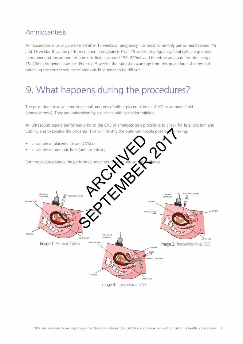
The procedures involve removing small amounts of either placental tissue (CVS) or amniotic fluid
(amniocentesis). They are undertaken by a clinician with specialist training.
An ultrasound scan is performed prior to the CVS or amniocentesis procedure to check for fetal position and
viability and to localise the placenta. This will identify the optimum needle position for taking:
• a sample of placental tissue (CVS) or
• a sample of amniotic fluid (amniocentesis).
Both procedures should be performed under continuous ultrasound guidance.
9. What happens during the procedures?
CVS and amniocentesis information for health professionals HP_CVS_AMNIO V1 July 096
Uterine wall
Placenta
Amniotic �uid
Ultrasound transducer Needle and syringe
Uterine wall
Placenta
Amniotic �uid
Ultrasound transducer
Uterine wall
Needle and syringe
Placenta
Amniotic �uid Bladder
Ultrasound transducer
Speculum
Bladder
Bladder
Image 1: Amniocentesis
Uterine wall
Placenta
Amniotic �uid
Ultrasound transducer Needle and syringe
Uterine wall
Placenta
Amniotic �uid
Ultrasound transducer
Uterine wall
Needle and syringe
Placenta
Amniotic �uid Bladder
Ultrasound transducer
Speculum
Bladder
Bladder
Image 2: Transabdominal CVS
Uterine wall
Placenta
Amniotic �uid
Ultrasound transducer Needle and syringe
Uterine wall
Placenta
Amniotic �uid
Ultrasound transducer
Uterine wall
Needle and syringe
Placenta
Amniotic �uid Bladder
Ultrasound transducer
Speculum
Bladder
Bladder
Image 3: Transcervical CVSCVSCVS is usually performed transabdominally (TA) between 11 and12 weeks of pregnancy. However, on rare occasions the procedure cannot be done either because the placenta is posterior or the uterus is retroverted. Strategies such as asking the woman to fill and empty her bladder or waiting another week may be helpful but if this fails, alternative options are to offer transcervical (TC) CVS or amniocentesis. This should be fully discussed with the woman to ensure all options are provided along with any associated risks.
It’s important to note that the risk of miscarriage associated with TC CVS is about 2–3% and given the choice, the woman may opt for an amniocentesis which has a lower risk of miscarriage.
Local anaesthetic may be given prior to the procedure.
Immediately before the procedure, the woman’s abdomen is cleaned to ensure that the procedure can take place in the most sterile conditions possible.
Gel is then applied over the woman’s abdomen to facilitate continuous ultrasound guidance.
During the TA procedure, a needle is passed through the abdomen and into the placenta under continuous ultrasound guidance. Small fragments of placental tissue are then aspirated.
After the procedure, the woman may wish to rest for a short period in the clinic.
AmniocentesisAmniocentesis is performed transabdominally (TA).
For an amniocentesis, local anaesthetic may be given prior to the procedure. However, research suggests that most women will not notice a beneficial analgesic effect and therefore the use of anaesthetic is not common during amniocentesis.
Immediately before the procedure, the woman’s abdomen is cleaned to ensure that the procedure can take place in the most sterile conditions possible.
Gel is then applied over the woman’s abdomen to carry out continuous ultrasound guidance.
ARCHIVED
SEPTEMBER 2017
NHS Fetal Anomaly Screening Programme Chorionic villus sampling (CVS) and amniocentesis – Information for health professionals8
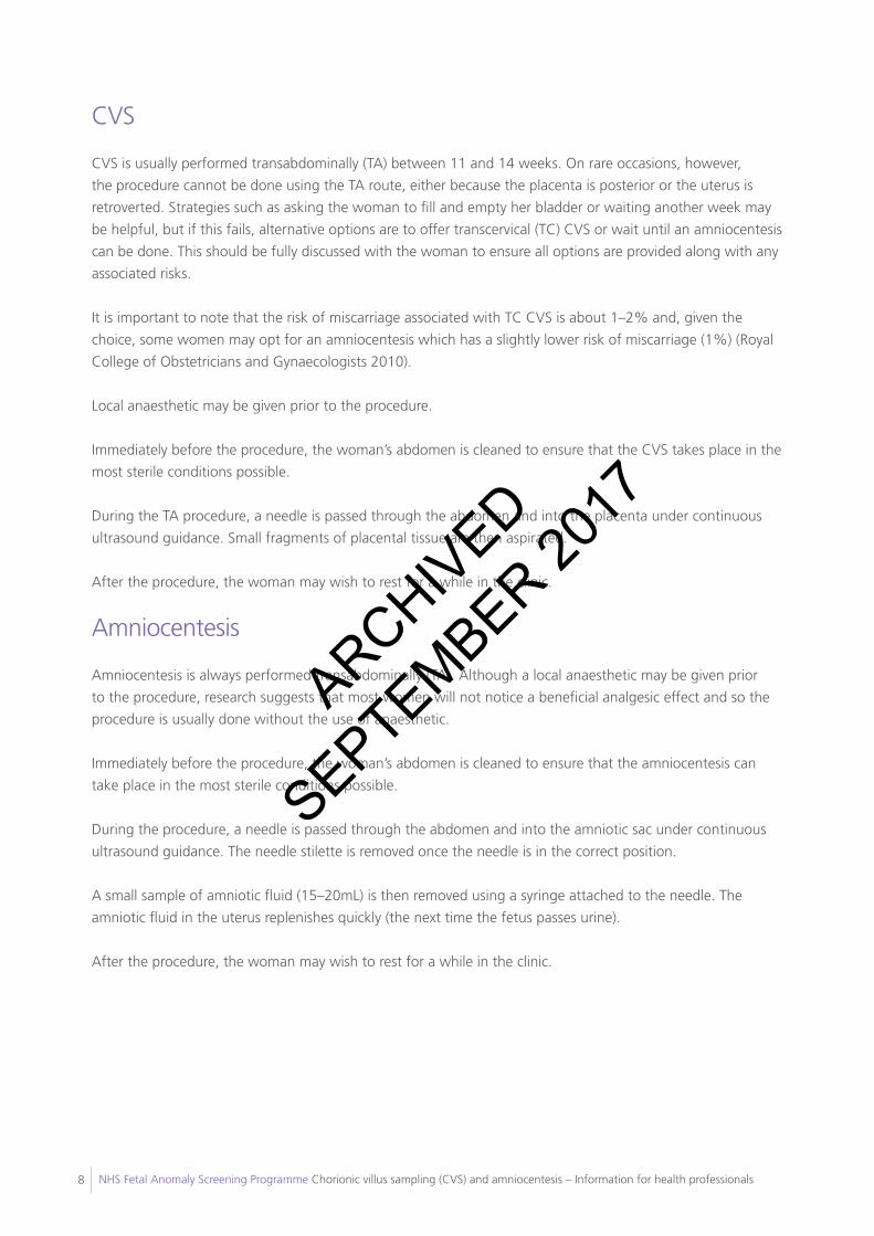
CVS
CVS is usually performed transabdominally (TA) between 11 and 14 weeks. On rare occasions, however,
the procedure cannot be done using the TA route, either because the placenta is posterior or the uterus is
retroverted. Strategies such as asking the woman to fill and empty her bladder or waiting another week may
be helpful, but if this fails, alternative options are to offer transcervical (TC) CVS or wait until an amniocentesis
can be done. This should be fully discussed with the woman to ensure all options are provided along with any
associated risks.
It is important to note that the risk of miscarriage associated with TC CVS is about 1–2% and, given the
choice, some women may opt for an amniocentesis which has a slightly lower risk of miscarriage (1%) (Royal
College of Obstetricians and Gynaecologists 2010).
Local anaesthetic may be given prior to the procedure.
Immediately before the procedure, the woman’s abdomen is cleaned to ensure that the CVS takes place in the
most sterile conditions possible.
During the TA procedure, a needle is passed through the abdomen and into the placenta under continuous
ultrasound guidance. Small fragments of placental tissue are then aspirated.
After the procedure, the woman may wish to rest for a while in the clinic.
Amniocentesis
Amniocentesis is always performed transabdominally (TA). Although a local anaesthetic may be given prior
to the procedure, research suggests that most women will not notice a beneficial analgesic effect and so the
procedure is usually done without the use of anaesthetic.
Immediately before the procedure, the woman’s abdomen is cleaned to ensure that the amniocentesis can
take place in the most sterile conditions possible.
During the procedure, a needle is passed through the abdomen and into the amniotic sac under continuous
ultrasound guidance. The needle stilette is removed once the needle is in the correct position.
A small sample of amniotic fluid (15–20mL) is then removed using a syringe attached to the needle. The
amniotic fluid in the uterus replenishes quickly (the next time the fetus passes urine).
After the procedure, the woman may wish to rest for a while in the clinic.
ARCHIVED
SEPTEMBER 2017
NHS Fetal Anomaly Screening Programme Chorionic villus sampling (CVS) and amniocentesis – Information for health professionals 9
Discomfort either during or after the procedure varies from woman to woman and in each pregnancy. Some
of the sensations women have reported include:
• period type pain during or after the procedure,
• a sharp, stinging sensation when the sample needle is inserted into the abdominal skin,
• pressure-type feeling either in the lower abdomen or in the lower back or vagina as the needle is removed.
CVS
The whole procedure usually takes about 10 minutes. An appointment for CVS will sometimes take slightly
longer than amniocentesis because, although the procedures are similar, more elaborate preparation may be
needed for CVS.
Amniocentesis
The whole procedure usually takes about 10 minutes. The time taken can depend on the position and
movements of the fetus and whether this hinders access to amniotic fluid pools.
Agreement should be reached between the healthcare professional and the woman about how the test
results will be given. The healthcare professional should check that the woman’s correct telephone number is
recorded on the hospital system and that she has been given the healthcare professional’s contact details.
The woman should also be advised who to contact in case of any complications post procedure. If she
experiences one or more of the following signs or symptoms, she should contact a hospital or a healthcare
professional:
• Feeling generally unwell (shivery, nauseous, abdominal discomfort)
• Pyrexia
• Persistent bleeding from the vagina
• Persistent lower abdominal/back pain
• Clear watery type loss (not urine) from the vagina
• Offensive smelling discharge from the vagina.
10. Discomfort during the procedure
11. Length of the procedure
12. Post procedure careARCHIVED
SEPTEMBER 2017
NHS Fetal Anomaly Screening Programme Chorionic villus sampling (CVS) and amniocentesis – Information for health professionals10
Occasionally the procedure cannot be performed on the day of the appointment for a variety of reasons. In
these cases, the woman should be offered another appointment.
Occasionally, in either procedure, an adequate sample may not be obtained.
The cytogenetic laboratory may also fail to obtain a result despite receiving an adequate sample. This is
relatively unusual, but if it does happen a further or alternative procedure will be recommended.
As a health professional, you should find out from your local service the failure rate for this procedure.
The woman may want a partner, a friend or a family member to attend the appointment with her.
Most hospitals advise that it is safe to eat and drink as normal before and after CVS or amniocentesis providing
the pregnancy is under 24 weeks gestation.
13. Barriers to the completion of the procedure
14. Bringing a partner or friend
15. Eating and drinking
ARCHIVED
SEPTEMBER 2017
NHS Fetal Anomaly Screening Programme Chorionic villus sampling (CVS) and amniocentesis – Information for health professionals 11
Direct injury to the fetus from either CVS or amniocentesis is very rare because continuous ultrasound guidance
is used. It is not possible, however, to prevent the fetus moving towards the needle during an amniocentesis.
Miscarriage rate after CVS or amniocentesis
Because CVS and amniocentesis are invasive procedures, miscarriage is a possible complication. The risk of
causing miscarriage by CVS is about 1% but the total miscarriage rate following CVS is between 1 and 2%. The
risk of causing miscarriage by amniocentesis is about 1%.
Although the procedure-related risks are similar, the post CVS miscarriage rate is higher than the post
amniocentesis miscarriage rate because the background risk of miscarriage (whether the procedure was done or
not) is higher in early pregnancy when a CVS is normally carried out.
These miscarriage rates are derived from national clinical audits. Local or individual data are of variable quality
and the number of procedures performed may not be large enough to reach statistical significance and should
not usually be used. This is particularly relevant if a woman is seeking reassurance that the miscarriage rate is
lower than quoted above.
Cause of miscarriage after CVS or amniocentesis
The exact cause of miscarriage following a CVS or amniocentesis is unknown. It is possible that miscarriage
following either procedure could be due to ruptured amniotic membranes, infection or bleeding.
Women offered a CVS or amniocentesis will often have risk factors for miscarriage such as maternal age,
abnormal blood tests or abnormal scan findings. It is therefore difficult to estimate the background risk of
pregnancy loss for the groups choosing to have invasive tests.
Miscarriages that would have occurred even if the procedure had not been undertaken will still occur. Pregnancy
outcomes following CVS and amniocentesis should be audited locally and more information on this issue may
become available in the future.
Reducing the risk of miscarriage
Clinical experience suggests that miscarriage as a consequence of the procedure can occur up to two weeks
following the procedure and that the risk diminishes after three weeks.
Some doctors advise that women should rest for a couple of days post procedure, avoiding intercourse, any
heavy lifting or strenuous exercise. There is no evidence to support this. Resting in bed is not necessary. It is
considered good practice to advise women to arrange for someone to drive them home after the procedure.
16. Do the procedures harm the fetus?
ARCHIVED
SEPTEMBER 2017
NHS Fetal Anomaly Screening Programme Chorionic villus sampling (CVS) and amniocentesis – Information for health professionals12
What is the difference between QF-PCR and karyotyping?
Quantitative fluorescent polymerase chain reaction (QF-PCR) marker (‘rapid’ test analysis) and karyotyping
are two types of cytogenetic tests. QF-PCR marker analysis is a laboratory process that does not require cell
culture whereas karyotyping does. PCR results can therefore be obtained more quickly.
PCR testing usually only looks for three specific defined chromosome conditions in the fetus: Trisomy 21
(Down’s syndrome), Trisomy 18 (Edwards’ syndrome) and Trisomy 13 (Patau’s syndrome). Normally there
are two copies of each chromosome, but if these syndromes are present there is an extra copy of that
chromosome in each cell. If monosomy XO (Turner’s syndrome) is suspected from the scan, a PCR test will be
performed to look at the sex chromosomes.
The PCR result is usually available within 3 working days (the national target).
Occasionally, karyotyping results from CVS may be inconclusive because of an abnormal cell line confined
to the placenta. This is referred to as ‘confined placental mosaicism’. Further testing by amniocentesis may
be needed later in the pregnancy to investigate this result. The chance of a confined placental mosaicism is
approximately 1–2% (Lestou and Kalousek 1998).
QF-PCR result
QF-PCR is very accurate, but can only give information about the specific chromosomes being tested. A result
from a rapid test is nearly 100% accurate in confirming whether or not the fetus either does or does not have
Trisomy 21, Trisomy 13 or Trisomy 18. Other chromosomes may be tested for, but this must be specifically
requested and discussed with the relevant cytogenetic laboratory.
If another condition is suspected, a full karyotype can sometimes be offered. As a significant proportion
of abnormal scans are associated with karyotypes other than these trisomies, full karyotyping is usually
recommended when the indication is an abnormal scan.
A small number of amniotic fluid PCR samples fail to yield a PCR result, mainly because of maternal blood
contamination of the amniotic fluid.
Full karyotype result
Karyotyping involves growing the fetal cells floating in the amniotic fluid and making a preparation showing the
chromosomes, which are then examined under the microscope. The test looks at changes in the number and
appearance of all the chromosomes.
The cells take about 5−10 days to grow in the laboratory. Results from karyotyping therefore take longer to
obtain than results from PCR (usually 10−14 days).
17. Cytogenetic results
ARCHIVED
SEPTEMBER 2017
NHS Fetal Anomaly Screening Programme Chorionic villus sampling (CVS) and amniocentesis – Information for health professionals 13
Further guidance on cytogenetic testing can be found in the NHS FASP Working Standards document available
from www.fetalanomaly.screening.nhs.uk/standardsandpolicies.
Does a normal karyotype mean there is nothing ‘wrong’ with the fetus?
Many serious diseases are not genetic and could never be found with a chromosome test. Not all serious genetic
diseases will be detected by a chromosome test alone because some chromosome changes are so small that they
may go unnoticed when viewed under the microscope. Some can be found with special tests if it is known what
to test for and if specifically requested.
The full karyotype test will not detect:
• alterations in single genes, such as cystic fibrosis (each chromosome contains thousands of genes);
• microdeletions (loss of small segments of a chromosome); or
• other small changes in chromosomes.
A ‘normal’ chromosome result therefore means that the fetus appears to have normal chromosomes but does
not rule out all abnormalities.
The physical development of the fetus is not shown by this test. The 18+0 to 20+6 weeks Fetal Anomaly
ultrasound scan is performed to detect structural anomalies in the fetus. However, a scan cannot detect all
structural abnormalities either.
It is a good idea for health professionals assisting in these procedures to gain more information from the relevant
genetic departments. ARCHIVED
SEPTEMBER 2017
NHS Fetal Anomaly Screening Programme Chorionic villus sampling (CVS) and amniocentesis – Information for health professionals14
A small number of women will unfortunately receive abnormal test results and those few will need to make
choices about the treatment path they want to follow.
If the results show that the fetus has one of the disorders being tested for, the woman and her partner should
be given the opportunity to discuss this fully with the specialist midwife, obstetrician/fetal medicine specialist
or clinical geneticist as soon as possible.
After an abnormal result, the woman may also be referred to a consultant paediatrician, consultant geneticist
or genetic counsellor for further information and counselling. Although some disorders are treatable, others
are not.
Whatever the outcome, the woman must feel assured that the health professionals caring for her will support
her decision to:
• continue the pregnancy and use the information received to help prepare for the birth and care of her baby;
• continue the pregnancy and consider adoption; or
• terminate the pregnancy.
Some women who make an informed decision to terminate their pregnancy find it helpful to talk with a
health professional or a counsellor about their experience afterwards.
It may also be helpful to recommend the support groups listed at the end of this leaflet. These are also
available in the parent information leaflets ‘Amniocentesis test – information for parents’ and ‘Chorionic
villus sampling (CVS) – information for parents’ available from www.fetalanomaly.screening.nhs.uk/
publicationsandleaflets.
Websites maintained by these support groups often have useful information about the various conditions and
describe the experiences of women faced with difficult choices following diagnosis of chromosome and other
abnormalities.
If a woman decides to have a termination of pregnancy for an abnormal result, she may have the choice of
having either a surgical or medically induced termination. The method selected will depend, however, on the
nature of the anomaly and how advanced the gestation is.
18. Abnormal results
ARCHIVED
SEPTEMBER 2017
NHS Fetal Anomaly Screening Programme Chorionic villus sampling (CVS) and amniocentesis – Information for health professionals 15
Trisomy 21 (Down’s syndrome)
• Trisomy 21 is the most common chromosomal condition detected.
• Trisomy 21 occurs when there is an additional chromosome 21.
• Babies with Trisomy 21 can have multiple problems such as cardiac defects and severe learning difficulties.
However, some adults with Trisomy 21 are able to lead semi-independent lives.
• The birth incidence of this condition in an unscreened population is about 1 in 560 live births, but is
strongly linked to maternal age and is therefore different in different geographical areas.
• As women get older, the risk of having a pregnancy affected with Trisomy 21 becomes greater. However,
since women under the age of 35 have more children, most Trisomy 21 affected babies are born to
younger women.
Neither karyotyping nor the PCR test can predict how severe the condition will be in any given individual. An
ultrasound scan at 18+0 to 20+6 weeks may detect physical abnormalities that are associated with Trisomy 21,
such as cardiac defects, but should not be used as the primary screening test.
Trisomy 18 (Edwards’ syndrome)
• Trisomy 18 is a rare chromosome abnormality. It is less common than Trisomy 21.
• Trisomy 18 occurs when there is an additional chromosome 18.
• The incidence of Trisomy 18 at birth is 1 in 1500.
• Babies with Trisomy 18 can have multiple problems such as cardiac defects, renal abnormalities and severe
developmental delay. Many affected fetuses miscarry or die during later pregnancy, but when born alive,
life expectancy for the majority of babies with Trisomy 18 is usually limited to a few weeks and rarely
beyond one year.
Trisomy 13 (Patau’s syndrome)
• Trisomy 13 is a rare chromosome abnormality. It is less common than Trisomy 21.
• The incidence of this condition is 1 in 3000 live births.
• Trisomy 13 occurs when there is an additional chromosome 13.
• Most affected fetuses miscarry; liveborn babies with this condition usually do not live beyond the first
weeks of life and few survive beyond one year.
• Babies with Trisomy 13 can have multiple, severe problems such as cardiac defects, brain abnormalities
and severe renal abnormalities. Some of these abnormalities can be seen on ultrasound scan from 11
weeks onwards.
19. Examples of chromosome abnormalities that can be detected using CVS or amniocentesis
ARCHIVED
SEPTEMBER 2017
NHS Fetal Anomaly Screening Programme Chorionic villus sampling (CVS) and amniocentesis – Information for health professionals16
Sickle cell and thalassaemia
Sickle cell disease is a variable set of inherited conditions affecting the haemoglobin that can cause chronic
anaemia, jaundice, acute pain (crisis), organ damage, infections and strokes in children and adults.
Alpha and beta thalassaemia major are inherited blood conditions that affect the quantity of haemoglobin
produced. Alpha thalassaemia major is incompatible with life. Beta thalassaemia major results in severe anaemia.
Inheritance of an altered gene from both parents results in a disorder and inheritance of only one altered gene
results in a healthy carrier.
All pregnant women receiving abnormal haemoglobinopathy results should receive specialist obstetric and
haematological advice.
More information on sickle cell diseases and thalassaemia major is available from the NHS Sickle Cell &
Thalassaemia Screening Programme at www.sct.screening.nhs.uk.
Other conditions
There are numerous other abnormal results which will rarely be obtained, usually as a result of sophisticated
testing for specific conditions. All abnormal results should be given and explained by specialists, such as those in
Fetal Medicine Units or Clinical Genetics Units.
ARCHIVED
SEPTEMBER 2017
NHS Fetal Anomaly Screening Programme Chorionic villus sampling (CVS) and amniocentesis – Information for health professionals 17
20. Further information, charities and support groups
Antenatal Results and Choices (ARC)
73 Charlotte Street
London W1T 4PN
Helpline: 0207 631 0285
Email: [email protected]
Website: www.arc-uk.org
Antenatal Results and Choices (ARC) provides information and support to parents before, during and after
antenatal screening and diagnostic tests, especially those making difficult decisions about testing, or about
continuing or ending a pregnancy after a diagnosis. ARC offers ongoing support whatever decisions are made.
Down’s Syndrome Association (DSA)
Website: www.downs-syndrome.org.uk
Langdon Down Centre
2a Langdon Park
Teddington
TW11 9PS
Phone: 0845 230 0372
Email: [email protected]
The aim of the Down’s Syndrome Association (DSA) is to help people with Down’s syndrome lead full and
rewarding lives.
The Miscarriage Association
17 Wentworth Terrace
Wakefield WF1 3QW
Tel: 01924 200799
Email: [email protected]
Website: www.miscarriageassociation.org.uk
The Miscarriage Association is a registered national charity in England & Wales (no. 1076829) and in Scotland
(no. SC039790) and a company limited by guarantee (no. 3779123), working across England, Northern
Ireland, Scotland and Wales. It was founded in 1982 by a group of people who had experienced miscarriage
and continues to offer support and information to anyone affected by the loss of a baby in pregnancy, to raise
awareness and to promote good practice in medical care.
ARCHIVED
SEPTEMBER 2017
NHS Fetal Anomaly Screening Programme Chorionic villus sampling (CVS) and amniocentesis – Information for health professionals18
Healthtalk online
PO Box 428
Witney
Oxon OX28 9EU
Email: [email protected]
Website: www.healthtalkonline.org
The Health Experience Research Group has created a unique database of personal and patient experiences
through in-depth qualitative research into over 60 different illnesses and health conditions. The results of their
research are published on two websites (www.healthtalkonline.org and www.youthhealthtalk.org) which are
aimed at patients, their carers, family and friends, doctors, nurses and other health professionals. Their target is
to complete at least 100 conditions within the next 5–10 years.
They have also recently started two social networking sites for people to add their own experiences of health and
illness at www.myhealthtalk.org and www.myyouthhealthtalk.org. The websites are run by the DIPEx Charity.
Sickle Cell Society54 Station Road
London NW10 4UA
Tel: 0208 961 7795
Email: [email protected]
Website: www.sicklecellsociety.org
The Sickle Cell Society provides information, counselling and care for people with sickle cell disorders and their
families.
UK Thalassaemia Society Website: www.ukts.org
19 The Broadway
Southgate Circus
London
N14 6PH
Phone: 020 8882 0011
Email: [email protected]
The Society raises the awareness and the health education of the communities of the UK at risk of Thalassaemia.
ARCHIVED
SEPTEMBER 2017
NHS Fetal Anomaly Screening Programme Chorionic villus sampling (CVS) and amniocentesis – Information for health professionals 19
Antenatal Screening Wales and NHS Fetal Anomaly Screening Programme (2008) Amniocentesis and chorionic
villus sampling: Policy, standards and protocols. April.
de Ruiter A, Mercey D, Anderson J et al. (2008) British HIV Association and Children’s HIV Association
guidelines for the management of HIV infection in pregnant women 2008. HIV Medicine 9:452–502.
Lestou VS, Kalousek DK (1998) Confined placental mosaicism and intrauterine fetal growth. Arch Dis Child
Fetal Neonatal Ed. 79(3):F223–6.
Royal College of Obstetricians and Gynaecologists (2005) Amniocentesis and chorionic villus sampling.
London: RCOG.
Royal College of Obstetricians and Gynaecologists (2006) Amniocentesis; what you need to know. Available
from: www.rcog.org.uk/womens-health/clinical-guidance/amniocentesis-what-you-need-know
Royal College of Obstetricians and Gynaecologists (2006) Chorionic villus sampling (CVS): what you need to
know. Available from:
www.rcog.org.uk/womens-health/clinical-guidance/chorionic-villus-sampling-cvs-what-you-need-know
Royal College of Obstetricians and Gynaecologists (2010) Green-top Guidelines No. 8, Amniocentesis and
Chorionic Villus Sampling. Available from:
www.rcog.org.uk/womens-health/clinical-guidance/amniocentesis-and-chorionic-villus-sampling-green-top-8
21. Sources and references
ARCHIVED
SEPTEMBER 2017
NHS Fetal Anomaly Screening ProgrammeUK National Screening CommitteeTelephone: 0845 527 7910Email: [email protected]
This information has been produced on behalf of the NHS Fetal Anomaly Screening Programme. The leaflet has been developed through consultation with the NHS Fetal Anomaly Screening Programme expert groups and is based on text originally developed by Antenatal Screening Wales.
All of our publications can be found online at www.fetalanomaly.screening.nhs.uk
NHS staff can reproduce any information in this booklet. Please ensure you have permission to reuse images. Amendments must be discussed with the original author.
To order copies of this leaflet please email: [email protected] or call 0191 4969735
If you have any comments on this booklet or enquiries for the programme please contact us at the address below:
Innovation CentreRennes DriveUniversity of ExeterExeter, EX4 4RN
ARCHIVED
SEPTEMBER 2017
Top Related