


Languages
Pages
Legal

3
Childhood Pancreatitis
Ali E. Abdelbasit Department of Paediatric Surgery, Soba University Hospital,
University of Khartoum, Sudan
1. Introduction
Most of the available English literature on pancreatitis is adult based. The last few decades have witnessed increasing interest in childhood pancreatitis and attention to research into the subject. Many recent published works have focused on various aspects of pancreatitis. The incidence of childhood pancreatitis is a rare event though it appears to be increasing. Despite its rarity, pancreatitis in children deserves special attention because of the significant morbidity and mortality associated with it. The management of childhood pancreatitis has also witnessed remarkable development in the last few decades. The improvement in the imaging resolutions, biochemical tests, endoscopic procedures and the intensive care facilities have resulted in better outcome.
This chapter aims at providing an up-to-date review of childhood pancreatitis. It includes the aspects of aetiological factors, pathophysiology, classification, typical and atypical clinical presentation with abdominal and extra-abdominal manifestations of the disease and its complications, applicability of severity scores, the diagnostic and prognostic investigations, various methods of medical and surgical treatment - with indications thereof - touching on the pros and cons of the different methods, and the overall prognosis.
2. Aetiology
2.1 Acute pancreatitis
The causes of acute pancreatitis in children are diverse (Table 1) and unlike in the adults where the majority of the cases are due to alcoholism or gallstone disease, most of the cases in children are due to pancreaticobiliary tract anomalies, biliary tract stones, trauma, drugs and a plethora of systemic diseases. In a considerable proportion of up to 25% of the patients, no aetiological factor is identified and is therefore labeled as idiopathic (Haddock et al., 1994; Uretsky et al., 1999; Mehta & Gittes, 2005; Hebra et al, 2009; Werlin, 2003).
2.1.1 Pancreaticobiliary anomalies
Pancreaticobiliary malunion (PBMU) accounts for approximately 6-33% of cases of pancreatitis in children. PBMU is used synonymously with common pancreaticobiliary channel, anomalous pancreaticobiliary channel, pancreaticobiliary maljunction, anomalous pancreaticobiliary ductal union or long common pancreaticobiliary channel, with or without
www.intechopen.com
Pancreatitis – Treatment and Complications
30
a choledochal cyst or common bile duct (CBD) dilatation. PBMU is complicated by bile reflux into the pancreatic duct predisposing to pancreatitis. Common channel and pancreatic duct obstruction by protein plugs, biliary stones or sludge may result in pancreatitis or jaundice. Presence of a choledochal cyst may produce pancreatitis by direct mechanical pancreatic duct compression (Miyano, 2006).
Pancreas divisum is a persistence of the developmental pancreatic ductal system due to
failure of fusion of the dorsal (Wirsung) and the ventral (Santorini) ducts. As a result, the
exocrine drainage of the pancreatic body and tail is impaired due to the narrow minor duct
and its papilla. The resultant flow impediment may cause recurrent episodes of acute
pancreatitis. The anomaly is encountered in approximately 4-11% of patients and, as such, is
one of the most common congenital anomalies of the pancreas (Miyano, 200; Mehta & Gittes,
2005). Annular pancreas was reported to cause recurrent acute pancreatitis in children
(Hwang et al 2010, Ohno & Kanematsu, 2008).
2.1.2 Biliary stone disease
Choledocho- or cholcystolithiasis are uncommon in children; however, when they exist they cause pancreatitis secondary to transient pancreatic duct obstruction. Biliary stones may result from haemolytic disorders such as hereditary spherocytosis, B thalassemia and sickle cell disease. Occasionally, biliary stones may complicate obesity or cholestasis due to biliary dyskinesia or prolonged use of total parenteral nutrition (TPN) (Mehta & Gittes 2005).
2.1.3 Trauma
Trauma is a major cause of pancreatitis in children. By virtue of the position of the pancreas against the lumbar spine, accidents or violent acts leading to blunt abdominal trauma in children and adolescents - typically as in bicycle handle bar injuries- may cause acute pancreatitis. The severity of the trauma influences the onset of symptoms, severity of clinical manifestations and course of the disease. In cases of ambiguity of the history, child abuse must be taken into consideration. Iatrogenic pancreatitis may be experienced following pancreatic injury that occur during the course of abdominal surgery or may complicate endoscopic retrograde cholangiopancreatography ERCP (Miyano, 2006; Mehta & Gittes, 2005; Uretsky et al., 1999; Ibrahim et al., 2011).
2.1.4 Systemic infections
Infection with viruses including Mumps, Rubella, Coxsackie B and Rotavirus can be incriminated in the aetiology of pancreatitis. Also, generalised bacterial sepsis may cause pancreatitis (Mehta & Gittes, 2005).
2.1.5 Systemic diseases and metabolic abnormalities
Including hypercalcaemia, hyperlipidaemia, hypertriglyceridaemia, Reye syndrome and
Kawasaki’s disease, may be associated with cases of acute pancreatitis. Cystic fibrosis with
inspissation of pancreatic secretions within the pancreatic ducts may cause recurrent acute
pancreatitis (Uretsky et al., 1999; Mehta & Gittes, 2005).
www.intechopen.com
Childhood Pancreatitis
31
2.1.6 Drugs
Use of a wide range of drugs, including corticosteroids, HIV therapeutic agents,
immunosuppressants and cytotoxic drugs, has been associated with incidents of acute
pancreatitis accounting for 8-25% of cases (Miyano, 2006).
Pancreatitis may be induced by the administration of corticosteroids and valproic acid.
Perhaps young children are more commonly affected when valproic acid is incriminated.
Bai found that there was a higher frequency of valproic acid-associated pancreatitis in
children younger than 11 years of age (Bai et al., 2011).
Didanosine (Nucleoside analogue) and Zalcitabine used in the treatment of human
immunodeficiency viral (HIV) infection were incriminated as causative agents of acute
pancreatitis (Ridout & Lakhoo, 2011). Butler et al found that pancreatitis developed in 7% of
95 children with HIV who received the reverse transcriptase inhibitor Dideoxyinosine (ddI)
at 60 to 540mg/m2 per day for a mean of 56 weeks. They also noted that pancreatitis
occurred only in patients who received the highest dose levels and resolved in all of the
affected children upon withdrawal of the drug (Butler et al, 1993).
Immunosuppressive drugs such as Azathioprine and Mercaptopurine were incriminated as
causative factors in approximately 5% of cases. The causative effect of Immunosuppressants
can be reversed by early use of high doses of the synthetic protease inhibitor gabexate
mesylate (Miyano, 2006).
The use of the cytotoxic drug L asparaginase in leukaemia regimens was associated with a
marked increase in acute pancreatitis in childhood oncology clinics (Nydegger et al., 2006).
Ifosfamide used in the treatment of paediatric solid tumours is known to have serious
adverse effects, including acute pancreatitis as a rare complication of therapy (Garg et al.,
2010). Tetracycline, erythromycin, metronidazole and nitrofurantoin are among other drugs
reported to induce acute pancreatitis (Kuhls, 2004).
Alcohol abuse is an uncommon cause of acute pancreatitis in children, however, it was
reported to be implicated in older children and adolescents (Camp et al., 1994; Uretsky et al.,
1999).
2.1.7 Miscellaneous
Acute pancreatitis can be caused by hypovolaemia. The disease was related to
hypovolaemia in one third (33%) of 21 children following cardiopulmonary bypass or severe
GI bleeding (Berney, 1996).
Liver transplantation was recently reported as a possible cause for acute pancreatitis, with
an incidence of 1.9% and a mortality rate of 43% (Miyano, 2006).
Early antenatal destruction of pancreas, Shwachman Diamond syndrome, is an autosomal
recessive disorder recently mapped to the centromeic region of chromosome 7. Children
suffering from this condition have pancreatic insufficiency at birth, generally with very low
serum trypsinogen levels, indicative of nearly total exocrine pancreatic atrophy by birth
(Miyano, 2006). Hereditary pancreatitis may present with acute recurrent pancreatitis
(Aamarapurkar, 2001).
www.intechopen.com
Pancreatitis – Treatment and Complications
32
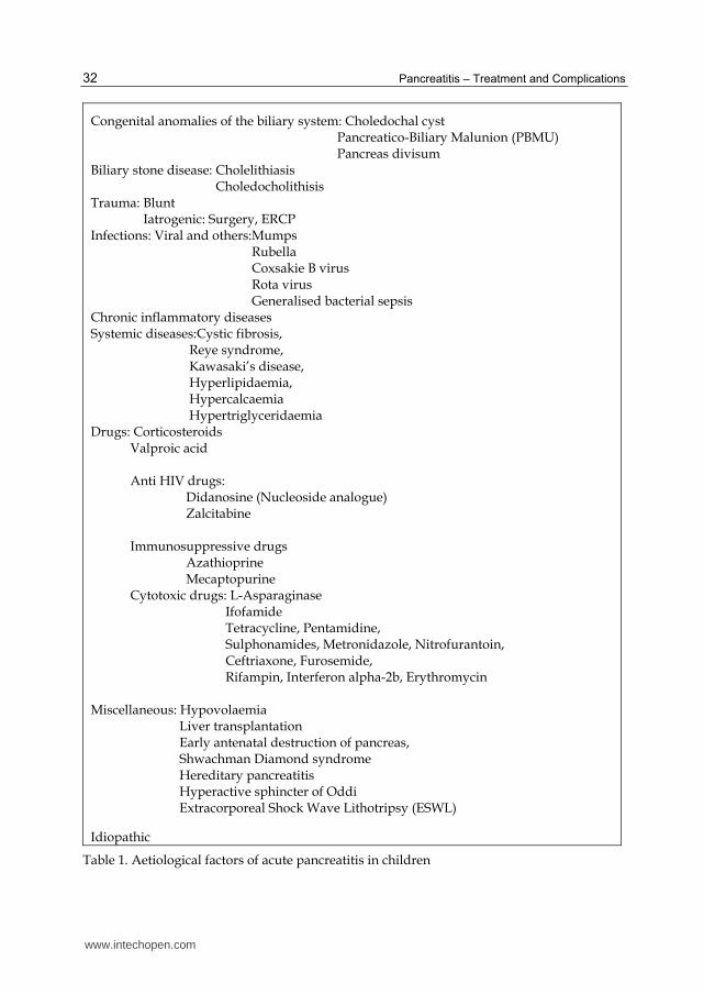
Congenital anomalies of the biliary system: Choledochal cyst Pancreatico-Biliary Malunion (PBMU) Pancreas divisum Biliary stone disease: Cholelithiasis Choledocholithisis Trauma: Blunt Iatrogenic: Surgery, ERCP Infections: Viral and others:Mumps Rubella Coxsakie B virus Rota virus Generalised bacterial sepsis Chronic inflammatory diseases Systemic diseases:Cystic fibrosis, Reye syndrome, Kawasaki’s disease, Hyperlipidaemia, Hypercalcaemia Hypertriglyceridaemia Drugs: Corticosteroids Valproic acid Anti HIV drugs: Didanosine (Nucleoside analogue) Zalcitabine Immunosuppressive drugs Azathioprine Mecaptopurine Cytotoxic drugs: L-Asparaginase Ifofamide Tetracycline, Pentamidine, Sulphonamides, Metronidazole, Nitrofurantoin, Ceftriaxone, Furosemide, Rifampin, Interferon alpha-2b, Erythromycin Miscellaneous: Hypovolaemia Liver transplantation Early antenatal destruction of pancreas, Shwachman Diamond syndrome Hereditary pancreatitis Hyperactive sphincter of Oddi Extracorporeal Shock Wave Lithotripsy (ESWL)
Idiopathic
Table 1. Aetiological factors of acute pancreatitis in children
www.intechopen.com
Childhood Pancreatitis
33
2.2 Chronic pancreatitis
In children, the most common causes of chronic and chronic relapsing pancreatitis are trauma, hereditary, systemic disease and malformations of the pancreaticobiliary ductal system such as pancreas divisum and annular pancreas, and choledocholithiasis.
Chronic pancreatitis could either be calcifying (commoner) or obstructive (less common).
2.2.1 Calcifying chronic pancreatitis
The most common cause of calcifying pancreatitis is hereditary or familial pancreatitis. Comfort and Steinberg gave the first description of hereditary pancreatitis in 1952 (Comfort & Steinberg, 1952). Since then a number of cases of hereditary pancreatitis have been reported. The mode of inheritance strongly suggested was an autosomal dominant trait (Aamarapurkar, 2001). The gene mutations identified in early-onset idiopathic chronic pancreatitis (ICP) are the serine protease inhibitor, Kazal type 1 (SPINK1) gene mutations, and the cystic fibrosis transmembrane regulator (CFTR) gene mutations. Recently, Cationic trypsinogen protease serine 1 PRSS1 gene mutations were detected in "idiopathic pancreatitis" and SPINK1 gene mutations in idiopathic and tropical pancreatitis. It is also very likely that SPINK1 gene mutations are important in tropical calcific pancreatitis (TCP) (see below) (DiMagno M & E., 2003). As many as 10% of patients with "idiopathic pancreatitis" may have hereditary pancreatitis if finding a PRSS1 gene mutation without a family history is considered an acceptable diagnostic criterion (DiMagno M & E, 2003).
The association of CFTR gene mutations and chronic pancreatitis is well known. Even in classic cystic fibrosis (CF) with pancreatic functional preservation, the prevalence of pancreatitis may be 17% or higher because such patients have recurrent, severe abdominal pain that may be due to unrecognized pancreatitis (DiMagno M & E., 2003).
Gene sequencing of the 7q35 chromosome region revealed a strong association of the
(p.R122 H) mutation of the PRSS1 gene encoding cationic trypsinogen with hereditary
pancreatitis (Teich et al., 2006). Developments in genetics have shown that in the majority of
patients, hereditary pancreatitis is usually associated with expression of one of two specific
mutations in the cationic trypsinogen PRSS1 gene, specifically R122H or N29I. Further
sequencing of the PRSS1 gene in the proband of families without these common mutations
revealed R122C and N29T mutations in independent families that segregated with the
disease in an autosomal dominant fashion. The R122C mutation eliminates the arginine
autolysis site as with R122H mutations. The N29T mutation may also enhance
intrapancreatic trypsin activity as has been demonstrated in vitro (Pfutzer et al., 2002).
Further mutations of this gene were discovered in patients with hereditary or idiopathic
chronic pancreatitis. In vitro the mutations increase autocatalytic conversion of trypsinogen
to active trypsin and thus probably cause premature intrapancreatic trypsinogen activation
in vivo. The clinical presentation is highly variable, but most affected mutation carriers have
relatively mild disease (Teich et al, 2006).
Tropical calcific pancreatitis is a type of pancreatitis seen exclusively in tropics. Though the exact aetiology of tropical chronic pancreatitis is obscure, malnutrition with protein deficiency, cassava toxicity, impaired immune response, viral infection and genetic susceptibility have been considered as various factors in the aetiopathogenesis
www.intechopen.com
Pancreatitis – Treatment and Complications
34
(Aamarapurkar, 2001). It is also very likely that SPINK1 gene mutations are important in tropical calcific pancreatitis (TCP), a disease associated with malnutrition and cassava consumption in Afro-Asian countries that is characterized by onset at an early age and variable familial clustering (DiMagno M & E., 2003)..
2.2.2 Obstructive chronic pancreatitis
Encompasses the other causes of chronic pancreatitis in children which include congenital anomalies of the pancreaticobiliary duct system such as pancreas divisum, pancreaticobiliary malunion and annular pancreas. Trauma is also a major cause leading to chronic pancreatitis, particularly when fibrosis complicates pancreatic duct disruption, resulting in pancreatic duct stricture and, consequently, obstructive chronic pancreatitis. Other aetilogical factors include biliary disease, systemic inflammatory and metabolic diseases (table 2). Hypertriglyceridaemia and hyper parathyroidism, though rare, were reported to cause chronic pancreatitis in children (Aamarapurkar, 2001).
Idiopathic fibrosing pancreatitis is a rare condition that affects children and adolescents. It can be a cause of recurrent abdominal pain and obstructive jaundice (Harb & Naon, 2005).
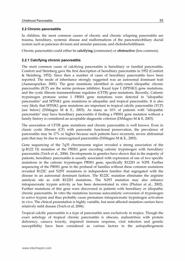
Biliary disease Congenital anomalies of the biliary system: Pancreas divisum PBMU Choledochal cyst Biliary stone disease: Cholelithiasis Choledocholithisis
Hereditary pancreatitis Systemic and metabolic diseases: Hyperlipoproteinaemia Hyperparathyroidism Hypercalcaemia Hypertriglycaedaemia Cystic fibrosis Inborn errors of metabolism Systemic lupus erythematosus Haemophilia Shwachman Diamond Syndrome Infection
Chronic inflammatory diseases Crohn’s disease Ulcerative colitis Chronic fibrosing pancreatitis Juvenile tropical pancreatitis Tropical calcific pancreatitis Idiopathic chronic pancreatitis Idiopathic fibrosing pancreatitis
Table 2. Aetiological factors of chronic pancreatitis in children:
www.intechopen.com
Childhood Pancreatitis
35
3. Pathophysiology
The pancreas matures after birth and by 2 years of age it is functioning in the same way as an adult pancreas. Due to its excellent reserve, the immature pancreas does not seem to affect the healthy children significantly, however, it can have a major impact on very ill or malnourished children (Durie, 2011).
3.1 Acute pancreatitis
3.1.1 Acinar cell injury
Acute pancreatitis originates from acinar cell injury due to factors such as trauma, drugs, metabolic disorders and infection which lead to premature intraductal activation of proenzymes, specifically trypsinogen to trypsin. The resultant autodigestion of the pancreatic tissue and accompanying aggressive immune response cause the pathological process of pancreatitis and subsequent complications (Uretsky et al.,1999; Nydegger et al., 2006)
3.1.2 Proenzyme activation
Inappropriate, premature proenzyme activation in the pancreas, and consequently autodigestion of pancreatic tissue, may occur due to either ductal flow obstruction (structural) with extravasation of enzyme rich ductal fluid into the parenchyma of the pancreas, reflux of duodenal enterokinase into the pancreas or failure in feed-back control (hereditary).
3.1.3 Inflammatory mediators
Once activated, elastase, phospholipase A2 and superoxide free radicles in addition to release from the pancreas of other active mediators including cytokines such as tumour necrosis factor-alpha; and vasoactive substances such as histamines, prostaglandins, kinins and kallikreins, are thought to play an important role as mediators of tissue damage (Weber & Adler, 2003; Mehta & Gittes, 2005).
Along with premature activation of digestive enzymes, disturbances of intracellular calcium, and activation of transcription factors such as NF-κB characterize the initial phase of acute pancreatitis. Depending on the severity of pancreatic injury, the impact may not be confined to the pancreas. Pancreatic enzymes can cause damage at distant sites either by vascular dissemination or by the release of proinflammatory mediators and the recruitment of immune cells which may expand the local disturbances to a systemic inflammatory response associated with diffuse tissue damage throughout the body and failure of distant organs such as lungs or kidneys, a form of multiple system organ failure (MSOF). Patients with severe acute pancreatitis are, therefore, likely to have a high incidence of refractory sepsis and death (Weber & Adler, 2003; Mehta & Gittes, 2005).
3.1.4 Role of cytokines
Cytokines themselves do not induce pancreatitis but rather mediate the progression of pancreatitis. Cellular immune responses are the major determinants of severity. The most important step for the development of severe pancreatitis appears to be the activation of the
www.intechopen.com
Pancreatitis – Treatment and Complications
36
T helper cell type 1 (Th1) response. Pancreatic production of proinflammatory cytokines such as TNF-alpha, interleukin IL-1B, IL-6 and IL-8 modulates local injury, systemic inflammatory response and distant organ failure. This modulation, coupled with pancreatic necrosis, determine the outcome from acute pancreatitis. The TNFa, IL1B, IL6 & IL8 released from granulocytes and macrophages also reach the liver through the portosystemic circulation and act on Kupffer cells that produce more cytokines and acute phase proteins. Other factors determining the severity of pancreatitis include free radicles, pancreatic glutathione, ischaemia, chemokines and neurokines. Because the onset of cytokines action follows immediately after the onset of pancreatitis and peaks after 36-48 hours, cytokine antagonists therapy represent a potential therapeutic target and therefore is of intense interest (Nydegger et al, 2006; Berney et al, 1999).
3.2 Chronic pancreatitis
Chronic pancreatitis is an inflammatory disorder that results in anatomical changes that include chronic inflammatory cell infiltration and gland fibrosis, with loss of exocrine function (malabsorption) and endocrine function (diabetes mellitus). In susceptible children, repeated episodes of acute inflammation may lead to chronic pancreatitis, the so called necrosis-fibrosis hypothesis of Comfort et al. and, accordingly, it is likely that there is significant overlap in the underlying pathophysiology of these conditions (Nydegger et al, 2006).
Whitcomb has proposed a sentinel acute pancreatitis event SAPE which acts as a priori step toward the development of chronic pancreatitis. The acute injury must be sufficiently severe to attract monocytes and to cause infiltration, differentiation and proliferation of pancreatic stellate cells. For fibrosis to occur there must be recurrent acinar injury resulting in chemocytokine release that then stimulates stellate cells (Whitcomb 1999, 2004).
Genetics: Cationic trypsinogen gene Protease Serine 1 (PRSS1) gene accounts for 65% of trypsinogen and is one of the most abundant molecules produced by acinar cells. In chronic pancreatitis, it has been suggested that mutations of the cationic trypsinogen PRSS1 gene lead to an alteration in the trypsin recognition site that prevents deactivation of trypsin within the pancreas. Consequently, autodigestion occurs resulting in pancreatitis. The genetic mutations responsible for hereditary pancreatitis have been isolated to chromosome 7q35. The most common hereditary pancreatitis causing mutations in PRSS1 gene are R122H and N29I (Pfutzeret al., 2002; Teich et al, 2006; Nydegger et al., 2006). These two mutations in the cationic trypsinogen gene appeared to allow prematurely activated trypsinogen to cause acinar cell auto-digestion and acute pancreatitis. Chronic pancreatitis in these patients certainly arises from recurrent acute pancreatitis (Aamarapurkar, 2001). Several other mutations in the PRSS1 gene have been identified including mutations in codons 16, 22, 23 and 122. This discovery of genetic mutations associated with hereditary pancreatitis support the hypothesis that intrapancreatic activation of pancreatic zymogens is crucial to the pathogenesis of acute pancreatitis. The mutant trypsin in hereditary pancreatitis is resistant to lysis, remains active and causes auto-digestion of the pancreas and episodes of acute pancreatitis. Median age at onset of disease is 11 years in patients with the N29I mutation and 10 years in those with the R122H. Approximately half of patients with hereditary pancreatitis develop chronic pancreatitis (Nydegger et al., 2006).
www.intechopen.com
Childhood Pancreatitis
37
Trypsin can exert a negative feed-back, self inactivation to protect the pancreas from auto-digestion. The serine protease inhibitor SPINK1 produces a similar pancreas sparing effect by inhibition of prematurely activated trypsinogen. This gene may have a role in tropical pancreatitis, an idiopathic form of pancreatitis seen in southern Asia and parts of Africa and probably related to malnutrition, together with cassava intake. SPINK1 mutations were also shown to be associated with a subtype of tropical pancreatitis, fibrocalculous pancreatic diabetes. SPINK1 mutations alone probably do not cause pancreatitis but act as disease modifier. The SPINK1 mutations are relatively common, being present in approximately 2% of the general population. However, the frequency of SPINK1 mutations in populations with idiopathic chronic pancreatitis is markedly increased, approximately 25%, an evidence that an association exists between the SPINK1 mutations and pancreatitis (Nydegger et al., 2006).
Cystic fibrosis transmembrane regulator (CFTR): An association between idiopathic chronic pancreatitis and CFTR gene mutations was reported. Several mild CFTR mutations were found to be associated with idiopathic pancreatitis. Available data suggest that patients with two severe mutations have classical cystic fibrosis (CF), those with a single mutation are carriers and those who are compound heterozygotes with one severe and one mild mutation are at risk of developing pancreatitis (Ridout & Lakhoo, 2011; Nydegger, 2006).
4. Classification
Pancreatitis can be classified as:
- Acute pancreatitis: Simple Necrotizing Haemorrhagic
- Acute recurrent pancreatitis - Chronic pancreatitis: Calcific or Obstructive - Chronic relapsing pancreatitis
5. Clinical presentation
5.1 Acute pancreatitis
Diagnosis is based on symptoms and signs accompanied by a three fold increase in either amylase or lipase. Presentation is variable in children and symptoms may range from mild abdominal pain to severe systemic involvement, metabolic disturbances and shock. Abdominal pain, anorexia, nausea and vomiting are the most frequent symptoms (Hebra et al., 2009; Mehta & Gittes, 2005). The pain can be of a sudden or insidious onset with slow and gradual progression with increasing intensity. Although the pain is usually epigastric in origin, right and left upper abdominal quadrants are infrequently involved. Central and lower abdominal pain may also be encountered (Uretsky et al, 1999). The typical radiation of pain to the back observed in adults is not a common feature in children. Food intake exacerbates pain and vomiting (Haddock et al, 1994) while drawing the knees up to the chest relieves the pain (Uretsky et al, 1999). Extracellular fluid losses can be enormous (Mehta & Gittes, 2005).
On examination, the child may be ill, irritable, quiet or a combination of these. Movement aggravates the abdominal pain, therefore children typically tend to assume a still, supine
www.intechopen.com
Pancreatitis – Treatment and Complications
38
position, or they may lie on their side or doubled up with the hips and knees flexed (Hebra et al, 2009). Tachycardia, mild fever, hypotension, jaundice and abdominal signs such as diffuse tenderness, rebound tenderness, guarding, distension and decreased or absent bowel sounds may be elicited (Nydegger et al., 2006, Hebra et al., 2009, Ibrahim & Gabr, 2011). Parotid enlargement may be observed occasionally (Haddock et al, 1994). In severe cases of necrotizing or haemorrhagic pancreatitis, haemorrhage may dissect from around the pancreas along the abdominal wall tissue planes appearing as discoloration either in the flanks (Grey Turner’s sign) or at the umbilicus (Cullen’s sign). These echymoses may take 1-2 days to develop (Mehta & Gittes, 2005). These signs of haemorrhagic pancreatitis are seldom present in children (Nydegger et al, 2006). Evidence of pleural effusion or dyspnoea, with or without acute respiratory distress syndrome, may be found on examination of the chest (Uretsky et al, 1999, Ali et al, 2010). Recurrent acute pancreatitis is seen in 10% of children after the initial acute episode. It is more likely in children with structural anomalies, idiopathic and familial causes (Nydegger et al, 2006).
5.2 Chronic pancreatitis
Unlike acute pancreatitis in which complete reversal of the inflammatory changes occurs upon resolution of the episodes, chronic pancreatitis is characterised by permanent structural changes in the pancreas associated with varying degrees of pancreatic dysfunction and, accordingly, increased risk of developing pancreatic insufficiency, adenocarcinoma, and pancreatic pseudocysts. Patients with this disease typically present with chronic or recurrent upper abdominal pain with significant morbidity. The pain is most commonly described as epigastric, deep or radiating towards the back and accompanied by nausea and vomiting. It is often relieved by sitting and leaning forward and may increase following meal. It may commence as intermittent attacks with periods of well-being and then become more continuous, others may have little or no pain. Common other associated clinical features include weight loss and diabetes mellitus. Paediatric data on prevalence and incidence of exocrine and endocrine failure are limited, however, steatorrhea and weight loss will not occur until pancreatic exocrine function has been reduced to around 2% of the normal output. Because insulin and glucagons secreting cells are destroyed in chronic pancreatitis, the diabetes in these patients can be particularly refractory to treat (Nydegger et al, 2006; Mehta & Gittes 2005).
Tropical calcific pancreatitis is characeritized by recurrent episodes of abdominal pain, severe malnutrition, and ketosis resistant diabetes. Ten per cent of patients with tropical calcific pancreatitis develop pancreatic cancer. Almost one third of patients with tropical calcific pancreatitis get recurrent episodes of pain in childhood and may develop diabetes in childhood (Aamarapurkar, 2001).
6. Investigations
6.1 Acute pancreatitis
Serum amylase levels are usually, but not invariably elevated. Normal levels do not exclude the diagnosis of acute pancreatitis. Also, the degree of serum amylase elevation does not correlate with severity of the disease. Goh found that serum amylase levels were elevated in all the 11 patients in his study. Their serum amylase levels ranged from 190 to 1370 U/L
www.intechopen.com
Childhood Pancreatitis
39
(Median 512.5). One third of them had amylase levels lower than 4 times the upper limit of normal, ie. 440 U/L. (Goh et al., 2003). Amplified elevations of serum amylase are suggestive of development of a pseudocyst or other complications of pancreatitis.
Amylase is excreted in the urine when the level of hyperamylasemia exceeds the tubular reabsorptive capacity. Therefore, moderately elevated levels of serum amylase may not be detectable in the urine. It is important to note that other conditions may cause hyperamylasemia or hyperamylasuria. These include salivary inflammation or trauma, intestinal disease including perforation, ischaemia, necrosis or inflammation, renal failure and macroamylasemia (Mehta & Gittes, 2005). To differentiate the likely underlying cause, a three fold, or more, elevation of serum amylase concentration is strongly in favour of the diagnosis of acute pancreatitis. The half life of amylase is approximately 10 hours and the value frequently becomes normal after 2-5 days.
Initial serum amylase level does not correlate with the severity of pancreatic injury. Schmittenbecher noted a longer lasting elevation of serum amylase in patients with traumatic pancreatitis (Schmittenbecher et al., 1996).
Elevated level of pleural effusion fluid-amylase in children with respiratory symptoms secondary to pancreatitis is a useful diagnostic test (Segura, 2004). Thoracic pancreatic pseudocyst as a complication of traumatic pancreatitis has been reported (Ali et al, 2010).
Lipase: Unlike amylase, lipase is produced only in the pancreas and its measurement is particularly helpful for distinguishing pancreatic trauma from salivary trauma. In children with acute pancreatitis, the serum lipase level is usually elevated for many days. Therefore, lipase levels have been proposed as a more specific test of pancreatic tissue damage. An important caveat to note here is that the degree of lipase elevation has little correlation with the extent of the pancreatitis and that intestinal perforation does cause an elevation of lipase through reabsorption via the peritoneum. Urinary lipase levels may remain elevated for a few days longer than serum levels (Mehta & Gittes, 2005).
Calcium and Glucose: Hypocalcaemia was reported in 10-15% of children with pancreatitis (Goh et al., 2003; Uretsky et al., 1999) while hypoglycaemia was noted in 25% of cases during the acute attack (Uretsky et al., 1999).
The blood glucose level measurement is a key investigation when chronic pancreatitis is suspected. Persistently elevated blood glucose level indicates significant pancreatic endocrine-reserve impairment.
Haematological: Elevated total white blood cell counts (with increased band counts) are usually present (Goh et al., 2003). An increased haematocrit secondary to haemoconcentration that occurs after volume depletion may be found.
Genetics: The majority of pancreatic diseases are associated with genetic polymorphism. Early use of genetic testing is likely to play a critical role in early diagnosis and prognosis of pancreatic diseases (Whitcomb, 2004).
The levels of cationic trypsinogen gene and SPINK1 mutations are said to be a more sensitive early marker of pancreatic inflammation (Uretsky et al., 1999).
Gene testing for CFTR gene is helpful in the diagnosis of pancreatitis in cases thought to be related to cystic fibrosis.
www.intechopen.com
Pancreatitis – Treatment and Complications
40
Imaging: plain abdominal radiographs may reveal obscured psoas margins, a sentinel loop of a dilated duodenum and a gasless segment of mid transverse colon. The latter is caused by local spasm with proximal dilatation known as colon cut-off sign due to inflammation of the adjacent pancreas. Pancreatic calcifications suggest chronic pancreatitis. In cases of tropical calcific pancreatitis, dense calcification is seen in pancreatic parenchyma (Mehta & Gittes, 2005).
Plain chest x rays should be performed in all patients with acute pancreatitis to look for evidence of pleural effusion and pulmonary oedema (Mehta & Gittes, 2005). One fifth of children show evidence of pleural effusion (Uretsky et al., 1999).
Ultrasound Scan (US): is one of the most useful imaging tools in childhood pancreatitis (Haddock et al., 1994). Abdominal US may show pancreatic swelling or a decrease in pancreatic echogenicity due to oedema or peri-pancreatic fluid collection. These findings per se are not sufficient to coin the diagnosis of acute pancreatitis or estimate its severity. The main use of US is to demonstrate gallstones as a possible cause of pancreatitis and to follow up the treatment by serial observation of improvement in oedema or peripancreatic fluid collection (Mehta & Gittes, 2005).
Abdomominal CT scan: provides images of the pancreas superior to those obtained by US scan in as far as determining the size of the pancreas, the degree of oedema and the presence of fluid collection is concerned. Due to the much better resolution, the size and anatomy of the pancreatic duct can often be delineated much more accurately with CT than with US. The presence of complications such as pancreatic abscess or pseudocyst may also be determined. Dynamic CT pancreatography, using an intravenous bolus of contrast with rapid scanning in fine cuts through the pancreas, has the advantage of differentiating perfused from non-perfused (necrotic) pancreas. It has the ability to provide a precise assessment of the percentage of and distribution of pancreatic perfusion. Furthermore, CT scan can be used for interventional procedures for diagnosis or drainage of fluid collections (Mehta & Gittes, 2005).
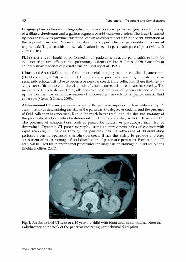
Fig. 1. An abdominal CT scan of a 10 year old child with blunt abdominal trauma. Note the radiolucency at the neck of the pancreas indicating parenchymal disruption
www.intechopen.com
Childhood Pancreatitis
41
ERCP: although the literature suggest that the complication rates with ERCP are higher in children than in adults, ERCP is being used increasingly in children. ERCP can provide detailed information on the pancreatic duct. It is particularly helpful for its diagnostic and therapeutic roles in the management of children with acute obstructive biliary pancreatitis. ERCP and pancreatic duct stenting can also be beneficial in trauma patients in whom a ductal injury is suspected or a pancreatic pseudocyst has formed (Mehta & Gittes, 2005, Ali et al., 2010). In cases of pancreas divisum, however, cannulation of the minor papilla is possible in less than half of the cases because of its small size and the angle at which it meets the duodenum (Miyano, 2006).
MRCP: the magnetic resonance cholangio-pancreatography is a useful, non invasive
diagnostic tool for evaluating the biliary tree and the pancreatic duct. This technique
precludes the risk of complications encountered with ERCP. The study is cheaper and
employs no radiation or administration of contrast media which is routinely performed with
ERCP. MRCP is not without disadvantages. As an essentially diagnostic study, it does not
facilitate therapeutic intervention and it has a tendency to overestimate the stenosis of the
main pancreatic duct in patients with pancreatitis. Despite the recognized disadvantages,
MRCP is now the initial imaging study of choice in the evaluation of pancreatic ductal
anatomy in children with unexplained or recurrent pancreatitis (Mehta & Gittes, 2005).
6.2 Chronic pancreatitis
In children with suspected chronic pancreatitis the following investigations are suggested:
- Sweat test and gene testing for CFTR - Testing for cationic trypsinogen and SPINK1 mutations - Assessment for autoimmune causes - Biochemical investigations for diabetes mellitus, lipase, electrolytes and stools for
evidence of malabsorption - Imaging to exclude structural (congenital or acquired) abnormalities. In children with
chronic pancreatitis, changes on imaging studies are usually encountered - Pancreatic biopsy, considered to be a gold standard for diagnosis in adults, is rarely if
ever required in children (Nydegger et al., 2006).
7. Severity scores
Many scoring systems were used to assess the severity of acute pancreatitis in adults, however, there is no widely adopted robust severity scoring system that is specifically targeting children (Nydegger et al., 2006). The applicability of adult scoring systems such as Ranson's and the Imrie prognostic criteria, the Glasgow and modified Glasgow scores and the Acute Physiology and Chronic Health Evaluation (APACHE) II score have not been validated in children. An alternative severity scoring system has been suggested by other researchers. This group has compared a scoring system designed specifically to paediatric patients with selected adult scoring systems (DeBanto et al., 2002). This system compares eight variables to predict severe outcome and mortality during an episode of acute pancreatitis. An episode of pancreatitis is considered to be severe if the patient died, had surgery on the pancreas, developed a pseudocyst or abscess or infected necrosis, or met criteria for multiple organ dysfunction. The admission criteria considered are:
www.intechopen.com
Pancreatitis – Treatment and Complications
42
- Age <7 yrs
- Weight <23Kg
- WBC (Admission) >18.5/cmm
- LDH (Admission) >2000 IU/L
- 48hr trough albumin <2.6mg/dl
- 48hr trough calcium <8.3mg/dl
- 48hr fluid sequestration >75ml/kg/48hrs
- 48hr rise in blood urea nitrogen >5mg/dl
Each factor is allocated a score of 1 point and higher total scores are associated with an
increased chance of severe pancreatitis and mortality.
Scores of 5-7 points showed 80% severe outcome and 10% mortality
2-4 points showed 38.5 severe outcome and 5.8% mortality
0-2 points showed 8.6% severe outcome and 1.4% mortality
When the cutoff for predicting severe outcome was set at 3 criteria, this new system had a
sensitivity of 70%, a –ve predictive value of 91%, a specificity of 79% and a +ve predictive
value of 45%. The system probably performs better in identifying those patients who are at
risk for severe pancreatitis than the existing systems used in adults (DeBanto et al., 2002).
When compared with the Ranson’s and Glasgow systems used in adult patients with
pancreatitis, this system showed a better sensitivity (70%) versus Ranson (30%) and
Glasgow (35%) scores and a better negative predictive value (91% vs 85% and 85%
respectively) (DeBanto et al., 2002). In trauma, the grade of pancreatic injury was an
independent predictor of both pancreatic complications (Kao et al., 2003).
Some researchers associated the severity of pancreatitis to the aeiology. Haddock found that
severe pancreatitis was most commonly associated with systemic disease (22 of 51; 43.1%)
and trauma (13 of 51; 25.4%), beside associated factors such as significantly higher body
weight, higher frequency of dyspnoea and pleural effusion, and lower serum calcium and
albumin levels (Haddock et al., 1994; Chang et al., 2011).
Many classification systems were adopted for grading of disease severity based on various
factors. Examples of these classification systems include:
- Marseille classification which relies on grading of biopsy specimens.
- Cambridge classification with more emphasis on imaging to provide grading and
severity scoring, Computed tomography (CT) was more accurate than ultrasound in
evaluation of the severity of pancreatitis.
- TIGAR-O system: more recently, the Toxic, Idiopathic, Genetic, Autoimmune,
Recurrent severe associated chronic pancreatitis and Other systems has been developed
and allows multiple risk factors to be assessed in an individual patient either as a risk
factor or as an aetiology (Nydegger et al., 2006).
- Atlanta criteria: though this is still controversial with lack of consensus (Berney et al.,
1999; Bollen et al., 2007).
Overall, it is relevant to state that up till now commonly used scoring systems have limited
ability to predict disease severity in children and adolescents with acute pancreatitis.
www.intechopen.com
Childhood Pancreatitis
43
Careful and repeated evaluations are essential in managing these patients as they may
develop major complications without early signs (Lautz et al., 2011).
8. Treatment
8.1 Acute pancreatitis
8.1.1 Conservative treatment
Treatment of acute pancreatitis is primarily and essentially supportive. It aims at limiting
exocrine pancreatic secretion and monitoring for acute and long term complications. Key
features in the empirical therapeutic regimen commonly used in treating patients with acute
pancreatitis are: aggressive IV fluid administration to replace the profound fluid deficit and
to maintain a good hydration as indicated by adequate urine output (2ml/kg/hr), usually
measured with the aid of an indwelling urinary catheter. Pain control should be achieved
with parenteral analgesia. Adequate analgesia is critical to minimize the additional stress
from pain. Meperidine (Demerol) is thought to be a better analgesic in pancreatitis because
morphine is well known to cause spasm of the sphincter of Oddi which in turn is known to
increase pancreatic duct pressure and potentially worsen the pancreatitis (Mehta & Gittes,
2005). It is also important to be certain of the diagnosis of pancreatitis before giving the
patient significant doses of narcotics because the ability to diagnose serious non-pancreatic
problems, such as intestinal ischaemia or perforated ulcer, may be lost. It is imperative to
have a very low threshold for transferring the patient to an intensive care unit and provide
constant monitoring to avoid development of profound hypovolemia. Initially, bowel
should be kept at rest with NG suction. Most mild to moderate cases will settle if food and
drink is withheld for a few days. Proton pump inhibitors (PPI) or H2 receptor antagonists
administration helps to prevent exposure of the duodenal secretin producing cells to gastric
acid, which is a potent stimulator of pancreatic secretion. These antagonists also may help
prevent stress ulceration seen in patients with pancreatitis. While clinical trials have shown
improved outcome in acute pancreatitis by using long acting somatostatin analogues
octreotides, controversy still exists concerning the use of prophylactic antibiotics. In
general, mild to moderate cases probably do not benefit from antibiotics. More severe cases
of pancreatitis, however, may benefit because of the high rate of sepsis. Prophylactic
antibiotics are indicated when there is pancreatic necrosis or when infection is either
suspected or documented in severe acute pancreatitis. Studies have related prophylactic
antibiotic administration for 10-14 days in adult patients with severe acute pancreatitis and
proven pancreatic tissue necrosis with decrease in superinfection of necrotic tissue and
mortality (Nydegger et al., 2006; Mehta & Gittes, 2005; Miyano, 2006)). Some advantages
have been demonstrated with the use of imipenem with reduction in the incidence of
pancreatic sepsis in patients with necrotizing pancreatitis. Because the onset of cytokine
action follows immediately after the onset of pancreatitis and peaks after 36-48 hours,
cytokine antagonist therapy represents a potential therapeutic target and therefore is of
intense interest (Nydegger et al., 2006). In children with relapsing attacks of pancreatitis we
would advocate similar management and also include investigations to exclude structural,
metabolic and hereditary pancreatitis (Nydegger et al., 2006). Nutrition is a crucial factor in
the management of patients with pancreatitis. Early positive nitrogen balance has been
shown to improve survival rates. This need for aggressive nutrition should come in the form
www.intechopen.com
Pancreatitis – Treatment and Complications
44
of early TPN administration. The TPN should include lipid formulations. To minimise the
causal association of hyperlipidaemia and pancreatitis, it is recommended that serum lipid
levels should be monitored to maintain triglyceride levels no greater than 500 mg/dl (Mehta
& Gittes, 2005).
The results of several controlled studies in adult patients with acute pancreatitis, support enteral nutrition. It costs less than TPN by approximately 1/4. Total enteral nutrition moderates the acute phase response and improves disease severity and clinical outcome despite unchanged pancreatic imaging findings (Windsor et al., 1998; Nydegger et al., 2006). Although there are sparse paediatric data, enteral feeding has been widely adopted. Perhaps a nasojejunal feeding with high protein and low-fat formula through a tube inserted under radiological or endoscopic guidance can be implemented until the child is ready to commence oral intake (Nydegger et al., 2006). Naso-jejunal feeding may be particularly useful in places where TPN availability is considered to be a luxury!.
It is imperative to note that despite the known advantage of nutritional support in facilitating resolution of pancreatitis, quite often early commencement of enteral feeding results in relapse of acute abdominal pain and elevation of amylase levels.
In general, the resumption of enteral nutrition should be cautious, usually after complete resolution of abdominal pain and preferably after normalization of the serum enzyme levels (Mehta & Gittes 2005).
8.1.2 Surgical treatment
Surgery for pancreatic disorders in children is rarely required. Surgical intervention in children with acute pancreatitis is even less practiced. It should be restricted to patients with complications such as severe necrotizing pancreatitis requiring debridement or patients with other complications associated with acute pancreatitis including pancreatic ascites, pancreatic abscess collections not amenable for percutaneous drainage and pancreatic pseudocyst (Rabinovich et al., 2006) (see below).
In traumatic pancreatitis, as most children with a solid-organ abdominal injury can be managed conservatively, the pancreas is no exception. Non-operative management of low grade pancreatic injury is widely accepted. Management of major pancreatic parenchymal or ductal injury in children, however, remains controversial (Haddock et al., 1994; Stringer et al., 2005). Initial nonoperative management of injuries of the proximal pancreatic duct is a common practice and allows for the formation and uneventful delayed drainage of a pseudocyst, rather than the risks of early radical interventions (Jobst et al., 1999). If there is delayed or no response to conservative management, ERCP with sphincterotomy and/or stenting of injured pancreatic duct may be attempted as it has reportedly had a proven efficacy in resolving the symptoms in such patients (Cay et al., 2005; Canty & Weinman, 2001; Ali, 2010). Distal duct injuries are best managed by prompt spleen-sparing distal pancreatectomy (Jobst et al., 1999). Removal of impacted stones in gallstone pancreatitis in children should be performed endoscopically (Mehta & Gittes, 2005).
When discovered incidentally during the course of laparotomy for other suspected causes of
peritonitis, it is imperative to palpate the gallbladder (GB) for stones. In the presence of
gallstones and mild pancreatitis, cholecystectomy can be performed. However, with severe
www.intechopen.com
Childhood Pancreatitis
45
pancreatitis, cholecystostomy may be the safer option which can provide an access to the
biliary calculi later. Cholecystectomy can be done at a later stage when the patient’s
condition is favourable. If no gallstones are present, but the patient has severe necrotizing
pancreatitis, limited debridement and/or leaving large sump drains in place is probably
adequate (Mehta & Gittes, 2005).
Overall, early pancreatic lavage, pancreatic drainage, and pancreatic resection have not been shown to improve survival rates in cases of severe pancreatitis (Mehta & Gittes, 2005) and early intervention for pancreatic injury, in the absence of clinical deterioration or major ductal injury, is not recommended either (Keller et al., 1997). There is also an iatrogenic dimension to the problem as the rarity of pancreatic surgery in children caused an unfamiliarity that can be associated with contracting significant morbidity and mortality.
When comparing traumatic and non-traumatic pancreatitis, Schmittenbecher and co-researchers found that more patients with traumatic pancreatitis were operated on in 86% of cases and the rate of pseudocysts reached 61.5% whereas non-traumatic pancreatitis required surgical intervention in 50% and developed pseudocysts in 17% of cases. In non-traumatic pancreatitis it is recommended that surgery should be avoided and reserved for complications. Exceptions are obstructions of the pancreaticobiliary ducts which need early removal to prevent chronicity of the disease and functional loss of the organ (Schmittenbecher, 1996). In their study, Stringer et al summarized the indications for surgery in acute pancreatitis in persistent pseudocysts and treatment of an underlying cause of pancreatitis (Stringer et al., 2005).
Key points in the management of acute pancreatitis
IV Fluids ICU admission Pain control NGT aspiration PPI or H2 receptor-blockers Somatostatin analogues Prophylactic antibiotics Cytokine antagonists Adequate nutrition Endoscopic intervention Surgical intervention
8.2 Chronic pancreatitis
Management of chronic pancreatitis is directed towards identification of the aetiological
factors, diagnosis of the condition and hence implementation of the appropriate therapy.
Due to the structural changes associated with chronic pancreatitis, many more surgical
indications exist here when compared with acute pancreatitis. As appropriate, conservative
management consists of provision of medications, analgesia, antibiotics and nutrition.
Emphasis should be made on treatment of pancreatic exocrine insufficiency, therefore,
digestive problems related to failure of the pancreas require enzyme replacement therapy
www.intechopen.com
Pancreatitis – Treatment and Complications
46
with meals as well as fat soluble vitamins supplements (Durie, 2010). Endocrine
insufficiency, particularly diabetes mellitus, should be controlled.
Indications for operative intervention include unsuccessful conservative medical therapy, intractable pain, narcotic addiction, impaired nutrition and poor weight gain.
The goal of surgery in these patients is to relieve pain by provision of adequate analgesia and to preserve the exocrine and endocrine functions of the pancreas through decompression of pancreatic ducts and restoration of adequate pancreatic drainage.
Surgical options include lateral longitudinal pancreaticojejunostomy (Puestow procedure),
distal pancreatectomy with Roux-en-Y pancreaticojejunostomy (Duval procedure), ERCP
sphincterotomy or operative sphincteroplasty. In cases of intractable and refractory disease,
a few pediatric patients with chronic pancreatitis and chronic abdominal pain were
successfully treated with total pancreatectomy and islet cell transplantation (Hebra et al.,
2009; Bellin et al., 2008).
Surgical intervention is indicated for the management of congenital anatomic defects, such
as pancreas divisum. The primary goal of treatment of pancreas divisum associated with
pancreatitis is to establish adequate drainage of the duct of Santorini. This can be achieved
with accessory papilla sphincteroplasty. Correct use of this procedure demands that
intrapancreatic ductal obstruction be ruled out by pancreatography. The progression of
disease to pancreatic insufficiency can be arrested when the obstruction is relieved early.
Endoscopic stenting with or without sphincterotomy has been described, however, the
technique requires particular skill with the endoscope in cannulation of the small ducts
encountered in children. Re-stenosis and recurrence of symptoms have been reported
following endoscopic sphincterotomy.
If chronic pancreatitis has developed in the presence of a dilated duct, longitudinal
pancreaticojejunostomy (Puestow) should be performed. The indication for the use of a
direct ductal decompression procedure is evidence of pancreatic ductal ectasia with
multiple intrapancreatic duct strictures, particularly when symptomatic (Stringer et al.,
2005). An important precautionary measure in any drainage procedure is preservation of
existing endocrine function by avoiding major pancreatic resection. Longitudinal
pancreaticojejunostomy (Puestow technique) in adults has been successful in eliminating or
reducing pain intensity from chronic pancreatitis in 70 to 97% of patients. In children, some
authors maintain that the Puestow procedure improves pancreatic function, decreases
hospitalization and increases body weight toward ideal. Others have reported that distal
pancreatectomy and pancreatico-jejunostomy are effective whereas longitudinal
pancreaticojejunostomy is ineffective (Miyano, 2006). Traditionally, these procedures were
performed with open surgery. A minimally invasive approach to the longitudinal
pancreaticojejunostomy using robotic surgery in a child has been reported recently (Meehan
& Sawin, 2011). Due to this controversy regarding appropriate operative procedures in
paediatric cases, further consideration is warranted.
Subtotal or total pancreatectomy is associated with considerable morbidity and mortality
and is reserved for patients with intractable pain who have diffuse parenchymal damage
without ductal dilatation. The procedure is not generally indicated in children.
www.intechopen.com
Childhood Pancreatitis
47
DuBay et al. reported that in the treatment of complicated hereditary pancreatitis (HP) in children, the modified Puestow procedure (longitudinal pancreaticojejunostomy) improves the quality of life by improving pancreatic function, decreasing hospitalizations, and increasing the percentile ideal body weight. Direct pancreatic duct localization during the procedure reduces morbidity rate than localization via distal pancreatectomy. Surgery performed in the early stage of complicated disease may preserve pancreatic function (DuBay et al., 2000). Likewise, surgery for chronic relapsing pancreatitis is done to relieve pain, treat complications or both. Adequate surgical decompression, ultimately, can prevent
disease recurrence (Clifton et al., 2007; Bellin et al., 2008).
In children suffering from chronic pancreatitis complicated by diabetes mellitus, pancreatectomy and islet autotransplantation can prevent or reduce the severity of diabetes in about 75% of patients. Furthermore, outcome is noted to be better in younger than older children (Bellin et al., 2008).
Treatment of chronic calcific pancreatitis includes control of diabetes, relief of pain with analgesics, pancreatic enzyme replacements, endoscopic or surgical decompression of dilated ducts and removal of pancreatic calculi (Aamarapurkar, 2001).
The role of laparoscopy: laparoscopy has been reported to be used for common bile duct
(CBD) exploration in obstructive recurrent pancreatitis (Shah et al., 1997). The commoner
use of laparoscopy is for cholecystectomy when gallstones are incriminated in childhood
pancreatitis. Laparoscopy has also been used in the treatment of pancreatic pseudocysts (see
below).
Surgery of the pancreas is generally of 3 types,
1. Sphincteroplasty
2. Pancreatic drainage via longitudinal pancreaticojejunostomy (Puestow)
or end to end pancreaticojejunostomy (Duval)
or pancreatogastrostomy (Smith)
3. Pancreatectomy: partial or total
9. Complications
Expected local complications of childhood pancreatitis include necrotizing pancreatitis,
haemorrhagic pancreatitis, pseudocyst and pancreatic fistula. While systemically, as cases of
severe pancreatitis progress, close monitoring of the patients should be exercised for signs of
development of hypovolaemic shock and of multiorgan system failure. Pleural effusion and
pulmonary oedema can progress to severe adult respiratory distress syndrome with
hypoxia requiring endotracheal intubation. The tense abdominal distention associated with
pancreatitis, either due to ileus or ascites, frequently contributes to the hypoventilation.
Hypocalcemia, hypomagnesemia, anaemia from haemorrhage, hyperglycemia, renal failure
and late sepsis can be seen in these patients and require close monitoring and treatment.
9.1 Acute haemorrhagic pancreatitis
Most of the complications related to acute pancreatitis tend to occur in the first two weeks of
onset of pain; and secondary infection and necrosis account for 70-80% of deaths (Munoz &
www.intechopen.com
Pancreatitis – Treatment and Complications
48
Katerndahl, 2000). Zhu et al. noted that pancreatic damage or pancreatic necrosis in critically
ill children is characterized by acute onset, profound severity, short course and multiple
organ failure. It may be asymptomatic in early stage, and can be easily missed (Zhu et al.,
2011).
Acute hemorrhagic pancreatitis is a rare event in children. This serious complication of
acute pancreatitis may be attended with a mortality rate approaching 50% because of shock,
systemic inflammatory response syndrome with multiple organ dysfunction, acute
respiratory distress syndrome (ARDS), disseminated intravascular coagulation (DIC),
massive gastrointestinal bleeding and systemic sepsis or peritonitis. Clinical manifestations
related to hemorrhagic pancreatitis may include Grey Turner sign (bluish discoloration of
the flanks) or Cullen sign (discoloration of the periumbilical region) because of blood
extravasation in the fascial planes of the abdominal wall. Additional signs include pleural
effusions, haematemesis, melaena, and coma (Mehta & Gittes, 2005; Miyano 2006)
9.2 Pancreatic pseudocysts
9.2.1 Definition
Pancreatic pseudocysts are localized collections of pancreatic secretions with no epithelial
lining. They develop as a complication of pancreatitis where they were reported to
complicate 10-23% of the acute episodes. When associated with abdominal trauma, the
frequency rate of pseudocyst identification is higher than 50% (Hebra et al., 2009). The other
common causes of pseudocysts in children are pancreatic duct obstruction, infection and,
rarely, drug induced acute pancreatitis such as with valproic acid (Miyano, 2006; Mehta &
Gittes, 2005).
9.2.2 Pathology
As a result of extravasation of digestive pancreatic enzymes, pseudocysts develop in the
lesser sac between the pancreas posteriorly and the stomach anteriorly. The cavity is lined
by fibroelastic connective tissue capsule due to inflammatory reaction on the surrounding
organs such as the pancreas, stomach, duodenum, colon, small bowel and omentum. An
acute pseudocyst develops a thick fibrous wall in 4 to 6 weeks (Mehta & Gittes, 2005). After
6 weeks, a pancreatic pseudocyst is considered mature. Cysts may vary in size and if greater
than 6 cm in size, resolution with conservative medical management is doubtful (Yoder et
al., 2009). Cyst fluid is clear or straw coloured in most cases and may contain toothpaste-like
debris. The amylase level of the cyst fluid is typically higher than 50000 U/mL (Miyano,
2006).
9.2.3 Diagnosis
A pancreatic pseudocyst formation should be suspected in patients with a history of
abdominal trauma or pancreatitis followed by clinical or radiological appearance of an
upper abdominal mass. Common associated symptoms and signs include persistent
abdominal pain, tenderness, abdominal mass, gastric outlet obstruction, anorexia, nausea
and vomiting. Occasionally, gastrointestinal haemorrhage, weight loss, jaundice, chest pain,
fever and ascites may be encountered (Miyano, 2006; Yoder et al., 2009).
www.intechopen.com
Childhood Pancreatitis
49
Pseudocysts may be acute or chronic. The acute pseudocyst has an irregular wall on CT
scan, is tender and usually follows a recent episode of acute pancreatitis or trauma. Chronic
pseudocysts are usually spherical with a thick wall and they are commonly seen in patients
with chronic pancreatitis. It is important to distinguish these two types of pseudocysts
because 50% of acute pseudocysts resolve without therapy, whereas chronic pseudocysts
rarely do (Mehta & Gittes, 2005).
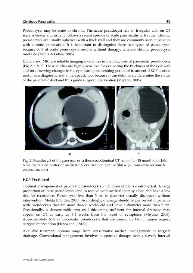
US, CT and MRI are reliable imaging modalities in the diagnosis of pancreatic pseudocysts
(Fig 2, a & b). These studies are highly sensitive for evaluating the thickness of the cyst wall
and for observing changes in the cyst during the ensuing period of treatment. ERCP is often
useful as a diagnostic and a therapeutic tool because it can definitively determine the status
of the pancreatic duct and thus guide surgical intervention (Miyano, 2006).
a) b)
Fig. 2. Pseudocyst of the pancreas on a thoracoabdominal CT scan of an 18 month old child. Note the related posterior mediastinal cyst seen on picture film a. (a, transverse section; b, coronal section)
9.2.4 Treatment
Optimal management of pancreatic pseudocysts in children remains controversial. A large
proportion of these pseudocysts tend to resolve with medical therapy alone and have a low
risk for recurrence. Pseudocysts less than 5 cm in diameter usually disappear without
intervention (Mehta & Gittes, 2005). Accordingly, drainage should be performed in patients
with pseudocysts that are more than 6 weeks old and have a diameter more than 5 cm.
Occasionally, a demonstrable cyst wall thickening sufficient for internal drainage may
appear on CT as early as 3-4 weeks from the onset of symptoms (Miyano, 2006).
Approximately 60% of pancreatic pseudocysts that are caused by blunt trauma require
surgical intervention (Hebra et al., 2009).
Available treatment options range from conservative medical management to surgical
drainage. Conventional management involves supportive therapy over a 6-week interval
www.intechopen.com
Pancreatitis – Treatment and Complications
50
during which time the cyst may either resolve spontaneously or the cyst wall undergoes
fibrous maturation, potentially allowing internal surgical drainage to the stomach or jejunum.
Octreotide acetate, a long acting somatostatin analogue, has been shown to be successful in reducing exocrine function and, thereby, helps resolving pancreatic pseudocysts in children (Miyano, 2006; Mehta & Gittes, 2005).
If the patient can not withstand major surgery, has an infected or an immature cyst, percutaneous external surgical drainage is preferred. U/S or CT guided percutaneous drainage is the treatment of choice for infected pseudocysts because these cysts typically have thin, weak walls not amenable to internal drainage. However, when compared with internal drainage, external drainage is generally associated with adverse consequences such as fistula formation and a higher recurrence rate (Miyano, 2006).
Internal drainage, especially transgastric cysto-gastrostomy is widely used. There is significant evidence indicating that it is effective in the treatment of mature pancreatic pseudocysts (Miyano, 2006; Mehta & Gittes, 2005).
Roux-en-Y cysto-jejunostomy is an alternative internal drainage procedure and is associated with less complications and recurrence rates.
Though less commonly used than other operative procedures, contemplating cysto-duodenostomy may be effective when a cyst is intimately adherent to the duodenum.
Either pancreatico-duodenectomy or distal pancreatectomy, as indicated, is effective in treating patients with pseudocysts in selected cases. Pseudocysts in the body or tail of the pancreas or those involving the head and uncinate process of the pancreas that are not amenable to internal drainage are rare and may require distal or proximal pancreatic resection, respectively (Miyano, 2006).
Minimally invasive drainage approaches to pancreatic pseudocysts were reported. Strategies for cysto-gastrostomy include transoesophago-gastric endoscopic cysto-gastrostomy and percutaneous drainage (Mehta & Gittes, 2005). The other minimally invasive technique is laparoscopic transgastric cysto-gastrostomy or laparoscopic cysto-jejunostomy. There are few reports on the use of laparoscopy in the management of pancreatic pseudocysts (Saad et al., 2005; Sietz et al.,2006). In a multi-institutional retrospective review of 13 patients with a mean age of 10.4 years and mean weight of 52.1 kg who underwent laparoscopic cysto-gastrostomy with no conversions, the authors concluded that a laparoscopic approach to pancreatic cysto-gastrostomy for chronic pseudocysts proved to be safe and effective. Techniques varied, but 92% of patients had complete resolution with minimal morbidity and rapid recovery. Laparoscopic cysto-gastrostomy can be adopted as an appropriate first-line treatment for chronic pseudocysts in children (Yoder et al., 2009).
Both endoscopic and laparoscopic drainage approaches are safe, efficient and effective, however, the two procedures should be performed at institutions with significant experience with these techniques to minimize serious potential risks.
Treatment of persistent Pancreatic pseudocysts:
- Internal drainage (preferred) - Excision (distal pseudocysts only) - External drainage (infected or immature cysts)
www.intechopen.com
Childhood Pancreatitis
51
9.2.5 Pseudocyst complications
Untreated pseudocysts may present with persistence of symptoms or become complicated
with major haemorrhage, infection or cyst rupture. Haemorrhage can occur as a result of
cyst pressure and erosion into an adjacent blood vessel and may be controlled with
emergency angiography and embolization. For infected or ruptured pseudocysts, early
intervention with external drainage is indicated (Miyano, 2006; Mehta & Gittes, 2005)
9.3 Pancreatic ascites
Pancreatic ascites is uncommon but may follow trauma or pancreatic surgery. Leakage of
pancreatic fluid from a damaged major pancreatic duct results in ascites. Treatment is
initially conservative and consists of bowel rest, TPN administration and use of long-acting
somatostatin analogue. In many cases ascites resolves with this treatment. If not, ERCP or
MRCP should be performed to determine the site of the ductal injury and, accordingly, the
appropriate course of surgical intervention (Mehta & Gittes. 2005).
9.4 Pleural effusion
Occasionally, pancreatic pleural effusion may develop following severe pancreatitis. A
significantly elevated level of pleural fluid amylase/lipase with concentrations higher than
those obtained from patient’s serum sample, may be encountered following traumatic
pancreatitis. Intrathoracic pancreatic pseudocyst has been reported following traumatic
pancreatitis where, eventually, persistent pancreatic ductal leak was successfully managed
with ERCP stenting of the pancreatic duct (Ali et al., 2010).
9.5 Pancreatic abscess
Infection may complicate necrotic pancreatic tissue or peripancreatic fluid collection
resulting in pancreatic abscess. Abscess increases the mortality rate of pancreatitis threefold
and is an absolute indication for surgical intervention. Persistence of fever and leukocytosis
in a child with pancreatitis for more than 7 days is an indication for CT guided needle
aspiration to differentiate pancreatic abscess from an uninfected pancreatic fluid collection.
Dynamic CT pancreatography is a useful test in showing pancreatic necrosis which is a
likely predisposing factor for the subsequent development of a pancreatic abscess. The
definitive therapy for pancreatic abscess entails surgical debridement and adequate abscess
cavity drainage (Mehta & Gittes, 2005).
9.6 Pancreatic fistula
Pancreatic fistula develops following surgical procedures in the pancreas. Most low output
fistulas close spontaneously but may drain for several months. Long acting somatostatin
analogue is beneficial in decreasing the fistula output and accelerating the rate of closure.
Fistula closure can be facilitated by adequate nutrition, with TPN if enteral feeding results in
high-volume output, and by ensuring that the fistula tract does not become obstructed. In
persistent fistulas surgical Roux-en-Y jejunostomy to the leak point is recommended (Mehta
& Gittes, 2005).
www.intechopen.com
Pancreatitis – Treatment and Complications
52
9.7 Hyperglycaemia
Both hyperglycaemia and diabetes can occur in children with pancreatitis. In a recent study, researchers looked at 176 patients who were up to 21 years of age and who were hospitalized with acute pancreatitis, acute recurrent pancreatitis, and chronic pancreatitis, excluding those with known pre-existing diabetes or cystic fibrosis before presentation with pancreatitis. Severe pancreatitis was associated with hyperglycaemia, and eight patients developed diabetes requiring insulin by the time of discharge. The authors noted that while adults tend to develop diabetes after chronic pancreatitis, children can develop diabetes due to a single episode of acute pancreatitis (Raman et al., 2011).
9.8 Adenocarcinoma of the pancreas
In patients with chronic pancreatitis, it has been reported that there is a 4% life risk of developing pancreatic adenocarcinoma. This risk may be as high as 40% in patients with hereditary pancraetitis (Nydegger et al., 2006).
10. Prognosis
Uncomplicated acute pancreatitis in children usually have excellent prognosis. The outcome also depends on the co-morbid conditions (Werlin 2003). The mortality rate is approximately 10% in mild disease and up to 90% in cases of necrotizing or haemaorrhagic pancreatitis (Uretsky 1999).
11. Conclusion
Pancreatitis in children is an uncommon event, however, it is attended with significant morbidity and mortality. The presentation may range from transient mild symptoms to life threatening, overwhelming sepsis, multiple organ system failure and death. Prompt institution of appropriate diagnostic and therapeutic measures is crucial to achieve good clinical results. Conservative management and minimally invasive surgical and endoscopic procedures are usually sufficient to treat most of the patients. Children rarely need surgical intervention for the treatment of pancreatitis and its complications. Further developments are anticipated in the field of research, diagnostic tools, disease severity scoring and therapeutic methods to improve the outcome.
12. References
Aamarapurkar, D. (2001). Chronic pancreatitis, cited on 15/07/2011, available from www.bhj/journal/2001_4301_jan/sp_76.htm
Ali, A.; Tan, H.; Gent, R.; Davidson, G. & Roberts-Thompson, I. (2010). Intra- thoracic pancreatic pseudocyst. A rare complication of traumatic pancreatitis. Pediatr Surg Int, Vol.26, No.8, pp. 859-861
Bai, H.; Ma, M.; Orabi, A.; Park, A.; Latif, S.; Bhandari, V.; Husain, S. Novel Characterization of Drug-Associated Pancreatitis in Children. J Pediatr Gastroenterol Nutr. (Jun 2011) 14. DOI:10.1097/MPG.0b013e318228574e. Retrieved on 18/07/2011 from http:// pubget.com/paper/21681111
www.intechopen.com

Childhood Pancreatitis
53
Bellin, M.; Carlson, A.; Kobayashi, T.; et al. (2008) Outcome after pancreatectomy and islet autotransplantation in a pediatric population, J Pediatr Gastroenterol Nutr, Vol.47, No.1, (Jul 2008), pp.37-44
Berney, T.; Belli, D.; Bugmann, P.; Beghetti, M.; Morel, P. & LeCoultre, C. (1996). Influence of severe underlying pathology and hypovolemic shock on the development of acute pancreatitis in children, J Pediatr Surg, Vol.31, No.9, (September 1996), pp. 1256-1261
Berney, T.; Gasche, Y.; Robert, J.; Jenny, A.; Mensi, N.; Grau, G.; et al. (1999). Serum profile of interleukin-6, interleukin-8, and interleukin-10 in patients with severe and mild acute pancreatitis, Pancreas, Vol.18, No.4, (May 1999), pp. 371-377
Bollen, T.; Santvoort, H.; Besselink, M.; van Leeuwen, M.; Horvath, K.; Freeny, P. & Gooszen, H. (2007). The Atlanta classification of acute pancreatitis revisited. Br J Surg, Vol.95, No.1, (Jan 2008), pp. 6-21
Butler, K.; Venzon, D.; Henry, N.; Husson, R.; Mueller, B.; Balis, F.; Jacobsen, F.; Lewis, L. & Pizzo, P. (1993). Pancreatitis in human immunodeficiency virus-infected children receiving dideoxyinosine, Pediatrics, Vol.91, No.4, (April 1993), pp 747-751
Camp, J.; Polley, T. & Coran, A. (1994). Pancreatitis in children: diagnosis and etiology in 57 patients, Pediatr Surg Int, Vol. 9, No.7, (August 1994), pp 492-497
Canty, T. & Weinman, D. (2001). Treatment of pancreatic duct disruption in children by an endoscopically placed stent. J Pediatr Surg, Vol.36, No.2, (Feb 2001), pp. 345-348
Cay, A.; Imamoglu, M.; Bektas, O.; Ozdemir, O.; Arslan, M. & Sarihan, H. (2005). Nonoperative treatment of traumatic pancreatic duct disruption in children with an endoscopically placed stent, J Pediatr Surg, Vol.40, No.12, (2005), pp. e9-e12
Chang, Y.; Chao, H.; Kong, M.; Hsia, S.; Lai, M. & Yan, D. (2011). Acute pancreatitis in children, Acta Paediatrica, Vol.100, No.5, (May 2011), pp. 740–744
Clifton, M.; Pelayo, J.; Cortes, R.; Grethel, E.; Wagner, A.; Lee, H.; Harrison, M.; Farmer, D. & Nobuhara, K. (2007). Surgical treatment of childhood recurrent pancreatitis, J Pediatr Surg, Vol.42, No.7, (July 2007), pp. 1203-1207
Comfort, M. & Steinberg, A. (1952). Pedigree of family with hereditary chronic relapsing pancreatitis, Gastroenterology, No.21, pp 54-63
DeBanto, J.; Goday, P.; Pedroso, M.; et al. (2002). Acute pancreatitis in children. Am J Gastoenterol, Vol.97, No.7, (July 2002), pp. 1726-1731
DiMagno, M. & DiMagno, E. (2003). Chronic pancreatitis, Current Opinion in Gastroenterology, Vol.19, No.5, (September 2003), pp. 451-457
DuBay, D.; Sandler, A.; Kimura, K.; Bishop, W.; Eimen, M. & Soper, R. (2000). The modified Puestow procedure for complicated hereditary pancreatitis in children. J Pediatr Surg, Vol.35, No.2, (Feb 2000), pp. 343-348
Durie, P. (2010). Inherited pancreatic disorders of childhood. Cited 16/07/2011, available from www.pancreasfoundation.org/2010/04/inherited-pancreatic-disorders-of-childhood/
Garg, R.; Agarwala, S. & Bhatnagar, V. (2010). Acute pancreatitis induced by ifosfamide therapy, J Pediatr Surg, Vol.45, No.10, (October 2010), pp. 2071-2073
Goh, S.; Chui, C. & Jacobson, A. (2003). Childhood acute pancreatitis in a children’s hospital, Singapore Med J, Vol.44, No.9, pp. 453-456
Haddock, G., Coupar, G., Youngson, G., MacKinlay G., & Raine P. (1994). Acute pancreatitis in children: A 15-year review, J Pediatr Surg, Vol. 29, No.6, pp. 719-722
www.intechopen.com
Pancreatitis – Treatment and Complications
54
Harb, R. & Naon, H. (2005). Idiopathic fibrosing pancreatitis in a 3-year-old girl: a case report and review of the literature, J Pediatr Surg, Vol.40, No.8, (August 2005), pp 1335-1340
Hebra, A.; Adams, S. & Thomas, P. (2009). Pediatric pancreatitis and pancreatic pseudocyst treatment and management. Medscape Reference. 21 Oct 2009. available from www.emedicine.medscape.com/article/933256-treatment
Hwang, S.; Paik, C.; Lee, K. Chung, W.; Jang, U. & Yang, J. (2010). Recurrent acute pancreatitis caused by an annual pancreas in a child, Gastrointest Endosc, Vol.72, No.4, (October 2010), pp. 848-849
Ibrahim, M.; Gabr, K.; Abdulrazik, M.; Fahmy, H. & El-Booq, Y. (2011). Acute pancreatitis in children: an experience with 50 cases, Annals of Pediatric Surgery, Vol.7, No.2, (April 2011), pp. 72–75
Jobst, M.; Canty, T. & Lynch, F. (1999). Management of pancreatic injury in pediatric blunt abdominal trauma, J Pediatr Surg, Vol.34, No.5, (May 1999), pp. 818-824
Kao, L.; Bulger, E.; Parks, D.; Byrd, G. & Jurkovich, G. (2003). Predictors of morbidity after traumatic pancreatic injury, J Trauma, Vol.55, No.5, pp 898-905
Keller, M.; Stafford, P. & Vane, D. (1997). Conservative management of pancreatic trauma in children, Journal of Trauma-Injury Infection & Critical Care, Vol.42, No.6, (June 1997), pp. 1097-1100
Kuhls, TL. (2004). Pancreatitis, in: Textbook of pediatric infectious diseases. Feigin RD. Cherry, Demmler, Kaplan (Eds). pp. 694-701, Saunders, ISBN 0-7216-9329-6, Philadelphia.
Lautz, T.; Chin, A. & Radhakrishnan, J. (2011). Acute pancreatitis in children: spectrum of disease and predictors of severity, J Pediatr Surg, Vol.46, No.6, (June 2011), pp. 1144-1149
Meehan, J. & Sawin, R. (2011). Robotic lateral pancreaticojejunostomy (Puestow), J Pediatr Surg, Vol.46, No.6, (June 2011), pp. e5-e8
Mehta, S. & Gittes G. (2005). Lesions of the pancreas and spleen. In: Pediatric Surgery, Ashcraft KW, Holcomb GW, Murphy JP, pp (639-645), Elsevier Saunders, ISBN 0721602223.
Miyano, T. (2006). The Pancreas, In: Pediatric Surgery, Grosfeld JL, O’Neil JA Jr, Fonkalsrud EW & Coran AG, pp (1671-1678), Mosby-Elsevier, ISBN 9780323028424, Philadelphia 2006.
Munoz, A. & Katerndahl, D. (2000). Diagnosis and management of acute pancreatitis. Am Fam Physician, No.62, pp. 164-174
Nydegger, A.; Couper, R. & Oliver, M. (2006). Childhood pancreatitis. J Gastroenterology and Hepatology, No.21, pp 499-509
Ohno, Y. & Kanematsu, T. (2008). Annular pancreas causing localized recurrent pancreatitis in a child: Report of a case, Surgery Today, Vol.38, No.11, pp. 1052-1055
Pfützer, R.; Myers, E.; Applebaum-Shapiro, S.; Finch, R.; Ellis, I.; Neoptolemos, J.; Kant, J. & Whitcomb, D. (2000). Novel cationic trypsinogen (PRSS1) N29T and R122C mutations cause autosomal dominant hereditary pancreatitis, Gut, Vol.50, No.2, (Feb 2002), pp. 271-272
Rabinovich, A.; Rescorla, F.; Howard, T.; Grosfeld, J. & Lillemoe, K. (2006) Pancreatic Disorders in Children: Relationship of Postoperative Morbidity and the Indication for Surgery, The American Surgeon, Vol.72, No.7, (July 2006), pp. 641-643
www.intechopen.com
Childhood Pancreatitis
55
Raman, V.; Loar, R.; Renukuntia, V.; et al. Hyperglycemia and Diabetes Mellitus in Children With Pancreatitis. Journal of Pediatrics (04/01/2011) Vol. 158, No. 4, P. 612; March - 16 - 2011
Ridout, A. & Lakhoo, K. (2011). Pancreatitis, in: Paediatric surgery: A comprehensive text for Africa, Ameh EA, Bickler SW, Lakhoo K, Nwomeh BC, Poenaru D. Global Help. Pp. 507-511. ISBN 978-1-60189-091-7. www.global-help.org/publications/books/help_ pedsurgeryafrica 00introductions.pdf
Saad, D.; Gow, K.; Cabbabe S.; Heiss, K. & Wulkan, M. (2005). Laparoscopic cystogastrostomy for the treatment of pancreatic pseudocysts in children. J Pediatr Surg, Vol.40, No.11, (November 2005), pp. e13-e17
Schmittenbecher, P.; Rapp, P. & Dietz, H. (1996). Traumatic and nontraumatic pancreatitis in pediatric surgery, Eur J Pediatr Surg, Vol.6, No.2, (April 1996), pp. 86-91
Segura, R. (2004). Useful clinical biological markers in diagnosis of pleural effusion in children, Paediatr Respir Rev, No.5 Suppl A:S205-12
Seitz, G.; Warmann, S.; Kirschner, H.; Haber, H.; Schaefer, J. & Fuchs, J. (2006). Laparoscopic cystojejunostomy as a treatment option for pancreatic pseudocysts in children—a case report. J Pediatr Surg, Vol.41, No.12, (December 2006), pp. e33-e35
Shah, R.; Chen, M.; Lobe, T.; Bufo, A.; Gross, E. & Whitington, G. (1997). Laparoscopic common duct exploration in a child with recurrent pancreatitis due to primary fungus ball in the terminal common bile duct, J Laparoendosc Adv Surg Tech A, Vol.7, No.1, pp. 63-67
Stringer, M.; Davison, S.; McClean, P.; Rajwal, S.; Puntis, J.; Sheridan, M.; Ramsden, W. & Woodley, H. (2005). Multidisciplinary Management of Surgical Disorders of the Pancreas in Childhood, Journal of Pediatric Gastroenterology & Nutrition, Vol.40, No.3, (March 2005), pp. 363-367 Teich, N.; Rosendahl, J.; Tóth, M.; Mössner, J. & Sahin-Tóth, M. (2006). Mutations of human cationic trypsinogen (PRSS1) and chronic pancreatitis. Hum Mutat, Vol.27, No.8, (Aug 2006), pp. 721-30
Uretsky, G.; Goldschmiedt, M. & James, K. (1999). Childhood Pancreatitis. American Family Physician. 1 May 1999. Available from www.aafp.org/afp/990501ap/2507.html
Weber, C. & Adler, G. (2003). Acute pancreatitis. Current Opinion in Gastroenterology, Vol.19, No.5, (September 2003), pp. 447-450
Werlin, S. (2003). Pancreatitis in children. J Pediatr Gastroenterol Nutr, Vol. 37, No.5, (November 2003), pp. 591-595
Whitcomb, D.; Gorry, M.; Preston, R.; Furey, W.; Sossenheimer, M.; Ulrich, C.; et al. (1996). Hereditary pancreatitis is caused by a mutation in the cationic trypsinogen gene, Nat Genet, No.14, pp. 141-145
Whitcomb, D. (1999). Hereditary pancreatitis: new insights into acute and chronic pancreatitis, Gut, No.45, pp. 317-22
Whitcomb, D. (2004). Value of genetic testing in the management of pancreatitis, Gut, No.53, pp. 1710-1717
Windsor, A.; Kanwar, S.; LI, A.; Barnes, E.; Guthrie, J.; Spark J.; Welsh, F.; Guillou, P.; Reynolds, J. (1998). Compared with parenteral nutrition, enteral feeding attenuates the acute phase response and improves disease severity in acute pancreatitis, Gut, No.42, pp. 431-435
www.intechopen.com
Pancreatitis – Treatment and Complications
56
Yoder, S.; Rothenberg, S.; Tsao, K.; Wulkan, M.; Ponsky, T.; Peter, S.; Ostlie D. & Kane, T. (2009). Laparoscopic Treatment of Pancreatic Pseudocysts in Children, J Laparoendosc Adv Surg Tech A. No.19 (April 2009) (Suppl 1) S37–S40
Zhu, Y.; Liu, F.; Zhou, X.; Gao, X.; Xu, Z. & Du, Y. (2011). Clinical and pathologic characteristics of pancreatic necrosis in critically ill children, World J Emerg Med, No.2, pp 111-116
www.intechopen.com
Pancreatitis - Treatment and ComplicationsEdited by Prof. Luis Rodrigo
ISBN 978-953-51-0109-3Hard cover, 212 pagesPublisher InTechPublished online 02, March, 2012Published in print edition March, 2012
InTech EuropeUniversity Campus STeP Ri Slavka Krautzeka 83/A 51000 Rijeka, Croatia Phone: +385 (51) 770 447 Fax: +385 (51) 686 166www.intechopen.com
InTech ChinaUnit 405, Office Block, Hotel Equatorial Shanghai No.65, Yan An Road (West), Shanghai, 200040, China
Phone: +86-21-62489820 Fax: +86-21-62489821
Pancreatitis may be acute or chronic. Although they can be caused by similar aetiologies, they tend to followdistinct natural histories. Around 80% of acute pancreatitis (AP) diagnoses occur as secondary to gallstonedisease and alcohol misuse. This disease is commonly associated with the sudden onset of upper abdominalthat is usually severe enough to warrant the patient seeking urgent medical attention. Overall, 10 to 25% of APepisodes are classified as severe, leading to an associated mortality rate of 7 to 30%. Treatment isconservative and consists of general medical support performed by experienced teams, sometimes in ICUs.Although most cases of acute pancreatitis are uncomplicated and resolve spontaneously, the presence ofcomplications has significant prognostic importance. Necrosis, hemorrhage, and infection convey rates of up to25%, 50%, and 80% mortality, respectively. Other complications such as pseudocyst formation,pseudoaneurysm formation, or venous thrombosis increase morbidity and mortality to a lesser degree. Thepresence of pancreatic infection must be avoided.
How to referenceIn order to correctly reference this scholarly work, feel free to copy and paste the following:
Ali E. Abdelbasit (2012). Childhood Pancreatitis, Pancreatitis - Treatment and Complications, Prof. LuisRodrigo (Ed.), ISBN: 978-953-51-0109-3, InTech, Available from:http://www.intechopen.com/books/pancreatitis-treatment-and-complications/childhood-pancreatitis
Top Related