


Languages
Pages
Legal

1
Title Page
Case Report
Tuberculosis-Associated Hemophagocytic lymphohistiocytosis in adolescent diagnosed
by PCR
Ju-Hee Seo, M.D.1, Jun Ah Lee, M.D.
1, Dong Ho Kim, M.D.
1, Joongbum Cho, M.D.
2*, Jung
Sub Lim, M.D.1
Department of Pediatrics, Korea Cancer Center Hospital, Seoul1
Department of Pediatrics, Samsung Medical Center, Sungkyunkwan University School of
Medicine2
Running title : Tuberculosis-associated hemophagocytic lymphohistiocytosis
Correspondence to: Jung Sub Lim, M.D. Ph.D.
Department of Pediatrics, Korea Cancer Center Hospital,
Gongneung-dong 215, Nowon-gu, Seoul 139-706, Republic of Korea.
Tel.: +82 2 970 1224; fax: +82 2 6008 5748; e-mail: [email protected]
*Joongbum Cho, M.D. took part in treatment of patient in Korea Cancer Center Hospital and
moved work to Samsung Medical Center thereafter.
2
Abstract
We present the case of a 14-year-old female patient diagnosed with tuberculosis-associated
hemophagocytic lymphohistiocytosis. The patient presented with weight loss, malaise,
fatigue, prolonged fever, generalized lymphadenopathy. Laboratory investigation revealed
pancytopenia (white blood cells 2020 cells/µL, hemoglobin 10.2 g/dL, platelets 52000
cells/µL), hypertriglyceridemia (229 mg/dL), and hyperferritinemia (1420 ng/mL). Bone
marrow biopsy showed a hypocellular bone marrow with high numbers of histiocytes and
marked hemophagocytosis, diagnosed with hemophagocytic lymphohistiocytosis. Polymerase
chain reaction with both the bone marrow aspiration and sputum samples revealed the
presence of Mycobacterium tuberculosis. Anti-tubercular therapy with immune modulation
therapy including dexamethasone and intravenous immunoglobulin was initiated. The results
of all laboratory tests including bone marrow biopsy and polymerase chain reaction with both
the bone marrow aspiration and sputum samples were normalized after treatment. Early bone
marrow biopsy and the use of techniques such as polymerase chain reaction can avoid delays
in diagnosis and can improve the chance of survival among patients with tuberculosis-
associated hemophagocytic lymphohistiocytosis.
Key words: Hemophagocytic lymphohistiocytosis, Adolescent, Tuberculosis, Polymerase
chain reaction
3
Introduction 1
Hemophagocytic lymphohistiocytosis (HLH) is an uncommon disorder characterized by a 2
dysregulation of the activation and proliferation of macrophages, leading to uncontrolled 3
phagocytosis of platelets, erythrocytes, lymphocytes, and their hematopoietic precursors 4
throughout the reticuloendothelial system including in the bone marrow 1, 2)
. 5
HLH was first described by Scott and Robb-Smith in 1939. They characterized by an acute 6
onset and a progressively fatal course with fever, hepatosplenomegaly, lymphadenopathy, 7
pancytopenia, and widespread histiocytic infiltration. HLH was known to associate with 8
various stimuli, such as infections, malignant neoplasms, and several other immune disorders. 9
Of these, infection plays an important role in the etiology of the syndrome, and more than 10
half of documented pediatric HLH cases occurred in countries in East Asia including Korea 1,
11
3). 12
Mycobacterium tuberculosis is an important pathogen in Asia where the prevalence of 13
tuberculosis is still high4)
, although Epstein-Barr virus (EBV) is known to be the most 14
common cause of HLH among children. Over 50 cases of tuberculosis associated HLH have 15
been published internationally 5-7)
. Also several cases of tuberculosis associated HLH 16
reported in adult, but not children in Korea 8)
. The scarcity of reports on this condition may 17
be due to the difficulties associated with diagnosing Mycobacterium tuberculosis in HLH 18
patients, who exhibit a high mortality rate. Here, we report the successful treatment of a 19
female adolescent patient with HLH caused by tuberculosis, which was diagnosed early using 20
polymerase chain reaction (PCR). 21
22
23
4
Case report 24
A 14-year-old girl was admitted to the hospital with fever, chills, malaise, fatigue, and a 25
dull pain in the right subcostal area. She had lost 5kg in bodyweight in the preceding 3 26
months. Ten days prior to presentation, she had complained of a remittent feve, with 27
temperatures of up to 39.5°C. Two days prior to presentation, a dry cough and epistaxis had 28
developed. There was no known history of contact with tuberculosis patients. Physical 29
examination revealed a temperature of 38.8°C, cutaneous pallor, and mild tenderness on the 30
right upper quadrant of the abdomen. Lymphadenopathy was detected on the both sides of the 31
neck (a single matted lymph nodes in both side, more than 20 x 10 mm in the size that were 32
firm with mild tenderness but not erytherma). There was no splenomegaly or hepatomegaly. 33
Examination of the other systems revealed no abnormalities. 34
Laboratory investigations undertaken at the initial assessment revealed microcytic 35
hypochromic anemia (hemoglobin 10.2 g/dL), leukocytopenia (2020 cells/µL), 36
thrombocytopenia (52000 cells/µL) with poikilocytosis and atypical lymphocytosis identified 37
in the peripheral smears. The erythrocyte sedimentation rate was increased to 26 mm/h and 38
the C-reactive protein level was slightly increased to 0.86 mg/L (normal range <0.50 mg/L). 39
Liver function tests revealed mild abnormalities (alanine transaminase, 54 U/L; aspartate 40
aminotransferase, 153 U/L). We also observed mild hypocalcemia (8.0 mg/dL) with normal 41
phosphorous (2.9 mg/dL) and alkaline phosphatase levels (61.0 U/L). Serum lactate 42
dehydrogenase was 1535 U/L (reference range, 240-480 U/L). The chest radiograph did not 43
reveal any suspicious abnormalities usually associated with pulmonary tuberculosis or 44
pneumonia. 45
During observation, the fever persisted with a remittent pattern. Pancytopenia was 46
aggravated, and neutropenia was particularly aggravated (neutrophil count, 520 cells/µL). We 47
5
evaluated the potential causes of the fever of unknown origin (FUO). We started treatment 48
with cefepime for neutropenic fever on the second day. Further laboratory tests revealed 49
elevated levels of lactate dehydrogenase (1690 IU/L), hypertriglyceridemia (229 mg/dL), and 50
hyperferritinemia (1420 ng/mL). Multiple lymph node enlargements were detected on 51
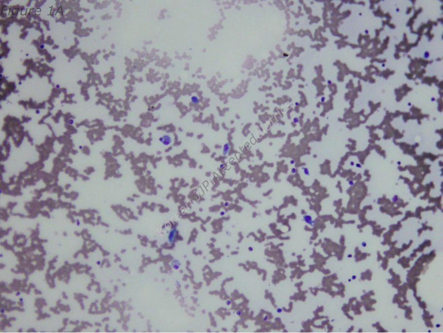
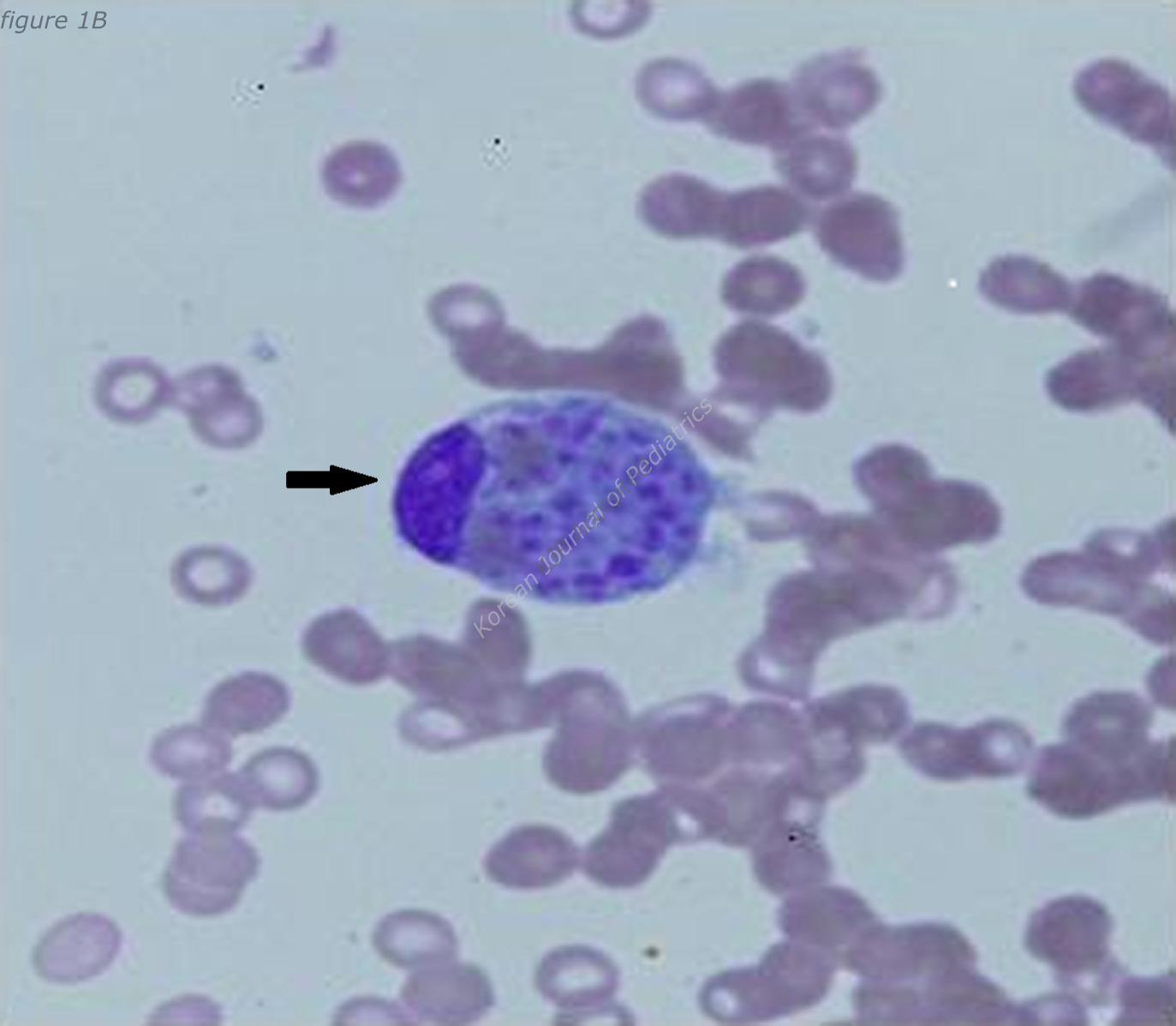
abdominal computed tomography (CT). We noted a hypocellular bone marrow with high 52
numbers of histiocytes and marked hemophagocytosis on the bone marrow biopsy performed 53
on day 7 after hospitalization (Fig. 1A, B). The ratio of CD3CD4/CD3CD8 was reduced to 54
0.53 (normal range, 0.9–3.6) while CD16+CD56+ (NK cell activity) was within the normal 55
range. However, caseous granuloma was not noted in the bone marrow specimen. The 56
Mantoux test, acid-fast bacterium (AFB) stain and culture of sputum revealed negative at that 57
time. Results of several virus tests, including hepatitis A virus(negative for IgM and IgG) , 58
cytomegalovirus (negative for IgM, positive for IgG), EBV (negative for VCA IgM, EA IgM 59
and IgG, positive for VCA IgG and EBNA IgG), and human immunodeficiency virus tests 60
(negative for Anti-HIV), didn’t suggest acute infection. Only the venereal disease research 61
laboratory (VDRL) test result was positive. However, the Treponema pallidum 62
Hemagglutination Assay (TPHA) and fluorescent treponemal antibody-absorption (FTA-ABS) 63
test results were both negative. Results of fluorescent antinuclear antibody (FANA) test was 64
positive (1:1280) but those of the rheumatoid arthritis factor test and anti-smith antibody 65
assay were both negative. 66
On the basis of these findings, a diagnosis of HLH was made and treatment with high-dose 67
dexamethasone (0.3 mg/kg/day for 7 days and tapering thereafter) and intravenous 68
immunoglobulin (0.5 g/kg/day for 2days) began on day 8 after hospitalization. At that time, 69
the patient’s activated partial thromboplastin time began to prolong to 44.3 s (normal range, 70
29.0–42.0 s) and fibrin degradation product was positive. After immune modulation, the fever 71
6
subsided on day 9 after hospitalization, pancytopenia improved and ferritin level decreased to 72
within the normal range. The PCR tests (AdvanSure TM
TB/NTM real-time PCR using 73
Mycobacterium tuberculosis comlex specific IS6110 and ITS specific primer) with both the 74
bone marrow aspiration and sputum samples were reported to be positive for tuberculosis on 75
day 10 after hospitalization. An anti-tubercular therapy regimen including 4 drugs–isoniazid 76
300㎎/day, rifampicin 450㎎/day, ethambutol 500㎎/day, and pyrazinamide 100㎎/day–was 77
immediately started. 78
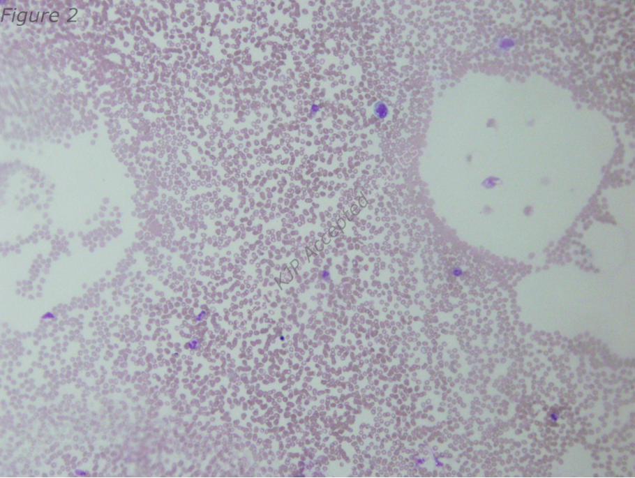
On day 17 after hospitalization, the results of all laboratory tests were normalized. After 3 79
weeks of follow-up, cellularity was restored to normal level and no hemophagocytosis was 80
observed at bone marrow biopsy (Fig.2). Results of PCR with both the bone marrow 81
aspiration and sputum samples were also negative for tuberculosis. The previously detected 82
multiple lymph node enlargements were decreased in size on the follow-up abdominal CT. 83
After 6 months of anti-tubercular therapy, the patient was healthy with no signs of disease. 84
85
86
7
Discussion 87
Here, we report a rare case of tuberculosis-associated HLH in a female adolescent diagnosed 88
using PCR performed with both the bone marrow and sputum samples. The patient was 89
successfully treated with immune modulation and anti-tubercular therapy. 90
In this case, we suspected disseminated tuberculosis based on clinical findings. Tuberculosis 91
is widespread in Asian countries, including Korea, and is one of the important causes of FUO 92
4, 5, 9). The clinical features of disseminated tuberculosis vary widely and are not often 93
detected early. As the clinical symptoms of tuberculosis, which include fever, anorexia, 94
weight loss, and weakness, are nonspecific and do not readily suggest a precise diagnosis. 95
Pulmonary symptoms may or may not be present. If tuberculosis involves the extrapulmonary 96
sites, generalized adenopathy, hepatomegaly, and splenomegaly may present. Only 10-20% of 97
patients with disseminated tuberculosis have a history of contact with tuberculosis. Definitive 98
physical signs of disseminated tuberculosis are also rare. Fever may be the only sign of 99
disseminated tuberculosis. Thus, disseminated tuberculosis should be suspected as one of the 100
most frequent cause of FUO, especially in children, the elderly, and any group with decreased 101
cell-mediated immunity. Most of the tuberculosis-associated HPS cases in the literature were 102
diagnosed with tuberculosis simultaneously. Furthermore, disseminated tuberculosis may be 103
diagnosed after a long delay following the initial manifestation of hemophagocytosis 10)
. 104
Our patient had a fever for over 17 days, as well as pancytopenia, hypofibrinogenemia, 105
hemophagocytosis of the bone marrow, and hyperferritinemia; which fulfilled the diagnostic 106
criteria for HLH according to the 2004 Histiocyte Society Protocol entitled hemophagocytic 107
lymphohistiocytosis 1)
. Usually, symptoms of HPS include fever, hepatosplenomegaly, 108
lymphadenopathy, and pancytopenia due to widespread histiocytic infiltration of the 109
reticuloendothelial system, including the bone marrow. Furthermore, HLH is known to have 110
8
an acute onset and a progressively increasing mortality risk as the condition develops, with an 111
overall mortality rate of 52% 1, 2)
. One review reported 53 cases of tuberculosis associated 112
HLH and 28 (52.8%) patients survived 7)
. In another report of patients with immune 113
deficiency, only 1 (16.7%) of the 6 patients survived 11)
. HPA-associated mortality is usually 114
due to underlying disease or pancytopenia-associated complications. Eliopoulos et al reported 115
that cause of death in tuberculosis-associated HLH include immunological disturbance and 116
disseminated tuberculosis 12)
. 117
However, confirming the presence of tuberculosis is difficult because mycobacterial loads in 118
the initial stage are very low and require several weeks of incubation before the organism can 119
be isolated and identified, in addition, the sample yield is low. Thus, the Centers for Disease 120
Control and Prevention (CDC) recommended nucleic acid amplification test that aid early 121
diagnosis 13)
. Recently, one study examined the diagnostic utility of PCR for samples of bone 122
marrow aspirate from patients with diverse clinical symptoms 14)
. Using PCR, tuberculosis 123
was detected in 70% of patients while the culture test detected tuberculosis in only 3.3% of 124
patients. Clinical improvement with anti-tubercular therapy was observed in 85% of the 125
patients with positive PCR results. In another study of 2296 patients with suspected 126
tuberculosis, the sensitivity, specificity, and positive predictive values of PCR were 97.2%, 127
100%, and 100% for smear-positive specimens and 75.3%, 97.0%, and 47.5% for smear-128
negative specimens 15)
. 129
One study reported the survival rate among 29 patients who received therapy was 65.5% (12 130
out of 20 patients received a combination of immunomodulatory and anti-tubercular therapy 131
and 7 out of 9 patients received anti-tubercular therapy alone) whereas no one who received 132
no treatment survived. Moreover, the most failure of treatment resulted from the initiation of 133
therapy late in the course of the illness 5)
. Therefore, early diagnosis and anti-tubercular 134
9
therapy besides the immunomodulatory therapy are important. 135
To our knowledge, this is the first case of tuberculosis-associated HPS diagnosed by PCR 136
and successfully treated using anti-tubercular therapy in Korean children. Considering that 137
tuberculosis-associated HLH showed high mortality rate without anti-tubercular therapy and 138
early bacteriological diagnosis of tuberculosis is difficult, we recommend tuberculosis PCR 139
test for the early differential diagnosis of HLH patients. 140
141
142

10
Conflict of interest 143
The authors have no potential conflicts of interest to report.144
11
REFERENCES
1. Rouphael NG, Talati NJ, Vaughan C, Cunningham K, Moreira R, Gould C. Infections
associated with haemophagocytic syndrome. Lancet Infect Dis 2007;7:814-22.
2. Verbsky JW, Grossman WJ. Hemophagocytic lymphohistiocytosis: diagnosis,
pathophysiology, treatment, and future perspectives. Ann Med 2006;38:20-31.
3. Fisman DN. Hemophagocytic syndromes and infection. Emerg Infect Dis 2000;6:601-
8.
4. Corbett EL, Watt CJ, Walker N, Maher D, Williams BG, Raviglione MC, et al. The
growing burden of tuberculosis: global trends and interactions with the HIV epidemic.
Arch Intern Med 2003;163:1009-21.
5. Brastianos PK, Swanson JW, Torbenson M, Sperati J, Karakousis PC. Tuberculosis-
associated haemophagocytic syndrome. Lancet Infect Dis 2006;6:447-54.
6. Balasubramanian S, Kaarthigeyan K, Aparna V, Srinivas S. Tuberculosis associated
hemophagocytic syndrome in infancy. Indian Pediatr 2008;45:593-5.
7. Shea YF, Chan JF, Kwok WC, Hwang YY, Chan TC, Ni MY, et al. Haemophagocytic
lymphohistiocytosis: an uncommon clinical presentation of tuberculosis. Hong Kong
Med J 2012;18:517-25.
8. Kim HI, Kim SW, Chang HH, Lee JM, Kim NS, Kwon KT, et al. Causes and Risk
Factors of Mortality in Adult Patients with Hemophagocytic Syndrome. Infect
Chemother 2012;44:51-5.
9. Cunha BA, Krakakis J, McDermott BP. Fever of unknown origin (FUO) caused by
miliary tuberculosis: diagnostic significance of morning temperature spikes. Heart
Lung 2009;38:77-82.
10. Dilber E, Erduran E, Kalyoncu M, Aynaci FM, Okten A, Ahmetoglu A.
12
Hemophagocytic syndrome as an initial presentation of miliary tuberculosis without
pulmonary findings. Scand J Infect Dis 2002;34:689-92.
11. Baraldes MA, Domingo P, Gonzalez MJ, Aventin A, Coll P. Tuberculosis-associated
hemophagocytic syndrome in patients with acquired immunodeficiency syndrome.
Arch Intern Med 1998;158:194-5.
12. Eliopoulos G, Vaiopoulos G, Kittas C, Fessas P. Tuberculosis associated
hemophagocytic syndrome complicated with severe bone marrow failure and
disseminated intravascular coagulation. Nouv Rev Fr Hematol 1992;34:273-6.
13. Diagnostic Standards and Classification of Tuberculosis in Adults and Children. This
official statement of the American Thoracic Society and the Centers for Disease
Control and Prevention was adopted by the ATS Board of Directors, July 1999. This
statement was endorsed by the Council of the Infectious Disease Society of America,
September 1999. Am J Respir Crit Care Med 2000;161:1376-95.
14. Escobedo-Jaimes L, Cicero-Sabido R, Criales-Cortez JL, Ramirez E, Romero M,
Rivero V, et al. Evaluation of the polymerase chain reaction in the diagnosis of
miliary tuberculosis in bone marrow smear. Int J Tuberc Lung Dis 2003;7:580-6.
15. Michos AG, Daikos GL, Tzanetou K, Theodoridou M, Moschovi M, Nicolaidou P, et
al. Detection of Mycobacterium tuberculosis DNA in respiratory and nonrespiratory
specimens by the Amplicor MTB PCR. Diagn Microbiol Infect Dis 2006;54:121-6.

13
Figure legend
Fig. 1A, B. Mature histiocyte-engulfing hematic cells was observed on bone marrow
aspiration. (A x200, B x1000).
Fig. 2. Bone marrow biopsy presented normal cellularity and no hemophagocytosis. (x200)
Top Related