


Languages
Pages
Legal

Breaking the diffraction limit of light-sheetfluorescence microscopy by RESOLFTPatrick Hoyera,b,c, Gustavo de Medeirosc, Bálint Balázsc,d, Nils Norlinc, Christina Besirc, Janina Hannea,b,e,Hans-Georg Kräussliche, Johann Engelhardta,b, Steffen J. Sahlf, Stefan W. Hella,b,f,1, and Lars Hufnagelc,1
aOptical Nanoscopy Division, German Cancer Research Center, 69120 Heidelberg, Germany; bBioquant, 69120 Heidelberg, Germany; cCell Biology andBiophysics Unit, European Molecular Biology Laboratory, 69117 Heidelberg, Germany; dFaculty of Information Technology and Bionics, Pázmány PéterCatholic University, 1083 Budapest, Hungary; eDepartment of Infectious Diseases, Virology, University of Heidelberg, 69120 Heidelberg, Germany;and fDepartment of NanoBiophotonics, Max Planck Institute for Biophysical Chemistry, 37077 Göttingen, Germany
Edited by Xiaowei Zhuang, Harvard University/Howard Hughes Medical Institute, Cambridge, MA, and approved February 25, 2016 (received for reviewNovember 11, 2015)
We present a plane-scanning RESOLFT [reversible saturable/switchableoptical (fluorescence) transitions] light-sheet (LS) nanoscope, which fun-damentally overcomes the diffraction barrier in the axial direction viaconfinement of the fluorescent molecular state to a sheet of subdiffrac-tion thickness around the focal plane. To this end, reversibly switchablefluorophores located right above and below the focal plane are trans-ferred to a nonfluorescent state at each scanning step. LS-RESOLFTnanoscopy offers wide-field 3D imaging of living biological specimenswith low light dose and axial resolution far beyond the diffractionbarrier. We demonstrate optical sections that are thinner by 5–12-foldcompared with their conventional diffraction-limited LS analogs.
light-sheet microscopy | RESOLFT | optical nanoscopy | 3D |live-cell imaging
Far-field nanoscopy (1, 2) methods discern features within sub-diffraction distances by briefly forcing their molecules to two
distinguishable states for the time period of detection. Typically,fluorophores are switched between a signaling “on” and a non-signaling (i.e., dark) “off” state. Depending on the switching andfluorescence registration strategy used, these superresolution tech-niques can be categorized into coordinate-stochastic and coordinate-targeted approaches (2). The latter group of methods, comprisingthe so-called RESOLFT [reversible saturable/switchable optical(fluorescence) transitions] (1, 3–7) approaches, have been realizedusing patterns of switch-off light with one or more zero-intensitypoints or lines, to single out target point (zero-dimensional) or line(1D) coordinates in space where the fluorophores are allowed toassume the on state. The RESOLFT idea can also be implementedin the inverse mode, by using switch-on light and confining the offstate. In any case, probing the presence of molecules in new sets ofpoints or lines at every scanning step produces images.Owing to the nature of the on and off states involved––first excited
electronic and ground state––stimulated emission depletion (STED)(3) and saturated structured illumination microscopy (SSIM) (8),which both qualify as variants of the RESOLFT principle, typicallyapply light intensities in the range of MW/cm2 and above. Especiallywhen imaging sensitive samples where photoinduced changes mustbe avoided, RESOLFT is preferably realized with fluorophoreswhich lead to the same factor of resolution improvement at muchlower intensities of state-switching light. Reversibly switchable fluo-rescent proteins (RSFPs) are highly suitable for this purpose (4–7, 9),as transitions between their metastable on and off states require 5orders of magnitude lower threshold intensities than STED/SSIM toguarantee switch-off. Suitable spectral properties, relatively fast mil-lisecond switching kinetics, and high photostability of recently de-veloped yellow-green-emitting RSFPs like rsEGFP (5), rsEGFP2 (7),and rsEGFP(N205S) (10) compared with early RSFPs have indeedenabled RESOLFT nanoscopy in living cells and tissues. To date,RSFP-based RESOLFT has achieved resolution improvements byfactors of 4–5 in rsEGFP2-labeled samples (7). To further reduce theimaging time, massive parallelization of scanning has been reported
(10). However, the diffraction-limited axial resolution and lack ofbackground suppression restrict applications to thin samples.Imaging applications typically require careful tuning of imaging
parameters including speed, contrast, photosensitivity, and spatialresolution, depending on the information that is sought. Light-sheetfluorescence microscopy (LSFM) (11–15) stands out by its ability tobalance most of these parameters for 3D imaging of living specimens.Recently reenacted as the selective plane illumination microscope(13), this microscopy mode has sparked increasing interest notablybecause of its short acquisition times in 3D imaging and low photo-toxicity in living specimens. It excites fluorophores only in a thindiffraction-limited slice of the sample, perpendicular to the directionof fluorescence detection. The LS is generated by a cylindrical lenswhich focuses an expanded laser beam in only one direction onto thespecimen or into the back-focal plane of an illumination objective.Alternatively, a single beam is quickly moved as a “virtual” LS (16)across a specimen section.In such conventional LSFM imaging, the lateral resolution is
determined by the numerical aperture (N.A.) of the detectionobjective (17), whereas axial resolution is given by the LS thickness,provided the latter is thinner than the axial extent of the point-spread function describing the imaging process from the focalplane of the detecting lens to the camera. In a previous study, theaxial resolution of LSFM was pushed to the diffraction limit byusing the full aperture of the illumination objective with Gaussianbeams; this was carried out for practically useful combinations of
Significance
Light-sheet fluorescence microscopy (LSFM) is an imaging mo-dality in which a sample is illuminated from the side by a beamengineered into a wide and relatively thin “sheet.” This allowshighly parallelized planewise scanning of volumes with inherentoptical sectioning, offering a good balance between spatial andtemporal resolution with reduced photostress. Unfortunately,the axial extent of the illuminated section is ultimately limitedby diffraction. Here, we show that a RESOLFT [reversible satu-rable/switchable optical (fluorescence) transitions] strategyneutralizes the resolution-limiting role of diffraction in LSFM.While other LS strategies exist, which run into new hard axiallimits of resolution, LS-RESOLFT is conceptually diffraction-un-limited and can be developed toward molecular-scale resolution.
Author contributions: P.H., S.J.S., S.W.H., and L.H. designed research; P.H. performed re-search; G.d.M., B.B., N.N., C.B., J.H., H.-G.K., and J.E. contributed new reagents/analytictools; P.H. analyzed data; and P.H., S.J.S., and S.W.H. wrote the paper.
Conflict of interest statement: S.W.H. benefits through patents on the basic RESOLFTconcept held by the Max Planck Society.
This article is a PNAS Direct Submission.
Freely available online through the PNAS open access option.1To whom correspondence may be addressed. Email: [email protected] or [email protected].
This article contains supporting information online at www.pnas.org/lookup/suppl/doi:10.1073/pnas.1522292113/-/DCSupplemental.
3442–3446 | PNAS | March 29, 2016 | vol. 113 | no. 13 www.pnas.org/cgi/doi/10.1073/pnas.1522292113
N.A. (e.g., 0.8 for both illumination and detection objectives)permissible in light of the geometrical constraints given by theobjective lens dimensions (18). High-N.A. illumination comes withshort Rayleigh ranges of Gaussian beams, which inherently limit thefield of view (FOV) along the direction of illumination. ScannedBessel beams for diffraction-limited excitation with a virtual LS (19–21) typically offer larger FOVs (22), but side lobes broaden thescanned LS in the axial direction and contribute to phototoxicityoutside of the focal plane of detection (20). A more complex ap-proach has used Bessel-beam excitation in combination with struc-tured illumination to obtain near-isotropic (but still diffraction-limited) resolution as measured on fluorescent beads (20), albeit atthe cost of acquisition time and reduced contrast due to fluorescencegenerated by the side lobes. In different work, axial resolution has alsobeen improved about fourfold by acquiring two complementary or-thogonal views of the sample using two alternating LSs, followed bycomputationally fusing image information with a deconvolution in-corporating both views (23). LS approaches have also helped sup-press out-of-focus background for single-molecule imaging in biologicalsituations (e.g., in ref. 24), including at superresolution (25–27).Slight axial resolution improvement beyond the diffraction barrier
has been demonstrated by overlapping a Gaussian excitation LS witha STED LS featuring a zero-intensity plane (28). Due to scatteringand possibly additional aberrations caused by the wavelength dif-ference between excitation and STED light, the maximal achievableresolution in biological specimens was severely limited. This was thecase even in fixed samples. A successful application of LS-STED toliving cells or organisms has not been reported. The relatively highaverage STED laser power required for high resolution gains callsfor developing a coordinate-targeted superresolution LS approachwith low-power operation, meaning a concept that does not solelyrely on changing the way the light is directed to––or collected from––
the sample, but a concept that harnesses an “on–off” transition forimproved feature separation.
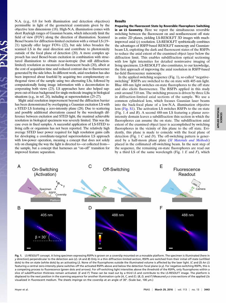
ResultsPreparing the Fluorescent State by Reversible Fluorophore Switchingin an LS Geometry. Here we report the simultaneous reversibleswitching between the fluorescent on and nonfluorescent off statein entire 2D planes, yielding LS-RESOLFT 3D images with much-improved axial (z) resolution. LS-RESOLFT symbiotically combinesthe advantages of RSFP-based RESOLFT nanoscopy and Gaussian-beam LS, exploiting the dark and fluorescent states of the RSFPsto reduce the axial extent of the examined object layer below thediffraction limit. This enables subdiffraction optical sectioningwith low light intensities for detailed noninvasive imaging ofliving specimens. LS-RESOLFT also constitutes, to our knowledge,the first approach of improving the axial resolution in RSFP-basedfar-field fluorescence nanoscopy.In the applied switching sequence (Fig. 1), so-called “negative-
switching” RSFPs are switched to the on state with 405-nm light.Blue 488-nm light switches on-state fluorophores to the off stateand also elicits fluorescence. The RSFPs applied in this studyemit around 510 nm. The switching process is driven by three LSsin diffraction-limited axial sections of the sample. We use acommon cylindrical lens, which focuses Gaussian laser beamsinto the back-focal plane of a low-N.A. illumination objectivelens (Fig. S1). The activation LS switches RSFPs to the on state(Fig. 1 A and B). A second 488-nm LS featuring a planar zero-intensity domain leaves a subdiffraction thin section in which thefluorophores can assume the on state. The subdiffraction axialextent of the examined object layer is accomplished by switchingfluorophores in the vicinity of this plane to the off state. Evi-dently, this plane is made to coincide with the focal plane ofdetection (Fig. 1 C and D). The off-switching pattern is gener-ated by a half-moon phase plate (SI Materials and Methods)placed in the collimated off-switching beam. In the next step ofthe sequence, the remaining on-state fluorophores are read outby a third LS of the same wavelength (Fig. 1 E and F), which
On-Switching
β = 30˚
405 nm
SpecimenRSFP
x
z
Platform
Coverslip
y
B
A
(Activation)
488 nm
Camera
F
EFluorescence
max
0
488 nm
D
COff-Switching
Readout
Fig. 1. LS-RESOLFT concept. A living specimen expressing RSFPs is grown on a coverslip mounted on a movable platform. The specimen is illuminated (here iny direction) perpendicular to the detection axis (z). (A and B) Only in a thin diffraction-limited section, RSFPs are switched from their initial off state (unfilleddots) to the on state (white dots) by an activating LS. None of the fluorophores outside the illuminated volume is affected by the laser light. (C and D) An LSfeaturing a central zero-intensity plane switches off the activated RSFPs above and below the detection focal plane (x-y). For negative-switching RSFPs, this isa competing process to fluorescence (green dots and arrows). For off-switching light intensities above the threshold of the RSFPs, only fluorophores within aslice of subdiffraction thickness remain activated. (E and F) These can be read out by a third LS and contribute to the LS-RESOLFT image. The platform isdisplaced to the next position in the scanning sequence for another illumination cycle (A, C, and E). (B, D, and F) Measured y-z cross-sections of the applied LSsvisualized in fluorescent medium. The sheets impinge on the coverslip at an angle of 30°. (Scale bar, 100 μm.)
Hoyer et al. PNAS | March 29, 2016 | vol. 113 | no. 13 | 3443
APP
LIED
PHYS
ICAL
SCIENCE
S
also switches them off again. Perpendicular to the illumination, ahigh-N.A. detection objective lens collects fluorescence in a wide-field manner. A tube lens focuses the detected light onto an sCMOS(scientific complementary metal-oxide-semiconductor) camera chip(lateral sampling of 108.3 nm). Images are recorded by displacing(scanning) the platform with the mounted coverslip and the sampleat a design angle of 30° with respect to the illumination axis throughthe stationary LSs. The above acquisition cycle is repeated for eachsection of the sample, with the step size of the piezoelectric stagedefining the axial sampling of the specimen (Fig. S2). The Rayleighrange along the illumination axis and the width of the LSs (Fig. S3)determine the FOV per plane; the maximal scan range of the pi-ezoelectric stage defines the detectable volume.
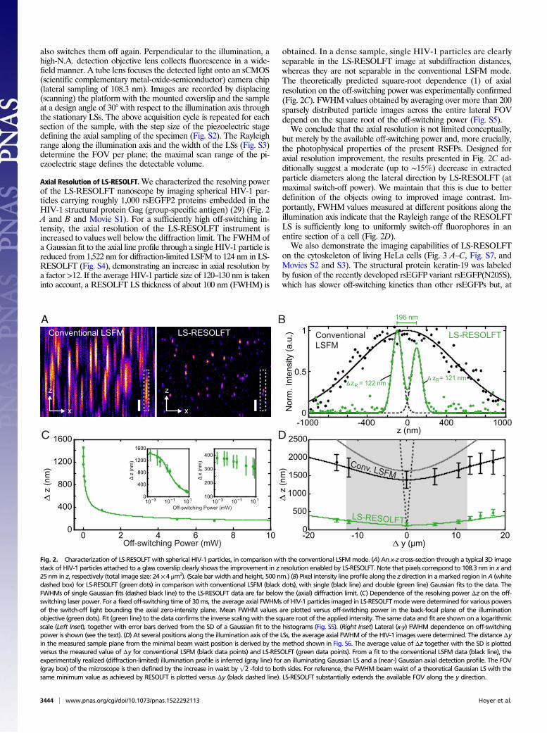
Axial Resolution of LS-RESOLFT.We characterized the resolving powerof the LS-RESOLFT nanoscope by imaging spherical HIV-1 par-ticles carrying roughly 1,000 rsEGFP2 proteins embedded in theHIV-1 structural protein Gag (group-specific antigen) (29) (Fig. 2A and B and Movie S1). For a sufficiently high off-switching in-tensity, the axial resolution of the LS-RESOLFT instrument isincreased to values well below the diffraction limit. The FWHM ofa Gaussian fit to the axial line profile through a single HIV-1 particle isreduced from 1,522 nm for diffraction-limited LSFM to 124 nm in LS-RESOLFT (Fig. S4), demonstrating an increase in axial resolution bya factor >12. If the average HIV-1 particle size of 120–130 nm is takeninto account, a RESOLFT LS thickness of about 100 nm (FWHM) is
obtained. In a dense sample, single HIV-1 particles are clearlyseparable in the LS-RESOLFT image at subdiffraction distances,whereas they are not separable in the conventional LSFM mode.The theoretically predicted square-root dependence (1) of axialresolution on the off-switching power was experimentally confirmed(Fig. 2C). FWHM values obtained by averaging over more than 200sparsely distributed particle images across the entire lateral FOVdepend on the square root of the off-switching power (Fig. S5).We conclude that the axial resolution is not limited conceptually,
but merely by the available off-switching power and, more crucially,the photophysical properties of the present RSFPs. Designed foraxial resolution improvement, the results presented in Fig. 2C ad-ditionally suggest a moderate (up to ∼15%) decrease in extractedparticle diameters along the lateral direction by LS-RESOLFT (atmaximal switch-off power). We maintain that this is due to betterdefinition of the objects owing to improved image contrast. Im-portantly, FWHM values measured at different positions along theillumination axis indicate that the Rayleigh range of the RESOLFTLS is sufficiently long to uniformly switch-off fluorophores in anentire section of a cell (Fig. 2D).We also demonstrate the imaging capabilities of LS-RESOLFT
on the cytoskeleton of living HeLa cells (Fig. 3 A–C, Fig. S7, andMovies S2 and S3). The structural protein keratin-19 was labeledby fusion of the recently developed rsEGFP variant rsEGFP(N205S),which has slower off-switching kinetics than other rsEGFPs but, at
C D
B
Δ z = 121 nmRΔz = 122 nmRz
x
Conventional LSFM Conventional LSFM
LS-RESOLFT LS-RESOLFT
196 nm
0.5
1
0-1000
2500
2000
1500
1000
500
0-20 20-10 0 10
Off-switching Power (mW)
Off-switching Power (mW)
0 2 4 6 8 10
1000-400 4000z (nm)
Nor
m. I
nten
sity
(a.u
.)
∆ z
(nm
)
∆ z
(nm
)
∆ y (μm)
z
x
A
Conv. LSFM
LS-RESOLFT400
0
800
1200
1600
Δz
(nm
)
Δx
(nm
)
10−3 10−1 101 10−3 10−1 101100
200
300
400
0
400
800
1200
1600
Fig. 2. Characterization of LS-RESOLFT with spherical HIV-1 particles, in comparison with the conventional LSFMmode. (A) An x-z cross–section through a typical 3D imagestack of HIV-1 particles attached to a glass coverslip clearly shows the improvement in z resolution enabled by LS-RESOLFT. Note that pixels correspond to 108.3 nm in x and25 nm in z, respectively (total image size: 24 ×4 μm2). (Scale bar width and height, 500 nm.) (B) Pixel intensity line profile along the z direction in amarked region inA (whitedashed box) for LS-RESOLFT (green dots) in comparison with conventional LSFM (black dots), with single (black line) and double (green line) Gaussian fits to the data. TheFWHMs of single Gaussian fits (dashed black line) to the LS-RESOLFT data are far below the (axial) diffraction limit. (C) Dependence of the resolving power Δz on the off-switching laser power. For a fixed off-switching time of 30ms, the average axial FWHMs of HIV-1 particles imaged in LS-RESOLFTmodewere determined for various powersof the switch-off light bounding the axial zero-intensity plane. Mean FWHM values are plotted versus off-switching power in the back-focal plane of the illuminationobjective (green dots). Fit (green line) to the data confirms the inverse scalingwith the square root of the applied intensity. The same data and fit are shown on a logarithmicscale (Left Inset), together with error bars derived from the SD of a Gaussian fit to the histograms (Fig. S5). (Right Inset) Lateral (x-y) FWHM dependence on off-switchingpower is shown (see the text). (D) At several positions along the illumination axis of the LSs, the average axial FWHMof the HIV-1 images were determined. The distance Δyin the measured sample plane from the minimal beam waist position is derived by the method shown in Fig. S6. The average value of Δz together with the SD is plottedversus the measured value of Δy for conventional LSFM (black data points) and LS-RESOLFT (green data points). From a fit to the conventional LSFM data (black line), theexperimentally realized (diffraction-limited) illumination profile is inferred (gray line) for an illuminating Gaussian LS and a (near-) Gaussian axial detection profile. The FOV(gray box) of the microscope is then defined by the increase in waist by
ffiffiffi
2p
-fold to both sides. For reference, the FWHM beam waist of a theoretical Gaussian LS with thesame minimum value as achieved by RESOLFT is plotted versus Δy (black dashed line). LS-RESOLFT substantially extends the available FOV along the y direction.
3444 | www.pnas.org/cgi/doi/10.1073/pnas.1522292113 Hoyer et al.
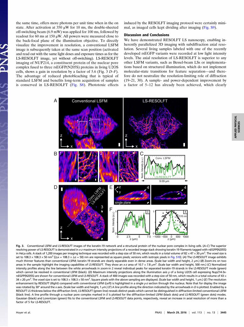
the same time, offers more photons per unit time when in the onstate. After activation at 350 μW for 10 ms, the double-sheetedoff-switching beam (6.9 mW) was applied for 100 ms, followed byreadout for 60 ms at 150 μW. All powers were measured close tothe back-focal plane of the illumination objective. To directlyvisualize the improvement in resolution, a conventional LSFMimage is subsequently taken at the same scan position (activatedand read out with the same light doses and exposure times as for theLS-RESOLFT image, yet without off-switching). LS-RESOLFTimaging of NUP214, a constituent protein of the nuclear porecomplex fused to three rsEGFP(N205S) proteins in living U2OScells, shows a gain in resolution by a factor of 5.6 (Fig. 3 D–F).The advantage of reduced photobleaching that is typical ofstandard LSFM and benefits long-term acquisition of samplesis conserved in LS-RESOLFT (Fig. S8). Phototoxic effects
induced by the RESOLFT imaging protocol were certainly mini-mal, as imaged cells kept dividing after imaging (Fig. S9).
Discussion and ConclusionsWe have demonstrated RESOLFT LS nanoscopy, enabling in-herently parallelized 3D imaging with subdiffraction axial reso-lution. Several living samples labeled with one of the recentlydeveloped rsEGFP variants were recorded at low light intensitylevels. The axial resolution of LS-RESOLFT is superior to anyother LSFM variants, such as Bessel-beam LSs or implementa-tions based on structured illumination, which do not implementmolecular-state transitions for feature separation––and there-fore do not neutralize the resolution-limiting role of diffraction(19–21, 30). A sample- and power-dependent improvement bya factor of 5–12 has already been achieved, which clearly
Conventional LSFM
C
A
B
21
z
x
LS-RESOLFT
z
x
Conv. LSFM
Norm
. Inten
sity (
a.u.)
2
LS-RESOLFT
1
D E
F
−1000 −500 0 500 10000
1 4
z (μm)0 2.5 7.55
0
1 3
z (μm)
Norm
. Inten
sity (
a.u.)
zy
4
z
x
4
Conv.
LSFM
LS-R
ESOLFT
single plane3
271 nm
1516 nm
9.09 μm
Fig. 3. Conventional LSFM and LS-RESOLFT images of the keratin-19 network and a structural protein of the nuclear pore complex in living cells. (A–C) The superiorresolving power of LS-RESOLFT is demonstrated in x-zmaximum intensity projections of a recorded image stack showing keratin-19 filaments taggedwith rsEGFP(N205S)in HeLa cells. A stack of 1,200 images per imaging technique was recordedwith a step size of 50 nm, which results in a total volume of 82 × 47 × 30 μm3. The voxel size isset to 108.3 × 108.3 × 50 nm3 [(Δx = 108.3 × Δz = 50) nm are represented as square pixels; versions with isotropic pixels in Fig. S10]. (A) The LS-RESOLFT image exhibitsmuch thinner features than conventional LSFM; keratin-19 strands are clearly separable even in dense areas. (Scale bar width and height, 2 μm.) (B) Zoom-ins on twoareas in the sample highlight the imaging capabilities of LS-RESOLFT. They show an x-z area of 10.7 × 7.8 μm2. (Scale bar width and height, 500 nm.) (C) Normalizedintensity profiles along the line between the white arrowheads in zoom-in 2 reveal individual peaks for separated keratin-19 strands in the LS-RESOLFT mode (green)which cannot be resolved in conventional LSFM (black). (D) Maximum intensity projections along the illumination axis y of a living U2OS cell expressing Nup214-3x-rsEGFP(N205S) are shown for conventional LSFM and LS-RESOLFT. A stack of 400 images was recorded with a step size of 50 nm, which results in a total volume of 43 ×34 × 20 μm3. The voxel size is set to 108.3 × 108.3 × 50 nm3. Square pixels with the above sampling are displayed. (Scale bar width and height, 1 μm.) (E) The resolutionenhancement by RESOLFT (Right) compared with conventional LSFM (Left) is highlighted in a single y-z section through the nucleus. Note that for display the imagewas rotated by 30° around the x axis. (Scale bar width and height, 1 μm.) (F) A line profile along the direction indicated by the arrowheads inD is plotted. Enabled by aRESOLFT LS thickness below the diffraction limit, LS-RESOLFT (green line) reveals distinct peaks which cannot be distinguished in diffraction-limited conventional LSFM(black line). A line profile through a nuclear pore complex marked in E is plotted for the diffraction-limited LSFM (black dots) and LS-RESOLFT (green dots) modes.Gaussian (black) and Lorentzian (green) fits to the conventional LSFM and LS-RESOLFT data points, respectively, reveal an increase in axial resolution of more than afactor of 5 for LS-RESOLFT.
Hoyer et al. PNAS | March 29, 2016 | vol. 113 | no. 13 | 3445
APP
LIED
PHYS
ICAL
SCIENCE
S
outperforms the previous STED-LS approach, where a gain inaxial resolution by <1.5-fold was reported for dye-filled particlesonly, and at much higher light doses (28).Clearly, the spatial resolution (i.e., information) increase––as
it necessitates a greater number of sampling steps in space to satisfyNyquist, and as it requires time to perform the state switching––must be inevitably accompanied by an overall somewhat reducedacquisition rate. This is a common inherent feature of all nanoscopyapproaches separating neighboring fluorophores by on–off transi-tions (2). As in all RESOLFT approaches, however, spatial reso-lution is––advantageously––readily and freely tunable (compare Fig.2C), and fewer steps may be sampled in space at accordingly fasterrates per volume stack. At ∼100 ms per frame, recording speed inthis first demonstration is up to about an order of magnitude slowerthan conventional LS imaging, which reads out coarser fluorescentvolumes. Note that the physical reduction by LS-RESOLFT of thespecimen section in which the fluorescent on state remains allowedper scanning step implies on average fewer fluorophores present persection. Examination of typical images with LS-RESOLFT revealedsignal-to-background ratios between ∼3:1 and ∼12:1 for imagingexperiments with HIV-1 particles depending on label incorporation,and ∼7:1 to ∼12:1 for keratin strands (comparing to directly neigh-boring dark regions)––at the conservatively chosen frame durationsin these proof-of-principle experiments. Ongoing improvements inswitchable fluorophore characteristics, notably in terms of fluores-cence quantum yield and switching speed, are expected to fur-ther improve temporal resolution of the RESOLFT approachesin general, including this highly parallelized LS variant.The capability to create optical sections of ∼100-nm thickness
is similar to the axial resolving power of a typical TIRF (totalinternal reflection fluorescence) microscope, yet with a decisivedifference: Whereas TIRF produces only one slice that is tightlyconfined to the coverslip surface, LS-RESOLFT offers sub-diffraction axial resolution in any slice throughout the entirespecimen and can thus be applied to a broad range of (not just)cell-biological questions. Due to the low off-switching intensities,LS-RESOLFT allows live-cell imaging of biological samples overextended time periods. Large FOVs are possible, as entire planesare being captured at once, without the need for sequential
lateral readout. Acquisition time is thus reduced by a factor givenby the number of pixels per FOV compared with confocal “z-doughnut” RESOLFT strategies achieving similar xyz resolutionfor identical volumes (SI Materials and Methods). Because off-switching and readout are performed at the same wavelength,potential chromatic aberrations are inherently avoided. Re-cordings with spectrally distinct RSFPs will in the future allowcolocalization studies in subdiffraction volumes. Looking ahead, LS-RESOLFT can be equipped with an option to additionallysuperresolve along the lateral dimensions. Because the presentLS-RESOLFT design employs independent beam paths for il-lumination and detection of the sample, the same concepts whichlead to conceptually diffraction-unlimited lateral resolution anduse wide-field detection can also be applied to LS-RESOLFT.One possible approach combines LS-RESOLFT with the pre-viously demonstrated parallelized 2D RESOLFT technique (10).
Materials and MethodsDetailed descriptions of the optical setup, the image acquisition and rep-resentation, sample preparation including mammalian cell culture, as well asa discussion of the acquisition speed gains enabled by the highly parallelizedLS readout, are provided in SI Materials and Methods. In brief, LS-RESOLFTnanoscopy at low light levels was demonstrated with custom-built optics,featuring two water-dipping objective lenses in perpendicular configuration:an illumination objective (10×/0.3 N.A.) to create three LSs for activation,off-switching, and readout of RSFPs, and a detection objective (40×/0.8 N.A.)for collection of sample fluorescence in the wide field. Laser beam pathswere coaligned, expanded, and focused by a common cylindrical lens intothe back aperture of the illumination objective. The activation (405 nm) andreadout (488 nm) LSs featured conventional geometries. The switch-offbeam (488 nm) was modulated by a phase retardation plate (Fig. S1) toproduce, after focusing by the objective, an off-switching light distributionwith a central plane of minimal intensity (the “zero”). Scanning of the specimenthrough the LSs acquired images plane by plane.
ACKNOWLEDGMENTS. We thank Jan Ellenberg and Anna Szymborska-Mell(both European Molecular Biology Laboratory) for vectors, and Barbara Müller(Heidelberg University) for vectors and cloning, and all of them, as well as TimGrotjohann (Max Planck Institute for Biophysical Chemistry), for helpful discus-sions. S.W.H. acknowledges the Federal Ministry of Education and Research forfunding this work within the project STEDlight (FKZ:13N11173).
1. Hell SW (2003) Toward fluorescence nanoscopy. Nat Biotechnol 21(11):1347–1355.2. Hell SW (2009) Microscopy and its focal switch. Nat Methods 6(1):24–32.3. Hell SW, Wichmann J (1994) Breaking the diffraction resolution limit by stimulated emis-
sion: Stimulated-emission-depletion fluorescence microscopy. Opt Lett 19(11):780–782.4. Hofmann M, Eggeling C, Jakobs S, Hell SW (2005) Breaking the diffraction barrier in
fluorescence microscopy at low light intensities by using reversibly photoswitchableproteins. Proc Natl Acad Sci USA 102(49):17565–17569.
5. Grotjohann T, et al. (2011) Diffraction-unlimited all-optical imaging and writing witha photochromic GFP. Nature 478(7368):204–208.
6. Brakemann T, et al. (2011) A reversibly photoswitchable GFP-like protein with fluo-rescence excitation decoupled from switching. Nat Biotechnol 29(10):942–947.
7. Grotjohann T, et al. (2012) rsEGFP2 enables fast RESOLFT nanoscopy of living cells. eLife 1:e00248.8. Gustafsson MGL (2005) Nonlinear structured-illumination microscopy: Wide-field
fluorescence imaging with theoretically unlimited resolution. Proc Natl Acad Sci USA102(37):13081–13086.
9. Schwentker MA, et al. (2007) Wide-field subdiffraction RESOLFT microscopy usingfluorescent protein photoswitching. Microsc Res Tech 70(3):269–280.
10. Chmyrov A, et al. (2013) Nanoscopy with more than 100,000 ‘doughnuts’. NatMethods 10(8):737–740.
11. Voie AH, Burns DH, Spelman FA (1993) Orthogonal-plane fluorescence optical sectioning:Three-dimensional imaging of macroscopic biological specimens. J Microsc 170(Pt 3):229–236.
12. Fuchs E, Jaffe J, Long R, Azam F (2002) Thin laser light sheet microscope for microbialoceanography. Opt Express 10(2):145–154.
13. Huisken J, Swoger J, Del Bene F, Wittbrodt J, Stelzer EHK (2004) Optical sectioningdeep inside live embryos by selective plane illumination microscopy. Science 305(5686):1007–1009.
14. Dodt H-U, et al. (2007) Ultramicroscopy: Three-dimensional visualization of neuronalnetworks in the whole mouse brain. Nat Methods 4(4):331–336.
15. Wu Y, et al. (2011) Inverted selective plane illumination microscopy (iSPIM) enablescoupled cell identity lineaging and neurodevelopmental imaging in Caenorhabditiselegans. Proc Natl Acad Sci USA 108(43):17708–17713.
16. Keller PJ, Stelzer EHK (2008) Quantitative in vivo imaging of entire embryos with digitalscanned laser light sheet fluorescence microscopy. Curr Opin Neurobiol 18(6):624–632.
17. Engelbrecht CJ, Stelzer EH (2006) Resolution enhancement in a light-sheet-basedmicroscope (SPIM). Opt Lett 31(10):1477–1479.
18. Capoulade J,WachsmuthM, Hufnagel L, KnopM (2011) Quantitative fluorescence imagingof protein diffusion and interaction in living cells. Nat Biotechnol 29(9):835–839.
19. Fahrbach FO, Simon P, Rohrbach A (2010) Microscopy with self-reconstructing beams.Nat Photonics 4(11):780–785.
20. Planchon TA, et al. (2011) Rapid three-dimensional isotropic imaging of living cellsusing Bessel beam plane illumination. Nat Methods 8(5):417–423.
21. Gao L, et al. (2012) Noninvasive imaging beyond the diffraction limit of 3D dynamicsin thickly fluorescent specimens. Cell 151(6):1370–1385.
22. Durnin J, Miceli JJ, Jr, Eberly JH (1988) Comparison of Bessel and Gaussian beams. OptLett 13(2):79–80.
23. Wu Y, et al. (2013) Spatially isotropic four-dimensional imaging with dual-view planeillumination microscopy. Nat Biotechnol 31(11):1032–1038.
24. Siebrasse JP, Kaminski T, Kubitscheck U (2012) Nuclear export of single nativemRNAmoleculesobserved by light sheet fluorescence microscopy. Proc Natl Acad Sci USA 109(24):9426–9431.
25. Zanacchi FC, et al. (2011) Live-cell 3D super-resolution imaging in thick biologicalsamples. Nat Methods 8(12):1047–1049.
26. Zhao ZW, et al. (2014) Spatial organization of RNA polymerase II inside a mammaliancell nucleus revealed by reflected light-sheet superresolution microscopy. Proc NatlAcad Sci USA 111(2):681–686.
27. Galland R, et al. (2015) 3D high- and super-resolution imaging using single-objectiveSPIM. Nat Methods 12(7):641–644.
28. Friedrich M, Gan Q, Ermolayev V, Harms GS (2011) STED-SPIM: Stimulated emission de-pletion improves sheet illumination microscopy resolution. Biophys J 100(8):L43–L45.
29. Carlson LA, et al. (2008) Three-dimensional analysis of budding sites and released virussuggests a revised model for HIV-1 morphogenesis. Cell Host Microbe 4(6):592–599.
30. Chen BC, et al. (2014) Lattice light-sheet microscopy: Imaging molecules to embryos athigh spatiotemporal resolution. Science 346(6208):1257998.
31. Lampe M, et al. (2007) Double-labelled HIV-1 particles for study of virus-cell interaction.Virology 360(1):92–104.
32. Müller B, et al. (2004) Construction and characterization of a fluorescently labeled in-fectious human immunodeficiency virus type 1 derivative. J Virol 78(19):10803–10813.
3446 | www.pnas.org/cgi/doi/10.1073/pnas.1522292113 Hoyer et al.
Top Related