


Languages
Pages
Legal


Brainstem
By
Dr. Bhushan R. Kavimandan

Development
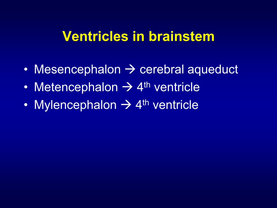
Ventricles in brainstem
• Mesencephalon cerebral aqueduct
• Metencephalon 4th ventricle
• Mylencephalon 4th ventricle
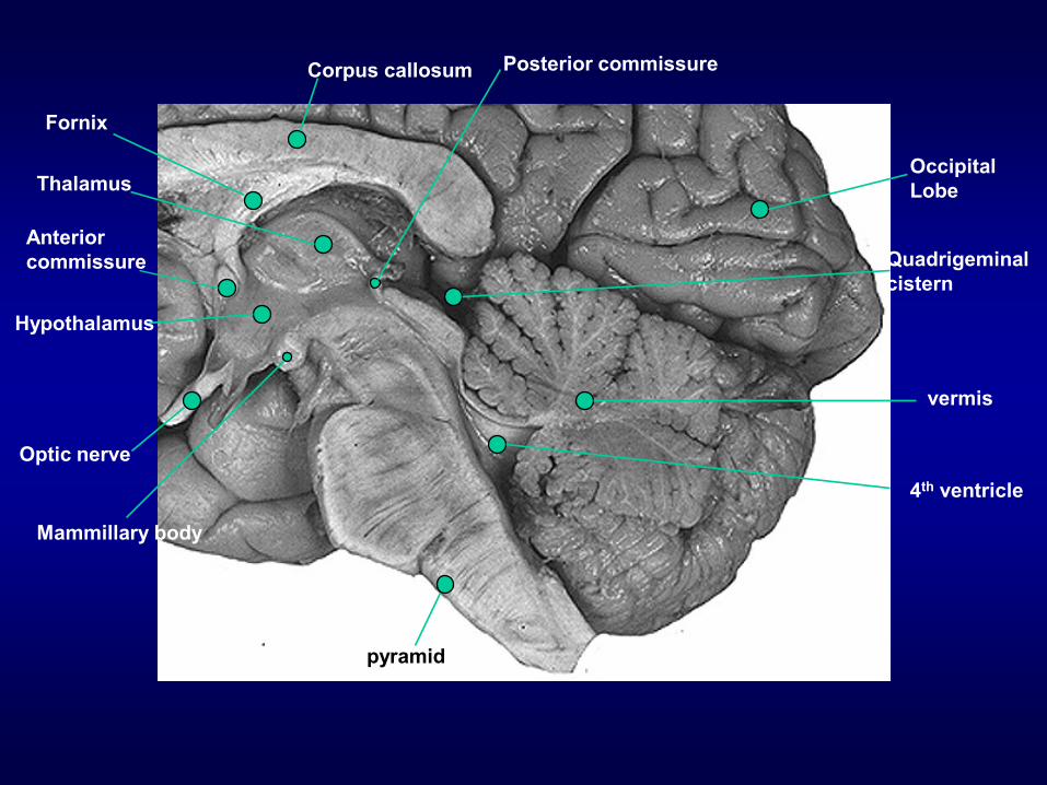
vermis
Occipital
LobeThalamus
Corpus callosum
Hypothalamus
Fornix
Anterior
commissure
Optic nerve
4th ventricle
Posterior commissure
pyramid
Mammillary body
Quadrigeminal
cistern
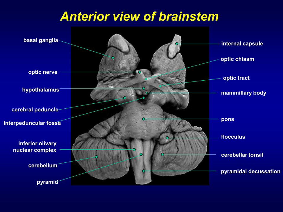
internal capsulebasal ganglia
optic nerveoptic tract
hypothalamusmammillary body
cerebral peduncle
interpeduncular fossa
flocculus
inferior olivary
nuclear complex
cerebellum
cerebellar tonsil
pyramid
pyramidal decussation
pons
optic chiasm
Anterior view of brainstem
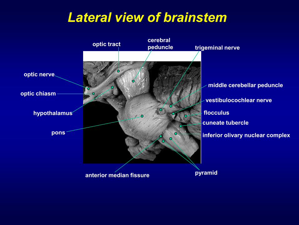
optic tract
optic nerve
hypothalamus
optic chiasm
middle cerebellar peduncle
trigeminal nerve
vestibulocochlear nerve
flocculus
cuneate tubercle
inferior olivary nuclear complex
anterior median fissure pyramid
pons
cerebral
peduncle
Lateral view of brainstem

Middle cerebellar
peduncle
Superior colliculus
Inferior colliculusCerebral
peduncle
Superior cerebellar
peduncle
Inferior cerebellar
peduncle
Medulla
4th ventricle
Posterior view of brainstem
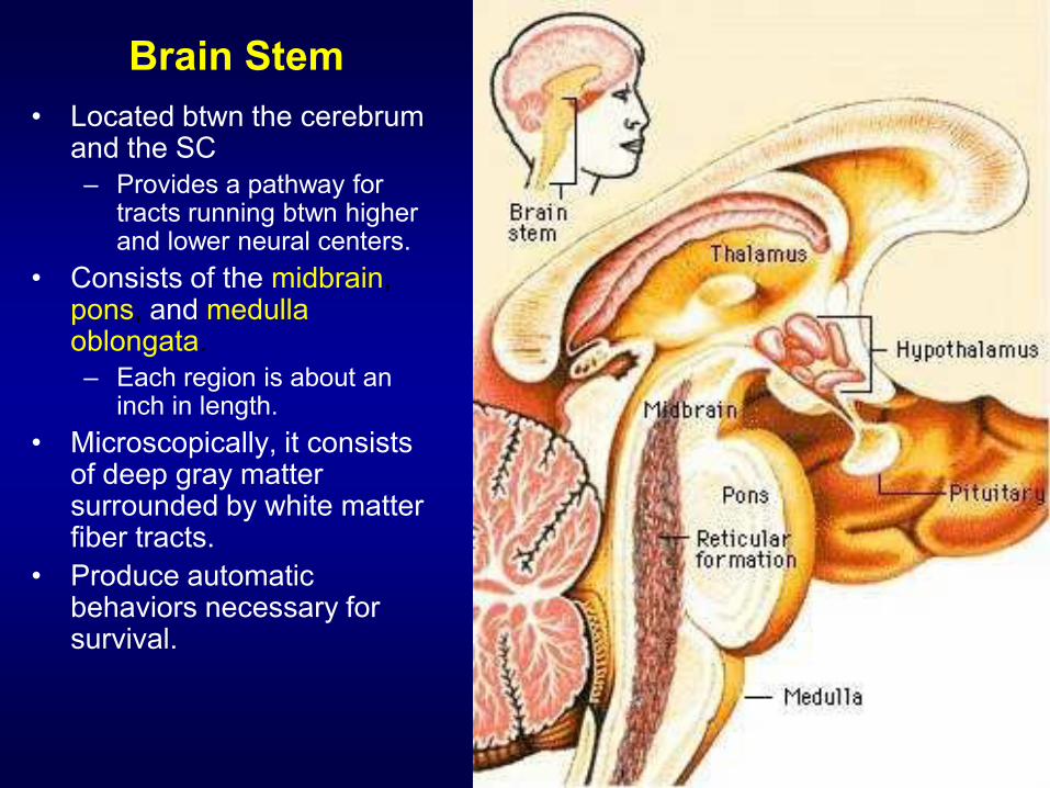
Brain Stem
• Located btwn the cerebrum and the SC
– Provides a pathway for tracts running btwn higher and lower neural centers.
• Consists of the midbrain, pons, and medullaoblongata.
– Each region is about an inch in length.
• Microscopically, it consists of deep gray matter surrounded by white matter fiber tracts.
• Produce automatic behaviors necessary for survival.

Brainstem: 3 major divisions
•Midbrain
•Pons
•Medulla
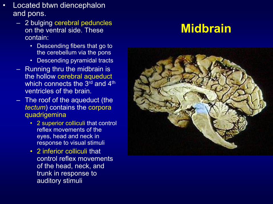
Midbrain
• Located btwn diencephalon and pons.
– 2 bulging cerebral peduncleson the ventral side. These contain:
• Descending fibers that go to the cerebellum via the pons
• Descending pyramidal tracts
– Running thru the midbrain is the hollow cerebral aqueductwhich connects the 3rd and 4th
ventricles of the brain.
– The roof of the aqueduct (thetectum) contains the corpora quadrigemina
• 2 superior colliculi that control reflex movements of the eyes, head and neck in response to visual stimuli
• 2 inferior colliculi that control reflex movements of the head, neck, and trunk in response to auditory stimuli

•Cranial nerves 3&4
(oculomotor and trochlear)
exit from the midbrain
•Midbrain also contains the
headquarters of the
reticular activating system
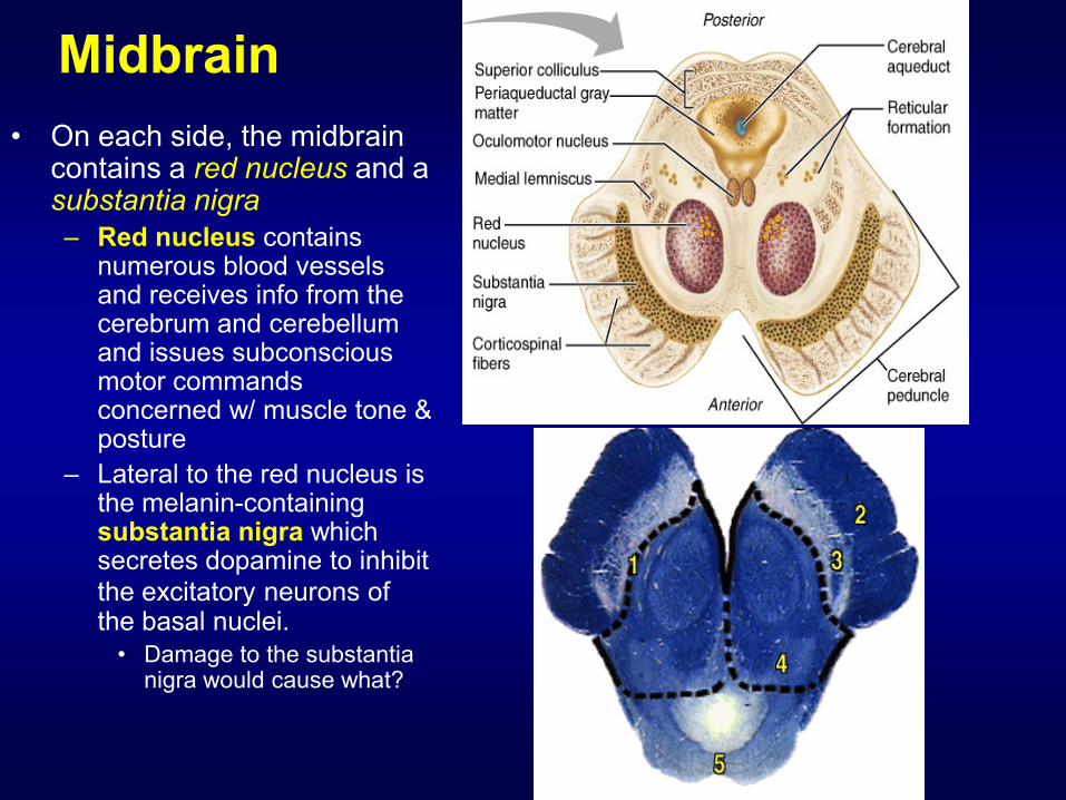
Midbrain
• On each side, the midbrain contains a red nucleus and asubstantia nigra
– Red nucleus contains numerous blood vessels and receives info from the cerebrum and cerebellum and issues subconscious motor commands concerned w/ muscle tone & posture
– Lateral to the red nucleus is the melanin-containing substantia nigra which secretes dopamine to inhibit
the excitatory neurons of the basal nuclei.
• Damage to the substantia nigra would cause what?

Pons• Literally means “bridge”
• Wedged btwn the midbrain & medulla.
• Contains:
– Sensory and motor nuclei for 4 cranial nerves
• Trigeminal (5), Abducens (6), Facial (7), and Auditory/Vestibular (8)
– Respiratory nuclei:
• Apneustic & pneumotaxic centerswork w/ the medulla to maintain respiratory rhythm
– Nuclei & tracts that process and relay info to/from the cerebellum
– Ascending, descending, and transverse tracts that interconnect other portions of the CNS

Medulla Oblongata
• Most inferior region of the brain stem.
• Becomes the spinal cord at the level of the foramen magnum.
• Ventrally, 2 ridges (themedullary pyramids) are visible.
– These are formed by the large motor corticospinal tracts.
– Right above the medulla-SC junction, most of these fibers
cross-over (decussate).

Medulla Oblongata• Nuclei in the medulla are
associated w/ autonomic control, cranial nerves, and motor/sensory relay.
• Autonomic nuclei:
– Cardiovascular centers
• Alter the rate and force of cardiac contractions
• Alter the tone of vascular smooth muscle
– Respiratory rhythmicity centers
• Receive input from the pons
– Additional Centers
• Emesis, deglutition, coughing, hiccupping, and sneezing
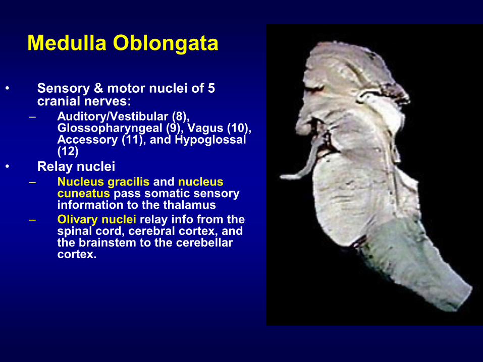
Medulla Oblongata
• Sensory & motor nuclei of 5 cranial nerves:
– Auditory/Vestibular (8), Glossopharyngeal (9), Vagus (10), Accessory (11), and Hypoglossal (12)
• Relay nuclei– Nucleus gracilis and nucleus
cuneatus pass somatic sensory information to the thalamus
– Olivary nuclei relay info from the spinal cord, cerebral cortex, and the brainstem to the cerebellar cortex.
Top Related