


Languages
Pages
Legal

A CASE REPORT
76� PRACTICAL�GASTROENTEROLOGY� •� JUNE�2015
A CASE REPORT
Biliary Tubulopapillary Adenoma with Concurrent Biliary Stone Presenting with Pruritus and Obstructive Jaundice: A Case Report and Review of Literatureby Jehangir Ansari, Padmini Krishnamurthy
Jehangir Ansari MD, Padmini Krishnamurthy, MD Department of Gastroenterology, Wright State University, Boonshoft School of Medicine, Dayton, OH
His abdominal examination was unremarkable. His liver enzymes revealed an alanine aminotransferase (ALT) of 127 IU/L, aspartate aminotransferase (AST) of 98 IU/L, alkaline phosphatase of 499 IU/L and a total bilirubin of 3.0 mg/dl. Computed tomography (CT) of the abdomen revealed a 3.4 cm enhancing mass in the distal common bile duct (CBD) with severe extra- and intrahepatic biliary dilation (Figure 1). Carbohydrate antigen 19-9 was 24.1 U/ml. Positron emission test (PET) scan demonstrated no abnormal uptake in the CBD. He underwent endoscopic retrograde cholangiopancreatography (ERCP) which showed diffuse biliary dilation and a large filling defect in the mid CBD with irregular ductal margins (Figure 2). Balloon sweep resulted in the passage of small clumps of soft tissue in addition to a large stone and sludge. Histology of the soft tissue suggested intraductal papillary neoplasm of the CBD. Endoscopic ultrasound (EUS) showed localized CBD mass with possible malignant cells on fine needle aspirate. Subsequently, he underwent a Whipple procedure where a 3.5 cm pedunculated polyposis mass was found in the CBD.
INTRODUCTION
Biliary papillary tumors account for 11% of all biliary ductal tumors1 and have malignant potential.2 They can be intrahepatic or extrahepatic in location3,4,5
and may be associated with tubular adenomas elsewhere in the gastrointestinal tract.6 Several cases of biliary papillary tumors have been reported from the far east.2-
4,6-13 with few reported case series from the Western population.1,5 The term, biliary intraductal papillary tumor, has been used interchangeably with intraductal adenoma.6,7,14 Here, we present a rare case of benign biliary intraductal tubulopapillary adenoma with review of literature about biliary intraductal papillary tumors.
CASEA 75 year old Caucasian male presented with a four month history of pruritus and weight loss. He had a past medical history of stage II prostate cancer in remission after being treated with hormone therapy.
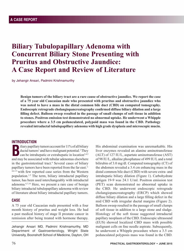
Benign tumors of the biliary tract are a rare cause of obstructive jaundice. We report the case of a 75 year old Caucasian male who presented with pruritus and obstructive jaundice who was noted to have a mass in the distal common bile duct (CBD) on computed tomography. Endoscopic retrograde cholangiopancreatography confirmed diffuse biliary dilation and a large filling defect. Balloon sweep resulted in the passage of small clumps of soft tissue in addition to stones. Positron emission test demonstrated no abnormal uptake. He underwent a Whipple procedure where a 3.5 cm pedunculated, polypoid mass was found in the CBD. Pathology revealed intraductal tubulopapillary adenoma with high grade dysplasia and microscopic mucin.

A CASE REPORT
PRACTICAL�GASTROENTEROLOGY� •� JUNE�2015� 77
Biliary Tubulopapillary Adenoma with Concurrent Biliary Stone Presenting with Pruritus and Obstructive Jaundice
A CASE REPORT
Biliary Tubulopapillary Adenoma with Concurrent Biliary Stone Presenting with Pruritus and Obstructive Jaundice: A Case Report and Review of Literature
The resection margins were clear with benign periductal and peripancreatic lymph nodes. Pathology of the mass showed tubulopapillary adenoma with high grade dysplasia of biliary and gastric epithelial subtypes and microscopic mucin (Figure.3). The patient has since recovered well from the surgery.
DISCUSSIONMost intraductal biliary tumors are malignant with only 6% being reported as benign.14 World Health Organization (WHO) has classified biliary epithelial tumors as adenomas with or without dysplasia and carcinomas.15 Adenomas are further classified, based on their pattern of growth, as papillary, tubular and tubulopapillary. Biliary papillary tumors are composed of papillary proliferation of atypical biliary epithelium along with delicate fibrovascular cores and present as sessile or pedunculated polypoid mass within the bile duct.6,14 . About 50% are multiple and termed as biliary papillomatosis.2,8 Biliary papillary tumors encompass both benign tumors (papillary adenoma) with varying degrees of dysplasia and malignant tumors (papillary cholangiocarcinoma) with the latter accounting for 74% to 83% of the cases.1,2,5,8 Of all the types of cholangiocarcinomas, the papillary type of cholangiocarcinoma accounts for 2.9-8.9% and has a better prognosis.5,16,17 Biliary papillary tumors are further classified pathologically based on their epithelial subtype into gastric, intestinal and pancreato-biliary subtypes with the pancreato-biliary subtype accounting for more than 50% of cases.1,5,8)
While the data is conflicting, biliary intraductal papillary neoplasm could also be termed as intraductal papillary mucinous neoplasm of the biliary tract or IPMN-B. They share certain common pathological features with intraductal mucinous neoplasm of pancreas or IPMN-P such as papillary proliferation, similar epithelial subtypes and mucin production.8 The terminology and histopathology of IPMN-B is not well defined by WHO.15 Mucin hypersecretion is seen in only 25-35% of IPMN-B while it is seen in most of cases of IPMN-P.5,8,9 In one review of case series of biliary tumors, 36% of tumors previously named as cystic, papillary or mucinous tumors were re-classified as IPMN-B based on macroscopic and histopathological criteria used for diagnosing IPMN-P.5 Immunohistochemical analysis of biliary papillary tumors generally show a better prognosis for MUC 2 staining than for MUC 1 staining tumors.1,8,9 Biliary papillary tumors differ from biliary
Figure 2. ERCP showing dilated CBD with tumor and stone
Figure 1. CT scan with Intrahepatic ductal dilation
Figure 3. Intraductal papillary tubuloadenoma with focal high grade dysplasia

78� PRACTICAL�GASTROENTEROLOGY� •� JUNE�2015
A CASE REPORT
Biliary Tubulopapillary Adenoma with Concurrent Biliary Stone Presenting with Pruritus and Obstructive Jaundice
cystadenoma by communication with the bile duct and absence of ovarian like stroma.18 Attempts have been made to study the inherent biology of biliary papillary tumors. In a study of genetic alterations, high level microsatellite instability was seen in 11.8% and low level microsatellite instability was seen in 35.3% of biliary papillary tumors.19
The median age for biliary papillary neoplasia is 68 years with a predilection for males.5 Cases of biliary papillary neoplasia from Asia have been associated with choledocholithiasis and parasitic infestations such as Clonorchis sinensis,2,24,25 but no such association has been found in the Western population.1 The common symptoms include abdominal pain,1,5 followed by obstructive jaundice,3,6 pruritus 7 and acute cholangitis.2 Bile duct stones may occur in association with biliary tumors which is believed to be the result of biliary stasis. Cases of bile duct rupture with implantation of tumor cells in the peritoneal space (pseudomyxoma peritonei) have been reported as well.3
Since it is slow-growing, it is possible to diagnose intraductal papillary neoplasia at an early stage, given the advancements in diagnostic procedures such as ERCP and cholangioscopy. CT scan and magnetic resonance imaging (MRI) often show hyperenhancement of the tumors within the bile duct.22 At cholangiography, the biliary ducts are noted to be dilated with intra luminal filling defects either due to the fixed or detached tumor with extrusion of soft tissue on balloon sweep .3,7,23 Mucin hypersecreting tumors may show mucin extruding out of the papilla.24,25 On cholangioscopy, sludge material is frequently noted to cover the papillary masses observed within the bile duct lumen. The masses are usually soft, friable and the surface color is bright yellow or pinkish.2 As it is difficult to detect malignant foci pre-operatively, diagnosis is usually made after radical resection such as Whipple procedure.6 Intra operative cholangioscopy may be needed to ascertain absence of macroscopic intraluminal extension of the tumors as they tend to spread along the epithelial surface of the bile duct.4 Elevated serum CA 19-9 is seen more frequently in mucin hypersecreting biliary papillary tumors likely due to cholestasis and cholangitis.2
Management of biliary papillary tumors is not well defined in the literature. As there is favorable prognosis after complete surgical resection and an inability to identify malignant foci pre-operatively, an aggressive surgical resection is recommended regardless of tumor size and extent.10-13 Types of
surgeries performed depend on location of tumor and vary from pancreatoduodenectomy to hepatic resection.1,5 Since biliary intraductal papillary neoplasia are adenomas, they can recur after surgical resection.14 There are no guidelines regarding frequency or mode of surveillance of remnant biliary tract after surgical resection of biliary papillary tumors. In a study from Asia, comparing biliary intraductal papillary neoplasia to non-papillary biliary tumors, the five year survival after resection for biliary papillary adenoma was 90% as compared to 50%, 0% and 58%, respectively for papillary-cholangiocarcinoma, non-papillary-cholangiocarcinoma and IPMN-P.8 In a case series from the Western population, IPMN-B was found to have a component of invasive carcinoma in 74% of the cases1 and median 5 year survival for invasive IPMN-B was reported to be about 38%.5
CONCLUSIONWe report a case of biliary intraductal tubulopapillary adenoma with high grade dysplasia which is a rare tumor in the western population and generally has a favorable prognosis as compared to non-papillary biliary tumors. Our case is also unique due to the concomitant presence of a common bile duct stone further emphasizing the possible predisposition of biliary intraductal papillary neoplasia to biliary stones. Further, we suggest that there is a need to reclassify biliary tumors to accommodate IPMN-B as a distinct entity similar to IPMN-P.
References
1. Rocha FG, Lee H, Katabi N, DeMatteo RP, Fong Y, D’Angelica MI, Allen PJ, Klimstra DS, Jarnagin WR. Hepatology. 2012 Oct; 56(4):1352-60. Epub 2012 Aug 27.
2. Lee SS, Kim MH, Lee SK, Jang SJ, Song MH, Kim KP, Kim HJ, Seo DW, Song DE, Yu E, Lee SG, Min Yl: Clinicopathologic review of 58 patients with biliary papillomatosis. Cancer 2004, 100:783-793.
3. Lim JH, Yoon KH, Kim SH, et al. Intraductal papillary muci-nous tumor of the bile ducts. Radio-Graphics 2004;24:53–66, discussion 66–67.
4. W.J. Sohn, S. Jo A huge intraductal papillary mucinous car-cinoma of the bile duct treated by right trisectionectomy with caudate lobectomy World J Surg Oncol, 7 (2009), p. 93.
5. Barton JG, Barrett DA, Maricevich MA, Schnelldorfer T, Wood CM, Smyrk TC, Baron TH, Sarr MG, Donohue JH, Farnell MB, et al. Intraductal papillary mucinous neoplasm of the biliary tract: a real disease? HPB (Oxford) 2009; 11:684–691.
6. Kim BS, Joo SH, Joo KR. Carcinoma in situ arising in a tubu-lovillous adenoma of the distal common bile duct: a case report World J Gastroenterol. 2008 Aug 7; 14(29):4705-8.
7. Chae BW, Chung JP, Park YN, Yoon DS, Yu JS, Lee SJ, Lee KS, Chung JB, Lee SI, Moon YM, et al. Villous adenoma of the bile ducts: a case report and a review of the reported cases in Korea. Yonsei Med J. 1999; 40:84–89.
(continued on page 80)

80� PRACTICAL�GASTROENTEROLOGY� •� JUNE�2015
A CASE REPORT
Biliary Tubulopapillary Adenoma with Concurrent Biliary Stone Presenting with Pruritus and Obstructive Jaundice
8. Zen Y, Fujii T, Itatsu K, Nakamura K, Minato H, Kasashima S, et al. Biliary papillary tumors share pathological features with intraductal papillary mucinous neoplasm of the pan-creas. Hepatology.2006;44:1333–1343.
9. Naito Y, Kusano H, Nakashima O, Sadashima E, Hattori S, Taira T. Intraductal neoplasm of the intrahepatic bile duct: clinicopathological study of 24 cases World Journal of Gastroenterology: WJG, 18 (2012), pp. 3673–3680.
10. Suh KS, Roh HR, Koh YT, Lee KU, Park YH, Kim SW. Clinicopathologic features of the intraductal growth type of peripheral cholangiocarcinoma. HEPATOLOGY 2000;31:12-17.
11. Paik KY, Heo JS, Choi SH, Choi DW.Intraductal papillary neoplasm of the bile ducts: the clinical features and surgical outcome of 25 cases. J Surg Oncol. 2008, 97:508-512.
12. Chen MF, Jan YY, Chen TC. Clinical studies of mucin-produc-ing cholangiocellular carcinoma: a study of 22 histopathology-proven Cases. Ann Surg 1998, 227:63-69.
13. Tajima Y, Kuroki T, Fukuda K, Tsuneoka N, Furui J, Kanematsu T. An intraductal papillary component is associated with pro-longed survival after hepatic resection for intrahepatic cholan-giocarcinoma.: Br J Surg. 2004, 91:99-104.
14. Hanafy M, McDonald P. Villous adenoma of the common bile duct. JRSoc Med 1993; 86: 603-604.
15. WHO Classification of Tumours of the Digestive System, Fourth Edition. 2010. Chapter 9. J. Albores-Saavedra, H.R. Menck, J.C. Scoazec, N. Soehendra, C. Wittekind, P.V.J. Sriram, B. Sripa. Tumors of the Gallbladder and Extrahepatic Bile Ducts. 203-217.
16. Suzuki A, Suzuki S, Sakaguchi T, et al. (2008) A case of mucin-producing cholangiocarcinoma arising from the right hepatic duct. Jpn J Gastroenterol Surg 41:206–211.
17. Jarnagin WR, Bowne W, Klimstra DS, Ben-Porat L, Roggin K, Cymes K, et al. Papillary phenotype confers improved survival after resection of hilar cholangiocarcinoma. Ann Surg 2005;241:703-712;
18. Zen Y, Fujii. Biliary cystic tumors with bile duct communica-tion: a cystic variant of intraductal papillary neoplasm of the bileduct Mod Pathol 19:1243-1254.
19. Abraham SC, Lee JH, Boitnott JK, Argani P, Furth EE, Wu TT.Microsatellite instability in intraductal papillary neoplasms of the biliary tract. Mod Pathol 2002; 15:1309-1317.
20. Chen TC, Nakanuma Y, Zen Y, et al. Intraductal papillary neopla-sia of the liver associated with hepatolithiasis.Hepatology2001; 34:651-658.
21. Kim YS, Myung ST, Kim SY, et al. Biliary papillomato-sis: clinical, cholangiographic and cholangioscopic findings.Endoscopy1998; 30:763-767.
22. Takanami K, Yamada T, Tsuda M, et al. (2011) Intraductal papillary mucininous neoplasm of the bile ducts: multimodal-ity assessment with pathologic correlation. Abdom Imaging 36:447–456.
23. Lim JH. Papillary neoplasms of the bile duct that mimic biliary stone disease. Radiographics. 2003 Mar-Apr;23(2):447-55.
24. Kokubo T, Itai Y, Ohtomo K, Itoh K, Kawauchi N, Minami M. Mucin-hypersecreting intrahepatic biliary neoplasms.Radiology1988; 168:609-614.
25. Lim JH, Yi CA, Lim HK, Lee WJ, Lee SJ, Kim SH. Radiological spectrum of intraductal papillary tumors of the bile ducts. Korean J Radiol2002; 3:57-63.
practicalgastro.com
CelebratingOver 3 D ecades
of Service
PRACTICAL GASTROENTEROLOGY
(continued from page 78)
Three member Gastroenterology
practice with a state of the art
Endoscopy Center and
Pathology Lab located in
Cary, North Carolina
Looking for a fourth partner
Bilary EUS preferred
E-mail CV to:[email protected]
JOB OPPORTUNITY
Top Related