


Languages
Pages
Legal

Author’s Accepted Manuscript
Feasibility study of silica bead thermoluminescencedetectors (TLDs) in an external radiotherapydosimetry audit programme
S.M. Jafari, G. Distefano, J. Lee, C. Gouldstone,H. Mayles, T. Jupp, A. Nisbet, C.H. Clark
PII: S0969-806X(17)30507-8DOI: http://dx.doi.org/10.1016/j.radphyschem.2017.07.021Reference: RPC7598
To appear in: Radiation Physics and Chemistry
Received date: 8 May 2017Accepted date: 25 July 2017
Cite this article as: S.M. Jafari, G. Distefano, J. Lee, C. Gouldstone, H. Mayles,T. Jupp, A. Nisbet and C.H. Clark, Feasibility study of silica beadthermoluminescence detectors (TLDs) in an external radiotherapy dosimetryaudit programme, Radiation Physics and Chemistry,http://dx.doi.org/10.1016/j.radphyschem.2017.07.021
This is a PDF file of an unedited manuscript that has been accepted forpublication. As a service to our customers we are providing this early version ofthe manuscript. The manuscript will undergo copyediting, typesetting, andreview of the resulting galley proof before it is published in its final citable form.Please note that during the production process errors may be discovered whichcould affect the content, and all legal disclaimers that apply to the journal pertain.
www.elsevier.com/locate/radphyschem
Feasibility study of silica bead thermoluminescence
detectors (TLDs) in an external radiotherapy dosimetry
audit programme
S. M. Jafari1,2*
, G. Distefano3, J. Lee
4, C. Gouldstone
5, H. Mayles
4, T. Jupp
3, A. Nisbet
1,3, C. H. Clark1,3,5
1Department of Physics, University of Surrey, Guildford, Surrey, GU2 7XH, UK
2Medical Physics Department, F-Level, Portsmouth Hospitals NHS Trust, Portsmouth, PO6 3LY, UK
3Department of Medical Physics, Royal Surrey County Hospital NHS Trust, Guildford, GU2 7XX Surrey, UK
4Clatterbridge Cancer Centre, Liverpool, UK
5National Physical Laboratory, Teddington, Middlesex TW11 0LW, U.K
*Corresponding author:
Abstract
Purpose
Investigating the feasibility of using low-cost commercially available silica beads as novel
thermo-luminescence dosimeters (TLD) for postal dosimetry audit.
Methods
A mail-based dosimetry audit was designed to assess the positional and dosimetric accuracy of
SABR-lung treatment delivery using alanine and EBT3-film, placed in a CIRS-anthropomorphic
thorax phantom. In conjunction, the silica beads were dosimetrically characterized as TLDs and
cross-calibrated against the alanine. A CT-scan of the phantom with pre-delineated volumes was
sent to 20 RT centres and used to create a SABR plan using local current protocols and
techniques. The silica beads were held in an insert, designed to match that of the alanine holder
and ionisation chamber to give the same measurement length. The doses determined by the silica
beads were compared to those measured by alanine, the local ionisation chamber, film and the
TPS calculation.
Results
The mean percentage difference between the doses measured by the silica beads and the
calculated doses by the TPS was found to be 0.7% and differed by 0.6%, 0.7 %, and 1.3 % from
the alanine, film and local ionisation chamber measurements respectively.
Conclusions
Results obtained with the silica beads agree well with those obtained from conventional detectors
including alanine, film and ionisation chambers. This together with the waterproof and inert
characteristics and minimal dose fading associated with silica bead TLDs confirm their potential
as a postal dosimetry audit tool in both water and plastic phantoms which could withstand
challenges of temperature and humidity variation, as well as postal service delays.
Keywords: Dosimetry audit, radiotherapy, SABR, Thermoluminescent detectors
1. Introduction
Radiotherapy audit provides an external independent assessment of the dosimetry and can help to
identify possible improvements and also find issues which may need addressing immediately [1,
2, 3, 4, 5, 6].
Although the use of thermoluminescence dosimeters (TLD) as the standard tool for postal dose
audits is common practice [7, 8, 3, 9] TLDs require improvements to face the new challenges
introduced by advanced radiotherapy techniques, and recently several other types of detector
have been used for audit including seven29 array (PTW-Freiburg GmbH, Germany) and
InLight® nanoDot OSLD system [4, 5, 10]. Advanced radiotherapy techniques very often use
small radiation fields with high dose gradients [11, 12] which introduce further challenges in
dosimetry. The common and well-established small TLDs, such as 1 mm3 LiF: Mg, Ti have been
designed for traditional radiotherapy practice with an upper dose range limit of approximately 10
Gy and therefore are not suitable for dose evaluation of high dose advanced techniques such as
SABR. Furthermore, for postal dosimetry audit of such techniques, especially when complex
dose distributions and field arrangements are used, the best detector would be a micro detector
with a linear response to a large dynamic dose range and ideally be a detector that also has a very
low fading, non-hygroscopic nature and is robust, as irradiation and postage return may take
some time [12, 13].
Silica beads have previously been characterised as novel high spatial resolution micro TLDs [14,
15, 16, 17, 18]. The particular silica beads employed have several potentially favourable physical
characteristics as dosimeters in postal dosimetry audit of advanced radiotherapy. They are small
in size (2 mm diameter and 1 mm thickness), chemically inert, inexpensive, readily available,
and reusable. They exhibit minimal fading compared with LiF: Mg, Ti TLDs, have a high
sensitivity and a large dynamic dose range that remains linear (R2 ≥ 0.999) from 1 cGy to 50 Gy
(i. e. the investigated dose range). The dose response has been found to be independent of dose
rate and incidence of beam angle which makes it suitable for delivery techniques such as VMAT
and non-coplanar beams. Although silica beads are silicon based TLDs and not soft-tissue
equivalent, they have shown a close to uniform response over the MV photon energy range
which is commonly used in advanced radiotherapy techniques. These properties suggest their
practical use as TL dosimeters for radiotherapy dosimetry.
In this work the feasibility of using silica bead TLDs for the lung radiotherapy postal dosimetry
audit is assessed in conjunction with the UK SABR Lung Consortium dose audit run by the
Consortium QA group. Silica beads were placed in the CIRS phantom where heterogeneity is
present and the performance of the silica beads in a non-equilibrium condition (near the
tissue/lung interfaces) was evaluated.
Stereotactic ablative body radiotherapy (SABR) is an advanced radiotherapy technique that uses
low fractionation, high dose treatments, stereotactically directed to the tumour. For example, in
SABR for lung a prescription dose of 18 Gy/fraction (in 3 fractions) to the 80% isodose is
typically prescribed [19]. In such a situation where a high dose is delivered in a few fractions,
there are fewer opportunities to correct or adapt the treatment dose. An error in any single
fraction would have a larger proportional impact and therefore, the dosimetric accuracy is
expected to have a greater effect on radiotherapy outcome. It is therefore essential that such
treatments be verified by undertaking appropriate quality assurance (QA). Dose errors in such
cases of the order of 5% can result in 10 to 20% changes in tumour control probability and up to
20 to 30% changes in normal tissue complication probabilities [20].
The audit was designed to assess the positional and dosimetric accuracy of SABR lung treatment
delivery across UK. This was achieved with the use of alanine pellets and EBT3 GafChromic
film dosimetry, placed in a CIRS lung phantom [21, 22]. 20 volunteer centres within the UK
collaborated with this feasibility study.
2. Material and methods
2.1 Silica beads A batch of transparent silica beads, 2 mm diameter and 1 mm thick outer shell (Petite: Stock#:
G42010, Mill Hill, Japan) with material composition (by weight): C-8.93%, O-42.18%, Na-
10.55%, Al-1.3%, Si-33.62%, K-1.09%, Ca-1.92%, Fe-0.37% [15] was selected for this work.
The silica beads were initially produced as jewellery silica beads and preparation was required
for them to be used as TLDs. The silica beads were acid washed in an ultrasonic bath in order to
remove the surface-applied colouration and were screened for variation in mass using an Ohaus
Adventurer Analytical balance. Overall, 280 silica beads of mass 3.4 ± 0.1 mg were used. For
each measurement, 20 silica beads were threaded along a cotton yarn, in order to have the same
effective length of measurement as a NE2571 graphite-walled cylindrical ionisation chamber (23
mm). In this arrangement, a high resolution dose profile measurement was obtained as well as
the mean value of all 20 silica beads were comparable to the dose measured by each centre’s
local ionisation chambers. They were then loaded into a custom-built insert.
2.2 Silica bead readout A Harshaw 4500 planchet TLD reader was employed to readout the silica beads at 300 ºC at a
ramp-rate of 35 ºC/s following a preheat treatment of 140 ºC for 10 s. To consider the post
radiation time delay for a potential postal service, readings were only obtained following a post-
irradiation delay of 10 days. Between each use, the beads were subsequently annealed for 1 h at
400 °C and then kept 24 h at room temperature under dark conditions to ensure they were
stabilised prior to the next irradiation. A detailed description of the preparation and annealing
procedure has previously been published [14].
2.3 Silica beads holder insert Given the phantom was designed for film and ionisation chamber dosimetry, custom made
inserts matching the external shape of the NE2571 type ionisation chamber were made from
tissue equivalent material (PEEK) in order to hold the silica bead TLDs and the alanine in an
identical measurement position. All the inserts were coded to keep track of individual silica
beads. Three inserts and a stem were packed in a black card box (figure 1) and posted to each
centre at the same time as the CIRS phantom, alanine and film were sent by the SABR
consortium audit team.
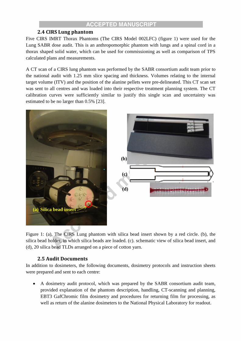
2.4 CIRS Lung phantom Five CIRS IMRT Thorax Phantoms (The CIRS Model 002LFC) (figure 1) were used for the
Lung SABR dose audit. This is an anthropomorphic phantom with lungs and a spinal cord in a
thorax shaped solid water, which can be used for commissioning as well as comparison of TPS
calculated plans and measurements.
A CT scan of a CIRS lung phantom was performed by the SABR consortium audit team prior to
the national audit with 1.25 mm slice spacing and thickness. Volumes relating to the internal
target volume (ITV) and the position of the alanine pellets were pre-delineated. This CT scan set
was sent to all centres and was loaded into their respective treatment planning system. The CT
calibration curves were sufficiently similar to justify this single scan and uncertainty was
estimated to be no larger than 0.5% [23].
Figure 1: (a), The CIRS Lung phantom with silica bead insert shown by a red circle. (b), the
silica bead holder, in which silica beads are loaded. (c). schematic view of silica bead insert, and
(d), 20 silica bead TLDs arranged on a piece of cotton yarn.
2.5 Audit Documents In addition to dosimeters, the following documents, dosimetry protocols and instruction sheets
were prepared and sent to each centre:
A dosimetry audit protocol, which was prepared by the SABR consortium audit team,
provided explanation of the phantom description, handling, CT-scanning and planning,
EBT3 GafChromic film dosimetry and procedures for returning film for processing, as
well as return of the alanine dosimeters to the National Physical Laboratory for readout.
(b)
(c)
(d)
Silica bead insert (a)
A GafChromic film irradiation record form
An alanine irradiation record form
Silica beads introductory document
Silica beads dosimetry instructions and irradiation record form
2.6 Measurement procedure
2.6.1 Silica beads calibration
This batch of silica beads has been dosimetrically characterised in the previous studies and the
details of characterisation measurements are published in earlier publications [14 and 15].
Linearity of dose response covering the range of radiotherapy dose levels (1cGy to 50Gy), had a
correlation coefficient of R2 ≥ 0.999; therefore, the large dose differences between reference
dose measurement and the audit dose levels can be considered to have a negligible calibration
error. Energy response was fairly uniform across the MV photon beams and the maximum
percentage difference compared to that obtained at 6 MV photons being -3.8% for 15 MV
photons [15]. The energy response is considered of relevance for the use of dosimeters in postal
audit situations where each institution may have slightly different quality index (QI) for their
respective photon energies thus ensuring that the calibration may still be considered valid when
same nominal beam energy is used.
The silica beads had a batch homogeneity of 8% (SD = 1) and so individual calibration factors
were determined. This relative calibration was performed in a 6 MV clinical photon beam
(TPR20/10 = 0.670) with a field size of 10 × 10 cm2. The calibration dose was 10 Gy at 5 cm
depth in a Solid-Water® phantom. This beam quality was chosen as all radiotherapy centres
employed 6 MV photon beams for their respective audit plans. A detailed description of the
calibration procedure has been published previously [17].
2.6.2 Absolute dose measurements using alanine dosimeters
For absolute dosimetry, the silica beads were cross calibrated against alanine dosimeters in
reference conditions. To perform absolute dosimetry with sufficient accuracy using alanine
pellets, a dose of greater than 10 Gy is ideally required [24]. At 10 Gy, the uncertainty associated
with the calibration of alanine dosimeters is 0.85% (SD = 1) and the pellet to pellet
reproducibility is 0.5% (SD = 1) [24, 25]. Centres were therefore requested to irradiate a set of
silica beads at a dose of 10 Gy at the same beam energy and under the same experimental
conditions. The local ionisation chamber was also given 10Gy under these conditions.
The irradiated alanine pellets were analysed by the National Physical Laboratory using an EPR
spectrometer and the results were reported in terms of dose (Gy).
2.6.3 SABR Plan
Each centre created a SABR plan on the scan of the CIRS lung phantom using their current
planning protocol and technique. The plan covered the Planning Target Volume (PTV), which
contained film, 9 alanine pellets and thus also the silica beads that in turn sit along the central
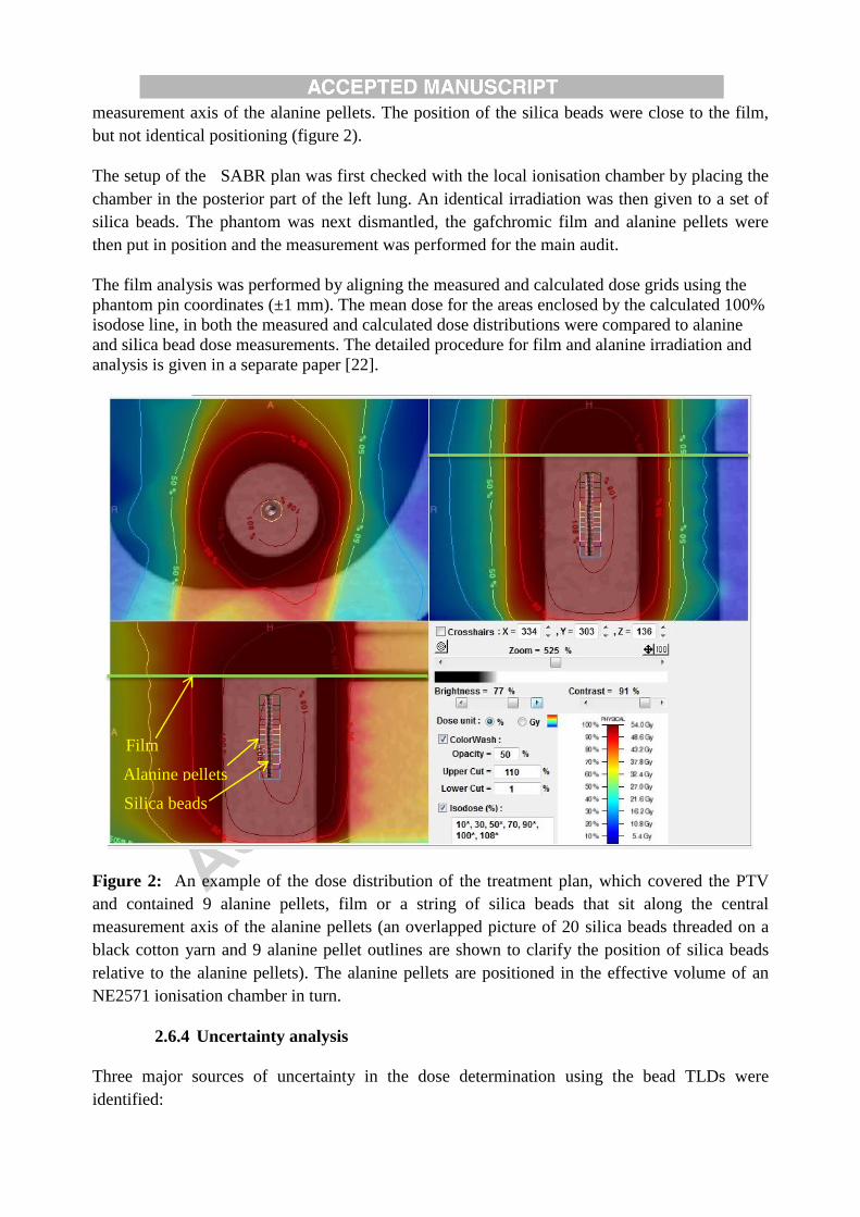
measurement axis of the alanine pellets. The position of the silica beads were close to the film,
but not identical positioning (figure 2).
The setup of the SABR plan was first checked with the local ionisation chamber by placing the
chamber in the posterior part of the left lung. An identical irradiation was then given to a set of
silica beads. The phantom was next dismantled, the gafchromic film and alanine pellets were
then put in position and the measurement was performed for the main audit.
The film analysis was performed by aligning the measured and calculated dose grids using the
phantom pin coordinates (±1 mm). The mean dose for the areas enclosed by the calculated 100%
isodose line, in both the measured and calculated dose distributions were compared to alanine
and silica bead dose measurements. The detailed procedure for film and alanine irradiation and
analysis is given in a separate paper [22].
Figure 2: An example of the dose distribution of the treatment plan, which covered the PTV
and contained 9 alanine pellets, film or a string of silica beads that sit along the central
measurement axis of the alanine pellets (an overlapped picture of 20 silica beads threaded on a
black cotton yarn and 9 alanine pellet outlines are shown to clarify the position of silica beads
relative to the alanine pellets). The alanine pellets are positioned in the effective volume of an
NE2571 ionisation chamber in turn.
2.6.4 Uncertainty analysis
Three major sources of uncertainty in the dose determination using the bead TLDs were
identified:
Film
Alanine pellets
Silica beads
uncertainties associated with the calibration of the silica bead TLDs consistency against
the alanine dosimeters
uncertainties in the determination of individual sensitivity correction factors for the silica
bead TLDs
the uncertainty associated with the silica bead TLD themselves: linearity, fading during
the readout process, and the TLD readout process
Dose response linearity, fading, and energy corrections factors are commonly applied for TLD
analysis [26]. However, the fading and energy correction factors were negligible for this work as
the absolute dose calibrations for the silica beads were performed on the day of read out and all
treatment plans utilised 6 MV energy. Small variations of quality index for this nominal energy
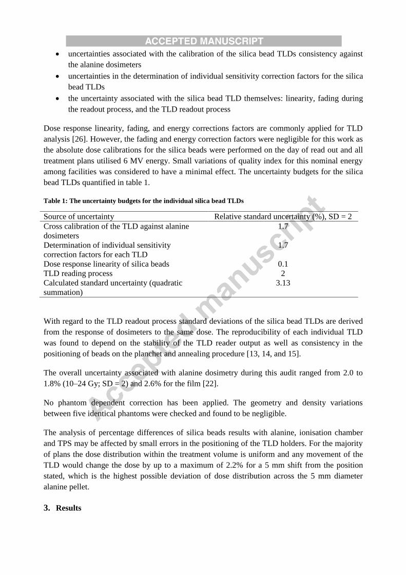
among facilities was considered to have a minimal effect. The uncertainty budgets for the silica
bead TLDs quantified in table 1.
Table 1: The uncertainty budgets for the individual silica bead TLDs
Source of uncertainty Relative standard uncertainty (%), SD = 2
Cross calibration of the TLD against alanine
dosimeters
1.7
Determination of individual sensitivity
correction factors for each TLD
1.7
Dose response linearity of silica beads 0.1
TLD reading process 2
Calculated standard uncertainty (quadratic
summation)
3.13
With regard to the TLD readout process standard deviations of the silica bead TLDs are derived
from the response of dosimeters to the same dose. The reproducibility of each individual TLD
was found to depend on the stability of the TLD reader output as well as consistency in the
positioning of beads on the planchet and annealing procedure [13, 14, and 15].
The overall uncertainty associated with alanine dosimetry during this audit ranged from 2.0 to
1.8% (10–24 Gy; SD = 2) and 2.6% for the film [22].
No phantom dependent correction has been applied. The geometry and density variations
between five identical phantoms were checked and found to be negligible.
The analysis of percentage differences of silica beads results with alanine, ionisation chamber
and TPS may be affected by small errors in the positioning of the TLD holders. For the majority
of plans the dose distribution within the treatment volume is uniform and any movement of the
TLD would change the dose by up to a maximum of 2.2% for a 5 mm shift from the position
stated, which is the highest possible deviation of dose distribution across the 5 mm diameter
alanine pellet.
3. Results
22 SABR treatment plans from 20 centres were measured. Delivery techniques varied amongst
the centres; 6 centres used conformal radiotherapy, 11 centres used Volumetric Modulated Arc
Therapy (VMAT) and 3 centres used CyberKnife. All centres employed 6 MV photon beams.
The silica bead results were analysed after correction for the calibration against the alanine
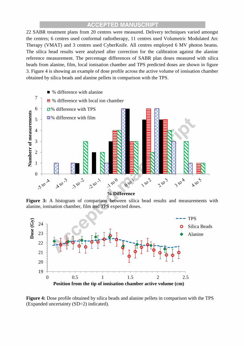
reference measurement. The percentage differences of SABR plan doses measured with silica
beads from alanine, film, local ionisation chamber and TPS predicted doses are shown in figure
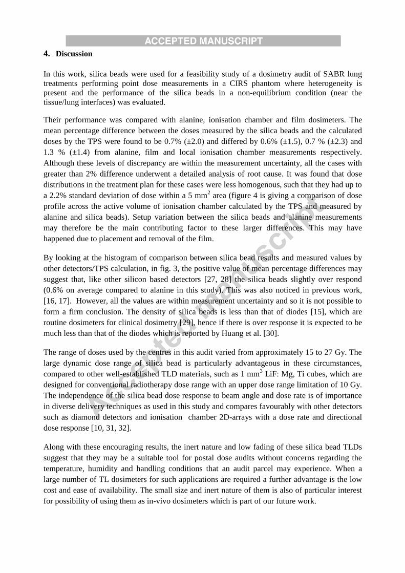
3. Figure 4 is showing an example of dose profile across the active volume of ionisation chamber
obtained by silica beads and alanine pellets in comparison with the TPS.
Figure 3: A histogram of comparison between silica bead results and measurements with
alanine, ionisation chamber, film and TPS expected doses.
Figure 4: Dose profile obtained by silica beads and alanine pellets in comparison with the TPS
(Expanded uncertainty (SD=2) indicated).
0
1
2
3
4
5
6
7
Nu
mb
err
of
mea
sure
men
ts
% Difference
% difference with alanine
% difference with local ion chamber
% difference with TPS
% difference with film
19
20
21
22
23
24
0 0.5 1 1.5 2 2.5
Dose
(G
y)
Position from the tip of ionisation chamber active volume (cm)
TPS
Silica Beads
Alanine
4. Discussion
In this work, silica beads were used for a feasibility study of a dosimetry audit of SABR lung
treatments performing point dose measurements in a CIRS phantom where heterogeneity is
present and the performance of the silica beads in a non-equilibrium condition (near the
tissue/lung interfaces) was evaluated.
Their performance was compared with alanine, ionisation chamber and film dosimeters. The
mean percentage difference between the doses measured by the silica beads and the calculated
doses by the TPS were found to be 0.7% (±2.0) and differed by 0.6% (±1.5), 0.7 % (±2.3) and
1.3 % (±1.4) from alanine, film and local ionisation chamber measurements respectively.
Although these levels of discrepancy are within the measurement uncertainty, all the cases with
greater than 2% difference underwent a detailed analysis of root cause. It was found that dose
distributions in the treatment plan for these cases were less homogenous, such that they had up to
a 2.2% standard deviation of dose within a 5 mm2 area (figure 4 is giving a comparison of dose
profile across the active volume of ionisation chamber calculated by the TPS and measured by
alanine and silica beads). Setup variation between the silica beads and alanine measurements
may therefore be the main contributing factor to these larger differences. This may have
happened due to placement and removal of the film.
By looking at the histogram of comparison between silica bead results and measured values by
other detectors/TPS calculation, in fig. 3, the positive value of mean percentage differences may
suggest that, like other silicon based detectors [27, 28] the silica beads slightly over respond
(0.6% on average compared to alanine in this study). This was also noticed in previous work,
[16, 17]. However, all the values are within measurement uncertainty and so it is not possible to
form a firm conclusion. The density of silica beads is less than that of diodes [15], which are
routine dosimeters for clinical dosimetry [29], hence if there is over response it is expected to be
much less than that of the diodes which is reported by Huang et al. [30].
The range of doses used by the centres in this audit varied from approximately 15 to 27 Gy. The
large dynamic dose range of silica bead is particularly advantageous in these circumstances,
compared to other well-established TLD materials, such as 1 mm3 LiF: Mg, Ti cubes, which are
designed for conventional radiotherapy dose range with an upper dose range limitation of 10 Gy.
The independence of the silica bead dose response to beam angle and dose rate is of importance
in diverse delivery techniques as used in this study and compares favourably with other detectors
such as diamond detectors and ionisation chamber 2D-arrays with a dose rate and directional
dose response [10, 31, 32].
Along with these encouraging results, the inert nature and low fading of these silica bead TLDs
suggest that they may be a suitable tool for postal dose audits without concerns regarding the
temperature, humidity and handling conditions that an audit parcel may experience. When a
large number of TL dosimeters for such applications are required a further advantage is the low
cost and ease of availability. The small size and inert nature of them is also of particular interest
for possibility of using them as in-vivo dosimeters which is part of our future work.
5. Conclusions
The silica beads have been evaluated for their use in postal dosimetry audit by undertaking a
feasibility study alongside an audit conducted at 20 radiotherapy centres within the UK assessing
positional and dosimetric accuracy of SABR lung treatment delivery. Their performance has
been compared to alanine, ionisation chamber and film dosimeters and doses predicted by the
treatment planning systems and agreed well with the above-mentioned dosimetry systems.
Dosimetric properties including linear response over a wide range of doses, low fading, dose rate
and angular independency in addition to their reusability, chemically inert nature and low cost is
advantageous for their application in postal dosimetry services. The results therefore confirm the
potential of the silica bead TLD systems as dosimeters for postal audit for such techniques as
SABR.
The results are encouraging and their application in postal dosimetry services is an example
where the low fading, non-hygroscopic nature and robustness is important where irradiation and
postage return may take some time. In particular, where more complex dose distributions and
field arrangements may be required to be measured [9, 12].
Acknowledgement
Authors would like to express a special appreciation and thanks to the Medical Physics
Department of the Royal Surrey County Hospital, the NPL Radiation Dosimetry group, and the
UK radiotherapy departments who volunteered to irradiate our dosimeters during the Lung
SABR national dosimetry audit; St Barts, Bristol, Clatterbridge, Guys and St. Thomas’, Hull,
Ipswich, Leeds, Leicester, Manchester, Middlesbrough, Mount Vernon, Newcastle, Norfolk &
Norwich, Nottingham, Oxford, Peterborough, Royal Marsden, Sheffield and UCLH radiotherapy
departments.
SJ would also like to convey thanks to the University of Surrey for the Overseas Research
Scholarship and Schlumberger Foundation for the Faculty for The Future scholarship.
References:
[1] Nisbet A., Thwaites D.I. 1997. A dosimetric intercomparison of electron beams in UK radiotherapy centres Phys
Med Biol, 42, 2393–2409.
[2] Thwaites D. I., Scalliet P., Leer J. W. and Overgaard J. 1995. Quality assurance in radiotherapy: European
society for therapeutic radiology and oncology advisory report to the commission of the European union
for the Europe against cancer programme Radiother. Oncol. 35 61–73
[3] Izewska J., Andreo P., Vatnitsky S., Shortt K.R. 2003. The IAEA/WHO TLD postal dose quality audits for
radiotherapy: a perspective of dosimetry practices at hospitals in developing countries Radiother Oncol, 69,
91–97.
[4] Clark C. H. et al., 2014. A multi-institutional dosimetry audit of rotational intensity-modulated radiotherapy
Radiother Oncol, 113, 272–278 http://dx.doi.org/10.1016/j.radonc.2014.11.015
[5] Ibbott G. S., Thwaites D. I. 2015. Audits for advanced treatment dosimetry in Journal of Physics: Conference
Series
[6] Clark C H, Aird E G A, Bolton S, Miles E A, Nisbet A, Snaith J A D, Thomas R A S, Venables K and Thwaites
D I, 2015. Radiotherapy dosimetry audit: three decades of improving standards and accuracy in UK clinical
practice and trials Br. J. Radiol. 88 20150251 [7] Aguirre JF, Tailor R, Ibbott G, Stovall M, Hanson, W. 2002. Thermoluminescence dosimetry as a tool for the
remote verification of output for radiotherapy beams: 25 years of experience. In: Proceedings of the
international symposium on standards and codes of practice in medical radiation dosimetry IAEA-CN-
96/82. Vienna: IAEA, 191–99.
[8] Ferreira IH, Dutreix A, Bridier A, Chavaudra J, Svensson H. 2000. The ESTRO-QUALity assurance network
(EQUAL). Radiother Oncol, 55: 273–84.
[9] Izewska J, Andreo P. 2000. The IAEA/WHO TLD postal programme for radiotherapy hospitals. Radiother
Oncol, 54: 65–72.
[10] Hussein M., Adams E. J., Jordan T. J., Clark C. H., Nisbet A., 2013. A critical evaluation of the PTW 2D-
ARRAY seven 29 and OCTAVIUS II phantom for IMRT and VMAT verification. J Appl Clin Med Phys.
14 274 – 92
[11] Benedict S.H. et al., 2010. Stereotactic body radiation therapy: the report of AAPM Task Group 101. Med
Phys, 37: 4078–101.
[12] Jain, P., Baker, A., Distefano, G., Scott, A.J., Webster, G.J., and Hatton, M.Q. Stereotactic ablative
radiotherapy in the UK: current status and developments. BJR. 2013, 86: 20130331
(http://dx.doi.org/10.1259/bjr.20130331)
[13] Izewska J., Svensson H., Ibbott G. 2002. Worldwide quality assurance networks for radiotherapy dosimetry.
IAEA-CN-96-76.
[14] Jafari, S. M., Bradley, D. A., Goldstone, C. A., Sharpe, P. H. G., Alalawi, A. I., Jordan, T. J., Clark, C. H.,
Nisbet, A. & Spyrou, N. M. 2014. Low-cost commercial glass beads as dosimeters in radiotherapy. Radiat.
Phys. Chem., 97; 95-101.
[15] Jafari, S. M., Jordan, T. J., Hussein, M., Bradley, D. A., Clark, C. H., Nisbet, A. & Spyrou, N. M. 2014. Energy
response of glass bead TLDs irradiated with radiation therapy beams. Radiation Physics and Chemistry,
104; 208–211.
[16] Jafari S.M., Alalawi A. I., Hussein M. , Alsaleh W., Najem M. A., Hugtenburg R.P., Bradley D.A., Spyrou
N.M., Clark C.H., Nisbet A. 2014. Glass beads and Ge-doped optical fibres as thermoluminescence
dosimeters for small field photon dosimetry. Phys. Med. Biol. 59 6875-6889.
[17] Jafari S. M., Jordan T. J., Distefano G., Bradley D. A., Spyrou N. M., Nisbet A., Clark C. H., 2014. Feasibility of
using glass bead thermo-luminescent dosimeters for radiotherapy treatment plan verification, BJR,
http://dx.doi.org/10.1259/bjr.20140804
[18 Jafari S.M., Bates N.M., Jupp T. Abdul Sani S.F., Nisbet A., Bradley D.A. 2016. Commercial glass beads as
TLDs in radiotherapy produced by different manufacturers. Radiation Physics and Chemistry, in press
[19] IPEM Group, 2010, National Radiotherapy Implementation. Clinical Review of the Evidence for SBRT. IPEM.
England : NHS, IPEM report
[20] Chetty I.J. et al. 2007. Report of the AAPM Task Group No. 105: Issues associated with clinical
implementation of Monte Carlo-based photon and electron external beam treatment planning. Med Phys 34:
4818-53
[21] Distefano G., Jafari S. M., Lee J., Gouldstone C., Mayles H., Clark C. H., UK SABR Consortium Lung
Dosimetry Audit; Absolute dosimetry results. European society for radiotherapy and oncology conference
(3rd ESTRO Forum) 24-28 April 2015 Barcelona Spain, Radiother. Oncol. Vol. 115, Supplement 1, April
2015, ISSN 0167-8140, p 96.
[22] Distefano G., Lee J., Jafari S. M., Gouldstone C., Baker C., Mayles H., , Clark C. H., 2016. A national
dosimetry audit for stereotactic ablative radiotherapy in lung. Radiother. Oncol. In press. [23]Husseina M., Tsang Y., Thomas R.A.S., Gouldstone C., Maughan D., Snaith J.A.D, Bolton S.C., Nisbet A.,
Clark C.H.. 2013. A methodology for dosimetry audit of rotational radiotherapy using a commercial
detector array Radiother. and Oncol., 108, (1), 78–85 [24] Sharpe P. H. G., Sephton J. P. 2006. Therapy level alanine dosimetry at the NPL “Alanine Dosimetry for
Clinical Applications” Proceedings of the 216th PTB Seminar” PTB, Braunschweig
[25] Budgell, G., Berresford, J., Trainer, M., Bradshaw, E., Sharpe, P., and Williams, P. 2011. A national dosimetric
audit of IMRT. Radiother Oncol. 99 246–252
[26] Kirby, T.H., Hanson, W.F., Jhonston, D.A., 1992. Uncertainty analysis of absorbed dose calculations from
thermoluminescence dosimeters. Med. Phys. 19 1427–1433.
[27] McKerracher, C., Thwaites, D.I., 2006. Notes on the construction of solid-state detectors. Radiother Oncol, 79
348–351.
[28] Lechner W., Palmans H., SÖlkner L., Grochowska P. and Georg D., 2013. Detector comparison for small field
output factor measurements in flattening filter free photon beams Radiother. Oncol. 109 356–60
[29] Griessbach I., Lapp M., Bohsung J., and Gademann G., 2005. Dosimetric characteristics of a new unshielded
silicon diode and its application in clinical photon and electron beams. Med. Phys. 32(12), 3750–3754.
[30] Huang K., Bice W. S. Jr. and Hidalgo-Salvatierra O. 2003. Characterization of an Invivo diode dosimetry
system for clinical use. J. Appl. Clin. Med. Phys. 4 132–42.
[31] Ade N., Nam T.L., Derry T.E., Mhlanga S.H., 2014. The dose rate dependence of synthetic diamond detectors
in the relative dosimetry of high-energy electron therapy beams Radiation Physics and Chemistry 98, 155–
162.
[32] Wolfsberger L. D., Wagar M., Nitsch P., Bhagwat M. S. And Zygmanski P., 2010. Angular dose dependence of
Matrixx TM and its calibration J. Appl. Clin. Med. Phys. 11 241-51
Highlights:
Silica beads have been used as novel micro-TLDs in a national dosimetry audit across
UK
The performance of the TLDs assessed in non-equilibrium conditions near tissue/lung
interfaces TLDs’ Results agreed well with those obtained from alanine, film and ionisation chamber
detectors
Top Related