

Languages
Pages
Legal


Adrenal InsufficiencyDr Rodney ItakiAnatomical Pathology Discipline
University of Papua New GuineaDivision of PathologySchool of Medicine & Health Sciences

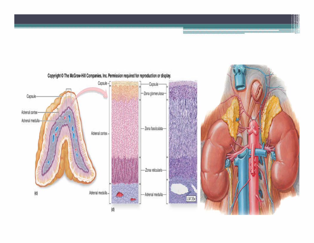
Normal Function – Adrenal Gland

• Zona Glomerulosa
• Zona Fasiculata
• Zona Reticularis

5
Steroid Hormones
• Glucocorticoids▫ CHO, lipid & fat metabolism▫ Increases blood glucose levels & gluconeogenesis▫ Increases protein breakdown▫ Inhibits protein synthesis
• Mineralcorticoids▫ Elecrolyte & fluid balance▫ Increases sodium & water retention▫ Regulated by renin & andiotensin
• Sex Steroids▫ Low synthesis in adrenals compared to gonads▫ Virilising hormones may be secreted

Adrenal gland Insufficiency• Can be caused by▫ Primary adrenal disease▫ Decreased adrenal stimulation from low ACTH
• Clinical Patterns of presentation▫ Primary acute adrenocortical insufficiency –Adrenal Ciris
▫ Primary chronic adrenocortical insufficiency –Addison disease
▫ Secondary adrenocortical insufficiency
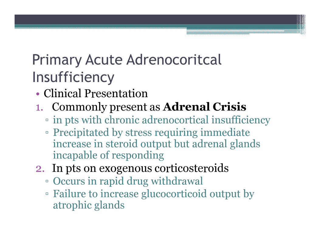
Primary Acute AdrenocoritcalInsufficiency• Clinical Presentation1. Commonly present as Adrenal Crisis▫ in pts with chronic adrenocortical insufficiency▫ Precipitated by stress requiring immediate increase in steroid output but adrenal glands incapable of responding
2. In pts on exogenous corticosteroids▫ Occurs in rapid drug withdrawal▫ Failure to increase glucocorticoid output by atrophic glands

Primary Acute AdrenocoritcalInsufficiency3. As a result from massive adrenal haemorrhage
destroying adrenal cortex

Adrenal Haemorrhage – adrenal crisis• New borns high risk – especially after prolonged and difficult labor.▫ Deficient in prothrombin for several days after birth increases risk of bleeding
• Pts on anticoagulant therapy• Post-surgical pts who develop DIC• Complication of sepsis – Waterhouse-
Friderichsen syndrome▫ N.meningitidis, pseudomonas, pneumococci,
H.influenzae or staphylococci infections

Waterhouse-Friderichsen Syndrome• Rapidly progressive hypotension leading to shock
• DIC with widespread purpura• Rapidly developing adrenocortical insufficienydue to massive bilateral haemorrhage
• High risk in children• Basis: direct bacterial seeding of small vessels in adrenal, endotoxin-induced vasculitis or hypersensitivity vasculitis

Waterhouse-Friderichsen Syndrome• Adrenal gland pathology: adrenals converted to sacs of clotted blood
• Histology: haemorrhage from medulla in thin walled sinusoids & extending to cotex
• Islands of cortical cells may be recognised

Morphology


Massive adrenal haemorrhage, resulting in primary acute adrenal insufficiency

Primary Chronic AdrenocorticalInsufficiency (Addison Disease)• Results from destruction of adrenal cortex• Clinical symptoms appear when 90% of cortical tissue has been damaged
• Causes:▫ Autoimmune disorder▫ Infections▫ Metastatic cancers

Autoimmune cause – Addison Disease• 60-70% of primary chronic adrenocorticalinsufficiency
• Can occur in sporadic cases or familial disorder• 50% of cases only adrenal gland affected• Other 50% adrenal gland + other endocrine gland affected▫ Thyroid (Hashimoto disease)▫ Type I DM▫ Parathyroid gland – idiopathic hypoparathyroidism▫ Pernicous anaemia

Addison Disease - Pathogenesis• Antibody directed against adrenal gland resulting in autoimmune adrenitis

Infective Causes of Addison Disease• TB common cause in PNG population• Fungal infections also can cause Addison Disease▫ Histoplasma capsulatum & coccidioides immitis

Metastatic Causes – Addison Disease• Breast• Lung• GIT• Melanoma• Haematopoietic neoplasms

Clinical Features – Addison Disease• Insidious onset▫ Symptoms appear when 90% of gland destroyed
• Progressive weakness• Easy fatigability• Non-specific GIT symptoms – anorexia, nausea, vomiting, wt loss & diarrhoea
• Hyperpigmentation of skin• Electrolyte abnormalities – High K & low Na from mineralocorticoid deficiency
• Hypoglycaemia – glucocorticoid deficiency

Features of Addison’s d.

Addison Disease - Morphology• Irregular shrunken adrenal glands – autoimmune ▫ Histology: scattered residual cortical cells in a collapsed network of tissue▫ Variable lympoid infiltrate present in cortex, may extend into subjacent medulla
• Infection – TB or fungal infection▫ Granulomatous inflammation
• Metastatic Ca▫ Adrenals enlarged▫ achitecture obscured

24
This is a caseating granuloma of tuberculosis in the adrenal gland. Tuberculosis used to be the most common cause of chronic
adrenal insufficiency. Now, idiopathic (presumably autoimmune) Addison's disease is much
more often the cause for chronic adrenal insufficiency.

Granulomatous inflammation

26
The pair of adrenals in the center are normal. Those at the top come from a patient with adrenal atrophy (with either
Addison's disease or long-term corticosteroid therapy). The adrenals at the bottom represent bilateral cortical hyperplasia.
This could be due to a pituitary adenoma secreting ACTH (Cushing's disease), or Cushing's syndrome from ectopic ACTH production, or idiopathic adrenal
hyperplasia.

Metastatic breast carcinoma affecting the adrenal gland and causing primary chronic adrenal insufficiency
Secondary Adrenocortical Insufficiency• Due to ▫ Disorder of hyothalamus & pituitary gland resulting in low ACTH synthesis and secretion
▫ Prolonged exogenous administration of glucocorticoids� Suppress ACTH production & adrenal function resulting
in adrenal atrophy
• Signs & symptoms similar to Addison Disease• NO SKIN PIGMENTATION• Mild electrolyte abnormaliy due to normal
aldosterone synthesis
Laboratory findings.1. A low serum Na level and a high serum K level together with a
characteristic clinical picture suggest the possibility of Addison’s disease.
2. Adrenal insufficiency can be specifically diagnosed by:• low levels of plasma glucocorticoids and mineralocorticoids, or
urinary 17 – hydroxycorticosteroid (17 – OHCS) or 17 – ketogenicsteroid (17 – KGS);
• demonstrating failure to increase plasma cortisol levels, or urinary 17 – OHCS or 17 – KGS excretion, upon administration of ACTH in patients with primary adrenal insufficiency
▫ (those with secondary adrenocortical insufficiency will have a significant increase in plasma cortisol or 24 - h urinary corticosteroid levels.)
3. To distinguish between primary and secondary adrenal insufficiency, have to find the level of plasma ACTH: primary shows increased, and secondary shows decreased level.

Diagnosis & Treatment• Diagnostic test▫ Synacthen test
• Treatment▫ Cortisol replacement� Hydrocortisone/Cortisone
▫ Aldosterone replacement� Fludrocortisone

Synacthen testPituitary
Cortisol
Adrenalgland
Synacthen(=synACTHen)
•Baseline cortisol may be normal in Addison’s disease•Synacthen test: uses synthetic ACTH analogue•Normal response: rise in cortisol

References: Robins Pathological Basis of Diseases
Download seminar notes at: www.pathologyatsmhs.wordpress.com
Top Related