


![A Case of Mediastinal Cystic Lymphangiomaousar.lib.okayama-u.ac.jp/files/public/5/53911/...1オ extend into the mediastinum [1];these mediasti-nal cystic lymphangiomas account for](https://static.fdocuments.net/doc/165x107/5f03171b7e708231d4077c7f/a-case-of-mediastinal-cystic-1i-extend-into-the-mediastinum-1these-mediasti-nal.jpg)
Languages
Pages
Legal

A Case of Mediastinal Cystic Lymphangioma
Hiroshi Suehisaa*, Tsuyoshi Uenoa, Shigeki Sawadaa, Motohiro Yamashitaa, and Norihiro Teramotob
Departments of aThoracic Surgery, and bPathology, National Hospital Organization Shikoku Cancer Center, Matsuyama 791-0280, Japan
A 35-year-old Japanese manʼs routine chest radiography revealed an abnormal opacity. Chest com-puted tomography and magnetic resonance imaging showed a 5.5cm in dia. cystic tumor located at the left anterior mediastinum. The tumor was suspected to be an asymptomatic thymic cyst, and we chose observation for the tumor. At the 3-year follow up, the cystic tumor had gradually enlarged to 7.5cm in dia. and we thus performed a surgical resection via left video-assisted thoracic surgery. An immu-nohistochemical analysis showed that the cystic tumor was not a thymic cyst but rather a mediastinal cystic lymphangioma. Mediastinal cystic lymphangiomas are very rare, and they are difficult to diag-nose preoperatively. Complete surgical resection is proposed for the treatment of such tumors.
Key words: mediastinal tumor, mediastinal cystic lymphangioma, thymic cyst
ymphangiomas are rare benign congenital mal-formations derived from the lymphatic system.
Most appear in the neck or axillae, and approximately 1オ extend into the mediastinum [1]; these mediasti-nal cystic lymphangiomas account for 0.7-4.5オ of all mediastinal tumors [2]. We encountered a rare case of mediastinal cystic lymphangioma that was diagino-sed postoperatively.
Case
A 35-year-old Japanese man underwent chest radiography as part of a routine medical examination, and the radiography revealed an abnormal opacity. A further examination was undertaken at the local hospi-tal. Chest computed tomography (CT) revealed a left anterior mediastinal tumor, and the patient was con-sequently referred to our hospital. He was on medica-tion for diabetes mellitus.
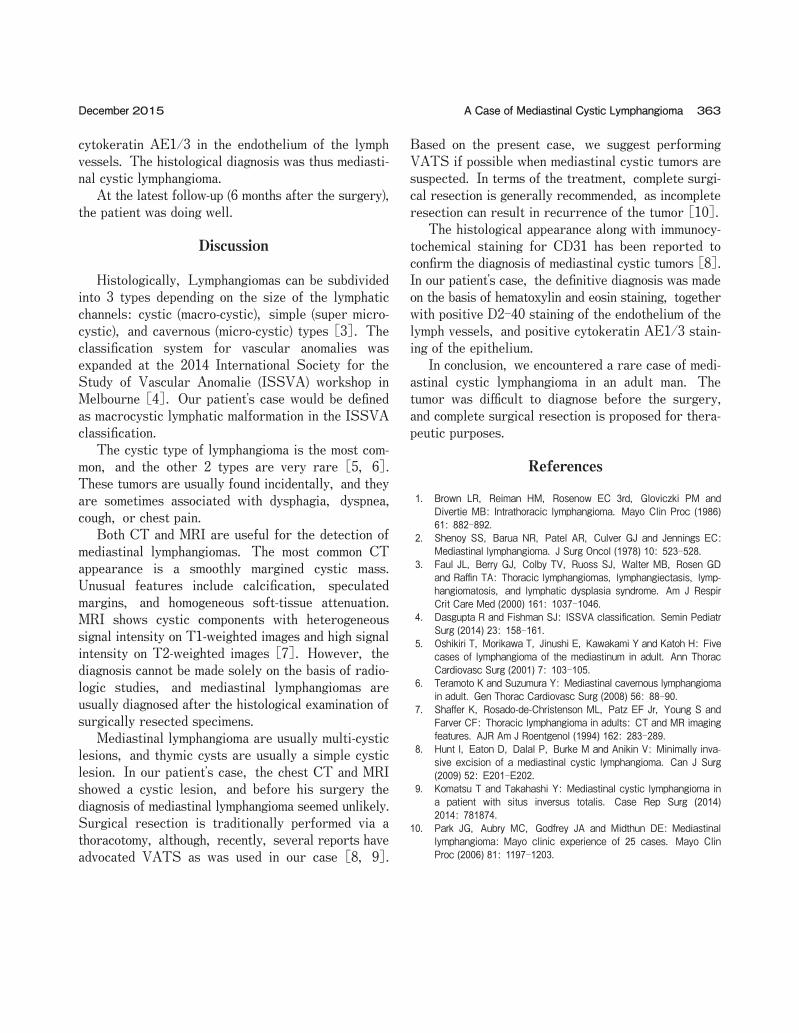
Chest CT showed a cystic tumor, 5.5cm in dia., with a smooth surface, round shape, and low attenua-tion area (Fig. 1). The cystic nature was confirmed upon magnetic resonance imaging (MRI), which showed a high-intensity area on T2-weighted images (Fig. 2). We highly suspected an asymptomatic thy-mic cyst, and we thus chose observation for the cystic tumor. After 3 years of follow-up, the cystic tumor had gradually enlarged to 7.5cm in dia., at which time we performed a surgical resection. With the patient under general anesthesia in the right hemi-lateral decubitus position, the tumor resection was performed via left video-assisted thoracic surgery (VATS) using one access window plus two ports. The resected tumor was soft and had serous effu-sion that was aspirated during the operation to allow for easy dissection. No obvious communication was found between the tumor and thoracic duct. The cystic tumor was not adhered to the left lung and could be separated from the pericardium by blunt dissection
L
Acta Med. Okayama, 2015Vol. 69, No. 6, pp. 361ン363CopyrightⒸ 2015 by Okayama University Medical School.
Case Report http ://escholarship.lib.okayama-u.ac.jp/amo/
Received March 20, 2015 ; accepted July 14, 2015.*Corresponding author. Phone : +81ン89ン999ン1111; Fax : +81ン89ン999ン1100E-mail : [email protected] (H. Suehisa)
Conflict of Interest Disclosures: No potential conflict of interest relevant to this article was reported.
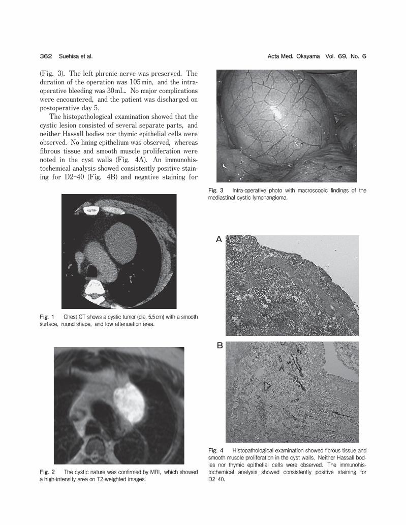
(Fig. 3). The left phrenic nerve was preserved. The duration of the operation was 105min, and the intra-operative bleeding was 30mL. No major complications were encountered, and the patient was discharged on postoperative day 5. The histopathological examination showed that the cystic lesion consisted of several separate parts, and neither Hassall bodies nor thymic epithelial cells were observed. No lining epithelium was observed, whereas fibrous tissue and smooth muscle proliferation were noted in the cyst walls (Fig. 4A). An immunohis-tochemical analysis showed consistently positive stain-ing for D2-40 (Fig. 4B) and negative staining for
362 Acta Med. Okayama Vol. 69, No. 6Suehisa et al.
Fig. 1 Chest CT shows a cystic tumor (dia. 5.5cm) with a smooth surface, round shape, and low attenuation area.
Fig. 2 The cystic nature was confirmed by MRI, which showed a high-intensity area on T2-weighted images.
A
B
Fig. 4 Histopathological examination showed fibrous tissue and smooth muscle proliferation in the cyst walls. Neither Hassall bod-ies nor thymic epithelial cells were observed. The immunohis-tochemical analysis showed consistently positive staining for D2-40.
Fig. 3 Intra-operative photo with macroscopic findings of the mediastinal cystic lymphangioma.
cytokeratin AE1/3 in the endothelium of the lymph vessels. The histological diagnosis was thus mediasti-nal cystic lymphangioma. At the latest follow-up (6 months after the surgery), the patient was doing well.
Discussion
Histologically, Lymphangiomas can be subdivided into 3 types depending on the size of the lymphatic channels: cystic (macro-cystic), simple (super micro- cystic), and cavernous (micro-cystic) types [3]. The classification system for vascular anomalies was expanded at the 2014 International Society for the Study of Vascular Anomalie (ISSVA) workshop in Melbourne [4]. Our patientʼs case would be defined as macrocystic lymphatic malformation in the ISSVA classification. The cystic type of lymphangioma is the most com-mon, and the other 2 types are very rare [5, 6]. These tumors are usually found incidentally, and they are sometimes associated with dysphagia, dyspnea, cough, or chest pain. Both CT and MRI are useful for the detection of mediastinal lymphangiomas. The most common CT appearance is a smoothly margined cystic mass. Unusual features include calcification, speculated margins, and homogeneous soft-tissue attenuation. MRI shows cystic components with heterogeneous signal intensity on T1-weighted images and high signal intensity on T2-weighted images [7]. However, the diagnosis cannot be made solely on the basis of radio-logic studies, and mediastinal lymphangiomas are usually diagnosed after the histological examination of surgically resected specimens. Mediastinal lymphangioma are usually multi-cystic lesions, and thymic cysts are usually a simple cystic lesion. In our patientʼs case, the chest CT and MRI showed a cystic lesion, and before his surgery the diagnosis of mediastinal lymphangioma seemed unlikely. Surgical resection is traditionally performed via a thoracotomy, although, recently, several reports have advocated VATS as was used in our case [8, 9].
Based on the present case, we suggest performing VATS if possible when mediastinal cystic tumors are suspected. In terms of the treatment, complete surgi-cal resection is generally recommended, as incomplete resection can result in recurrence of the tumor [10]. The histological appearance along with immunocy-tochemical staining for CD31 has been reported to confirm the diagnosis of mediastinal cystic tumors [8]. In our patientʼs case, the definitive diagnosis was made on the basis of hematoxylin and eosin staining, together with positive D2-40 staining of the endothelium of the lymph vessels, and positive cytokeratin AE1/3 stain-ing of the epithelium. In conclusion, we encountered a rare case of medi-astinal cystic lymphangioma in an adult man. The tumor was difficult to diagnose before the surgery, and complete surgical resection is proposed for thera-peutic purposes.
References
1. Brown LR, Reiman HM, Rosenow EC 3rd, Gloviczki PM and Divertie MB: Intrathoracic lymphangioma. Mayo Clin Proc (1986) 61: 882-892.
2. Shenoy SS, Barua NR, Patel AR, Culver GJ and Jennings EC: Mediastinal lymphangioma. J Surg Oncol (1978) 10: 523-528.
3. Faul JL, Berry GJ, Colby TV, Ruoss SJ, Walter MB, Rosen GD and Raffin TA: Thoracic lymphangiomas, lymphangiectasis, lymp-hangiomatosis, and lymphatic dysplasia syndrome. Am J Respir Crit Care Med (2000) 161: 1037-1046.
4. Dasgupta R and Fishman SJ: ISSVA classification. Semin Pediatr Surg (2014) 23: 158-161.
5. Oshikiri T, Morikawa T, Jinushi E, Kawakami Y and Katoh H: Five cases of lymphangioma of the mediastinum in adult. Ann Thorac Cardiovasc Surg (2001) 7: 103-105.
6. Teramoto K and Suzumura Y: Mediastinal cavernous lymphangioma in adult. Gen Thorac Cardiovasc Surg (2008) 56: 88-90.
7. Shaffer K, Rosado-de-Christenson ML, Patz EF Jr, Young S and Farver CF: Thoracic lymphangioma in adults: CT and MR imaging features. AJR Am J Roentgenol (1994) 162: 283-289.
8. Hunt I, Eaton D, Dalal P, Burke M and Anikin V: Minimally inva-sive excision of a mediastinal cystic lymphangioma. Can J Surg (2009) 52: E201-E202.
9. Komatsu T and Takahashi Y: Mediastinal cystic lymphangioma in a patient with situs inversus totalis. Case Rep Surg (2014) 2014: 781874.
10. Park JG, Aubry MC, Godfrey JA and Midthun DE: Mediastinal lymphangioma: Mayo clinic experience of 25 cases. Mayo Clin Proc (2006) 81: 1197-1203.
363A Case of Mediastinal Cystic LymphangiomaDecember 2015
Top Related