


Languages
Pages
Legal

Brain Meninges, Ventricles and CSF

Lecture Objectives• Describe the arrangement of the meninges and their relationship to brain and spinal cord.
• Explain the occurrence of epidural, subdural and subarachnoid spaces.
• Locate the principal subarachnoid cisterns, and arachnoid granulations.
• Describe the ventricles of brain and importance of their choroids plexus.
• Summarize the pathway of cerebrospinal fluid (CSF) circulation.• Locate the safe sites for the lumbar puncture.• Identify brain ventricles in CT scan, MRI and ventriculograms.

Cerebral Meninges
• Dura mater• Endosteal layer• Meningeal layer
• Arachnoid mater• Pia mater
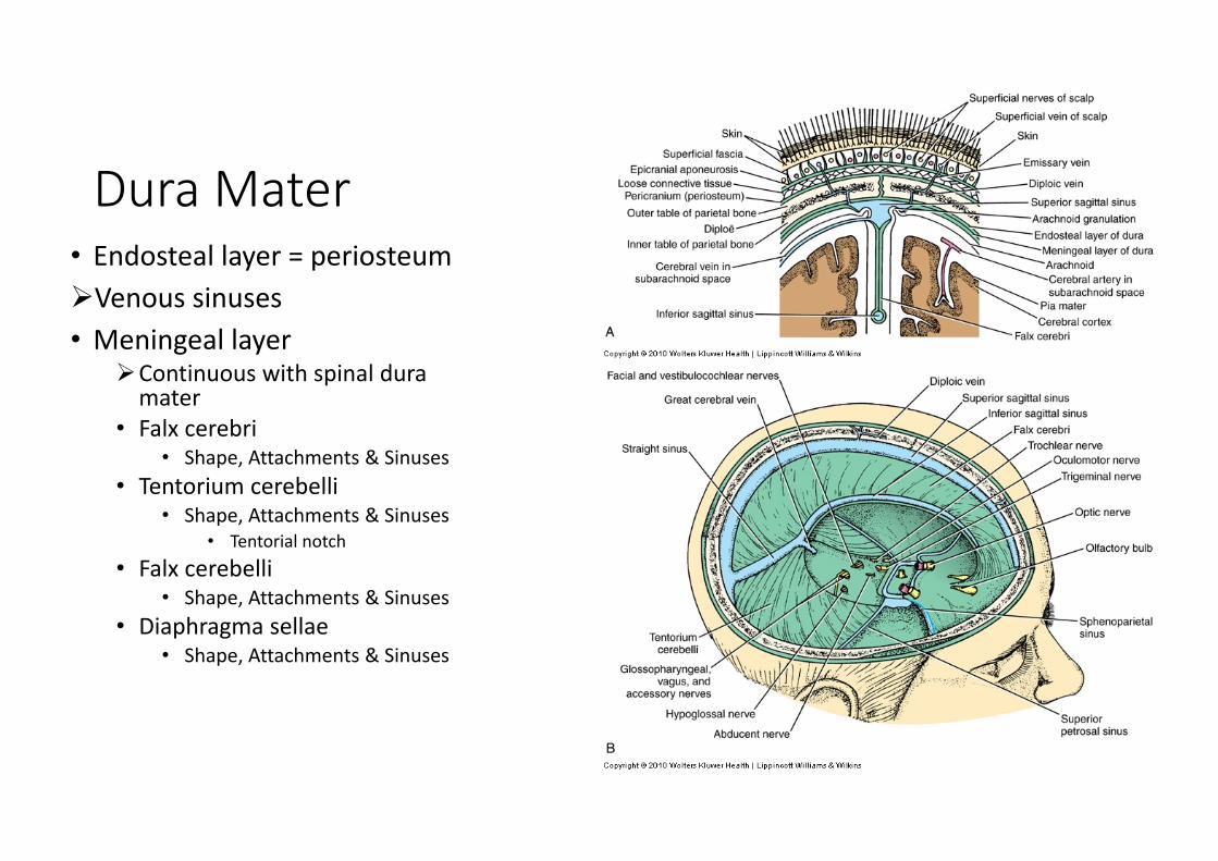
Dura Mater• Endosteal layer = periosteumVenous sinuses• Meningeal layer
Continuous with spinal duramater
• Falx cerebri• Shape, Attachments & Sinuses
• Tentorium cerebelli• Shape, Attachments & Sinuses
• Tentorial notch• Falx cerebelli
• Shape, Attachments & Sinuses• Diaphragma sellae
• Shape, Attachments & Sinuses

Dura Mater
• Nerve supply• Cranial nerves V & X
• Referral pain to the head from above the tentorium cerebelli
• Spinal nerves C1‐C3• Referral pain to the back of the head and neck from bellow tentorium
• Sympathetic
• Blood supply• Internal carotid, maxillary, ascending pharyngeal, occipital & vertebral aa
• Middle meningeal a.
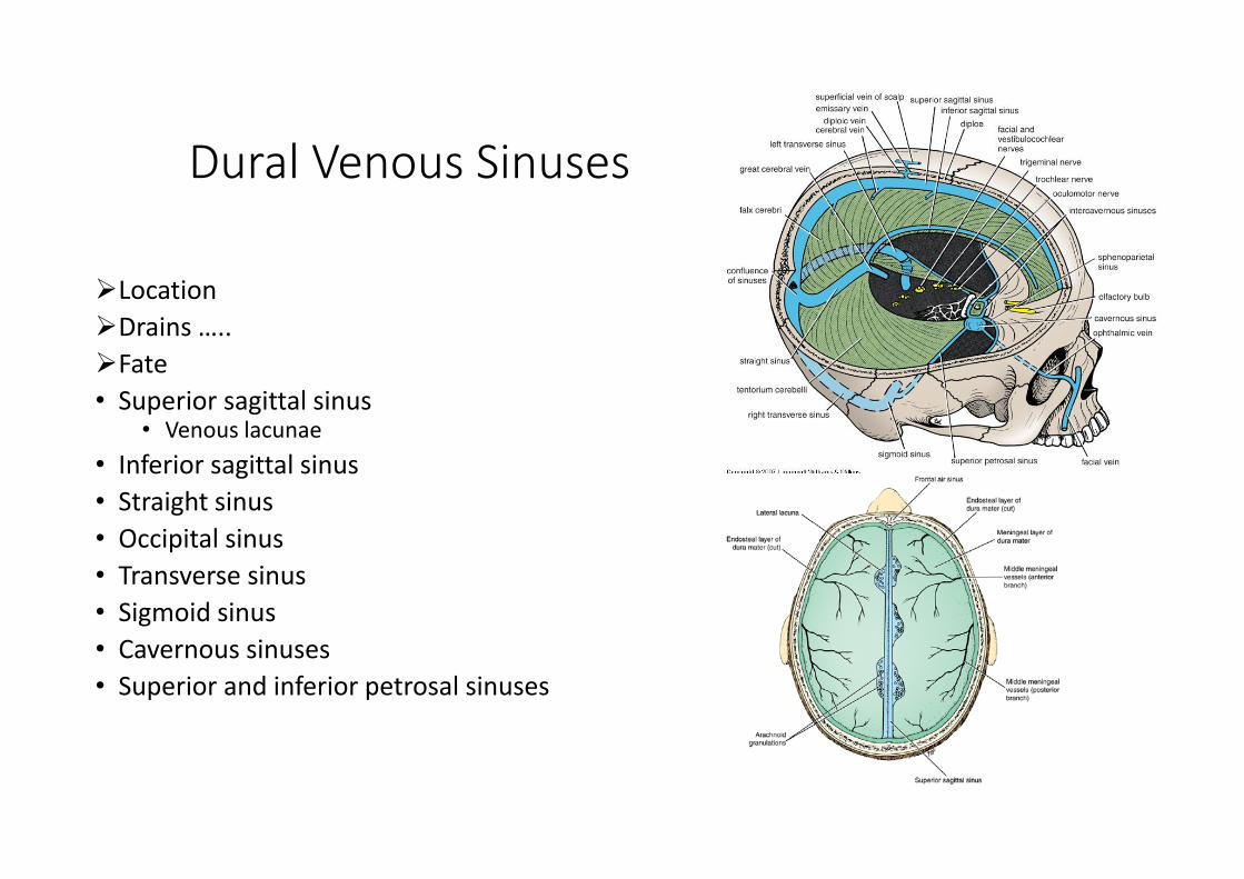
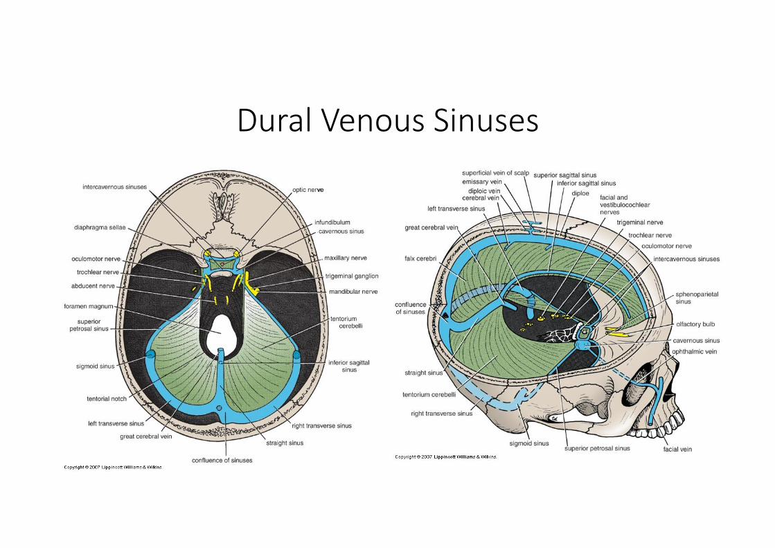
Dural Venous Sinuses
LocationDrains …..Fate• Superior sagittal sinus
• Venous lacunae• Inferior sagittal sinus• Straight sinus• Occipital sinus• Transverse sinus• Sigmoid sinus• Cavernous sinuses• Superior and inferior petrosal sinuses
Dural Venous Sinuses

Arachnoid Mater
• Subdural space• Subarachnoid space
• Cerebral BV & cranial nn.• CSF• Subarachnoid cisternae• Arachnoid villi
• Arachnoid granulations
• Fuse with epineurium at foramina• Except for optic nerve ‐ fuse with sclera

Pia Mater• Adheres closely to the brain
• Goes deep into the sulci• Fuse with nerves epineurium• Covers cerebral arteries entering brain substance• Specialized over the roofs of ventricles (tela choroidea)
• Contribute to formation of choroid plexuses
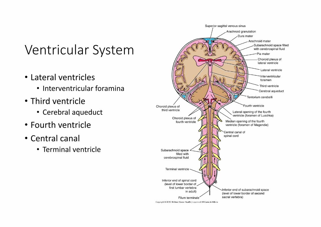
Ventricular System
• Lateral ventricles• Interventricular foramina
• Third ventricle• Cerebral aqueduct
• Fourth ventricle• Central canal
• Terminal ventricle

Lateral Ventricles• Location • Shape • Parts
• Body• Horns
• Anterior, posterior, inferior
• Choroid plexus

Lateral Ventricles• Relations
• Corpus callosum• Septum pellucidum• Fornix • Thalamus • Caudate nucleus
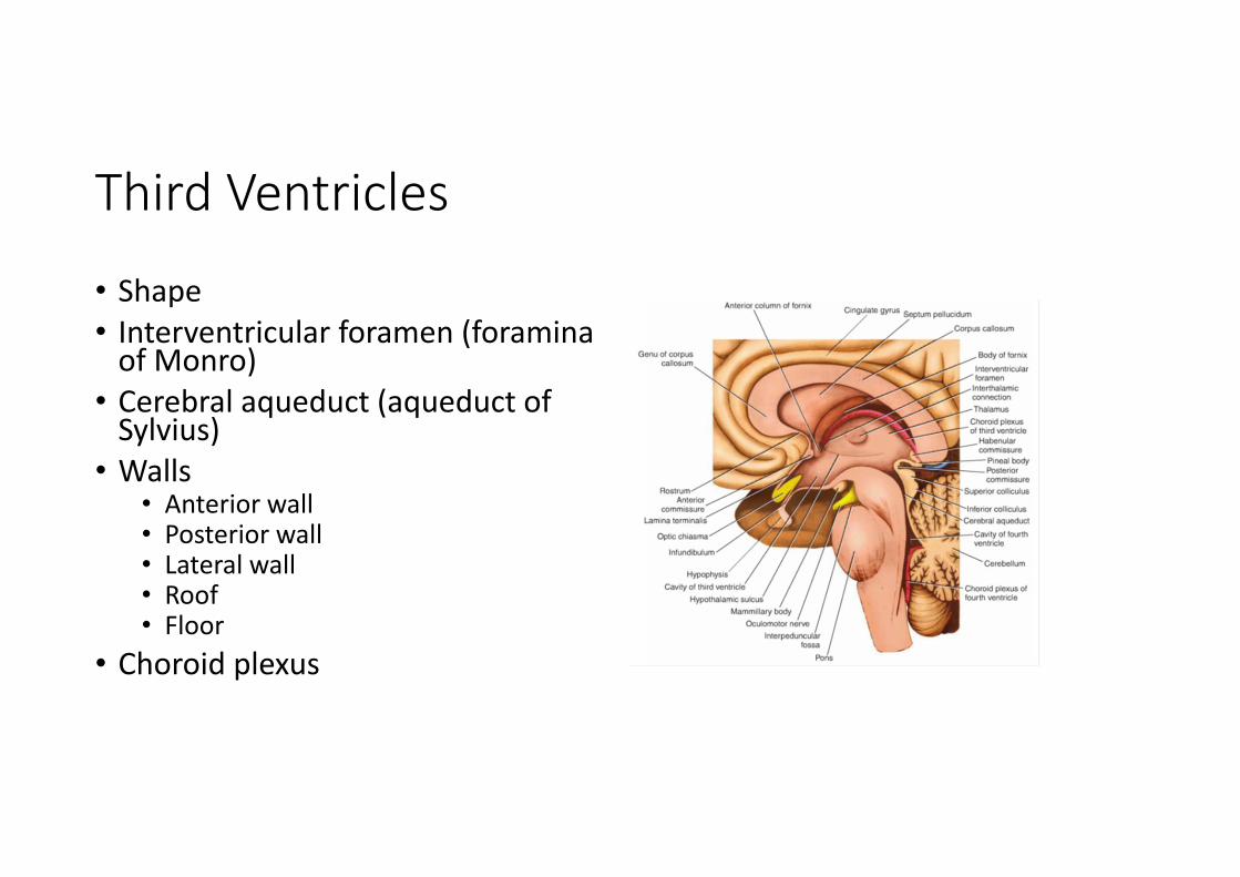
Third Ventricles
• Shape• Interventricular foramen (foramina of Monro)
• Cerebral aqueduct (aqueduct of Sylvius)
• Walls• Anterior wall• Posterior wall • Lateral wall• Roof• Floor
• Choroid plexus

Fourth Ventricle
• Shape• Relations• Walls
• Lateral walls• Roof (posterior wall)
• Superior and inferior medullary velum• Median aperture (foramen of Magendie)
• Lateral apertures (foramina of Luschka)• Choroid plexus
• Floor (rhomboid fossa)

Subarachnoid Cisterns
• Extended area of the subarachnoid space
• Subarachnoid cisterns• Cerebellomedullary cistern (cisterna magnum)• Median aperture
• Pontine cistern• Lateral apertures
• Interpeduncular cistern

Subarachnoid Cisterns

Cerebrospinal Fluid (CSF)
• 80‐150 ml (3‐5oz)• Clear liquid containing glucose, proteins, & ions• Functions
• mechanical protection • floats brain & softens impact with bony walls
• chemical protection• optimal ionic concentrations for action potentials
• circulation• nutrients and waste products to and from bloodstream

Origin of CSF
• Choroid plexus = capillaries covered by ependymal cells• 2 lateral ventricles, one within each cerebral hemisphere• Roof of 3rd ventricle• Roof of fourth ventricle


Drainage of CSF from Ventricles
• One median aperture & two lateral apertures allow CSF to exit from the interior of the brain

Flow of Cerebrospinal Fluid

Reabsorption of CSF
• Reabsorbed through arachnoid villi• grapelike clusters of arachnoid penetrate dural venous sinus
• 0.5 ml/min reabsorption rate = same as production rate
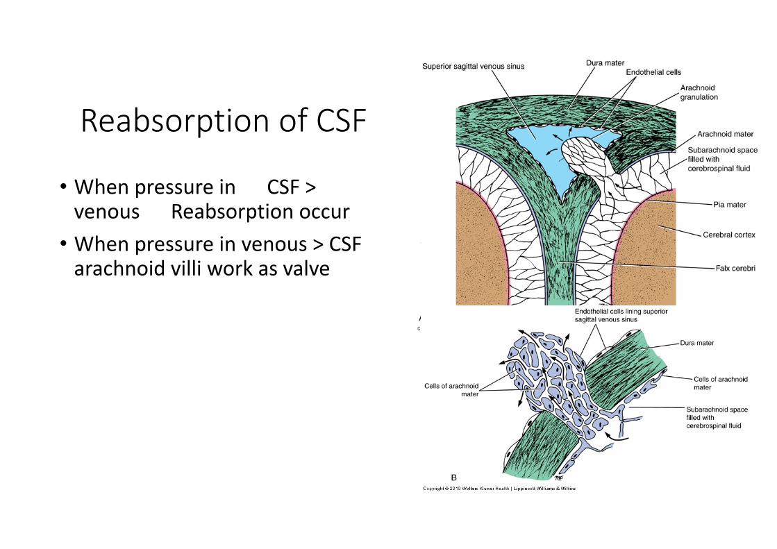
Reabsorption of CSF
• When pressure in CSF > venous Reabsorption occur
• When pressure in venous > CSF arachnoid villi work as valve
Hydrocephalus• Blockage of drainage of CSF (tumor, inflammation, developmental malformation, meningitis, hemorrhage or injury)
• Continued production cause an increase in pressure ‐‐‐ hydrocephalus
• In newborn or fetus, the fontanels allow this internal pressure to cause expansion of the skull and damage to the brain tissue
• Neurosurgeon implants a drain shunting the CSF to the veins of the neck or the abdomen
Leptomeningeal Disease
• Cancer metastasis through CSF• Originate from
• Primary CNS tumors• Secondary distant tumors through blood
• Symptoms may include headache, spine or radicular limb pain or sensory abnormalities, nausea and vomiting
Subarachnoid Hemorrhage
• Nontraumatic (spontaneous)• Blood in CSF• Mainly from aneurisms
• Arise at arterial branch points• 85% anterior circulation• 15% posterior circulation• 30% anterior comm., 25% posterior comm., 20% MCA
• The main symptom is a severe headache that starts suddenly (often called thunderclap headache)
• Traumatic• More common• Due to contusions and other traumatic injuries• Severe headache

Extensions of the Subarachnoid Space

Blood Brain Barrier
• protects cells from some toxins and pathogens• proteins & antibiotics can not pass but alcohol & anesthetics do
• Structure• tight junctions seal together epithelial cells
• continuous basement membrane• astrocyte processes covering capillaries

Blood Brain Barrier
• Areas without BBB• Area postrema in the floor of the fourth ventricle
• Areas in the hypothalamus
• Structure• Endothelial fenestrations

Blood Cerebrospinal Fluid Barrier
• Structure• Endothelial cells• BM of endothelial cells• Pale cells• BM of choroidal epithelial cells• Tight junctions seal the choroidalepithelial cells
Top Related