


Languages
Pages
Legal


43 Bile Duct Narrowing or Obstruction

CLINICAL IMAGAGINGAN ATLAS OF DIFFERENTIAL DAIGNOSIS
EISENBERG
DR. Muhammad Bin Zulfiqar PGR-FCPS III SIMS/SHL

• Fig GI 43-1 Cholangiocarcinoma causing severe narrowing of a long segment of the common hepatic duct (arrows).

• Fig GI 43-2 Klatskin tumor. Sclerosing cholangiocarcinomas arising at the junction of the right and left hepatic ducts (arrow).

• Fig GI 43-3 Ampullary carcinoma. Abrupt occlusion (arrow) of the distal common bile duct.

• Fig GI 43-4 Carcinoma of the head of the pancreas. Irregular narrowing of the common bile duct (arrows). The calcifications reflect underlying chronic pancreatitis.

• Fig GI 43-5 Extrinsic obstruction of the common bile duct (arrow) due to nodal metastases from carcinoma of the colon.

• Fig GI 43-6 Granular cell tumor. Focal stricture in the distal common hepatic duct (arrow).53

• Fig GI 43-7 Primary sclerosing cholangitis in a patient with chronic ulcerative colitis.
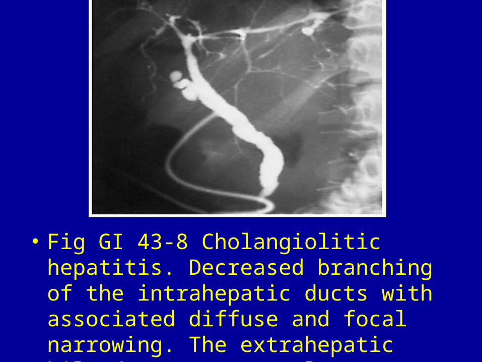
• Fig GI 43-8 Cholangiolitic hepatitis. Decreased branching of the intrahepatic ducts with associated diffuse and focal narrowing. The extrahepatic bile ducts are normal.54

• Fig GI 43-9 Chronic pancreatitis. Note the abrupt transition between the encased pipe stem segment and the dilated suprapancreatic portion of the common bile duct (arrow). Calcification suggestive of chronic pancreatitis can also be seen.

• Fig GI 43-10 Benign stricture of the common bile duct (arrow) related to previous biliary tract surgery.

• Fig GI 43-11 Congenital membranous diaphragm (web) of the common bile duct (arrow).


Top Related