


Languages
Pages
Legal

14
Catabolism of Organic Compounds
I Fermentations 37314.1 Energetic and Redox
Considerations 37314.2 Lactic and Mixed-Acid
Fermentations 37414.3 Clostridial and Propionic Acid
Fermentations 37714.4 Fermentations Lacking Substrate-
Level Phosphorylation 37914.5 Syntrophy 381
II Anaerobic Respiration 38314.6 Anaerobic Respiration:
General Principles 38314.7 Nitrate Reduction and
Denitrification 38414.8 Sulfate and Sulfur Reduction 38614.9 Acetogenesis 388
14.10 Methanogenesis 39014.11 Proton Reduction 39414.12 Other Electron Acceptors 39514.13 Anoxic Hydrocarbon Oxidation
Linked to Anaerobic Respiration 397
III Aerobic ChemoorganotrophicProcesses 40014.14 Molecular Oxygen as a Reactant
and Aerobic Hydrocarbon Oxidation 400
14.15 Methylotrophy and Methanotrophy 401
14.16 Sugar and Polysaccharide Metabolism 403
14.17 Organic Acid Metabolism 40614.18 Lipid Metabolism 406
Methanogens produce naturalgas (methane, CH4) and areable to do so because theycontain a series of unusualcoenzymes, such as thegreen-fluorescing F420, thatparticipate in biochemicalreactions unique to theseorganisms.

aData from Thauer, R. K., K. Jungermann, and K. Decker, 1977. Energy conservation inchemotrophic anaerobic bacteria. Bacteriol. Rev. 41: 100–180.bThe DG09 values shown here are for “standard conditions,” which are not necessarilythose of cells. Including heat loss, the energy costs of making an ATP are more like 60 kJ than 32 kJ, and the energy of hydrolysis of the energy-rich compounds shownhere is thus likely higher. But for simplicity and comparative purposes, the values in thistable will be taken as the actual energy released per reaction.
CHAPTER 14 • Catabolism of Organic Compounds 373
UN
IT 5
Figure 14.1 The essentials of fermentation. The fermentation productis excreted from the cell, and only a relatively small amount of the originalorganic compound is used for biosynthesis.
Organiccompound
Energy-richcompound
Fermentationproduct
ExcretionUptake
Oxidizedcompound
ADP
NAD+
NADH
ATP
Substrate-levelphosphorylation
In Chapter 13 we considered phototrophy and chemolithotro-
phy, strategies for energy conservation that do not use organic
compounds as electron donors. In this chapter we focus on
organic compounds as electron donors and the many ways in
which chemoorganotrophic microorganisms conserve energy. A
major focus will be on anaerobic forms of metabolism, because
novel strategies for anaerobic growth are a hallmark of prokary-
otic diversity. We end the chapter with a consideration of the aer-
obic catabolism of key organic compounds, primarily monomers
released from the degradation of macromolecules.
I Fermentations
Two broad metabolic processes for the catabolism of organic
compounds are fermentation and respiration. These processes
differ fundamentally in terms of oxidation–reduction (redox)
considerations and mechanism of ATP synthesis. In respiration,
whether aerobic or anaerobic, exogenous electron acceptors are
required to accept electrons generated from the oxidation of elec-
tron donors. In fermentation, this is not the case. Thus in respira-
tion but not fermentation we will see a common theme of electron
transport and the generation of a proton motive force.
We begin our exploration of organic catabolism with fermen-
tations. Compared with respirations, fermentations are typically
energetically marginal. However, we will see that a little free
energy can go a long way and that bacterial fermentative diversity
is both extensive and innovative.
14.1 Energetic and Redox ConsiderationsMany microbial habitats are anoxic (oxygen-free). In such envi-
ronments, decomposition of organic material occurs anaerobi-
cally. If adequate supplies of electron acceptors such as sulfate
(SO42-), nitrate (NO3
-), ferric iron (Fe3+), and others to be con-
sidered later are unavailable in anoxic habitats, organic com-
pounds are catabolized by fermentation (Figure 14.1). In Chapter
4 we discussed some key fermentations that yield alcohol or lactic
acid as products by way of the glycolytic pathway. There we
emphasized how fermentations are internally balanced redox
processes in which the fermentable substrate becomes both oxi-
dized and reduced.
An organism faces two major problems when it catabolizes
organic compounds for the purpose of energy conservation: (1)
ATP synthesis, and (2) redox balance. In fermentations, with rare
exception ATP is synthesized by substrate-level phosphorylation.
This is the mechanism in which energy-rich phosphate bonds
from phosphorylated organic compounds are transferred directly
to ADP to form ATP ( Section 4.7). The second problem,
redox balance, is solved by the production and subsequent excre-
tion of fermentation products generated from the original sub-
strate (Figure 14.1).
Energy-Rich Compounds and Substrate-Level PhosphorylationEnergy can be conserved by substrate-level phosphorylation
from many different compounds. However, central to an under-
standing of substrate-level phosphorylation is the concept of
energy-rich compounds. These are organic compounds that con-
tain an energy-rich phosphate bond or a molecule of coenzyme
A; the hydrolysis of either of these is highly exergonic ( Figure
4.12). Table 14.1 lists some energy-rich intermediates formed
during biochemical processes. The hydrolysis of most of the
compounds listed can be coupled to ATP synthesis (DG09 =-31.8 kJ/mol). In other words, if an organism can form one of
Table 14.1 Energy-rich compounds involved in substrate-levelphosphorylationa
Compound
Free energy of hydrolysis, �G09(kJ/mol)b
Acetyl-CoA -35.7
Propionyl-CoA -35.6
Butyryl-CoA -35.6
Caproyl-CoA -35.6
Succinyl-CoA -35.1
Acetyl phosphate -44.8
Butyryl phosphate -44.8
1,3-Bisphosphoglycerate -51.9
Carbamyl phosphate -39.3
Phosphoenolpyruvate -51.6
Adenosine phosphosulfate (APS) -88
N10-Formyltetrahydrofolate -23.4
Energy of hydrolysis of ATP (ATP S ADP + Pi) -31.8

UNIT 5 • Metabolic Diversity and Commercial Biocatalyses374
these compounds during fermentative metabolism, it can make
ATP by substrate-level phosphorylation.
Redox Balance, H2, and Acetate ProductionIn any fermentation there must be atomic and redox balance; the
total number of each type of atom and electrons in the products
of the reaction must balance those in the substrates. This is
obtained by the production and excretion from the cell of fer-
mentation products (Figure 14.1). In several fermentations,
redox balance is maintained by the production of molecular
hydrogen, H2. The production of H2 is associated with the activ-
ity of the iron–sulfur protein ferredoxin, a very low-potential
electron carrier, and is catalyzed by the enzyme hydrogenase, as
illustrated in Figure 14.2. H2 can also be produced from the C1
fatty acid formate. Either way, the H2 is then made available for
use by other organisms.
Many anaerobic bacteria produce acetate as a major or minor
fermentation product. The production of acetate and certain
other fatty acids (Table 14.1) is energy conserving because it
allows the organism to make ATP by substrate-level phosphory-
lation. The key intermediate generated in acetate production is
acetyl-CoA (Table 14.1), an energy-rich compound. Acetyl-CoA
can be converted to acetyl phosphate (Table 14.1) and the phos-
phate group of acetyl phosphate subsequently transferred to
ADP, yielding ATP. One of the precursors of acetyl-CoA is pyru-
vate, a major product of glycolysis. The conversion of pyruvate to
acetyl-CoA is a key oxidation reaction, and the electrons gener-
ated are used to form fermentation products or are released as
H2 (Figure 14.2).
MiniQuiz• What is substrate-level phosphorylation?
• Why is acetate formation in fermentation energeticallybeneficial?
14.2 Lactic and Mixed-AcidFermentations
Many fermentations are classified by either the substrate fer-
mented or the products formed. Table 14.2 lists some of the
major fermentations classified on the basis of products formed.
Note some of the broad categories, such as alcohol, lactic acid,
propionic acid, mixed acid, butyric acid, and acetogenic. Some
fermentations are described by the substrate fermented rather
than the fermentation product. For instance, some endospore-
forming anaerobic bacteria (genus Clostridium) ferment amino
acids, whereas others ferment purines and pyrimidines. Other
anaerobes ferment aromatic compounds (Table 14.3). Clearly, a
wide variety of organic compounds can be fermented. Certain
fermentations are carried out by only a very restricted group
of anaerobes; in some cases this may be only a single known
Figure 14.2 Production of H2 and acetate from pyruvate. At leasttwo mechanisms are known, one that produces H2 directly and the otherthat makes formate as an intermediate. When acetate is produced, ATPsynthesis is possible (Table 14.1).
H2
2 e–
Acetate(C2)
Acetyl~P
ATP
ADPHydrogenase
Ferredoxin
Pi
Acetyl-CoA + CO2
CO2
Acetyl-CoA + Formate
CoACoA
+Acetate(C2)
ATP+
2 H+
Pyruvate (C3)
Formatehydrogenlyase
Table 14.2 Common bacterial fermentations and some of the organisms carrying them out
Type Reaction Organisms
Alcoholic Hexose S 2 ethanol + 2 CO2 Yeast, Zymomonas
Homolactic Hexose S 2 lactate- + 2 H+ Streptococcus, some Lactobacillus
Heterolactic Hexose S lactate- + ethanol + CO2 + H+ Leuconostoc, some Lactobacillus
Propionic acid 3 Lactate- S 2 propionate- + acetate- + CO2 + H2O Propionibacterium, Clostridium propionicum
Mixed acida,b Hexose S ethanol + 2,3-butanediol + succinate2- + lactate- +acetate- + formate- + H2 + CO2
aNot all organisms produce all products. In particular, butanediol production is limited to only certain enteric bacteria. Reaction notbalanced.bStoichiometry shows major products. Other products include some acetate and a small amount of ethanol (butanol fermention only).
Butyric acidb Hexose S butyrate- + 2 H2 + 2 CO2 + H+ Clostridium butyricum
Butanolb 2 Hexose S butanol + acetone + 5 CO2 + 4 H2 Clostridium acetobutylicum
Caproate/Butyrate 6 Ethanol + 3 acetate- S 3 butyrate- + caproate- + 2 H2 + 4 H2O + H+ Clostridium kluyveri
Acetogenic Fructose S 3 acetate- + 3 H+ Clostridium aceticum
Enteric bacteria including Escherichia, Salmonella, Shigella, Klebsiella, Enterobacter

CHAPTER 14 • Catabolism of Organic Compounds 375
bacterium. A few examples are listed in Table 14.3. Many of these
bacteria can be considered metabolic specialists, having evolved
the capacity to catabolize a substrate not catabolized by other
bacteria.
We begin with two very common fermentations of sugars in
which lactic acid is a major product.
Lactic Acid FermentationThe lactic acid bacteria are gram-positive organisms that pro-
duce lactic acid as a major or sole fermentation product (
Section 18.1). Two fermentative patterns are observed. One,
called homofermentative, yields a single fermentation product,
lactic acid. The other, called heterofermentative, yields prod-
ucts in addition to lactate, mainly ethanol plus CO2.
Figure 14.3 summarizes pathways for the fermentation of glu-
cose by homofermentative and heterofermentative lactic acid
bacteria. The differences observed can be traced to the presence
or absence of the enzyme aldolase, a key enzyme of glycolysis
( Figure 4.14). Homofermentative lactic acid bacteria contain
aldolase and produce two molecules of lactate from glucose by
the glycolytic pathway (Figure 14.3a). Heterofermenters lack
aldolase and thus cannot break down fructose bisphosphate to
triose phosphate. Instead, they oxidize glucose 6-phosphate to 6-
phosphogluconate and then decarboxylate this to pentose phos-
phate. The pentose phosphate is then converted to triose
phosphate and acetyl phosphate by the key enzyme phos-
phoketolase (Figure 14.3b). The early steps in catabolism by het-
erofermentative lactic acid bacteria are those of the pentose
phosphate pathway (see Figure 14.38).
In heterofermenters, triose phosphate is converted to lactic
acid with the production of ATP (Figure 14.3). However, to
achieve redox balance the acetyl phosphate produced is used as
an electron acceptor and is reduced by NADH (generated dur-
ing the production of pentose phosphate) to ethanol. This
occurs without ATP synthesis because the energy-rich CoA
bond is lost during ethanol formation. Because of this, hetero-
fermenters produce only one ATP/glucose instead of the two
ATP/glucose produced by homofermenters. In addition, because
heterofermenters decarboxylate 6-phosphogluconate, they pro-
duce CO2 as a fermentation product; homofermenters do not
produce CO2. Thus a simple way of differentiating a homofer-
menter from a heterofermenter is to observe for the production
of CO2 in laboratory cultures.
Entner–Doudoroff PathwayA variant of the glycolytic pathway, called the Entner–Doudoroff
pathway, is widely distributed in bacteria, especially among
species of the pseudomonad group. In this pathway glucose
6-phosphate is oxidized to 6-phosphogluconic acid and NADPH;
the 6-phosphogluconic acid is dehydrated and split into pyruvate
and glyceraldehyde 3-phosphate (G-3-P), a key intermediate of
the glycolytic pathway. G-3-P is then catabolized as in glycolysis,
generating NADH and two ATP, and used as an electron acceptor
to balance redox reactions (Figure 14.3a).
Because pyruvate is formed directly in the Entner–Doudoroff
pathway and cannot yield ATP as can G-3-P (Figure 14.3), the
Entner–Doudoroff pathway yields only half the ATP of the gly-
colytic pathway. Organisms using the Entner–Doudoroff path-
way therefore share this physiological characteristic with
heterofermentative lactic acid bacteria that also use a variant of
the glycolytic pathway (Figure 14.3b). Zymomonas, an obligately
fermentative pseudomonad, and Pseudomonas, a nonfermentative
respiratory bacterium, are major genera that employ the Entner–
Doudoroff pathway ( Section 17.7).
Mixed-Acid FermentationsIn mixed-acid fermentations, characteristic of enteric bacteria
( Section 17.11), three different acids are formed from the fer-
mentation of glucose or other sugars—acetic, lactic, and succinic.
Ethanol, CO2, and H2 are also formed. Glycolysis is the pathway
used by mixed-acid fermenters, such as Escherichia coli, and we
outlined the steps in that pathway in Figure 4.14.
UN
IT 5
Table 14.3 Some unusual bacterial fermentations
Type Reaction Organisms
Acetylene 2 C2H2 + 3 H2O S ethanol + acetate- + H+ Pelobacter acetylenicus
Glycerol 4 Glycerol + 2 HCO3- S 7 acetate- + 5 H+ + 4 H2O Acetobacterium spp.
Resorcinol (aromatic) 2 C6H4(OH)2 + 6 H2O S 4 acetate- + butyrate- + 5 H+ Clostridium spp.
Phloroglucinol (aromatic) C6H6O3 + 3 H2O S 3 acetate- + 3 H+ Pelobacter massiliensisPelobacter acidigallici
Putrescine 10 C4H12N2 + 26 H2O S 6 acetate- + 7 butyrate- + 20 NH4+
+ 16 H2 + 13 H+Unclassified gram-positive nonsporulating anaerobes
Citrate Citrate3- + 2 H2O S formate- + 2 acetate- + HCO3- + H+ Bacteroides spp.
Aconitate Aconitate3- + H+ + 2 H2O S 2 CO2 + 2 acetate- + H2 Acidaminococcus fermentans
Glyoxylate 4 Glyoxylate- + 3 H+ + 3 H2O S 6 CO2 + 5 H2 + glycolate- Unclassified gram-negative bacterium
Benzoate 2 Benzoate- S cyclohexane carboxylate- + 3 acetate-
+ HCO3- + 3 H+
Syntrophus aciditrophicus

UNIT 5 • Metabolic Diversity and Commercial Biocatalyses376
Some enteric bacteria produce acidic products in lower
amounts than E. coli and balance redox in their fermentations by
producing larger amounts of neutral products. One key neutral
product is the four-carbon alcohol butanediol. In this variation of
the mixed-acid fermentation, butanediol, ethanol, CO2, and H2
are the main products observed (Figure 14.4). In the mixed-acid
fermentation of E. coli, equal amounts of CO2 and H2 are pro-
duced, whereas in a butanediol fermentation, considerably more
CO2 than H2 is produced. This is because mixed-acid fermenters
produce CO2 only from formic acid by means of the enzyme for-
mate hydrogenlyase (Figure 14.2):
HCOOH S H2 + CO2
By contrast, butanediol producers, such as Enterobacter aero-
genes, produce CO2 and H2 from formic acid but also produce
two additional molecules of CO2 during the formation of each
molecule of butanediol (Figure 14.4).
Because they produce fewer acidic products, butanediol fer-
menters do not acidify their environment as much as mixed-acid
fermenters do, and this is presumably a reflection of differences
in acid tolerance in the two groups that have significance for their
competitive success in nature.
MiniQuiz• How can homo- and heterofermentative lactic acid bacteria be
differentiated in pure cultures?
• Butanediol production leads to greater ethanol production thanin the mixed-acid fermentation of Escherichia coli. Why?
Figure 14.3 The fermentation of glucose in (a) homofermentative and (b) heterofermentative lactic
acid bacteria. Note that no ATP is made in reactions leading to ethanol formation in heterofermentativeorganisms.
Glucose Fructose 1,6-bisphosphate
Aldolase 2 Glyceraldehyde3-phosphate (G3-P)
Dihydroxyacetonephosphate
Pi
2 Pi
Pi
2 NADH 2 NAD+
2 1,3-Bisphospho-glyceric acid
Glucose 2 lactate– + 2H+ ΔG0′= –196 kJ(C6H12O6) 2(C3H5O3
–) (2 ATP)
2 G3-P
Glucose 6-phosphate
Glycer-aldehyde3-P
Phospho-ketolase
Acetylphosphate
6-Phosphogluconicacid
Ribulose5-phosphate + CO2
Xylulose5-phosphate
Acetaldehyde
Ethanol
2 Lactate
ADPATP
Glucose
ADPATP
ADPATP
2 ADP 2 ATP 2 ADP 2 ATP
2 Pyruvate
NADH NAD+
1,3-Bisphospho-glyceric acid
Glucose lactate– + ethanol + CO2 + H+ ΔG0′= –216 kJ(C6H12O6) (C3H5O3
–) (C2H5OH) (1 ATP)
Lactate
ADP ATPADP ATP
Pyruvate–
NADH NADH
NAD+
NAD+
(a) Homofermentative
(b) Heterofermentative

CHAPTER 14 • Catabolism of Organic Compounds 377
UN
IT 5
14.3 Clostridial and Propionic AcidFermentations
Species of the genus Clostridium are classical fermentative anaer-
obes ( Section 18.2). Different clostridia ferment sugars,
amino acids, purines and pyrimidines, and a few other com-
pounds. In all cases ATP synthesis is linked to substrate-level
phosphorylations either in the glycolytic pathway or from the
hydrolysis of a CoA intermediate (Table 14.1). We begin with
sugar-fermenting (saccharolytic) clostridia.
Sugar Fermentation by Clostridium SpeciesA number of clostridia ferment sugars, producing butyric acid as
a major end product. Some species also produce the neutral
products acetone and butanol, and Clostridium acetobutylicum is
a classic example of this. The biochemical steps in the formation
of butyric acid and neutral products from sugars are shown in
Figure 14.5.
Glucose is converted to pyruvate via the glycolytic pathway,
and pyruvate is split to yield acetyl-CoA, CO2, and H2 (through
ferredoxin) by the phosphoroclastic reaction (Figure 14.2). Some
of the acetyl-CoA is then reduced to butyrate or other fermenta-
tion products using NADH derived from glycolytic reactions as
electron donor. The actual products observed are influenced by
the duration and the conditions of the fermentation. During the
early stages of the butyric fermentation, butyrate and a small
amount of acetate are produced. But as the pH of the medium
drops, synthesis of acids ceases and acetone and butanol begin to
accumulate. However, if the pH of the medium is kept neutral by
buffering, there is very little formation of neutral products and
butyric acid production continues.
The accumulation of acidic products in the C. acetobutylicum
fermentation lowers the pH, and this triggers derepression of
genes responsible for solvent production. The production of
butanol is actually a consequence of the production of acetone.
For each acetone that is made, two NADH produced during gly-
colysis are not reoxidized as they would be if butyrate were pro-
duced (Figure 14.5). Because redox balance is necessary for any
Figure 14.4 Butanediol production and mixed-acid fermentations. Note how only one NADH, but twomolecules of pyruvate, are used to make one butanediol. This leads to redox imbalance and the productionof more ethanol by butanediol producers than by mixed-acid fermenters.
CH3C H
OH
CH3C COO–
O
CH3C COO–
Thiamine pyrophosphate(TPP) H
CH3C TPP
O
CH3C COO–
+
OH
TPP
CH3
C O
OH
CH3C H
CH3
C O
OH
CH3
C OHHPyruvate
Pyruvate
α-Acetolactate Acetoin 2,3-Butanediol
Overall reaction:
2 Pyruvate + NADH 2 CO2 + butanediol
GlucoseGlycolysis
LactateFormateSuccinateEthanol
Butanediol route, e.g.,Enterobacteraerogenes
Mixed-acidroute, e.g.,Escherichia coli
NADHCO2
CO2
Figure 14.5 The butyric acid and butanol/acetone fermentation. Allfermentation products from glucose are shown in bold (dashed lines indi-cate minor products). Note how the production of acetate and butyratelead to additional ATP by substrate-level phosphorylation. By contrast,formation of butanol and acetone reduces the ATP yield because thebutyryl-CoA step is bypassed. 2 H, NADH; Fdred, reduced ferredoxin.
+ +
Glucose
(CH3 C
Glycolysis
Pyruvate
Phosphoroclastic reaction
CO2 Fdred H2
Acetate
ADPAcetyl P Acetyl-CoA
Pi
Acetaldehyde
2 H
Ethanol
Acetyl-CoA
Acetoacetyl-CoA Acetoacetate
2 H
C CoA)
β-Hydroxybutyryl-CoA
H2O
Crotonyl-CoA
2 H
Butyryl-CoA
Butyraldehyde
2 H
ADP
ButyrateCOO–)
2 H
(CH3 CH2 CH2
CH2
(CH3
CH3 CH3
CH2 CH2
CH2OH)Butanol
CO2
Acetone
C
Isopropanol
2 H
O O
O
2 H
ATP
ATP
butyrate + 2 CO2 + 2 H2 + H+
ΔG0′= −264 kJ (3 ATP/glucose)Glucose
acetone + butanol + 5 CO2 + 4 H2 ΔG0′= −468 kJ (2 ATP/glucose)
2 Glucose
UNIT 5 • Metabolic Diversity and Commercial Biocatalyses378
fermentation to proceed, the cell then uses butyrate as an elec-
tron acceptor. Butanol and acetone are therefore produced in
equal amounts. Although neutral product formation helps the
organism keep its environment from becoming too acidic, there
is an energetic price to pay for this. In producing butanol, the cell
loses the opportunity to convert butyryl-CoA to butyrate and
thus ATP (Figure 14.5 and Table 14.1).
Amino Acid Fermentation by ClostridiumSpecies and the Stickland ReactionSome Clostridium species ferment amino acids. These are the
“proteolytic” clostridia, organisms that degrade proteins released
from dead organisms in nature. Some clostridia ferment individ-
ual amino acids, typically glutamate, glycine, alanine, cysteine,
histidine, serine, or threonine. The biochemistry behind these
fermentations is quite complex, but the metabolic strategy is
quite simple. In virtually all cases, the amino acids are catabo-
lized in such a way as to yield a fatty acid–CoA derivative, typi-
cally acetyl (C2), butyryl (C4), or caproyl (C6). From these, ATP is
produced by substrate-level phosphorylation (Table 14.1). Other
products of amino acid fermentation include ammonia (NH3)
and CO2.
Some clostridia ferment only an amino acid pair. In this situ-
ation one amino acid functions as the electron donor and is oxi-
dized, whereas the other amino acid is the electron acceptor
and is reduced. This coupled amino acid fermentation is known
as a Stickland reaction. For instance, Clostridium sporogenes
catabolizes a mixture of glycine and alanine; in this reaction
alanine is the electron donor and glycine is the electron accep-
tor (Figure 14.6). Amino acids that can function as donors or
acceptors in Stickland reactions are listed in Figure 14.6. The
products of the Stickland reaction are NH3, CO2, and a car-
boxylic acid with one fewer carbons than the amino acid that
was oxidized (Figure 14.6).
Many of the products of amino acid fermentation by clostridia
are foul-smelling substances, and the odor that results from
putrefaction is mainly a result of clostridial activity. In addition to
fatty acids, other odoriferous compounds produced include
hydrogen sulfide (H2S), methylmercaptan (from sulfur amino
acids), cadaverine (from lysine), putrescine (from ornithine), and
NH3. Purines and pyrimidines, released from the degradation of
nucleic acids, lead to many of the same fermentation products
and yield ATP from the hydrolysis of fatty acid–CoA derivatives
(Table 14.1) produced in their respective fermentative pathway.
Clostridium kluyveri FermentationAnother species of Clostridium also ferments a mixture of sub-
strates in which one is the donor and one is the acceptor, as in the
Stickland reaction. However, this organism, C. kluyveri, ferments
not amino acids but instead ethanol plus acetate. In this fermen-
tation, ethanol is the electron donor and acetate is the electron
acceptor. The overall reaction is shown in Table 14.2.
The ATP yield in the caproate/butyrate fermentation is low,
1 ATP/6 ethanol fermented. However, C. kluyveri has a selective
advantage over all other fermenters in its apparently unique abil-
ity to oxidize a highly reduced fermentation product (ethanol)
and couple it to the reduction of another common fermentation
product (acetate), reducing it to longer-chain fatty acids. The sin-
gle ATP produced in this reaction comes from substrate-level
phosphorylation during conversion of a fatty acid–CoA formed
in the pathway to the free fatty acid. The fermentation of
C. kluyveri is an example of a secondary fermentation, which is
essentially a fermentation of fermentation products. We see
another example of this now.
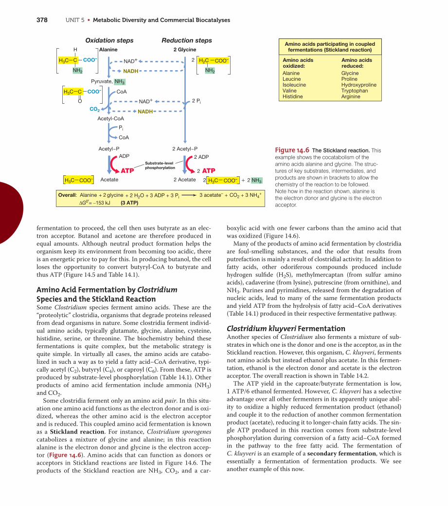
Figure 14.6 The Stickland reaction. Thisexample shows the cocatabolism of theamino acids alanine and glycine. The struc-tures of key substrates, intermediates, andproducts are shown in brackets to allow thechemistry of the reaction to be followed.Note how in the reaction shown, alanine isthe electron donor and glycine is the electronacceptor.
+
+ +
Oxidation steps Alanine
Acetyl~P 2 Acetyl~P
H
COO–
COO–
COO–
COO–
NH2 NH2
Pyruvate, NH3
C
CO2
Acetyl-CoA
O
CoA
Pi
CoA
Acetate
Reduction steps 2 Glycine
2 Acetate
2
ADP
NAD+
NADH
NAD+
NADH
2 2 NH3
Overall: Alanine + 2 glycine 3 acetate– CO2 3 NH4+
2 ADPSubstrate-levelphosphorylation
2 Pi
+ 2 H2O + 3 ADP + 3 Pi
H3C
H3C COO–H3C
H3C
H2CC Amino acidsoxidized:
Amino acidsreduced:
AlanineLeucineIsoleucineValineHistidine
GlycineProlineHydroxyprolineTryptophanArginine
Amino acids participating in coupledfermentations (Stickland reaction)
ATP 2 ATP
ΔG0′= −153 kJ (3 ATP)

CHAPTER 14 • Catabolism of Organic Compounds 379
UN
IT 5
Propionic Acid FermentationThe propionic acid bacterium Propionibacterium and some
related bacteria produce propionic acid as a major fermentation
product from either glucose or lactate. However, lactate, a fer-
mentation product of the lactic acid bacteria, is probably the
major substrate for propionic acid bacteria in nature, where these
two groups live in close association. Propionibacterium is an
important component in the ripening of Swiss (Emmentaler)
cheese, to which the propionic and acetic acids produced give the
unique bitter and nutty taste, and the CO2 produced forms bub-
bles that leave the characteristic holes (eyes) in the cheese.
Figure 14.7 shows the reactions leading from lactate to propi-
onate. When glucose is the starting substrate, it is first catabo-
lized to pyruvate by the glycolytic pathway. Then pyruvate,
produced either from glucose or from the oxidation of lactate, is
carboxylated to form methylmalonyl-CoA, leading to the forma-
tion of oxalacetate and, eventually, propionyl-CoA (Figure 14.7).
The latter reacts with succinate in a step catalyzed by the enzyme
CoA transferase, producing succinyl-CoA and propionate. This
results in a lost opportunity for ATP production from propionyl-
CoA but avoids the energetic costs of having to activate succinate
with ATP to form succinyl-CoA. The succinyl-CoA is then isomer-
ized to methylmalonyl-CoA and the cycle is complete; propionate
is formed and CO2 regenerated (Figure 14.7).
NADH is oxidized in the steps between oxalacetate and succi-
nate. Notably, the reaction in which fumarate is reduced to succi-
nate is linked to electron transport and the formation of a proton
motive force that yields ATP by oxidative phosphorylation. The
propionate pathway also converts some lactate to acetate plus
CO2, which allows for additional ATP to be made (Figure 14.7).
Thus, in the propionate fermentation both substrate-level and
oxidative phosphorylation occur.
Propionate is also formed in the fermentation of succinate by
the bacterium Propionigenium, but by a completely different
mechanism than that described here for Propionibacterium. Pro-
pionigenium, to be considered next, is phylogenetically and ecolog-
ically unrelated to Propionibacterium, but energetic aspects of its
metabolism are of considerable interest from the standpoint of
bioenergetics.
MiniQuiz• Compare the mechanisms for energy conservation in Clostridium
acetobutylicum and Propionibacterium.
• What are the substrates for the Clostridium kluyveri fermentation?In nature, where do these come from?
14.4 Fermentations Lacking Substrate-Level Phosphorylation
Certain fermentations yield insufficient energy to synthesize ATP
by substrate-level phosphorylation (that is, less than -32 kJ,
Table 14.1), yet still support growth. In these cases, catabolism of
the compound is linked to ion pumps that establish a proton
motive force or sodium motive force across the cytoplasmic
membrane. Examples of this include fermentation of succinate by
Propionigenium modestum and the fermentation of oxalate by
Oxalobacter formigenes.
Propionigenium modestumPropionigenium modestum was first isolated in anoxic enrich-
ment cultures lacking alternative electron acceptors and fed suc-
cinate as an electron donor. Propionigenium inhabits marine and
freshwater sediments, and can also be isolated from the human
oral cavity. The organism is a gram-negative short rod and, phy-
logenetically, is a species of Actinobacteria ( Section 18.4).
During studies of the physiology of P. modestum, it was shown to
require sodium chloride (NaCl) for growth and to catabolize suc-
cinate under strictly anoxic conditions:
Succinate2- + H2O S propionate- + HCO3-
DG09 = -20.5 kJ
This decarboxylation releases insufficient free energy to support
ATP synthesis by substrate-level phosphorylation (Table 14.1)
but sufficient free energy to pump a sodium ion (Na+) across the
cytoplasmic membrane from the cytoplasm to the periplasm.
Energy conservation in Propionigenium is then linked to the
sodium motive force that develops from Na+ pumping; a sodium-
translocating ATPase exists that uses the sodium motive force to
drive ATP synthesis (Figure 14.8a).
In a related decarboxylation reaction, the bacterium Malo-
nomonas, a species of Deltaproteobacteria, decarboxylates the C3
dicarboxylic acid malonate, forming acetate plus CO2. As for
Figure 14.7 The propionic acid fermentation of Propionibacterium.
Products are shown in bold. The four NADH made from the oxidation ofthree lactate are reoxidized in the reduction of oxalacetate and fumarate,and the CoA group from propionyl-CoA is exchanged with succinate dur-ing the formation of propionate.
Overall: 3 Lactate 2 propionate + acetate + CO2 + H2O
(3 ATP)
3 Lactate
3 Pyruvate
2 Propionate
Acetate + CO2
3 NADH
NADH
ADP
ADP2 CO2
2 CO2
2 Oxalacetate
2 Malate
2 H2O
2 NADH
2 NADH
2 Fumarate
2 Succinate
2 Succinyl~CoA 2 Methylmalonyl~CoA
2 Propionyl~CoA
CoA transfer
2 ATP
ATP
ΔG0′= –171 kJ

UNIT 5 • Metabolic Diversity and Commercial Biocatalyses380
Propionigenium, energy metabolism in Malonomonas is linked to
a sodium pump and sodium-driven ATPase. However, the mech-
anism of malonate decarboxylation is more complex than that of
Propionigenium and involves many additional proteins. Interest-
ingly, however, the energy yield of malonate fermentation by
Malonomonas is even lower than that of P. modestum, -17.4 kJ.
Sporomusa, an endospore-forming bacterium ( Section 18.2)
and also an acetogen (Section 14.9), is also capable of fermenting
malonate, as are a few other Bacteria.
Oxalobacter formigenesOxalobacter formigenes is a bacterium present in the intestinal
tract of animals, including humans. It catabolizes the C2 dicar-
boxylic acid oxalate, producing formate plus CO2. Oxalate
degradation by O. formigenes is thought to be important in
humans for preventing the accumulation of oxalate in the body,
a substance that can accumulate to form calcium oxalate kidney
stones. O. formigenes is a gram-negative strict anaerobe that is a
species of Betaproteobacteria. O. formigenes carries out the fol-
lowing reaction:
Oxalate2- + H2O S formate- + HCO3- DG09 = -26.7 kJ
As in the catabolism of succinate by P. modestum, insufficient
energy is available from this reaction to drive ATP synthesis by
substrate-level phosphorylation (Table 14.1). However, the reac-
tion supports growth of the organism because the decarboxyla-
tion of oxalate is exergonic and forms formate, which is excreted
from the cell. The internal consumption of protons during the
oxidation of oxalate and production of formate is, in effect, a pro-
ton pump. That is, a divalent molecule (oxalate) enters the cell
while a univalent molecule (formate) is excreted. The continued
exchange of oxalate for formate establishes a membrane potential
that is coupled to ATP synthesis by the proton-translocating
ATPase in the membrane (Figure 14.8b).
Energetics LessonsThe unique aspect of all of these decarboxylation-type fermenta-
tions is that ATP synthesis occurs without substrate-level phos-
phorylation or electron transport. Nevertheless, ATP synthesis
can occur because the small amount of energy released is cou-
pled to the pumping of an ion across the cytoplasmic membrane.
Organisms such as Propionigenium or Oxalobacter thus teach us
an important lesson in microbial bioenergetics: Energy conserva-
tion from reactions that yield less than -32 kJ is still possible if
the reaction is coupled to an ion pump. However, a minimal
requirement for an energy-conserving reaction is that it must
yield sufficient free energy to pump a single ion. This is estimated
to be about -12 kJ. Theoretically, reactions that release less
energy than this should not be able to drive ion pumps and
should therefore not be potential energy-conserving reactions.
However, as we will see in the next section, there are bacteria
known that push this theoretical limit even lower and whose
energetics are still incompletely understood. These are the syn-
trophs, prokaryotes living on the energetic “edge of existence.”
MiniQuiz• Why does Propionigenium modestum require sodium for growth?
• Of what benefit is the organism Oxalobacter to human health?
Figure 14.8 The unique fermentations of succinate and oxalate. (a) Succinate fermentation byPropionigenium modestum. Sodium export is linked to the energy released by succinate decarboxylation,and a sodium-translocating ATPase produces ATP. (b) Oxalate fermentation by Oxalobacter formigenes.Oxalate import and formate export by a formate–oxalate antiporter consume cytoplasmic protons. ATPsynthesis is linked to a proton-driven ATPase. All substrates and products are shown in bold.
ADP+ Pi
ATP ATPADP+ Pi
Propionate–
Succinate2–
(a) (b)
Na+
ATPaseH+
ATPase
Sodium-extrudingdecarboxylase
H2O
Oxalate2–
Succinate2–
Oxalate2–
Formate–
Formate–
Na+
Na+
Na+ H+
H+
Na+ Formate–oxalateantiporter
C—CC—CH2—CH2—C
–O
C—CH2—CH3
–O
O
–O
O
O
HCO3–
O–O–
OO
HCO3–
C
O
–O H
3–4 3–4

CHAPTER 14 • Catabolism of Organic Compounds 381
UN
IT 5
14.5 SyntrophyThere are many examples in microbiology of syntrophy, a meta-
bolic process in which two different organisms cooperate to
degrade a substance—and conserve energy doing it—that neither
can degrade alone. Most syntrophic reactions are secondary
fermentations (Section 14.3) in which organisms ferment the fer-
mentation products of other anaerobes. We will see in Section
24.2 how syntrophy is a key to the overall success of anoxic catab-
olism that leads to the production of methane (CH4). Here we
consider the microbiology and energetic aspects of syntrophy.
Table 14.4 lists some of the major groups of syntrophs and the
compounds they degrade. Many organic compounds can be
degraded syntrophically, including even aromatic and aliphatic
hydrocarbons. But the major compounds of interest in freshwa-
ter syntrophic environments are fatty acids and alcohols.
Hydrogen Consumption in Syntrophic ReactionsThe heart of syntrophic reactions is interspecies H2 transfer, H2
production by one partner linked to H2 consumption by the other.
The H2 consumer can be any one of a number of physiologically
distinct organisms: denitrifying bacteria, ferric iron–reducing
bacteria, sulfate-reducing bacteria, acetogens, or methanogens,
groups we will consider later in this chapter. Consider ethanol
fermentation to acetate plus H2 by a syntroph coupled to the pro-
duction of methane (Figure 14.9). As can be seen, the syntroph
carries out a reaction whose standard free-energy change (DG09) ispositive. However, the H2 produced by the syntroph can be used as
an electron donor by a methanogen in an exergonic reaction.
When the two reactions are summed, the overall reaction is exer-
gonic (Figure 14.9), and the free energy released is shared by both
organisms.
Another example of syntrophy is the oxidation of a fatty acid
such as butyrate to acetate plus H2 by the fatty acid–oxidizing
syntroph Syntrophomonas (Figure 14.10):
Butyrate- + 2 H2O S 2 acetate- + H+ + 2 H2
DG09 = +48.2 kJ
The free-energy change of this reaction is highly unfavorable, and
in pure culture Syntrophomonas will not grow on butyrate. How-
ever, if the H2 produced by Syntrophomonas is consumed by a
partner organism, Syntrophomonas grows on butyrate in cocul-
ture with the H2 consumer. Why is this so?
Energetics of H2 TransferBecause it is such a powerful electron donor for anaerobic respi-
rations, H2 is quickly consumed in anoxic habitats. In a syn-
trophic relationship, the removal of H2 by a partner organism
pulls the reaction in the direction of product formation and
thereby affects the energetics of the reaction. A review of the
principles of free energy given in Appendix 1 indicates that the
concentration of reactants and products in a reaction can have a
major effect on energetics. This is usually not an issue for most
fermentation products because they are not consumed to
extremely low levels. H2, by contrast, can be consumed to nearly
undetectable levels, and at these tiny concentrations, the energet-
ics of the reactions are dramatically affected.
For convenience, the DG09 of a reaction is calculated on the
basis of standard conditions—one molar concentration of
Table 14.4 Properties of major syntrophic bacteriaa
aAll syntrophs are obligate anaerobes.bSee Chapters 17 and 18.cNot all species can use all substrates listed.
Genus
Numberof knownspecies Phylogenyb
Substrates fermentedin coculturec
Syntrophobacter 4 Deltaproteobacteria Propionate (C3), lactate; somealcohols
Syntrophomonas 9 Firmicutes C4–C18 saturated/ unsaturated fattyacids; somealcohols
Pelotomaculum 2 Firmicutes Propionate, lactate, several alcohols;some aromaticcompounds
Syntrophus 3 Deltaproteobacteria Benzoate and severalrelated aromaticcompounds; somefatty acids andalcohols
Figure 14.9 Syntrophy: Interspecies H2 transfer. Shown is thefermentation of ethanol to methane and acetate by syntrophic associationof an ethanol-oxidizing syntroph and a H2-consuming partner (in thiscase, a methanogen). (a) Reactions involved. The two organisms sharethe energy released in the coupled reaction. (b) Nature of the syntrophictransfer of H2.
Ethanol fermentation:
Methanogenesis:
4 H2 + CO2 CH4 + 2 H2O
Coupled reaction:
2 CH3CH2OH + CO2 CH4 + 2 CH3COO– + 2 H+
Ethanol fermenter Methanogen
2 Acetate CH4
4 H2
2 CH3CH2OH + 2 H2O 4 H2 + 2 CH3COO– + 2 H+
ΔG0′= +19.4 kJ/reaction
ΔG0′= –130.7 kJ/reaction
ΔG0′= –111.3 kJ/reaction
Interspecies hydrogen transfer CO2
(a) Reactions
(b) Syntrophic transfer of H2
2 Ethanol

UNIT 5 • Metabolic Diversity and Commercial Biocatalyses382
Figure 14.10 Energetics of growth of Syntrophomonas in
syntrophic culture and in pure culture. (a) In syntrophic culture,growth requires a H2-consuming organism, such as a methanogen. H2 production is driven by reverse electron flow because the E09 of theFADH and NADH couples are more electropositive than that of 2 H+/H2.(b) In pure culture, energy conservation is linked to anaerobic respira-tion with crotonate reduction to butyrate. Inset: photomicrograph ofFISH-stained cells ( Section 16.9) of a fatty acid-degrading syn-trophic bacterium in association with a methanogen.
−
~
~
~
~
~~
Butyryl S–CoA
Butyrate
Crotonyl S–CoA
Acetoacetyl S–CoA
CoAtransfer
CoA
FADH H2
H2NADH
Acetate
Acetyl S–CoA
Acetyl~P Acetate ATP+
3-Hydroxybutyryl S–CoA
(a) Syntrophic culture
(b) Pure culture
2 acetate– + Sum: 2 Crotonate– + H2O butyrate– + H+ ΔG0′ = –340 kJ
2 acetate– + H++ 2 H2 ΔG0′= +48.2 kJ
Sum: Butyrate— + H2O
Acetyl S–CoA
Syntrophs
H.J
.M. H
arm
sen
1. Crotonate oxidation:
2. Crotonate reduction:
O–CH3HC CH + H2OC
O
2 acetate
+ H2 + H+
O–CH3HC CH + H2C
O
butyrate
Proton motive force
(ΔG = –18 kJ)
Energetics in SyntrophsEnergy conservation in syntrophs is probably based on both
substrate-level and oxidative phosphorylations. From biochemical
studies of syntrophs, substrate-level phosphorylation has been
shown to occur during the conversion of acetyl-CoA (generated
by beta-oxidation of ethanol or the fatty acid) to acetate (Figure
14.10a), although the -18 kJ of energy released (DG) is in theory
insufficient for this. However, the energy released is sufficient to
produce a fraction of an ATP, so it is possible that two rounds of
butyrate oxidation (Figure 14.10a) are necessary to couple to the
production of one ATP by substrate-level phosphorylation.
Besides the syntrophic lifestyle, many syntrophs can carry out
anaerobic respirations (Section 14.6) in pure culture by the dis-
proportionation of unsaturated fatty acids (disproportionation is
a process in which some molecules of a substrate are oxidized
while some are reduced). For example, crotonate, an intermedi-
ate in syntrophic butyrate metabolism (Figure 14.10a), supports
growth of Syntrophomonas. Under these conditions some of the
crotonate is oxidized to acetate and some is reduced to butyrate
(Figure 14.10b). Because crotonate reduction by Syntrophomonas
is coupled to the formation of a proton motive force, as occurs in
other anaerobic respirations that employ organic electron accep-
tors (such as fumarate reduction to succinate, Section 14.12), it is
possible that some step or steps in syntrophic metabolism
(Figure 14.10a) generate a proton motive force as well. Pumping
protons or some other ion would almost certainly be required for
benzoate- and propionate-fermenting syntrophs (Figure 14.10a
inset) whose energy yield (DG) is only about -5 kJ or so.
Regardless of how ATP is made during syntrophic growth, an
additional energetic burden occurs in syntrophy. During syn-
trophic metabolism, syntrophs produce H2 (E09 -0.42 V) from
more electropositive electron donors such as FADH (E09 -0.22 V)
and NADH (E09 -0.32 V), generated during fatty acid oxidation
reactions (Figure 14.10a); this cannot occur without an energy
input. Thus, some fraction of the ATP generated by Syn-
trophomonas during syntrophic growth must be consumed to
drive reverse electron flow reactions ( Section 13.4), yielding
H2 for the H2 consumer. When this energy drain is coupled to the
inherently poor energetic yields of syntrophic reactions, it is clear
that syntrophic bacteria are somehow making a living on a
severely marginal energy economy. Even today syntrophs pose a
significant challenge to our understanding of the minimal
requirements for energy conservation in bacteria.
Ecology of SyntrophsEcologically, syntrophic bacteria are key links in the anoxic por-
tions of the carbon cycle. Syntrophs consume highly reduced fer-
mentation products and release a key product for anaerobic H2
consumers. Without syntrophs, a bottleneck would develop in
anoxic environments in which electron acceptors other than CO2
were limiting ( Section 24.2). By contrast, when conditions
are oxic or alternative electron acceptors are abundant, syn-
trophic relationships are unnecessary. For example, if O2 or
NO3- is available as an electron acceptor, the energetics of the
fermentation of a fatty acid or an alcohol is so favorable that syn-
trophic relationships are unnecessary. Thus, syntrophy is charac-
products and reactants. By contrast, the related term DG is cal-
culated on the basis of the actual concentrations of products and
reactants present (Appendix 1 explains how to calculate DG). At
very low levels of H2, the energetics of the oxidation of ethanol or
fatty acids to acetate plus H2, a reaction that is endergonic under
standard conditions, becomes exergonic. For example, if the con-
centration of H2 is kept extremely low from consumption by the
partner organism, DG for the oxidation of butyrate by
Syntrophomonas yields -18 kJ (Figure 14.10a). As we learned in
Section 14.4, this relatively low energy yield can still support
growth of a bacterium.
CHAPTER 14 • Catabolism of Organic Compounds 383
UN
IT 5
teristic of anoxic catabolism in which methanogenesis or aceto-
genesis are the terminal processes in the microbial ecosystem.
Methanogenesis is a major process in anoxic wastewater
biodegradation, and microbiological studies of sludge granules
that form in such systems have shown the close physical relation-
ship that develops between H2 producer and H2 consumer in
such habitats (Figure 14.10a inset).
MiniQuiz• Give an example of interspecies H2 transfer. Why can it be said
that both organisms benefit from this process?
• Predict how ATP is made during the syntrophic degradation ofethanol shown in Figure 14.9.
II Anaerobic Respiration
In the next several sections we survey the major forms of anaer-
obic respiration and see the many ways by which prokaryotes
can conserve energy under anoxic conditions using electron
acceptors other than oxygen (O2).
14.6 Anaerobic Respiration: General Principles
We examined the process of aerobic respiration in Chapter 4. As
we noted there, O2 functions as a terminal electron acceptor,
accepting electrons that have traveled through an electron trans-
port chain. However, we also noted that other electron acceptors
could be used instead of O2, in which case the process is called
anaerobic respiration ( Section 4.12). Here we consider
some of these processes.
Bacteria that carry out anaerobic respiration produce electron
transport chains containing cytochromes, quinones, iron–sulfur
proteins, and the other typical electron transport proteins that
we have seen in aerobic respiration ( Section 4.9) and in pho-
tosynthesis and chemolithotrophy (Chapter 13). In some organ-
isms, such as the denitrifying bacteria, which are for the most
part facultative aerobes ( Section 5.17), anaerobic respiration
competes with aerobic respiration. In such cases, if O2 is present,
the bacteria respire aerobically, and genes encoding proteins nec-
essary for anaerobic respiration are repressed. However, when O2
is depleted from the environment, the bacteria respire anaerobi-
cally, and the alternate electron acceptor is reduced. Many other
organisms that carry out anaerobic respiration are obligate
anaerobes and are unable to use O2.
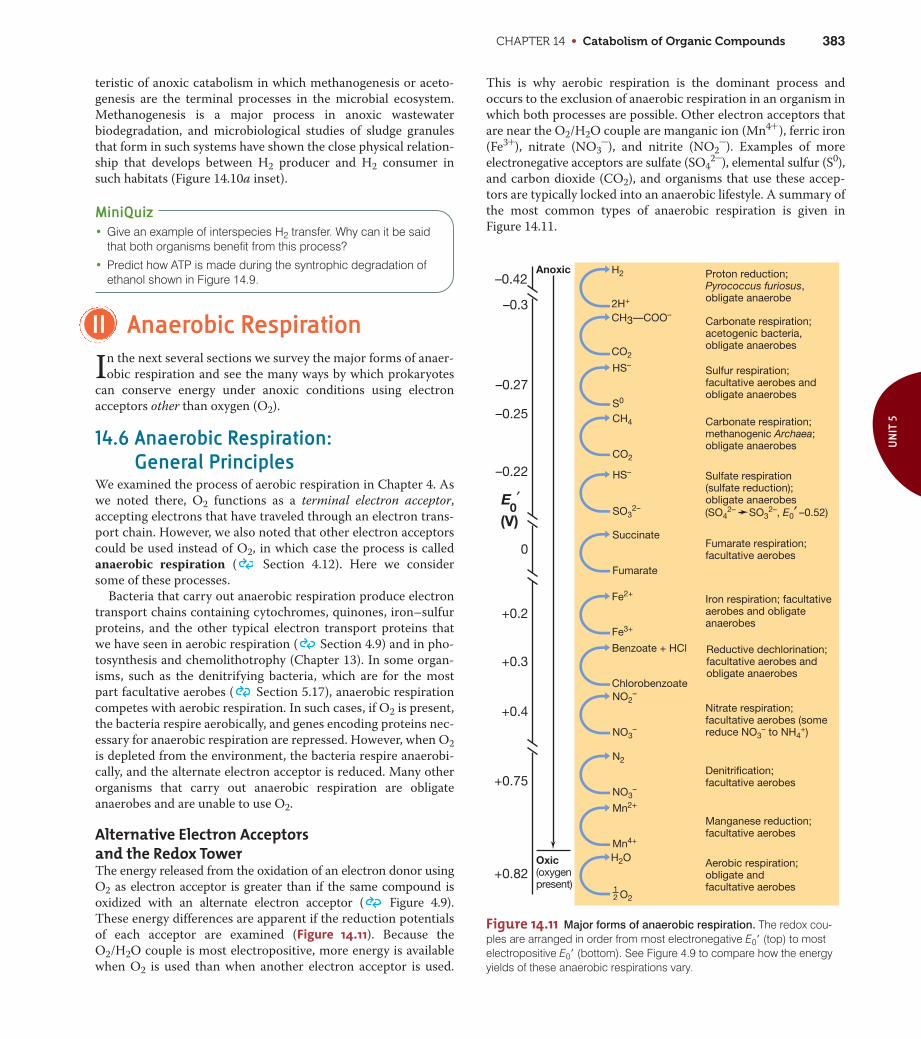
Alternative Electron Acceptors and the Redox TowerThe energy released from the oxidation of an electron donor using
O2 as electron acceptor is greater than if the same compound is
oxidized with an alternate electron acceptor ( Figure 4.9).
These energy differences are apparent if the reduction potentials
of each acceptor are examined (Figure 14.11). Because the
O2/H2O couple is most electropositive, more energy is available
when O2 is used than when another electron acceptor is used.
This is why aerobic respiration is the dominant process and
occurs to the exclusion of anaerobic respiration in an organism in
which both processes are possible. Other electron acceptors that
are near the O2/H2O couple are manganic ion (Mn4+ ), ferric iron
(Fe3+), nitrate (NO3-), and nitrite (NO2
-). Examples of more
electronegative acceptors are sulfate (SO42-), elemental sulfur (S0),
and carbon dioxide (CO2), and organisms that use these accep-
tors are typically locked into an anaerobic lifestyle. A summary of
the most common types of anaerobic respiration is given in
Figure 14.11.
Sulfur respiration;facultative aerobes andobligate anaerobes
Carbonate respiration;methanogenic Archaea;obligate anaerobes
Sulfate respiration(sulfate reduction);obligate anaerobes
Fumarate respiration;facultative aerobes
Nitrate respiration;facultative aerobes (somereduce NO3
– to NH4+)
Aerobic respiration;obligate andfacultative aerobes
Iron respiration; facultativeaerobes and obligateanaerobes
CH3—COO–
CO2
Carbonate respiration;acetogenic bacteria,obligate anaerobes
Proton reduction;Pyrococcus furiosus,obligate anaerobe
H2
2H+
HS–
SO32–
CH4
CO2
HS–
S0
Fe2+
Fe3+
Benzoate + HCl Reductive dechlorination;facultative aerobes andobligate anaerobes
Chlorobenzoate
H2O
O2
Succinate
Fumarate
NO2–
NO3–
Denitrification;facultative aerobes
N2
NO3–
Manganese reduction;facultative aerobes
Mn2+
Mn4+
–0.3
–0.42
–0.27
–0.25
–0.22
+0.4
+0.2
+0.3
+0.75
+0.82
Anoxic
Oxic(oxygenpresent)
0
(V)E
0′
(SO42– SO3
2–, E0′ –0.52)
12
Figure 14.11 Major forms of anaerobic respiration. The redox cou-ples are arranged in order from most electronegative E09 (top) to mostelectropositive E09 (bottom). See Figure 4.9 to compare how the energyyields of these anaerobic respirations vary.

UNIT 5 • Metabolic Diversity and Commercial Biocatalyses384
Figure 14.12 Steps in the dissimilative reduction of nitrate. Someorganisms can carry out only the first step. All enzymes involved arederepressed by anoxic conditions. Also, some prokaryotes are knownthat can reduce NO3
- to NH4+ in dissimilative metabolism. Note that
colors used here match those used in Figure 14.13.
Nitrous oxide N2O
Dinitrogen N2
Nitric oxide NO
Nitrite NO2–
Nitrate NO3–
Denitrification(Pseudomonasstutzeri)
Nitratereduction(Escherichiacoli)
Gases
Nitrate reductase
Nitrite reductase
Nitric oxide reductase
Nitrous oxide reductase
Assimilative and Dissimilative MetabolismInorganic compounds such as NO3
-, SO42-, and CO2 are
reduced by many organisms as sources of cellular nitrogen, sul-
fur, and carbon, respectively. The end products of such reduc-
tions are amino groups (—NH2), sulfhydryl groups (—SH), and
organic carbon compounds, respectively. When an inorganic
compound such as NO3-, SO4
2-, or CO2 is reduced for use in
biosynthesis, it is said to be assimilated, and the reduction
process is called assimilative metabolism. Assimilative metabo-
lism of NO3-, SO4
2-, and CO2 is conceptually and physiologically
quite different from the reduction of these electron acceptors for
the purposes of energy conservation in anaerobic metabolism. To
distinguish these two kinds of reductive processes, the use of
these compounds as electron acceptors for energy purposes is
called dissimilative metabolism.
Assimilative and dissimilative metabolisms differ markedly. In
assimilative metabolism, only enough of the compound (NO3-,
SO42-, or CO2) is reduced to satisfy the needs for biosynthesis,
and the products are eventually converted to cell material in the
form of macromolecules. In dissimilative metabolism, a large
amount of the electron acceptor is reduced, and the reduced
product is excreted into the environment. Many organisms carry
out assimilative metabolism of compounds such as NO3-, SO4
2-,
and CO2, whereas a more restricted group of primarily prokary-
otic organisms carry out dissimilative metabolism. As for elec-
tron donors, virtually any organic compound that can be
degraded aerobically can also be degraded under anoxic condi-
tions by one or more forms of anaerobic respiration. Moreover,
several inorganic substances can also be electron donors as long
as the E09 of their redox couple is more electronegative than that
of the acceptor couple in the anaerobic respiration.
MiniQuiz• What is anaerobic respiration?
• With H2 as an electron donor, why is the reduction of NO3- a
more favorable reaction than the reduction of S0?
14.7 Nitrate Reduction and Denitrification
Inorganic nitrogen compounds are some of the most common
electron acceptors in anaerobic respiration. Table 14.5 summa-
rizes the various forms of inorganic nitrogen with their oxidation
states. One of the most common alternative electron acceptors is
nitrate, NO3-, which can be reduced to nitrous oxide (N2O),
nitric oxide (NO), and dinitrogen (N2). Because these products of
nitrate reduction are all gaseous, they can easily be lost from the
environment, a process called denitrification (Figure 14.12).
Denitrification is the main means by which gaseous N2 is
formed biologically. As a source of nitrogen, N2 is much less
available to plants and microorganisms than is NO3-, so for agri-
cultural purposes, at least, denitrification is a detrimental process.
For sewage treatment ( Section 35.2), however, denitrification
is beneficial because it converts NO3- to N2. This transformation
decreases the load of fixed nitrogen in the sewage treatment efflu-
ent that can stimulate algal growth in receiving waters, such as
rivers and streams, or lakes ( Section 24.2).
Biochemistry of Dissimilative Nitrate ReductionThe enzyme that catalyzes the first step of dissimilative nitrate
reduction is nitrate reductase, a molybdenum-containing
membrane-integrated enzyme whose synthesis is repressed by
molecular oxygen. All subsequent enzymes of the pathway
(Figure 14.13) are coordinately regulated and thus also repressed
by O2. But, in addition to anoxic conditions, NO3- must also be
present before these enzymes are fully expressed.
The first product of nitrate reduction is nitrite (NO2-), and
the enzyme nitrite reductase reduces it to NO (Figure 14.13c).
Some organisms can reduce NO2- to ammonia (NH3) in a
dissimilative process, but the production of gaseous products—
denitrification—is of greatest global significance. This is because
denitrification consumes a fixed form of nitrogen (NO3-) and
produces gaseous nitrogen compounds, some of which are of
environmental significance. For example, N2O can be converted
Table 14.5 Oxidation states of key nitrogen compounds
Compound Oxidation state of N atom
Organic N (—NH2) -3
Ammonia (NH3) -3
Nitrogen gas (N2) 0
Nitrous oxide (N2O) +1 (average per N)
Nitric oxide (NO) +2
Nitrite (NO2-) +3
Nitrogen dioxide (NO2) +4
Nitrate (NO3-) +5
CHAPTER 14 • Catabolism of Organic Compounds 385
UN
IT 5
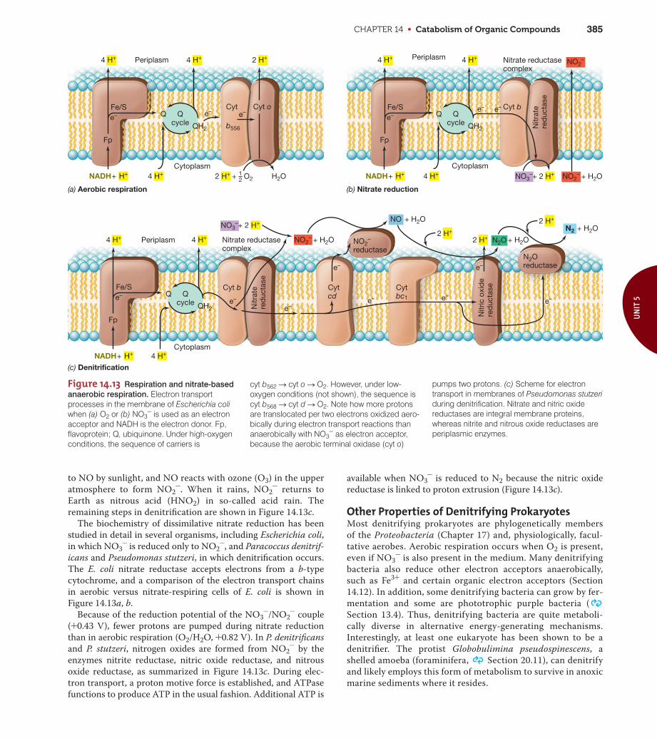
Figure 14.13 Respiration and nitrate-based
anaerobic respiration. Electron transportprocesses in the membrane of Escherichia coliwhen (a) O2 or (b) NO3
- is used as an electronacceptor and NADH is the electron donor. Fp,flavoprotein; Q, ubiquinone. Under high-oxygenconditions, the sequence of carriers is
(a) Aerobic respiration (b) Nitrate reduction
Cyt oCyt
b556
NO3–+ 2 H+
NO3–+ 2 H+
2 H+ + – O2
e–e–
e–e–e– e–
e–
e–e–
Qcycle
Periplasm Periplasm
Cytoplasm
Periplasm
Cytoplasm
Cytoplasm
Q
QH2
4 H+ 2 H+
4 H+
Qcycle
Q
QH2
4 H+
4 H+NADH + H+
Fp
Fe/Se–
e–
4 H+
NADH + H+
Fp
Fe/Se–
4 H+
Qcycle
Q
QH2
4 H+
4 H+NADH + H+
Fp
Fe/Se–
4 H+
H2O
Cyt b
Nitrate reductasecomplex
Nitrate reductasecomplex
Nitr
ate
red
ucta
se
Cyt b Cytcd
Cytbc1
Nitr
ate
red
ucta
se
NO2– + H2O
NO2–
+ H2O
+ H2O
+ H2O
+ H2O
(c) Denitrification
NO
N2O
N2
NO2–
reductaseN2Oreductase
Nitr
ic o
xid
ere
duc
tase
NO2–
2 H+
2 H+
2 H+
21
e–
to NO by sunlight, and NO reacts with ozone (O3) in the upper
atmosphere to form NO2-. When it rains, NO2
- returns to
Earth as nitrous acid (HNO2) in so-called acid rain. The
remaining steps in denitrification are shown in Figure 14.13c.
The biochemistry of dissimilative nitrate reduction has been
studied in detail in several organisms, including Escherichia coli,
in which NO3- is reduced only to NO2
-, and Paracoccus denitrif-
icans and Pseudomonas stutzeri, in which denitrification occurs.
The E. coli nitrate reductase accepts electrons from a b-type
cytochrome, and a comparison of the electron transport chains
in aerobic versus nitrate-respiring cells of E. coli is shown in
Figure 14.13a, b.
Because of the reduction potential of the NO3-/NO2
- couple
(+0.43 V), fewer protons are pumped during nitrate reduction
than in aerobic respiration (O2/H2O, +0.82 V). In P. denitrificans
and P. stutzeri, nitrogen oxides are formed from NO2- by the
enzymes nitrite reductase, nitric oxide reductase, and nitrous
oxide reductase, as summarized in Figure 14.13c. During elec-
tron transport, a proton motive force is established, and ATPase
functions to produce ATP in the usual fashion. Additional ATP is
available when NO3- is reduced to N2 because the nitric oxide
reductase is linked to proton extrusion (Figure 14.13c).
Other Properties of Denitrifying ProkaryotesMost denitrifying prokaryotes are phylogenetically members
of the Proteobacteria (Chapter 17) and, physiologically, facul-
tative aerobes. Aerobic respiration occurs when O2 is present,
even if NO3- is also present in the medium. Many denitrifying
bacteria also reduce other electron acceptors anaerobically,
such as Fe3+ and certain organic electron acceptors (Section
14.12). In addition, some denitrifying bacteria can grow by fer-
mentation and some are phototrophic purple bacteria (
Section 13.4). Thus, denitrifying bacteria are quite metaboli-
cally diverse in alternative energy-generating mechanisms.
Interestingly, at least one eukaryote has been shown to be a
denitrifier. The protist Globobulimina pseudospinescens, a
shelled amoeba (foraminifera, Section 20.11), can denitrify
and likely employs this form of metabolism to survive in anoxic
marine sediments where it resides.
cyt b562 S cyt o S O2. However, under low-oxygen conditions (not shown), the sequence iscyt b568 S cyt d S O2. Note how more protonsare translocated per two electrons oxidized aero-bically during electron transport reactions thananaerobically with NO3
- as electron acceptor,because the aerobic terminal oxidase (cyt o)
pumps two protons. (c) Scheme for electrontransport in membranes of Pseudomonas stutzeriduring denitrification. Nitrate and nitric oxidereductases are integral membrane proteins,whereas nitrite and nitrous oxide reductases areperiplasmic enzymes.

UNIT 5 • Metabolic Diversity and Commercial Biocatalyses386
MiniQuiz• For Escherichia coli, why is more energy released in aerobic
respiration than during NO3- reduction?
• How do the products of NO3- reduction differ between E. coli
and Pseudomonas?
• Where is the dissimilative nitrate reductase found in the cell?What unusual metal does it contain?
14.8 Sulfate and Sulfur ReductionSeveral inorganic sulfur compounds are important electron
acceptors in anaerobic respiration. A summary of the oxidation
states of key sulfur compounds is given in Table 14.6. Sulfate
(SO42-), the most oxidized form of sulfur, is a major anion in
seawater and is reduced by the sulfate-reducing bacteria, a
group that is widely distributed in nature. The end product of
sulfate reduction is hydrogen sulfide, H2S, an important natural
product that participates in many biogeochemical processes
( Section 24.3). Species in the genus Desulfovibrio have been
widely used for the study of sulfate reduction, and general prop-
erties of this and other sulfate-reducing bacteria are discussed in
Section 17.18.
Assimilative and Dissimilative Sulfate ReductionAgain, as with nitrogen, it is necessary to distinguish between
assimilative and dissimilative metabolism. Many organisms,
including plants, algae, fungi, and most prokaryotes, use SO42- as
a source for biosynthetic sulfur needs. The ability to use SO42-
as an electron acceptor for energy-generating processes, how-
ever, involves the large-scale reduction of SO42- and is restricted
to the sulfate-reducing bacteria. In assimilative sulfate reduction,
H2S is formed on a very small scale and is assimilated into
Table 14.6 Sulfur compounds and electron donors for sulfate reduction
Compound Oxidation state of S atom
Oxidation states of key sulfur compounds
Organic S (R—SH) -2Sulfide (H2S) -2Elemental sulfur (S0) 0Thiosulfate (—S–SO3
2-) -2/+6Sulfur dioxide (SO2) +4Sulfite (SO3
2-) +4Sulfate (SO4
2-) +6
Some electron donors used for sulfate reduction
H2 AcetateLactate PropionatePyruvate ButyrateEthanol and other alcohols Long-chain fatty acidsFumarate BenzoateMalate IndoleCholine Various hydrocarbons
organic form in sulfur-containing amino acids and other organic
sulfur compounds. By contrast, in dissimilative sulfate reduction,
H2S can be produced on a very large scale and is excreted from
the cell, free to react with other organisms or with metals to form
metal sulfides.
Biochemistry and Energetics of Sulfate ReductionAs the reduction potentials in Table A1.2 and Figure 14.11 show,
SO42- is a much less favorable electron acceptor than is O2 or
NO3-. However, sufficient free energy to make ATP is available
from sulfate reduction when an electron donor that yields NADH
or FADH is oxidized. Table 14.6 lists some of the electron donors
used by sulfate-reducing bacteria. Hydrogen (H2) is used by vir-
tually all species of sulfate-reducing bacteria, whereas use of the
other donors is more restricted. For example, lactate and pyru-
vate are widely used by species found in freshwater anoxic envi-
ronments, while acetate and longer-chain fatty acids are widely
used by marine sulfate-reducing bacteria. Many morphological
and physiological types of sulfate reducing bacteria are known,
and with the exception of Archaeoglobus ( Section 19.6), a
genus of Archaea, all known sulfate reducers are Bacteria (
Section 17.18).
The reduction of SO42- to H2S requires eight electrons and
proceeds through a number of intermediate stages. Sulfate is
chemically quite stable and cannot be reduced without first being
activated; SO42- is activated in a reaction requiring ATP. The
enzyme ATP sulfurylase catalyzes the attachment of SO42- to a
phosphate of ATP, forming adenosine phosphosulfate (APS ) as
shown in Figure 14.14. In dissimilative sulfate reduction, the
SO42- in APS is reduced directly to sulfite (SO3
2-) by the enzyme
APS reductase with the release of AMP. In assimilative reduction,
another phosphate is added to APS to form phosphoadenosine
phosphosulfate (PAPS) (Figure 14.14a), and only then is the SO42-
reduced. However, in both cases the product of sulfate reduction
is SO32-. Once SO3
2- is formed, H2S is generated from the activ-
ity of the enzyme sulfite reductase (Figure 14.14b).
During dissimilative sulfate reduction, electron transport reac-
tions lead to a proton motive force and this drives ATP synthesis by
ATPase. A major electron carrier in this process is cytochrome c3, a
periplasmic low-potential cytochrome (Figure 14.15). Cytochrome
c3 accepts electrons from a periplasmically located hydrogenase
and transfers these electrons to a membrane-associated protein
complex. This complex, called Hmc, carries the electrons across the
cytoplasmic membrane and makes them available to APS reductase
and sulfite reductase, cytoplasmic enzymes that generate sulfite and
sulfide, respectively (Figure 14.15).
The enzyme hydrogenase plays a central role in sulfate reduc-
tion whether Desulfovibrio is growing on H2, per se, or on an
organic compound such as lactate. This is because lactate is con-
verted through pyruvate to acetate (the latter is for the most part
excreted because Desulfovibrio is a non-acetate-oxidizing sulfate
reducer; Section 17.18) with the production of H2. The H2
produced crosses the cytoplasmic membrane and is oxidized by
the periplasmic hydrogenase to electrons, which are fed back into
the system, and protons, which establish the proton motive force
(Figure 14.15). Growth yields of sulfate-reducing bacteria suggest

CHAPTER 14 • Catabolism of Organic Compounds 387
UN
IT 5
Figure 14.14 Biochemistry of sulfate reduction: Activated sulfate.
(a) Two forms of active sulfate can be made, adenosine 59-phosphosul-fate (APS) and phosphoadenosine 59-phosphosulfate (PAPS). Both arederivatives of adenosine diphosphate (ADP), with the second phosphateof ADP being replaced by SO4
2-. (b) Schemes of assimilative and dissim-ilative sulfate reduction.
6 e– 6 e–
2 e–
APS (Adenosine 5′-phosphosulfate)
O P O
O
OH
S
O
O
O–
OHOH
HH H H
Adenine
Used in dissimilative metabolism
O P O
O
OH
S
O
O
O–CH2
OOH
HH H H
Adenine
Used in assimilative metabolism
P O–
O
OH
(a)
SO42–
PPi
APS
SO32–SO3
2–
H2S H2S
Excretion
ADP
APS kinase
Organic sulfur compounds(cysteine, methionine, and so on)
AMP PAP
NADPH
NADP+
(b)
PAPS (Phosphoadenosine 5′-phosphosulfate)
O
O
ATP sulfurylase
APSreductase
Sulfitereductase
Dissimilativesulfatereduction
Assimilativesulfatereduction
CH2
PAPS
ATPATP Figure 14.15 Electron transport and energy conservation in sulfate-
reducing bacteria. In addition to external H2, H2 originating from thecatabolism of organic compounds such as lactate and pyruvate can fuelhydrogenase. The enzymes hydrogenase (H2ase), cytochrome (cyt) c3, and a cytochrome complex (Hmc) are periplasmic proteins. A sepa-rate protein shuttles electrons across the cytoplasmic membrane fromHmc to a cytoplasmic iron–sulfur protein (FeS) that supplies electrons toAPS reductase (forming SO3
2-) and sulfite reductase (forming H2S,Figure 14.14b). LDH, lactate dehydrogenase.
Lactate Pyruvate
FADH
Out
In
APS2e–
6e–
Hmc
FeS
FeSH2ase
LDH
e–
8 e– H+3–4
ADP ATP
Acetate + CO2 + ATP
H2
4 H2 8 H+
cyt c3
SO42–
H2S
SO32–Ferredoxin
that a net of one ATP is produced for each SO42- reduced to HS-.
With H2 as electron donor, the reaction is
4 H2 + SO42- + H+ S HS- + 4 H2O DG09 = -152 kJ
When lactate or pyruvate is the electron donor, not only is ATP
produced from the proton motive force, but additional ATP can
be produced during the oxidation of pyruvate to acetate plus CO2
via acetyl-CoA and acetyl phosphate (Table 14.1 and Figure 14.2).
Acetate Use and AutotrophyMany sulfate-reducing bacteria can oxidize acetate to CO2 to
obtain electrons for SO42- reduction ( Section 17.18):
CH3COO- + SO42- + 3 H+ S 2 CO2 + H2S + 2 H2O
DG09 = -57.5 kJ
The mechanism for acetate oxidation in most species is the
acetyl-CoA pathway, a series of reversible reactions used by
many anaerobes for acetate synthesis or acetate oxidation. This
pathway employs the key enzyme carbon monoxide dehydro-
genase (Section 14.9). A few sulfate-reducing bacteria can also
grow autotrophically with H2. When growing under these condi-
tions, the organisms use the acetyl-CoA pathway for incorporat-
ing CO2 into cell material. The acetate-oxidizing sulfate-reducing
bacterium Desulfobacter lacks acetyl-CoA pathway enzymes and
oxidizes acetate through the citric acid cycle ( Figure 4.21),
but this seems to be the exception rather than the rule.
Sulfur DisproportionationCertain sulfate-reducing bacteria can disproportionate sulfur
compounds of intermediate oxidation state. Disproportionation
occurs when one molecule of a substance is oxidized while a second
molecule is reduced, ultimately forming two different products.
For example, Desulfovibrio sulfodismutans can disproportionate
thiosulfate (S2O32-) as follows:
S2O32- + H2O S SO4
2- + H2S DG09 = -21.9 kJ/reaction

UNIT 5 • Metabolic Diversity and Commercial Biocatalyses388
Note that in this reaction one sulfur atom of S2O32- becomes
more oxidized (forming SO42-), while the other becomes more
reduced (forming H2S). The oxidation of S2O32- by D. sulfodis-
mutans is coupled to proton pumping that is used by this organ-
ism to make ATP by ATPase. Other reduced sulfur compounds
such as sulfite (SO32-) and sulfur (S0) can also be disproportion-
ated. These forms of sulfur metabolism allow sulfate-reducing
bacteria to recover energy from sulfur intermediates produced
from the oxidation of H2S by sulfur chemolithotrophs that coex-
ist with them in nature and also from intermediates generated in
their own metabolism during SO42- reduction.
Phosphite OxidationAt least one sulfate-reducing bacterium can couple phosphite
(HPO3-) oxidation to SO4
2- reduction. The reaction is
chemolithotrophic, and the products are phosphate and sulfide:
4 HPO3- + SO4
2- + H+ S 4 HPO42- + HS- DG09 = -364 kJ
This bacterium, Desulfotignum phosphitoxidans, is an autotroph
and a strict anaerobe, which by necessity it must be because phos-
phite spontaneously oxidizes in air. The natural sources of phosphite
are likely to be organic phosphorous compounds called phos-
phonates, molecules generated from the anoxic degradation of
organic phosphorous compounds. Along with sulfur dispropor-
tionation (also a chemolithotrophic process) and H2 utilization,
phosphite oxidation underscores the diversity of chemolithotrophic
reactions carried out by sulfate-reducing bacteria.
Sulfur ReductionSome organisms produce H2S in anaerobic respiration, but are
unable to reduce SO42-; these are the elemental sulfur (S0) reduc-
ers. Sulfur-reducing bacteria carry out the reaction
S0 + 2 H S H2S
The electrons for this process can come from H2 or from various
organic compounds. The first sulfur-reducing organism to be
discovered was Desulfuromonas acetoxidans ( Section 17.18).
This organism oxidizes acetate, ethanol, and a few other com-
pounds to CO2, coupled with the reduction of S0 to H2S. Ferric
iron (Fe3+) also supports growth as an electron acceptor. The
physiology of dissimilative sulfur-reducing bacteria is not as well
understood as that of sulfate-reducing bacteria, but it is known
that sulfur reducers lack the capacity to activate sulfate to APS
(Figure 14.14), and presumably this prevents them from using
SO42- as an electron acceptor. Desulfuromonas contains high
levels of several cytochromes, including an analog of cytochrome
c3, a key electron carrier in sulfate-reducing bacteria. Because the
oxidation of acetate to CO2 releases less energy than that needed
to make an ATP by substrate-level phosphorylation, it is clear
that oxidative phosphorylation plays a major role in the energet-
ics of these organisms. A variety of other bacteria can use S0 as an
electron acceptor, including some species of the genera Wolinella
and Campylobacter. In culture some sulfur reducers including
Desulfuromonas can use Fe3+ as an electron acceptor, but S0 is
probably the major electron acceptor used in nature. It is the pro-
duction of H2S that connects the sulfur- and sulfate-reducing
bacteria in an ecological sense.
MiniQuiz• How is SO4
2- converted to SO32- during dissimilative sulfate
reduction? Physiologically, how does Desulfuromonas differ fromDesulfovibrio?
• Why is H2 of importance to sulfate-reducing bacteria?
• Give an example of sulfur disproportionation.
14.9 AcetogenesisCarbon dioxide, CO2, is common in nature and typically abun-
dant in anoxic habitats because it is a major product of the
energy metabolisms of chemoorganotrophs. Two major groups
of strictly anaerobic prokaryotes use CO2 as an electron acceptor
in energy metabolism. One of these groups is the acetogens, and
we discuss them here. The other group, the methanogens, will be
considered in the next section. H2 is a major electron donor for
both of these organisms, and an overview of their energy metabo-
lism, acetogenesis and methanogenesis, is shown in Figure 14.16.
Both processes are linked to ion pumps, of either protons (H+) or
sodium ions (Na+), as the mechanism of energy conservation, and
these pumps fuel ATPases in the membrane. Acetogenesis also
conserves energy in a substrate-level phosphorylation reaction.
Organisms and PathwayAcetogens carry out the reaction
4 H2 + H+ + 2 HCO3- S CH3COO- + 4 H2O
DG09 = -105 kJ
In addition to H2, electron donors for acetogenesis include C1
compounds, sugars, organic and amino acids, alcohols, and cer-
tain nitrogen bases, depending on the organism. Many acetogens
can also reduce nitrate (NO3-) and thiosulfate (S2O3
2-). How-
ever, CO2 reduction is probably the major reaction of ecological
significance.
A major unifying thread among acetogens is the pathway of
CO2 reduction. Acetogens reduce CO2 to acetate by the acetyl-
CoA pathway, the major pathway in obligate anaerobes for the
production or oxidation of acetate. Table 14.7 lists the major
groups of organisms that produce acetate or oxidize acetate via
the acetyl-CoA pathway. Acetogens such as Acetobacterium
woodii and Clostridium aceticum can grow either chemoor-
ganotrophically by fermentation of sugars (reaction 1) or
Figure 14.16 The contrasting processes of methanogenesis and
acetogenesis. Note the difference in free energy released in the reactions.
Proton or sodiummotive force (plussubstrate-levelphosphorylation foracetogens)
Acetogenesis(ΔG0′ = –105 kJ)
Methanogenesis(ΔG0′ = –136 kJ)
CH4 +
HCO3– + H+ 2 HCO3
– + H+
O
CH3—C—O–
+ 4 H2O3 H2O
4 H2
ATP

CHAPTER 14 • Catabolism of Organic Compounds 389
UN
IT 5
chemolithotrophically and autotrophically through the reduction
of CO2 to acetate with H2 (reaction 2) as electron donor. In either
case, the sole product is acetate:
(1) C6H12O6 S 3 CH3COO- + 3 H+
(2) 2 HCO3- + 4 H2 + H+ S CH3COO- + 4 H2O
Acetogens catabolize glucose by way of glycolysis, converting
glucose to two molecules of pyruvate and two molecules of
NADH (the equivalent of 4 H). From this point, two molecules of
acetate are produced:
(3) 2 Pyruvate- S 2 acetate- + 2 CO2 + 4 H
The third acetate of reaction (1) comes from reaction (2), using
the two molecules of CO2 generated in reaction (3), plus the four
H generated during glycolysis and the four H generated from the
oxidation of two pyruvates to two acetates [reaction (3)]. Starting
from pyruvate, then, the overall production of acetate can be
written as
2 Pyruvate- + 4 H S 3 acetate- + H+
Most acetogenic bacteria that produce and excrete acetate in
energy metabolism are gram-positive Bacteria, and many are
species of Clostridium or Acetobacterium (Table 14.7). A few
other gram-positive and many different gram-negative Bacteria
and Archaea use the acetyl-CoA pathway for autotrophic pur-
poses, reducing CO2 to acetate as a source of cell carbon.
The acetyl-CoA pathway functions in autotrophic growth for
certain sulfate-reducing bacteria and is also used by the
Table 14.7 Organisms employing the acetyl-CoA pathway
I. Pathway drives acetate synthesis for energy purposes
Acetoanaerobium noteraeAcetobacterium woodiiAcetobacterium wieringaeAcetogenium kivuiAcetitomaculum ruminisClostridium aceticumClostridium formicaceticumClostridium ljungdahliiMoorella thermoaceticaDesulfotomaculum orientisSporomusa paucivoransEubacterium limosum (also produces butyrate)Treponema primitia (from termite hindguts)
II. Pathway drives acetate synthesis for cell biosynthesis
AcetogensMethanogensSulfate-reducing bacteria
III. Pathway drives acetate oxidation for energy purposes
Reaction: Acetate + 2 H2O S 2 CO2 + 8 HGroup II sulfate reducers (other than Desulfobacter)
Reaction: Acetate S CO2 + CH4
Acetotrophic methanogens (Methanosarcina, Methanosaeta)
methanogens, most of which grow autotrophically on H2 + CO2
( Sections 19.3 and 14.10). By contrast, some bacteria
employ the reactions of the acetyl-CoA pathway primarily in
the reverse direction as a means of oxidizing acetate to CO2.
These include acetotrophic methanogens ( Section 19.3) and
sulfate-reducing bacteria ( Sections 17.18 and 14.8).
Reactions of the Acetyl-CoA PathwayUnlike other autotrophic pathways such as the Calvin cycle (
Section 13.12), the reverse citric acid cycle, or the hydroxypropi-
onate cycle ( Section 13.13), the acetyl-CoA pathway of CO2
fixation is not a cycle. Instead it catalyzes the reduction of CO2
along two linear pathways; one molecule of CO2 is reduced to the
methyl group of acetate, and the other molecule of CO2 is
reduced to the carbonyl group. The two C1 units are then com-
bined at the end to form acetyl-CoA (Figure 14.17).
A key enzyme of the acetyl-CoA pathway is carbon monox-
ide (CO) dehydrogenase. CO dehydrogenase contains the met-
als Ni, Zn, and Fe as cofactors. CO dehydrogenase catalyzes
the reaction
CO2 + H2 S CO + H2O
and the CO produced ends up as the carbonyl carbon of acetate
(Figure 14.17). The methyl group of acetate originates from the
reduction of CO2 by a series of reactions requiring the coen-
zyme tetrahydrofolate (Figure 14.17). The methyl group is then
Figure 14.17 Reactions of the acetyl-CoA pathway. Carbon monox-ide is bound to Fe and the CH3 group to nickel in carbon monoxidedehydrogenase. Note that the formation of acetyl-CoA is coupled to thegeneration of a Na+ motive force that drives ATP synthesis, and that ATP is also synthesized in the conversion of acetyl-CoA to acetate. THF,tetrahydrofolate; B12, vitamin B12 in an enzyme-bound intermediate.
~
H2O
Reduction of CO2 to methyl group
Reductionof CO2 tocarbonylgroup
CHOH2
THF
THF
2 H2
CH3 THF
B12
CH3 B12
MethylB12
CH3—C—O–
O
CO
Fe
Ni
CO2CO
H2
Formyltetrahydrofolate
CO dehydrogenaseCoA
CH3 C S–CoA
O
ADPAcetate
Methyltetrahydrofolate
Net: 4 H2 + 2 HCO3– + H+ acetate– + 4 H2O
ΔG0′= –105 kJ
Acetyl-CoA
CH3
Fe
NiNi
Na+ motive force
Fe
CO2
ATP
ATP
ATP

UNIT 5 • Metabolic Diversity and Commercial Biocatalyses390
transferred from tetrahydrofolate to an enzyme that contains
vitamin B12 as cofactor, and in the final step of the pathway, the
methyl group is combined with CO by the enzyme CO dehydro-
genase to form acetyl-CoA. Conversion of acetyl-CoA to acetate
plus ATP completes the reaction series (Figure 14.17).
Energy Conservation in AcetogenesisEnergy conservation in acetogenesis is the result of substrate-
level phosphorylation during the conversion of acetyl-CoA to
acetate plus ATP (Section 14.1). There is also an energy-conserving
step when a sodium motive force (analogous to a proton motive
force) is established across the cytoplasmic membrane during
the formation of acetyl-CoA. This energized state of the mem-
brane allows for energy conservation from a Na+-driven ATPase.
Recall that we saw a similar situation in the succinate fermenter
Propionigenium, where succinate decarboxylation was linked to
Na+ export and a Na+-driven ATPase (Section 14.4). Acetogens
need the ATP resulting from this reaction since the single ATP
made by substrate-level phosphorylation is consumed in the first
step of the acetyl-CoA pathway (Figure 14.17).
MiniQuiz• Draw the structure of acetate and identify the carbonyl group
and the methyl group. What key enzyme of the acetyl-CoApathway produces the carbonyl group of acetate?
• How do acetogens make ATP from the synthesis of acetate?
• If fructose catabolism by glycolysis yields only two acetates, how does Clostridium aceticum produce three acetates fromfructose?
14.10 MethanogenesisThe biological production of methane—methanogenesis—is car-
ried out by a group of strictly anaerobic Archaea called the
methanogens. The reduction of CO2 by H2 to form methane
(CH4) is a major pathway of methanogenesis and so we focus on
this process and compare it with other forms of anaerobic respi-
ration. We consider the basic properties, phylogeny, and taxon-
omy of the methanogens in Section 19.3; here we focus on their
biochemistry and bioenergetics. Methanogenesis is a unique
series of biochemical reactions that employs novel coenzymes.
Because of this, we begin by considering these coenzymes and
then move on to the actual pathway itself.
C1 Carriers in MethanogenesisMethanogenesis from CO2 requires the input of eight electrons,
and these electrons are added two at a time. This leads to inter-
mediary oxidation states of the carbon atom from +4 (CO2) to
-4 (CH4). The key coenzymes in methanogenesis can be divided
into two classes: (1) those that carry the C1 unit along its path of
enzymatic reduction (C1 carriers) and (2) those that donate elec-
trons (redox coenzymes) (Figure 14.18, and see Figure 14.20). We
consider the carriers first.
The coenzyme methanofuran is required for the first step of
methanogenesis. Methanofuran contains the five-membered
furan ring and an amino nitrogen atom that binds CO2 (Figure
14.18a). Methanopterin (Figure 14.18b) is a methanogenic coen-
zyme that resembles the vitamin folic acid and plays a role analo-
gous to that of tetrahydrofolate (a coenzyme that participates in
C1 transformations, see Figure 14.17) by carrying the C1 unit in
the intermediate steps of CO2 reduction to CH4. Coenzyme M
(CoM) (Figure 14.18c) is a small molecule required for the terminal
step of methanogenesis, the conversion of a methyl group (CH3)
to CH4. Although not a C1 carrier, the nickel (Ni2+)-containing
tetrapyrrole coenzyme F430 (Figure 14.18d) is also needed for the
terminal step of methanogenesis as part of the methyl reductase
enzyme complex (discussed later).
Redox CoenzymesThe coenzymes F420 and 7-mercaptoheptanoylthreonine phos-
phate (also called coenzyme B, CoB), are electron donors in
methanogenesis. Coenzyme F420 (Figure 14.18e) is a flavin deriv-
ative, structurally resembling the flavin coenzyme FMN (
Figure 4.15). F420 plays a role in methanogenesis as the electron
donor in several steps of CO2 reduction (see Figure 14.20). The
oxidized form of F420 absorbs light at 420 nm and fluoresces
blue-green. Such fluorescence is useful for the microscopic iden-
tification of a methanogen (Figure 14.19). CoB is required for
the terminal step of methanogenesis catalyzed by the methyl
reductase enzyme complex. As shown in Figure 14.18f, the struc-
ture of CoB resembles the vitamin pantothenic acid (which is
part of acetyl-CoA) ( Figure 4.12).
Methanogenesis from CO21 H2Electrons for the reduction of CO2 to CH4 come primarily from
H2, but formate, carbon monoxide (CO), and even certain alco-
hols can also supply the electrons for CO2 reduction in some
methanogens. Figure 14.20 shows the steps in CO2 reduction
by H2:
1. CO2 is activated by a methanofuran-containing enzyme and
reduced to the formyl level. The immediate electron donor is
the protein ferredoxin, a strong reductant with a reduction
potential (E09) near -0.4V.
2. The formyl group is transferred from methanofuran to an
enzyme containing methanopterin (MP in Figure 14.20). It is
subsequently dehydrated and reduced in two separate steps
(total of 4 H) to the methylene and methyl levels. The immedi-
ate electron donor is reduced F420.
3. The methyl group is transferred from methanopterin to an
enzyme containing CoM by the enzyme methyl transferase.
This reaction is highly exergonic and linked to the pumping of
Na+ across the membrane from inside to outside the cell.
4. Methyl-CoM is reduced to methane by methyl reductase; in
this reaction, F430 and CoB are required. Coenzyme F430
removes the CH3 group from CH3-CoM, forming a Ni2+–CH3
complex. This complex is reduced by CoB, generating CH4 and
a disulfide complex of CoM and CoB (CoM-S—S-CoB).
5. Free CoM and CoB are regenerated by the reduction of CoM-S—
S-CoB with H2.

CHAPTER 14 • Catabolism of Organic Compounds 391
UN
IT 5
Figure 14.18 Coenzymes of methanogenesis. The atoms shaded in brown or yellow are the sites ofoxidation–reduction reactions (brown in F420 and CoB) or the position to which the C1 moiety is attachedduring the reduction of CO2 to CH4 (yellow in methanofuran, methanopterin, and coenzyme M). The colorsused to highlight a particular coenzyme (CoB is orange, for example) are also in Figures 14.20–14.21 tofollow the reactions in each figure.
H
(a)
(b)
(c) (d)
(f)(e)
CH2 CH2 [ NH C CH2 CH2 CH]3 CH
OH2N CH2
Methanofuran
CH2 O
CH2
CH2
COO–
COO–COO–
CH2[ CHOH]3 CH2 O
CH2 O P O CH
OOH HO
ONH
HC
N
NH
H
O
O
HN
N
Methanopterin
CH2
CH2
CH3
CH3
COO–
O– COO–
H2N
CH2 CH2 S O–
O
O
HS
Coenzyme M (CoM)
H3C
COO–
CH2
CH2 CH2 COO–
CH2 COO–
CH3
CH2
H2NOC H2C
Ni+N N
NN
OCH2
CH2
O
HN
Coenzyme F430
–OOC H2C
COO–
COO–
Coenzyme B (CoB)
Ι. Coenzymes that function as C1 carriers, plus F430
Earlysteps
Middlesteps
Finalsteps
ΙΙΙΙ. Coenzymes that function as electron donors
O
O
O CH3
HS CH2 CH2 CH2 CH2 CH2 CH2 C NH CH CH O P O–
Coenzyme F420
Oxidized
Reduced
OH OH OH O
O–
CH3 O COO–
CH2
CH2
C O
NH
HC COO–
CH2
CH2
N NN
NH
O
O
HO
H
–2H +2H
COO–R
NN
NH
O
O
HO
H
NH
H2C CH CH CH CH2 O P O CH C NH CH
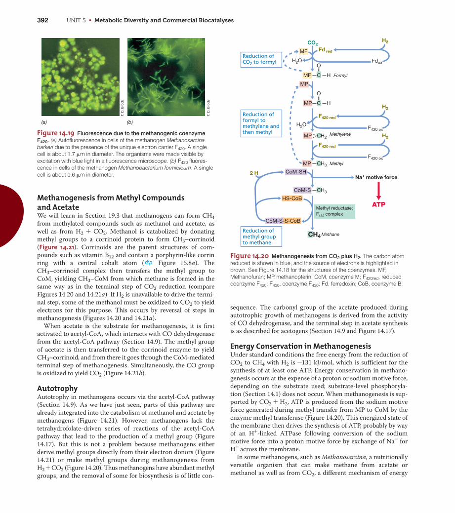
UNIT 5 • Metabolic Diversity and Commercial Biocatalyses392
Methanogenesis from Methyl Compounds and AcetateWe will learn in Section 19.3 that methanogens can form CH4
from methylated compounds such as methanol and acetate, as
well as from H2 + CO2. Methanol is catabolized by donating
methyl groups to a corrinoid protein to form CH3–corrinoid
(Figure 14.21). Corrinoids are the parent structures of com-
pounds such as vitamin B12 and contain a porphyrin-like corrin
ring with a central cobalt atom ( Figure 15.8a). The
CH3–corrinoid complex then transfers the methyl group to
CoM, yielding CH3–CoM from which methane is formed in the
same way as in the terminal step of CO2 reduction (compare
Figures 14.20 and 14.21a). If H2 is unavailable to drive the termi-
nal step, some of the methanol must be oxidized to CO2 to yield
electrons for this purpose. This occurs by reversal of steps in
methanogenesis (Figures 14.20 and 14.21a).
When acetate is the substrate for methanogenesis, it is first
activated to acetyl-CoA, which interacts with CO dehydrogenase
from the acetyl-CoA pathway (Section 14.9). The methyl group
of acetate is then transferred to the corrinoid enzyme to yield
CH3–corrinoid, and from there it goes through the CoM-mediated
terminal step of methanogenesis. Simultaneously, the CO group
is oxidized to yield CO2 (Figure 14.21b).
AutotrophyAutotrophy in methanogens occurs via the acetyl-CoA pathway
(Section 14.9). As we have just seen, parts of this pathway are
already integrated into the catabolism of methanol and acetate by
methanogens (Figure 14.21). However, methanogens lack the
tetrahydrofolate-driven series of reactions of the acetyl-CoA
pathway that lead to the production of a methyl group (Figure
14.17). But this is not a problem because methanogens either
derive methyl groups directly from their electron donors (Figure
14.21) or make methyl groups during methanogenesis from
H2 + CO2 (Figure 14.20). Thus methanogens have abundant methyl
groups, and the removal of some for biosynthesis is of little con-
sequence. The carbonyl group of the acetate produced during
autotrophic growth of methanogens is derived from the activity
of CO dehydrogenase, and the terminal step in acetate synthesis
is as described for acetogens (Section 14.9 and Figure 14.17).
Energy Conservation in MethanogenesisUnder standard conditions the free energy from the reduction of
CO2 to CH4 with H2 is -131 kJ/mol, which is sufficient for the
synthesis of at least one ATP. Energy conservation in methano-
genesis occurs at the expense of a proton or sodium motive force,
depending on the substrate used; substrate-level phosphoryla-
tion (Section 14.1) does not occur. When methanogenesis is sup-
ported by CO2 + H2, ATP is produced from the sodium motive
force generated during methyl transfer from MP to CoM by the
enzyme methyl transferase (Figure 14.20). This energized state of
the membrane then drives the synthesis of ATP, probably by way
of an H+-linked ATPase following conversion of the sodium
motive force into a proton motive force by exchange of Na+ for
H+ across the membrane.
In some methanogens, such as Methanosarcina, a nutritionally
versatile organism that can make methane from acetate or
methanol as well as from CO2, a different mechanism of energy
Figure 14.19 Fluorescence due to the methanogenic coenzyme
F420. (a) Autofluorescence in cells of the methanogen Methanosarcinabarkeri due to the presence of the unique electron carrier F420. A singlecell is about 1.7 �m in diameter. The organisms were made visible byexcitation with blue light in a fluorescence microscope. (b) F420 fluores-cence in cells of the methanogen Methanobacterium formicicum. A singlecell is about 0.6 �m in diameter.
(b)(a)T.
D. B
rock
T. D
. Bro
ck
Figure 14.20 Methanogenesis from CO2 plus H2. The carbon atomreduced is shown in blue, and the source of electrons is highlighted inbrown. See Figure 14.18 for the structures of the coenzymes. MF,Methanofuran; MP, methanopterin; CoM, coenzyme M; F420red, reducedcoenzyme F420; F430, coenzyme F430; Fd, ferredoxin; CoB, coenzyme B.
Reduction ofmethyl groupto methane
Reduction of CO2 to formyl
Reduction of formyl tomethylene andthen methyl
CH4
MF
H2O
H2O
MF C H
O
Formyl
MP
MP C H
O
MP CH2
H2
CH3
CH3
MP
CoM-S
CoM-SH
CoM-S-S-CoB
Methyl reductase;F430 complex
HS-CoB
Methane
F420 red
F420 red
F420 ox
H2
Fd red
Fdox
H2
F420 ox
Methylene
Methyl
Na+ motive force2 H
CO2
ATP
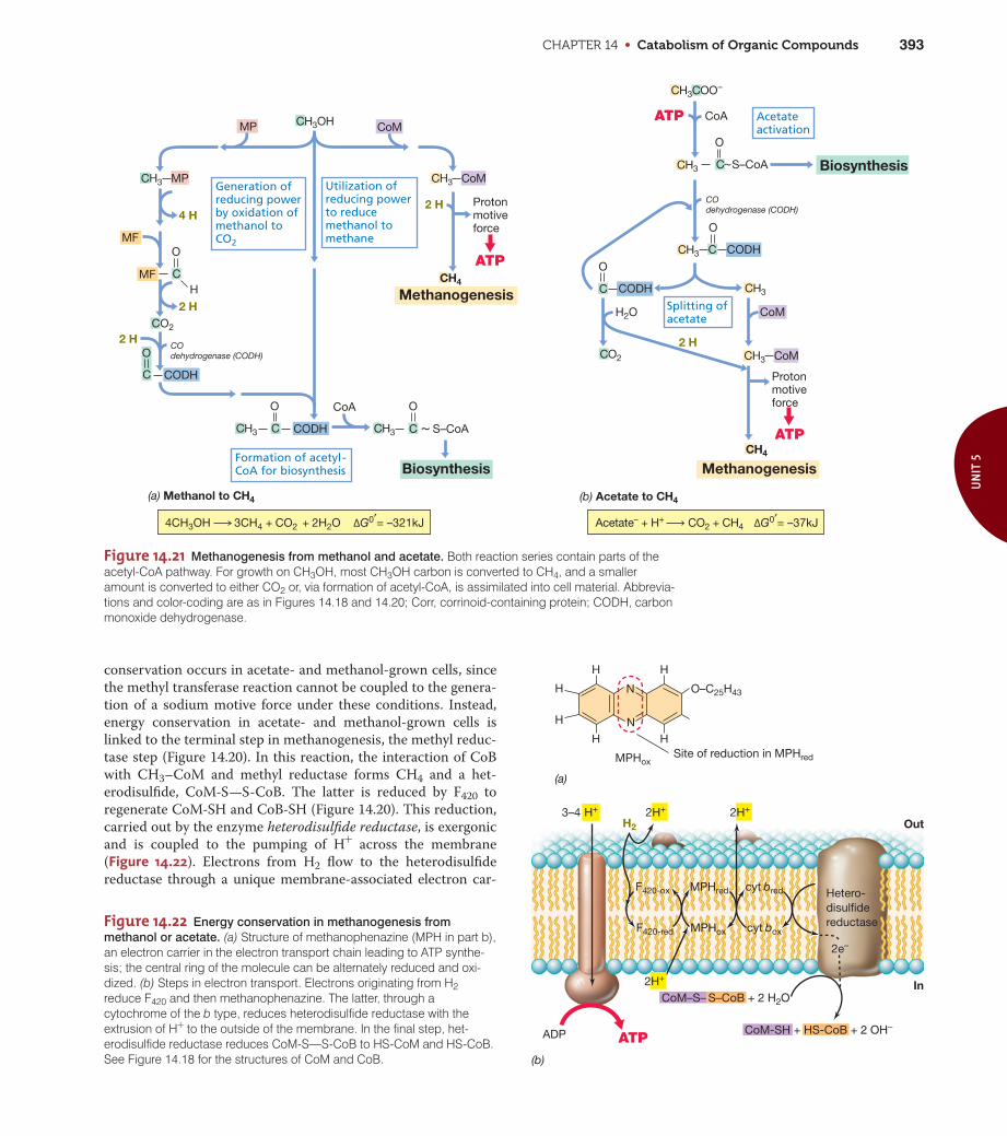
CHAPTER 14 • Catabolism of Organic Compounds 393
UN
IT 5
conservation occurs in acetate- and methanol-grown cells, since
the methyl transferase reaction cannot be coupled to the genera-
tion of a sodium motive force under these conditions. Instead,
energy conservation in acetate- and methanol-grown cells is
linked to the terminal step in methanogenesis, the methyl reduc-
tase step (Figure 14.20). In this reaction, the interaction of CoB
with CH3–CoM and methyl reductase forms CH4 and a het-
erodisulfide, CoM-S—S-CoB. The latter is reduced by F420 to
regenerate CoM-SH and CoB-SH (Figure 14.20). This reduction,
carried out by the enzyme heterodisulfide reductase, is exergonic
and is coupled to the pumping of H+ across the membrane
(Figure 14.22). Electrons from H2 flow to the heterodisulfide
reductase through a unique membrane-associated electron car-
Figure 14.21 Methanogenesis from methanol and acetate. Both reaction series contain parts of theacetyl-CoA pathway. For growth on CH3OH, most CH3OH carbon is converted to CH4, and a smalleramount is converted to either CO2 or, via formation of acetyl-CoA, is assimilated into cell material. Abbrevia-tions and color-coding are as in Figures 14.18 and 14.20; Corr, corrinoid-containing protein; CODH, carbonmonoxide dehydrogenase.
~
CH3OH
CH3COO–
CH3 MP
CH3 CH3
CH3 CoMCH3
CH3
CO2
CH3
CH4
CH4
MP
4 H
Generation ofreducing powerby oxidation of methanol to CO2MF
CMF
O
H
2 HCO2
2 H
C
O
CoA O
S–CoAC C
O
CODH
CODH
Formation of acetyl-CoA for biosynthesis Biosynthesis
(a) Methanol to CH4 (b) Acetate to CH4
Utilization of reducing power to reduce methanol to methane
Acetateactivation
Splitting ofacetate
2 H
Methanogenesis
CoA
C S–CoA
O
Biosynthesis
C CODH
O
COdehydrogenase (CODH)
COdehydrogenase (CODH)
C CODH
O
CoMH2O
Methanogenesis
2 H
CoM
ATP
ATP
Protonmotiveforce
ATP
Protonmotiveforce
4CH3OH 3CH4 + CO2 + 2H2O ΔG0′= –321kJ Acetate– + H+ CO2 + CH4 ΔG0′= –37kJ
~
CH3 CoM
Figure 14.22 Energy conservation in methanogenesis from
methanol or acetate. (a) Structure of methanophenazine (MPH in part b),an electron carrier in the electron transport chain leading to ATP synthe-sis; the central ring of the molecule can be alternately reduced and oxi-dized. (b) Steps in electron transport. Electrons originating from H2reduce F420 and then methanophenazine. The latter, through acytochrome of the b type, reduces heterodisulfide reductase with theextrusion of H+ to the outside of the membrane. In the final step, het-erodisulfide reductase reduces CoM-S—S-CoB to HS-CoM and HS-CoB.See Figure 14.18 for the structures of CoM and CoB.
H
H
H
N O–C25H43
H
NH
H
(a)
Site of reduction in MPHredMPHox
(b)
Out
InCoM–S– S–CoB + 2 H2O
Hetero-disulfidereductase
H2
H+ 2H+
ADP ATP
cyt bred
cyt box
MPHred
MPHox
F420-ox
F420-red
CoM-SH + HS-CoB + 2 OH–
2H+
2e–
2H+3–4

UNIT 5 • Metabolic Diversity and Commercial Biocatalyses394
rier called methanophenazine. This compound is reduced by F420
and then oxidized by a b-type cytochrome, and the latter is the
electron donor to the heterodisulfide reductase (Figure 14.22).
Cytochromes and methanophenazine are lacking in methanogens
that use only H2 + CO2 for methanogenesis.
In methanogens we thus see at least two mechanisms for
energy conservation: (1) a proton motive force linked to the
methylreductase reaction and used to drive ATP synthesis in
acetate- or methanol-grown cells, and (2) a sodium motive force
formed during methanogenesis from H2 + CO2.
MiniQuiz• What coenzymes function as C1 carriers in methanogenesis?
As electron donors?
• In methanogens growing on H2 + CO2, how is carbon obtainedfor cell biosynthesis?
• How is ATP made in methanogenesis when the substrates areH2 + CO2? Acetate?
14.11 Proton ReductionPerhaps the simplest of all anaerobic respirations is one carried
out by the hyperthermophile Pyrococcus furiosus. P. furiosus is a
species of Archaea and grows optimally at 100°C ( Section
19.5) on sugars and small peptides as electron donors. P. furio-
sus was originally thought to use the glycolytic pathway because
typical fermentation products such as acetate, CO2, and H2
were produced from glucose. However, analyses of sugar
metabolism in this organism revealed an unusual and enigmatic
situation.
During a key step of glycolysis, the oxidation of glyceralde-
hyde 3-phosphate forms 1,3-bisphosphoglyceric acid, an inter-
mediate with two energy-rich phosphate bonds, each of which
eventually yields ATP. In P. furiosus, this step is bypassed, yielding
3-phosphoglyceric acid directly from glyceraldehyde 3-phosphate
(Figure 14.23). This prevents P. furiosus from making ATP by
substrate-level phosphorylation at the 1,3-bisphosphoglyceric acid
to 3-phosphoglyceric acid step, one of two sites of energy conserva-
tion in the glycolytic pathway ( Figure 4.14). This yields P. furio-
sus a net of zero ATP from glycolytic steps that normally yield 2
ATP in other organisms. How can P. furiosus ferment glucose and
ignore the most important energy-yielding steps?
Protons as Electron AcceptorsThe riddle of energy conservation in P. furiosus revolves around
the oxidation of 3-phosphoglyceric acid. In glycolysis this acceptor
is NAD+, but in P. furiosus the protein ferredoxin is the electron
acceptor (Figure 14.23). Ferredoxin has a much more negative E09than that of NAD+/NADH, about the same as that of the 2 H+/H2
couple, –0.42 V. Ferredoxin is oxidized by transferring electrons to
protons to form H2 (Figure 14.23).
H2 is typically produced during the oxidation of pyruvate to
acetate plus CO2 (Figure 14.2). This allows for ATP to be synthe-
sized by substrate-level phosphorylation, and this also occurs in
P. furiosus (Figure 14.23). But in addition, the H2 released from
ferredoxin is coupled to the pumping of protons (H+) across the
membrane by a membrane-integrated hydrogenase. This estab-
lishes a proton motive force that drives ATP synthesis by ATPase
(Figure 14.23).
Although H+ reduction by P. furiosus does not employ an electron
transport chain per se, it can still be considered a form of anaerobic
respiration because protons function as a net electron acceptor. The
process differs from the proton pumping associated with decarboxy-
lation reactions, such as those of Oxalobacter, where the free energy
released during decarboxylation is coupled directly to H+ translo-
cation (Figure 14.8b). In P. furiosus, a proton is pumped during
hydrogenase activity, analogous to how terminal electron carriers
pump protons in aerobic or anaerobic respiratory processes (
Figures 4.19 and 14.13).
Growth Yields and EvolutionMeasurements of growth yields of P. furiosus on glucose indicate
that, despite being unable to conserve energy from the main reac-
tions in glycolysis, the organism actually synthesizes more ATP
from glucose than most other glucose fermenters! Two ATP are
produced by substrate-level phosphorylation during the conver-
sion of two acetyl-CoA to acetate, and about one additional ATP
is produced from H2 production by hydrogenase (Figure 14.23).
Whether H+ reduction by prokaryotes is more widespread
than that in P. furiosus is unknown. However, the ancient phy-
logeny of Pyrococcus ( Figure 19.1), coupled to its hot, anoxic
habitat, similar to that of early Earth ( Section 16.3), suggests
Glucose
G-3-P
Fdox
Fdred
OutIn
H+
H2
H+
3H+
3-PGA
PEPADP
ATP
ADP ADP
ATP ATP
Pyruvate
Acetate
Acetyl~S-CoA + CO2
Glycolysis Cytoplasmicmembrane
Hydrogenase
ATPase
Fdox
Fdred
3–4
Figure 14.23 Modified glycolysis and proton reduction in anaero-
bic respiration in the hyperthermophile Pyrococcus furiosus.
Hydrogen (H2) production is linked to H+ pumping by a hydrogenase thatreceives electrons from reduced ferredoxin (Fdred). All intermediates fromG-3-P downward in the pathway are present in two copies. Compare thisfigure with classical glycolysis in Figure 4.14. G-3-P, glyceraldehyde 3-phosphate; 3-PGA, 3-phosphoglycerate; PEP, phosphoenolpyruvate.

CHAPTER 14 • Catabolism of Organic Compounds 395
UN
IT 5
that proton reduction, a bioenergetic mechanism that requires
only a single membrane protein other than ATPase, might have
been a very early form of anaerobic respiration, perhaps even
nature’s first proton pump.
MiniQuiz• When fermenting glucose, how does Pyrococcus furiosus
overcome the loss of most of the ATP produced by otherglucose fermenters?
14.12 Other Electron AcceptorsIn addition to the electron acceptors for anaerobic respiration dis-
cussed thus far, ferric iron (Fe3+), manganic ion (Mn4+), chlorate
(ClO3–), perchlorate (ClO4
-), and various organic compounds are
important electron acceptors for bacteria in nature (Figure14.24). Diverse bacteria are able to reduce these acceptors, espe-
cially Fe3+, and many are able to reduce other acceptors, such as
nitrate (NO3–) and elemental sulfur (S0) (Sections 14.7 and 14.8),
as well.
Ferric Iron ReductionFerric iron is an electron acceptor for energy metabolism in cer-
tain chemoorganotrophic and chemolithotrophic prokaryotes.
Because Fe3+ is abundant in nature, its reduction supports a
major form of anaerobic respiration. The reduction potential of
the Fe3+/Fe2+ couple is somewhat electropositive (E09 = +0.2 V at
pH 7), and thus, Fe3+ reduction can be coupled to the oxidation
of several organic and inorganic electron donors. Electrons travel
through an electron transport chain that generates a proton
motive force and terminates in a ferric iron reductase system,
reducing Fe3+ to ferrous iron (Fe2+).
Much research on the energetics of Fe3+ reduction has been
done with the gram-negative bacterium Shewanella putrefaciens,
in which Fe3+-dependent anaerobic growth occurs with various
organic electron donors. Other important Fe3+ reducers include
Geobacter, Geospirillum, and Geovibrio, and several hyperther-
mophilic Archaea (Chapters 17–19). Geobacter metallireducens
has been a model for study of the physiology of Fe3+ reduction.
Geobacter oxidizes acetate with Fe3+ as an acceptor in a highly
exergonic reaction as follows:
Acetate- + 8 Fe3+ + 4 H2O S 2 HCO3- + 8 Fe2+ + 9 H+
DG09 = -809 kJ
Geobacter can also use H2 or other organic electron donors, includ-
ing the aromatic hydrocarbon toluene (see the Microbial Sidebar
“Microbially Wired” in Chapter 24). This is of environmental sig-
nificance because toluene from accidental spills or leakage from
hydrocarbon storage tanks often contaminates iron-rich anoxic
aquifers, and organisms such as Geobacter may be natural
cleanup agents in such environments. Anoxic hydrocarbon
metabolism is discussed in more detail shortly (Section 14.13).
Reduction of Manganese and Other Inorganic SubstancesManganese has several oxidation states, of which manganic
(Mn4+) and manganous (Mn2+) are the most relevant to micro-
bial energetics. S. putrefaciens and a few other bacteria grow
anaerobically on acetate or several other carbon sources with
Mn4+ as electron acceptor. The reduction potential of the
Mn4+/Mn2+ couple is extremely high (Figure 14.24); thus, several
compounds can donate electrons to Mn4+ reduction. This is also the
case for chlorate (Figure 14.24). Several chlorate and perchlorate-
reducing bacteria have been isolated, and most of them are facul-
tative aerobes and thus also capable of aerobic growth.
Other inorganic substances can function as electron acceptors
for anaerobic respiration. These include selenium and arsenic
compounds (Figure 14.24). Although usually not abundant in
natural systems, arsenic and selenium compounds are occasional
pollutants and can support anoxic growth of various bacteria.
The reduction of selenate (SeO42-) to selenite (SeO3
2-) and even-
tually to metallic selenium (Se0) is an important method of sele-
nium removal from water and has been used as a means of
cleaning—a process called bioremediation ( Section 24.8)—
selenium-contaminated soils. By contrast, the reduction of arse-
nate (AsO42-) to arsenite (AsO3
2-) can actually create a toxicity
problem. Some groundwaters flow through rocks containing
insoluble arsenate minerals. However, if the arsenate is reduced
to arsenite by bacteria, the arsenite becomes more mobile and
can contaminate groundwater. This has caused a serious problem
of arsenic contamination of well water in some developing coun-
tries, such as Bangladesh, in recent years.
Couple Reaction E0′
+0.03
+0.13
+0.14
+0.16
+0.20
+0.48
+0.80
+1.00
Manganic ion/Manganous ion
Mn4+ Mn2+2 e–
Selenate/Selenite
Se–O Se O + H2O
O O–
O–
O–
O
Ferric ion/Ferrous ion
Fe3+ Fe2+e–
Chlorate/Chloride
ClO3– Cl– + 3 H2O
6 H
Arsenate/Arsenite
As As O– + H2O
O– O–•
–O
O– O–
O
Fumarate/Succinate
O
CCC C–O –O
H
H
O O
CCH2 CH2C
O
O–O– 2 H
2 H
2 H
2 H
2 H
Dimethyl sulfoxide (DMSO)/Dimethyl sulfide (DMS)
CH3H3C S (CH3)2S + H2O
O
Trimethylamine-N-oxide (TMAO)/Trimethylamine (TMA)
CH3H3C N (CH3)3N + H2O
O
CH3
Figure 14.24 Some alternative electron acceptors for anaerobic
respirations. Note the reaction and E09 of each redox pair.

UNIT 5 • Metabolic Diversity and Commercial Biocatalyses396
Other forms of arsenate reduction are beneficial. For example,
the sulfate-reducing bacterium Desulfotomaculum can reduce
AsO43- to AsO3
3-, along with sulfate (SO42-) to sulfide (HS–).
During this process a mineral containing arsenic and sulfide
(As2S3, orpiment) precipitates spontaneously (Figure 14.25). The
mineral is formed both intracellularly and extracellularly, and
the process is an example of biomineralization, the formation of
a mineral by bacterial activity. In this case As2S3 formation also
functions as a means of detoxifying what would otherwise be a
toxic compound (arsenic), and such microbial activities may have
practical applications for the cleanup of arsenic-containing toxic
wastes and groundwater.
Organic Electron AcceptorsSeveral organic compounds can be electron acceptors in anaero-
bic respirations. Of those listed in Figure 14.24, the compound
that has been most extensively studied is fumarate, a citric acid
cycle intermediate, which is reduced to succinate. The role of
fumarate as an electron acceptor for anaerobic respiration
derives from the fact that the fumarate–succinate couple has a
reduction potential near 0 V (Figure 14.24), which allows cou-
pling of fumarate reduction to the oxidation of NADH, FADH, or
H2. Bacteria able to use fumarate as an electron acceptor include
Wolinella succinogenes (which can grow on H2 as electron donor
using fumarate as electron acceptor), Desulfovibrio gigas (a sulfate-
reducing bacterium that can also grow under non-sulfate-reducing
conditions), some clostridia, Escherichia coli, and many other
bacteria.
Trimethylamine oxide (TMAO) (Figure 14.24) is an important
organic electron acceptor. TMAO is a product of marine fish,
where it functions as a means of excreting excess nitrogen. Vari-
ous bacteria can reduce TMAO to trimethylamine (TMA), which
has a strong odor and flavor (the odor of spoiled seafood is due
primarily to TMA produced by bacterial action). Certain faculta-
tively aerobic bacteria are able to use TMAO as an alternate elec-
tron acceptor. In addition, several phototrophic purple nonsulfur
bacteria are able to use TMAO as an electron acceptor for anaer-
obic metabolism in darkness.
A compound similar to TMAO is dimethyl sulfoxide (DMSO),
which is reduced by bacteria to dimethyl sulfide (DMS). DMSO is
a common natural product and is found in both marine and
freshwater environments. DMS has a strong, pungent odor, and
bacterial reduction of DMSO to DMS is signaled by this charac-
teristic odor. Bacteria, including Campylobacter, Escherichia, and
many phototrophic purple bacteria, are able to use DMSO as an
electron acceptor in energy generation.
The reduction potentials of the TMAO/TMA and DMSO/
DMS couples are similar, near +0.15 V. This means that electron
transport chains that terminate with the reduction of TMAO or
DMSO must be rather short. As in fumarate reduction, in most
instances of TMAO and DMSO reduction cytochromes of the
b type (reduction potentials near 0 V) have been identified as ter-
minal oxidases.
Halogenated Compounds as Electron Acceptors:Reductive DechlorinationSeveral chlorinated compounds can function as electron accep-
tors for anaerobic respiration in the process called reductive
dechlorination (also called dehalorespiration). For example, the
bacterium Desulfomonile grows anaerobically with H2 or organic
compounds as electron donors and chlorobenzoate as an electron
acceptor that is reduced to benzoate and hydrochloric acid (HCl):
C7H4O2Cl- + 2 H S C7H5O2- + HCl
The benzoate produced in this reaction can then be catabolized
as an electron donor in energy metabolism. Besides Desulfomonile,
which is also a sulfate-reducing bacterium (Table 14.6), several
other bacteria can reductively dechlorinate, and some of these
are restricted to chlorinated compounds as electron acceptors
(Table 14.8).
Many of the chlorinated compounds used as electron acceptors
are toxic to fish and other animal life; by contrast, the products of
reductive dechlorination are often less toxic or even completely
nontoxic. For example, the bacterium Dehalococcoides reduces
tri- and tetrachloroethylene to the harmless gas ethene and
Dehalobacterium converts dichloromethane (CH2Cl2) into acetate
and formate (Table 14.8). Species of Dehalococcoides, which can
only use chlorinated compounds as electron acceptors for anaer-
obic respiration, also reduce polychlorinated biphenyls (PCBs).
PCBs are widespread organic pollutants that contaminate the
sediments of lakes, streams, and rivers, where they accumulate in
fish and other aquatic life. But removal of the chlorine groups
from these molecules reduces their toxicity and makes the mole-
cules available to further catabolism by other groups of anaerobic
bacteria, such as sulfate-reducing and denitrifying bacteria.
Thus, reductive dechlorination is not only a form of energy
metabolism, but also an environmentally significant process of
bioremediation. Many reductive dechlorinators are also capable
of reducing nitrate or various reduced sulfur compounds (Table
14.8), and thus the group consists of both specialist and oppor-
tunist species.
Figure 14.25 Biomineralization during arsenate reduction by the
sulfate-reducing bacterium Desulfotomaculum auripigmentum. Left,appearance of culture bottle after inoculation. Right, following growth fortwo weeks and biomineralization of arsenic trisulfide, As2S3. Center, syn-thetic sample of As2S3.
Dia
nne
K. N
ewm
an a
nd S
tep
hen
Tay
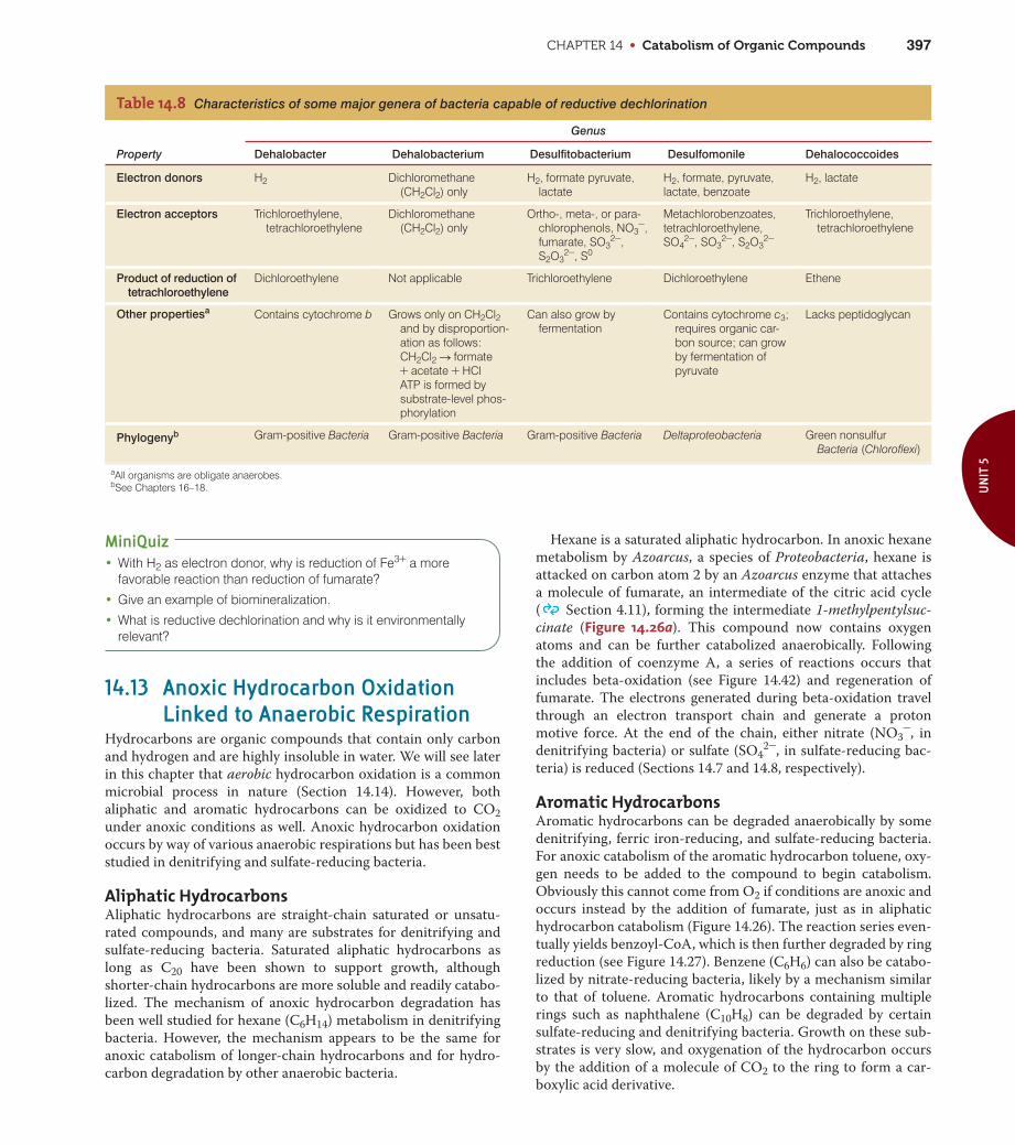
CHAPTER 14 • Catabolism of Organic Compounds 397
UN
IT 5
MiniQuiz• With H2 as electron donor, why is reduction of Fe3+ a more
favorable reaction than reduction of fumarate?
• Give an example of biomineralization.
• What is reductive dechlorination and why is it environmentallyrelevant?
14.13 Anoxic Hydrocarbon OxidationLinked to Anaerobic Respiration
Hydrocarbons are organic compounds that contain only carbon
and hydrogen and are highly insoluble in water. We will see later
in this chapter that aerobic hydrocarbon oxidation is a common
microbial process in nature (Section 14.14). However, both
aliphatic and aromatic hydrocarbons can be oxidized to CO2
under anoxic conditions as well. Anoxic hydrocarbon oxidation
occurs by way of various anaerobic respirations but has been best
studied in denitrifying and sulfate-reducing bacteria.
Aliphatic HydrocarbonsAliphatic hydrocarbons are straight-chain saturated or unsatu-
rated compounds, and many are substrates for denitrifying and
sulfate-reducing bacteria. Saturated aliphatic hydrocarbons as
long as C20 have been shown to support growth, although
shorter-chain hydrocarbons are more soluble and readily catabo-
lized. The mechanism of anoxic hydrocarbon degradation has
been well studied for hexane (C6H14) metabolism in denitrifying
bacteria. However, the mechanism appears to be the same for
anoxic catabolism of longer-chain hydrocarbons and for hydro-
carbon degradation by other anaerobic bacteria.
Hexane is a saturated aliphatic hydrocarbon. In anoxic hexane
metabolism by Azoarcus, a species of Proteobacteria, hexane is
attacked on carbon atom 2 by an Azoarcus enzyme that attaches
a molecule of fumarate, an intermediate of the citric acid cycle
( Section 4.11), forming the intermediate 1-methylpentylsuc-
cinate (Figure 14.26a). This compound now contains oxygen
atoms and can be further catabolized anaerobically. Following
the addition of coenzyme A, a series of reactions occurs that
includes beta-oxidation (see Figure 14.42) and regeneration of
fumarate. The electrons generated during beta-oxidation travel
through an electron transport chain and generate a proton
motive force. At the end of the chain, either nitrate (NO3-, in
denitrifying bacteria) or sulfate (SO42-, in sulfate-reducing bac-
teria) is reduced (Sections 14.7 and 14.8, respectively).
Aromatic HydrocarbonsAromatic hydrocarbons can be degraded anaerobically by some
denitrifying, ferric iron-reducing, and sulfate-reducing bacteria.
For anoxic catabolism of the aromatic hydrocarbon toluene, oxy-
gen needs to be added to the compound to begin catabolism.
Obviously this cannot come from O2 if conditions are anoxic and
occurs instead by the addition of fumarate, just as in aliphatic
hydrocarbon catabolism (Figure 14.26). The reaction series even-
tually yields benzoyl-CoA, which is then further degraded by ring
reduction (see Figure 14.27). Benzene (C6H6) can also be catabo-
lized by nitrate-reducing bacteria, likely by a mechanism similar
to that of toluene. Aromatic hydrocarbons containing multiple
rings such as naphthalene (C10H8) can be degraded by certain
sulfate-reducing and denitrifying bacteria. Growth on these sub-
strates is very slow, and oxygenation of the hydrocarbon occurs
by the addition of a molecule of CO2 to the ring to form a car-
boxylic acid derivative.
Table 14.8 Characteristics of some major genera of bacteria capable of reductive dechlorination
Genus
Property Dehalobacter Dehalobacterium Desulfitobacterium Desulfomonile Dehalococcoides
aAll organisms are obligate anaerobes.bSee Chapters 16–18.
Electron donors H2 Dichloromethane(CH2Cl2) only
H2, formate pyruvate, lactate
H2, formate, pyruvate, lactate, benzoate
H2, lactate
Electron acceptors Trichloroethylene, tetrachloroethylene
Dichloromethane(CH2Cl2) only
Ortho-, meta-, or para-chlorophenols, NO3
-,fumarate, SO3
2-,S2O3
2-, S0
Metachlorobenzoates,tetrachloroethylene,SO4
2-, SO32-, S2O3
2-
Trichloroethylene, tetrachloroethylene
Product of reduction of tetrachloroethylene
Dichloroethylene Not applicable Trichloroethylene Dichloroethylene Ethene
Other propertiesa Contains cytochrome b Grows only on CH2Cl2and by disproportion-ation as follows:CH2Cl2 S formate+ acetate + HClATP is formed bysubstrate-level phos-phorylation
Can also grow by fermentation
Contains cytochrome c3;requires organic car-bon source; can grow by fermentation of pyruvate
Lacks peptidoglycan
Phylogenyb Gram-positive Bacteria Gram-positive Bacteria Gram-positive Bacteria Deltaproteobacteria Green nonsulfur Bacteria (Chloroflexi)

UNIT 5 • Metabolic Diversity and Commercial Biocatalyses398
Figure 14.26 Anoxic catabolism of two hydrocarbons. (a) In anoxic catabolism of the aliphatic hydro-carbon hexane, the addition of fumarate provides the oxygen atoms necessary to form a fatty acid derivativethat can be catabolized by beta-oxidation (see Figure 14.42) to yield acetyl-CoA. Electrons (H) generatedfrom hexane catabolism are used to reduce sulfate or nitrate in anaerobic respirations. (b) Fumarate addi-tion during the anoxic catabolism of the aromatic hydrocarbon toluene forms benzylsuccinate.
H3C
Hexane
1-Methylpentylsuccinate
Acetyl-CoA
Benzylsuccinate
Propionyl-CoA
Anaerobicbenzoyl-CoA pathway
H2O
Succinyl-CoA
Addition offumarate
Removal of CO2and CoA transferto form succinate
Beta-oxidation
CH3
2 H
2 H
Fumarate
SuccinateH3C
CH3 COO–
COO–
HS— CoA
COO–
COO–
COO–
COO–
H3C
3 H3C C S CoA +
To NO3– or SO4
2–
reduction
To NO3– or SO4
2–
reduction
CH3 C S— CoA
O
O
12 H
4 H
H3C CH2 C
O
O
O
S CoA
CO2
6 CO2 + 24 H7 CO2
30 H
(a) Hexane catabolism
Fumarateaddition
Activationwith CoA
To anaerobicrespiration
COO–
COO–
COO–
C S— CoAO
COO–
COO–
COO–
S— CoA
+ HS— CoA
COO–
COO–
COO–
CoA transfer
(b) Toluene catabolism
CH3
~~
~
~
C S— CoAO ~ C S— CoAO ~
Besides the groups of anaerobes listed above, many other
groups of bacteria can catabolize aromatic hydrocarbons anaero-
bically, including fermentative and phototrophic bacteria. How-
ever, except for toluene, only aromatic compounds that contain an
O atom are degraded, and they are typically degraded by a com-
mon mechanism. When we examine the aerobic catabolism of
aromatic compounds (Section 14.14), we will see that the bio-
chemical mechanism occurs by way of ring oxidation (see Figure
14.30). By contrast, under anoxic conditions, the catabolism of
aromatic compounds proceeds by ring reduction. Benzoate catab-
olism by the “benzoyl-CoA pathway” has been the focus of much
of the work in this area, and the purple phototrophic bacterium
Rhodopseudomonas palustris, an organism capable of cataboliz-
ing a wide variety of aromatic compounds, has been a model
experimental organism (Figure 14.27). Benzoate catabolism in
this pathway begins by forming the coenzyme A derivative fol-
lowed by ring cleavage to yield fatty or dicarboxylic acids that can
be further catabolized to intermediates of the citric acid cycle.
Anoxic Oxidation of MethaneMethane (CH4) is the simplest hydrocarbon. In freshwater
ecosystems, methane is produced in anoxic sediments by
methanogens and then oxidized to CO2 by methanotrophs when
it reaches oxic zones. These methanotrophs require O2 for the
catabolism of CH4 because the first step in CH4 oxidation
employs a monooxygenase enzyme (see Section 14.14 and Figure
14.30). However, CH4 can also be oxidized under anoxic condi-
tions in marine and freshwater sediments.
In marine sediments, the anoxic oxidation of methane (AOM)
is catalyzed by cell aggregates that contain both sulfate-reducing

CHAPTER 14 • Catabolism of Organic Compounds 399
UN
IT 5
Figure 14.27 Anoxic degradation of benzoate by the benzoyl-CoA pathway. This pathway operates inthe purple phototrophic bacterium Rhodopseudomonas palustris and many other facultative bacteria, bothphototrophic and chemotrophic. Note that all intermediates of the pathway are bound to coenzyme A. Theacetate produced is further catabolized in the citric acid cycle.
COOH
Benzoate(C7)
CoAC S–CoA
O
4 H
O
H2O
O
OH
2 H 8 H
O
O
H2O
O
2 Acetate +propionyl-CoA
Benzoyl-CoA
COOH
Pimelyl-CoA(C7)
To citricacid cycle
Ringreduction
Ringcleavage
To anaerobicrespiration
Beta-oxidation
Formingbenzoyl-CoA
ATP
~ C S–CoA~ C S–CoA~ C S–CoA~ C S–CoA~
Figure 14.28 Anoxic methane oxidation. (a) Methane-oxidizing cellaggregates from marine sediments. The aggregates contain methano-trophic Archaea (red) surrounded by sulfate-reducing bacteria (green).Each cell type has been stained by a different FISH probe ( Section16.9). The aggregate is about 30 �m in diameter. (b) Mechanism for thecooperative degradation of CH4. An organic compound or some othercarrier of reducing power transfers electrons from methanotroph tosulfate reducer.
(a)
(b)
Ant
je B
oetiu
s an
d A
rmin
Gie
seke
CH4 CO2
SO H2S
e– Organic compounds
42-
Methanotrophic Archaea(ANME-types)
Sulfate-reducing Bacteria
CO2
bacteria and Archaea phylogenetically related to methanogens
(Figure 14.28). However, the archaeal component, called ANME
(anoxic methanotroph), of which there are several types, does
not function in the consortium as a methanogen, but instead as a
methanotroph, oxidizing CH4 as an electron donor. Electrons
from methane oxidation are transferred to the sulfate reducer,
which uses them to reduce SO42- to H2S (Figure 14.28b).
Details of the mechanism of AOM by the two organisms in the
consortia are unclear, but it is thought that the methanotroph
first activates CH4 in some way and then oxidizes it to CO2 by
reversing the steps of methanogenesis, a series of reactions that
would be highly endergonic (Section 14.10). Electrons are gener-
ated during the oxidative steps, but in what form the electrons
are released to the sulfate reducer is unknown. Electrons are not
released as H2. Instead, electrons from the oxidation of CH4 are
shuttled from the methanotroph to the sulfate reducer in some
organic intermediate, such as acetate, formate, or possibly as an
organic sulfide (Figure 14.28b).
Regardless of mechanism, AOM yields only a very small
amount of free energy:
CH4 + SO42- + H+ S CO2 + HS- + 2 H2O DG09 = -18 kJ
How this energy is split between the methanotroph and the sul-
fate reducer is unknown. Substrate-level phosphorylation is
unlikely, but as we have seen several times in this chapter, ion
pumps can operate at these low energy yields and probably play a
role in the energetics of AOM. In addition to oxidizing CH4, the
methanotrophic component of this consortium has been shown
to fix nitrogen ( Section 13.14), and it is possible that this fixed
nitrogen supports the nitrogen needs of the entire consortium.
AOM is not limited to sulfate-reducing bacteria consortia.
Methane-oxidizing denitrifying consortia are active in anoxic envi-
ronments where CH4 and NO3- coexist in significant amounts,
such as certain freshwater sediments. In laboratory enrichments of
these consortia some contain ANME-type methanotrophs while
others are totally free of Archaea. AOM linked to ferric iron (Fe3+)
or manganic ion (Mn4+) reduction also occurs. In both of these
cases ANME-type methanotrophs have been identified, but in
each system different ANME groups seem to predominate.
Notably, however, the free-energy yield of AOM using Mn4+ or
Fe3+ as electron acceptors is considerably more favorable than that
of SO42-, as would be expected from comparison of the reduction
potentials of these different redox couples (Figure 14.11).
A newly discovered denitrifying bacterium employs a remark-
able mechanism for anoxic methanotrophy not seen in any other
methanotrophic system. The organism, provisionally named
Methylomirabilis oxyfera because it is not yet in pure culture, oxi-
dizes CH4 with NO3- as an electron acceptor. During CH4 oxida-
tion, electrons reduce NO3- in steps we have previously seen in
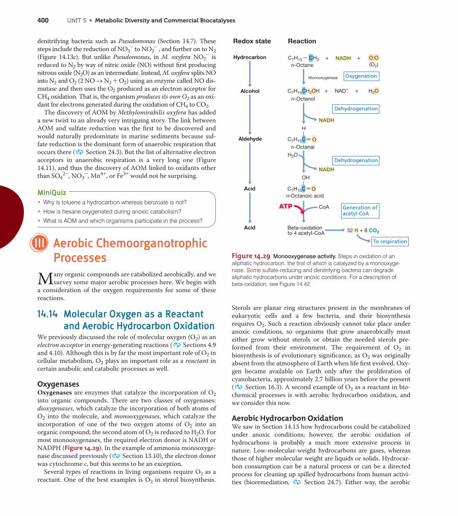
UNIT 5 • Metabolic Diversity and Commercial Biocatalyses400
denitrifying bacteria such as Pseudomonas (Section 14.7). These
steps include the reduction of NO3- to NO2
- , and further on to N2
(Figure 14.13c). But unlike Pseudomonas, in M. oxyfera NO2- is
reduced to N2 by way of nitric oxide (NO) without first producing
nitrous oxide (N2O) as an intermediate. Instead, M. oxyfera splits NO
into N2 and O2 (2 NO S N2 + O2) using an enzyme called NO dis-
mutase and then uses the O2 produced as an electron acceptor for
CH4 oxidation. That is, the organism produces its own O2 as an oxi-
dant for electrons generated during the oxidation of CH4 to CO2.
The discovery of AOM by Methylomirabilis oxyfera has added
a new twist to an already very intriguing story. The link between
AOM and sulfate reduction was the first to be discovered and
would naturally predominate in marine sediments because sul-
fate reduction is the dominant form of anaerobic respiration that
occurs there ( Section 24.3). But the list of alternative electron
acceptors in anaerobic respiration is a very long one (Figure
14.11), and thus the discovery of AOM linked to oxidants other
than SO42-, NO3
-, Mn4+, or Fe3+ would not be surprising.
MiniQuiz• Why is toluene a hydrocarbon whereas benzoate is not?
• How is hexane oxygenated during anoxic catabolism?
• What is AOM and which organisms participate in the process?
III Aerobic ChemoorganotrophicProcesses
Many organic compounds are catabolized aerobically, and we
survey some major aerobic processes here. We begin with
a consideration of the oxygen requirements for some of these
reactions.
14.14 Molecular Oxygen as a Reactantand Aerobic Hydrocarbon Oxidation
We previously discussed the role of molecular oxygen (O2) as an
electron acceptor in energy-generating reactions ( Sections 4.9
and 4.10). Although this is by far the most important role of O2 in
cellular metabolism, O2 plays an important role as a reactant in
certain anabolic and catabolic processes as well.
OxygenasesOxygenases are enzymes that catalyze the incorporation of O2
into organic compounds. There are two classes of oxygenases:
dioxygenases, which catalyze the incorporation of both atoms of
O2 into the molecule, and monooxygenases, which catalyze the
incorporation of one of the two oxygen atoms of O2 into an
organic compound; the second atom of O2 is reduced to H2O. For
most monooxygenases, the required electron donor is NADH or
NADPH (Figure 14.29). In the example of ammonia monooxyge-
nase discussed previously ( Section 13.10), the electron donor
was cytochrome c, but this seems to be an exception.
Several types of reactions in living organisms require O2 as a
reactant. One of the best examples is O2 in sterol biosynthesis.
Sterols are planar ring structures present in the membranes of
eukaryotic cells and a few bacteria, and their biosynthesis
requires O2. Such a reaction obviously cannot take place under
anoxic conditions, so organisms that grow anaerobically must
either grow without sterols or obtain the needed sterols pre-
formed from their environment. The requirement of O2 in
biosynthesis is of evolutionary significance, as O2 was originally
absent from the atmosphere of Earth when life first evolved. Oxy-
gen became available on Earth only after the proliferation of
cyanobacteria, approximately 2.7 billion years before the present
( Section 16.3). A second example of O2 as a reactant in bio-
chemical processes is with aerobic hydrocarbon oxidation, and
we consider this now.
Aerobic Hydrocarbon OxidationWe saw in Section 14.13 how hydrocarbons could be catabolized
under anoxic conditions; however, the aerobic oxidation of
hydrocarbons is probably a much more extensive process in
nature. Low-molecular-weight hydrocarbons are gases, whereas
those of higher molecular weight are liquids or solids. Hydrocar-
bon consumption can be a natural process or can be a directed
process for cleaning up spilled hydrocarbons from human activi-
ties (bioremediation, Section 24.7). Either way, the aerobic
Figure 14.29 Monooxygenase activity. Steps in oxidation of analiphatic hydrocarbon, the first of which is catalyzed by a monooxyge-nase. Some sulfate-reducing and denitrifying bacteria can degradealiphatic hydrocarbons under anoxic conditions. For a description ofbeta-oxidation, see Figure 14.42.
C7H15 CH3 + NADH + O:On-Octane
Oxygenation
To respiration
Dehydrogenation
Dehydrogenation
Generation ofacetyl-CoA
Hydrocarbon
Aldehyde
Acid
Acid
Alcohol
(O2)
C7H15CH2OH + NAD+ + H2O
NADH
C7H15C
H
O
NADH
32 H + 8 CO2
n-Octanoic acid
OH
H2O
CoA
Beta-oxidationto 4 acetyl-CoA
Monooxygenase
n-Octanol
n-Octanal
C7H15C O
ReactionRedox state
ATP

CHAPTER 14 • Catabolism of Organic Compounds 401
UN
IT 5
catabolism of hydrocarbons can be very rapid owing to the meta-
bolic advantage of having O2 available as an electron acceptor
compared with other acceptors of less positive reduction potential
(Figure 14.11).
Several bacteria and fungi can use hydrocarbons as electron
donors to support growth under aerobic conditions. The initial
oxidation step of saturated aliphatic hydrocarbons by these organ-
isms requires O2 as a reactant, and one of the atoms of the oxygen
molecule is incorporated into the oxidized hydrocarbon, typically
at a terminal carbon atom. This reaction is carried out by a
monooxygenase and a typical reaction sequence is shown in
Figure 14.29. The end product of the reaction sequence known as
beta-oxidation (see Figure 14.42) is acetyl-CoA, and this is oxi-
dized in the citric acid cycle along with the production of elec-
trons for the electron transport chain. The sequence is repeated
to progressively degrade long hydrocarbon chains, and in most
cases the hydrocarbon is oxidized completely to CO2.
Aromatic HydrocarbonsMany aromatic hydrocarbons can also be used as electron
donors aerobically by microorganisms. The metabolism of these
compounds, some of which contain several rings such as naph-
thalene or biphenyls, typically has as its initial stage the forma-
tion of catechol or a structurally related compound via catalysis
by oxygenase enzymes, as shown in Figure 14.30. Once catechol
is formed it can be further degraded and cleaved into com-
pounds that can enter the citric acid cycle: succinate, acetyl-
CoA, and pyruvate.
Several steps in the aerobic catabolism of aromatic hydrocar-
bons require oxygenases. Figure 14.30a–c shows four different
oxygenase-catalyzed reactions, one using a monooxygenase,
two using a ring-cleaving dioxygenase, and one using a ring-
hydroxylating dioxygenase. As in aerobic aliphatic hydrocarbon
catabolism, aromatic compounds, whether single or multiple
ringed, are typically oxidized completely to CO2 and electrons
enter an electron transport chain terminating with the reduc-
tion of O2 to H2O.
MiniQuiz• How do monooxygenases differ in function from dioxygenases?
• What is the final product of catabolism of a hydrocarbon?
• What fundamental difference exists in the anaerobic degradationof an aromatic compound compared with its aerobic metabolism?
14.15 Methylotrophy and MethanotrophyMethane (CH4) and many other C1 compounds can be catabolized
aerobically by methylotrophs. Methylotrophs are organisms that
use organic compounds that lack C—C bonds as electron donors
and carbon sources ( Section 17.6). The catabolism of com-
pounds containing only a single carbon atom, such as methane and
methanol (CH3OH), have been the best studied of these substrates.
We focus here on the physiology of methylotrophy, using CH4 as an
example.
Biochemistry of Methane OxidationThe steps in CH4 oxidation to CO2 can be summarized as
CH4 S CH3OH S CH2O S HCOO- S CO2
Methanotrophs are those methylotrophs that can use CH4.
Methanotrophs assimilate either all or one-half of their carbon
(depending on the pathway used) at the oxidation state of
formaldehyde (CH2O). We will see later that this affords a major
energy savings compared with the carbon assimilation of
autotrophs, which also assimilate C1 units, but exclusively from
CO2 rather than organic compounds.
Figure 14.30 Roles of oxygenases in
catabolism of aromatic compounds.
Monooxygenases introduce one atom of oxygenfrom O2 into a substrate, whereas diooxygenasesintroduce both atoms of oxygen. (a) Hydroxylationof benzene to catechol by a monooxygenase inwhich NADH is an electron donor. (b) Cleavage of catechol to cis,cis-muconate by an intradiolring-cleavage dioxygenase. (c) The activities of a ring-hydroxylating dioxygenase and an extradiolring-cleavage dioxygenase in the degradation oftoluene. The oxygen atoms that each enzymeintroduces are distinguished by different colors.Catechol and related compounds are commonintermediates in aerobic aromatic catabolism.Compare aerobic toluene catabolism to anoxictoluene catabolism shown in Figure 14.26b.
O:O
H2O
O
H
H
Benzene Benzene epoxide
H2O
OHH
OHH
OH
OH
Benzenediol Catechol
Benzenemonooxygenase
OH
OH
Catechol
O:O OOH
OHO
C
C
O
O
OH
OH
Monooxygenase
Dioxygenase
Catechol dioxetane (hypothetical)
Catechol1,2-dioxygenase
Methyl catechol2,3-dioxygenase
cis,cis-Muconate
(a)
(b)
Toluene
O:O OH
OH
O
C
C
OH
Sequential dioxygenases
Toluenedioxygenase
(c) CH3 CH3
OH
OH
CH3
O:O
H3C
NADH
NADH
NADH
NADH
O
OH

UNIT 5 • Metabolic Diversity and Commercial Biocatalyses402
Figure 14.31 Oxidation of methane by methanotrophic bacteria.
CH4 is oxidized to CH3OH by the enzyme methane monooxygenase(MMO). A proton motive force is established from electron flow in themembrane, and this fuels ATPase. Note how carbon for biosynthesiscomes from CH2O. Although not depicted as such, MMO is actually amembrane-associated enzyme and methanol dehydrogenase is periplas-mic. FP, flavoprotein; cyt, cytochrome; Q, quinone; MP, methanopterin.
2H
NADHH2OO2
Biosynthesis
CH4 CH3OH CH2O HCOO–
CO2
MMO
2 H+4 H+
+ 4 H+
e–
4 H+ H+Out
In
ADP
NADH
NADH
H2O
QFP
O2
ATP
NADH
cyt aa3cyt ccyt b
3–4
MP
12
Reactions and Bioenergetics of Aerobic MethanotrophyThe initial step in the aerobic oxidation of CH4 is carried out by
the enzyme methane monooxygenase (MMO). As we discussed in
Section 14.14, monooxygenases catalyze the incorporation of
oxygen atoms from O2 into carbon compounds (and into some
nitrogen compounds, Section 13.10), thereby preparing them
for further degradation. Methanotrophy has been especially well
studied in the bacterium Methylococcus capsulatus. This organism
contains two MMOs, one cytoplasmic and the other membrane-
integrated. The electron donor for the cytoplasmic MMO is
NADH, and NADH is probably the electron donor for the
membrane-integrated MMO as well (Figure 14.31).
In the MMO reaction, an atom of oxygen is introduced into
CH4, and CH3OH and H2O are the products. Reducing power for
the first step comes from later oxidative steps in the pathway.
CH3OH is oxidized by a periplasmic dehydrogenase, yielding
formaldehyde and NADH (Figure 14.31). Once CH2O is formed
it is oxidized to CO2 by either of two different pathways. One
pathway uses enzymes that contain the coenzyme tetrahydrofo-
late, a coenzyme widely involved in C1 transformations. The sec-
ond and totally independent pathway employs the coenzyme
methanopterin. Recall that methanopterin is a C1 carrier in inter-
mediate steps of the reduction of CO2 to CH4 by methanogenic
Archaea (Section 14.10 and Figure 14.20). Methanotrophs use a
methanopterin-containing reaction to drive the oxidation of
CH2O to formate plus NADH; formate is then oxidized to CO2 by
the enzyme formate dehydrogenase. However, regardless of the
CH2O oxidation pathway employed, electrons from the oxidation
of CH2O enter the electron transport chain, generating a proton
motive force from which ATP is synthesized (Figure 14.31).
C1 Assimilation into Cell MaterialAs will be discussed in Chapter 17, three phylogenetic groups of
methanotrophs are known and at least two distinct pathways for
C1 incorporation into cell material exist. The serine pathway,
utilized by type II methanotrophs, is outlined in Figure 14.32. In
this pathway, a two-carbon unit, acetyl-CoA, is synthesized from
one molecule of CH2O (produced from the oxidation of CH3OH,
Figure 14.31) and one molecule of CO2. The serine pathway
requires reducing power and energy in the form of two molecules
each of NADH and ATP, respectively, for each acetyl-CoA syn-
thesized. The serine pathway employs a number of enzymes of
the citric acid cycle and one enzyme, serine transhydroxymeth-
ylase, unique to the pathway (Figure 14.32).
The ribulose monophosphate pathway, used by type I
methanotrophs, is outlined in Figure 14.33. This pathway is more
efficient than the serine pathway because all of the carbon for cell
material is derived from CH2O. And, because CH2O is at the
same oxidation level as cell material, no reducing power is
needed. The ribulose monophosphate pathway requires one mol-
ecule of ATP for each molecule of glyceraldehyde 3-phosphate
(G-3-P) synthesized (Figure 14.33). Two G-3-Ps can be converted
into glucose by the glycolytic pathway. Consistent with the lower
Figure 14.32 The serine pathway for the assimilation of C1 units
into cell material by methylotrophic bacteria. The product of the path-way, acetyl-CoA, is used as the starting point for making new cell mate-rial. The key enzyme of the pathway is serine transhydroxymethylase.
CoA
Malyl-CoA
Acetyl-CoA To biosynthesis
Oxalacetate
Phosphoenolpyruvate
GlycerateNH2
NH3
NH3
Serine
Methylenetetrahydrofolate
HCHO
C1 substrate
Serinetranshydroxymethylase
Glyoxylate
Glycine
Hydroxypyruvate
NADH
NADH
OOH
HOOC CH2 CH2 C~S CoAMalate
O
O
O
CH3 C~S CoA
HOCH2 CH COOH
HC COOH
H2N CH2 COOH
HOOC C CH2 COOH
Formaldehyde
Overall: Formaldehyde + CO2 + 2 NADH + 2 ATP acetyl S–CoA + 2 H2O
CoA
CO2
ATP
ATP
~

CHAPTER 14 • Catabolism of Organic Compounds 403
UN
IT 5
Glyceraldehyde-3-P (3 C)
CH2OH
C O
C OHH
C OHH
CH2OPO32–
CH2OPO32–
CH2OPO32–
CH2OPO32–
CH2OPO32–
CH2OPO32–
CH2OH
C OHH
C O
C OHH
C OHH
3 Ribulose-5-P (15 C) 3 Hexulose-6-P (18 C)
HCHO
3 Formaldehyde
Hexulose-P-synthase
Isomerase
2 Fructose-6-P (12 C) + Fructose 1,6-bisphosphate (6 C)
C1 incorporation
CH2OH
C O
C
C OHH
C OHH
CHO
C OHHHHO
Biosynthesis
Sugarrearrangements
Overall: 3 Formaldehyde + ATP glyceraldehyde-3-P
C O
C
C OHH
C OHH
HHO
ATP
energy requirements of the ribulose monophosphate pathway, the
cell yield (grams of cells produced per mole of CH4 oxidized) of
type I methanotrophs is higher than for type II methanotrophs.
The enzymes hexulosephosphate synthase, which condenses
one molecule of formaldehyde with one molecule of ribulose
5-phosphate, and hexulose 6-P isomerase (Figure 14.33) are unique
to the ribulose monophosphate pathway. The remaining enzymes
of this pathway are widely distributed in bacteria. Finally, it should
also be noted that the substrate for the initial reaction in this path-
way, ribulose 5-phosphate, is very similar to the C1 acceptor in the
Calvin cycle, ribulose 1,5-bisphosphate ( Section 13.12), a sig-
nal that these two cycles likely share common evolutionary roots.
MiniQuiz• Why are the energy and reducing power requirements for the
ribulose monophosphate pathway different from those of theserine pathway?
• Why does the oxidation of CH4 to CH3OH require reducingpower?
• Which pathway, the Calvin cycle or the ribulose monophosphatepathway, requires the greater energy input? Why?
14.16 Sugar and PolysaccharideMetabolism
Sugars and polysaccharides are common substrates for chemoor-
ganotrophs, and we briefly consider their catabolism here.
Hexose and Polysaccharide UtilizationSugars containing six carbon atoms, called hexoses, are the most
important electron donors for many chemoorganotrophs and are
also important structural components of microbial cell walls,
capsules, slime layers, and storage products. The most common
sources of hexose in nature are listed in Table 14.9, from which it
can be seen that most are polysaccharides, although a few are
disaccharides. Cellulose and starch are two of the most abundant
natural polysaccharides.
Although both starch and cellulose are composed entirely of
glucose, the glucose units are bonded differently (Table 14.9), and
this profoundly affects their properties. Cellulose is more insolu-
ble than starch and is usually less rapidly digested. Cellulose
forms long fibrils, and organisms that digest cellulose are often
found attached directly to these fibrils (Figure 14.34). In this way
cellulase, the enzyme required to degrade cellulose, can contact
its substrate and begin the digestive process. Many fungi are able
to digest cellulose, and these are mainly responsible for the
decomposition of plant materials on the forest floor. Among
bacteria, however, cellulose digestion is restricted to relatively
few groups, of which the gliding bacteria Sporocytophaga and
Cytophaga (Figure 14.34 and Figure 14.35), clostridia, and actino-
mycetes are the most common.
Anoxic digestion of cellulose is carried out by a few
Clostridium species, which are common in lake sediments, ani-
mal intestinal tracts, and systems for anoxic sewage digestion.
Cellulose digestion is also a major process in the rumen of rumi-
nant animals where Fibrobacter and Ruminococcus species ac-
tively degrade cellulose ( Section 25.9).
Figure 14.33 The ribulose monophosphate pathway for assimila-
tion of C1 units by methylotrophic bacteria. Three molecules of CH2Oare needed to complete the cycle, with the net result being one moleculeof glyceraldehyde 3-phosphate. The key enzyme of this pathway is hexu-lose P-synthase. The sugar rearrangements require enzymes of the pen-tose phosphate pathway (Figure 14.38).
Cellulose fiber Bacteria
B. V
. Hof
sten
Figure 14.34 Cellulose digestion. Transmission electron micrographshowing attachment of the cellulose-digesting bacterium Sporocytophagamyxococcoides to cellulose fibers. Cells are about 0.5 �m in diameter.
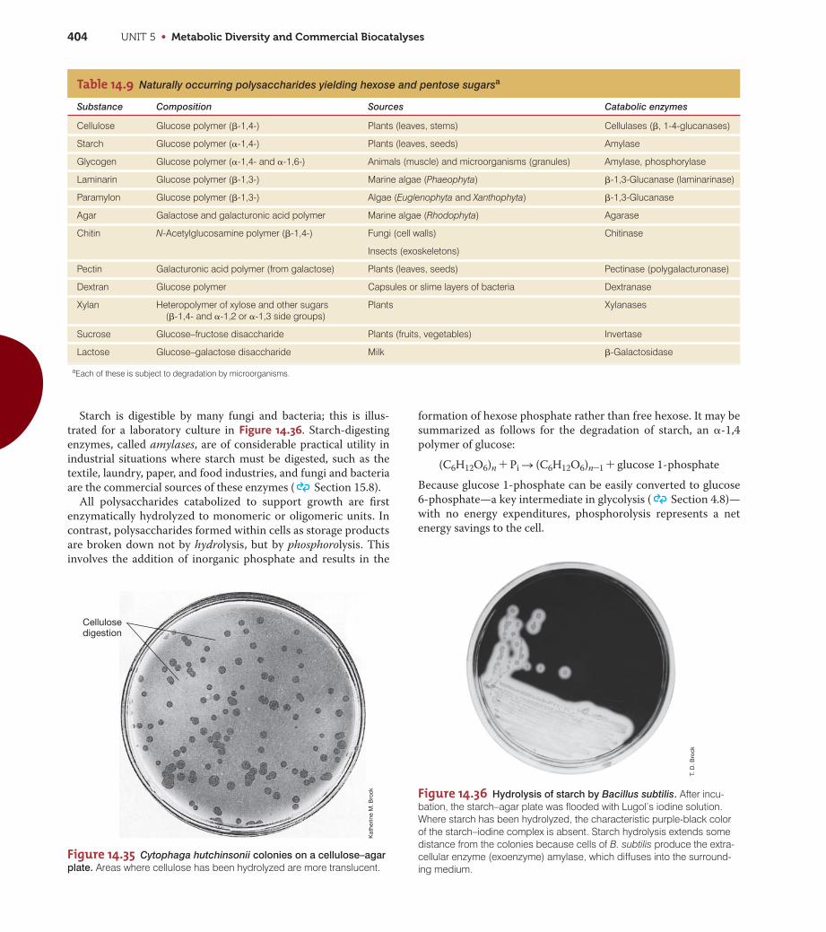
aEach of these is subject to degradation by microorganisms.
UNIT 5 • Metabolic Diversity and Commercial Biocatalyses404
Figure 14.35 Cytophaga hutchinsonii colonies on a cellulose–agar
plate. Areas where cellulose has been hydrolyzed are more translucent.
Kat
herin
e M
. Bro
ck
Cellulosedigestion
T. D
. Bro
ck
Table 14.9 Naturally occurring polysaccharides yielding hexose and pentose sugarsa
Substance Composition Sources Catabolic enzymes
Cellulose Glucose polymer (�-1,4-) Plants (leaves, stems) Cellulases (�, 1-4-glucanases)
Starch Glucose polymer (�-1,4-) Plants (leaves, seeds) Amylase
Glycogen Glucose polymer (�-1,4- and �-1,6-) Animals (muscle) and microorganisms (granules) Amylase, phosphorylase
Laminarin Glucose polymer (�-1,3-) Marine algae (Phaeophyta) �-1,3-Glucanase (laminarinase)
Paramylon Glucose polymer (�-1,3-) Algae (Euglenophyta and Xanthophyta) �-1,3-Glucanase
Agar Galactose and galacturonic acid polymer Marine algae (Rhodophyta) Agarase
Chitin N-Acetylglucosamine polymer (�-1,4-) Fungi (cell walls) Chitinase
Insects (exoskeletons)
Pectin Galacturonic acid polymer (from galactose) Plants (leaves, seeds) Pectinase (polygalacturonase)
Dextran Glucose polymer Capsules or slime layers of bacteria Dextranase
Xylan Heteropolymer of xylose and other sugars (�-1,4- and �-1,2 or �-1,3 side groups)
Plants Xylanases
Sucrose Glucose–fructose disaccharide Plants (fruits, vegetables) Invertase
Lactose Glucose–galactose disaccharide Milk �-Galactosidase
Starch is digestible by many fungi and bacteria; this is illus-
trated for a laboratory culture in Figure 14.36. Starch-digesting
enzymes, called amylases, are of considerable practical utility in
industrial situations where starch must be digested, such as the
textile, laundry, paper, and food industries, and fungi and bacteria
are the commercial sources of these enzymes ( Section 15.8).
All polysaccharides catabolized to support growth are first
enzymatically hydrolyzed to monomeric or oligomeric units. In
contrast, polysaccharides formed within cells as storage products
are broken down not by hydrolysis, but by phosphorolysis. This
involves the addition of inorganic phosphate and results in the
formation of hexose phosphate rather than free hexose. It may be
summarized as follows for the degradation of starch, an �-1,4
polymer of glucose:
(C6H12O6)n + Pi S (C6H12O6)n-1 + glucose 1-phosphate
Because glucose 1-phosphate can be easily converted to glucose
6-phosphate—a key intermediate in glycolysis ( Section 4.8)—
with no energy expenditures, phosphorolysis represents a net
energy savings to the cell.
Figure 14.36 Hydrolysis of starch by Bacillus subtilis. After incu-bation, the starch–agar plate was flooded with Lugol’s iodine solution.Where starch has been hydrolyzed, the characteristic purple-black colorof the starch–iodine complex is absent. Starch hydrolysis extends somedistance from the colonies because cells of B. subtilis produce the extra-cellular enzyme (exoenzyme) amylase, which diffuses into the surround-ing medium.
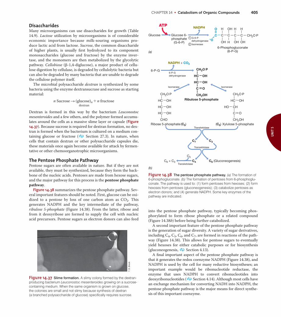
CHAPTER 14 • Catabolism of Organic Compounds 405
UN
IT 5
T. D
. Bro
ck
DisaccharidesMany microorganisms can use disaccharides for growth (Table
14.9). Lactose utilization by microorganisms is of considerable
economic importance because milk-souring organisms pro-
duce lactic acid from lactose. Sucrose, the common disaccharide
of higher plants, is usually first hydrolyzed to its component
monosaccharides (glucose and fructose) by the enzyme inver-
tase, and the monomers are then metabolized by the glycolytic
pathway. Cellobiose (�-1,4-diglucose), a major product of cellu-
lose digestion by cellulase, is degraded by cellulolytic bacteria but
can also be degraded by many bacteria that are unable to degrade
the cellulose polymer itself.
The microbial polysaccharide dextran is synthesized by some
bacteria using the enzyme dextransucrase and sucrose as starting
material:
n Sucrose S (glucose)n + n fructosedextran
Dextran is formed in this way by the bacterium Leuconostoc
mesenteroides and a few others, and the polymer formed accumu-
lates around the cells as a massive slime layer or capsule (Figure14.37). Because sucrose is required for dextran formation, no dex-
tran is formed when the bacterium is cultured on a medium con-
taining glucose or fructose ( Section 27.3). In nature, when
cells that contain dextran or other polysaccharide capsules die,
these materials once again become available for attack by fermen-
tative or other chemoorganotrophic microorganisms.
The Pentose Phosphate PathwayPentose sugars are often available in nature. But if they are not
available, they must be synthesized, because they form the back-
bone of the nucleic acids. Pentoses are made from hexose sugars,
and the major pathway for this process is the pentose phosphate
pathway.
Figure 14.38 summarizes the pentose phosphate pathway. Sev-
eral important features should be noted. First, glucose can be oxi-
dized to a pentose by loss of one carbon atom as CO2. This
generates NADPH and the key intermediate of the pathway,
ribulose 5-phosphate (Figure 14.38). From the latter, ribose and
from it deoxyribose are formed to supply the cell with nucleic
acid precursors. Pentose sugars as electron donors can also feed
into the pentose phosphate pathway, typically becoming phos-
phorylated to form ribose phosphate or a related compound
(Figure 14.38b) before being further catabolized.
A second important feature of the pentose phosphate pathway
is the generation of sugar diversity. A variety of sugar derivatives,
including C4, C5, C6, and C7, are formed in reactions of the path-
way (Figure 14.38). This allows for pentose sugars to eventually
yield hexoses for either catabolic purposes or for biosynthesis
(gluconeogenesis, Section 4.13).
A final important aspect of the pentose phosphate pathway is
that it generates the redox coenzyme NADPH (Figure 14.38), and
NADPH is used by the cell for many reductive biosyntheses; an
important example would be ribonucleotide reductase, the
enzyme that uses NADPH to convert ribonucleotides into
deoxyribonucleotides ( Section 4.14). Although most cells have
an exchange mechanism for converting NADH into NADPH, the
pentose phosphate pathway is the major means for direct synthe-
sis of this important coenzyme.
Figure 14.38 The pentose phosphate pathway. (a) The formation of6-phosphogluconate. (b) The formation of pentoses from 6-phosphoglu-conate. The pathway is used to: (1) form pentoses from hexoses; (2) formhexoses from pentoses (gluconeogenesis); (3) catabolize pentoses aselectron donors; and (4) generate NADPH. Some key enzymes of thepathway are indicated.
ATP
Glucose 6-phosphate
(G-6-P)
1 G-6-Pdehydrogenase
6-P-Gdehydrogenase
NADPH + CO2
NADPH
6-Phosphogluconate(6-P-G)
Isomerase Isomerase
Ribulose 5-phosphate
CH2O P
CH2OH
HC OH
HC OH
C O
Ribose 5-phosphate
CH2O P
CHO
HC OH
HC OH
HC OH
CH2O PC
H
OH
CCC–O
O
C
H
OH H
OH
H
OH
Xylulose 5-phosphateTransketolase
C3C7
C6 (Gluconeogenesis)C6 + C3 C4
C5
(C5) (C5)
CH2O P
CH2OH
HC OH
CHHO
C O
Transaldolase
Transketolase
Glucose
6-P-G
(a)
(b)
2 Isomerase
Figure 14.37 Slime formation. A slimy colony formed by the dextran-producing bacterium Leuconostoc mesenteroides growing on a sucrose-containing medium. When the same organism is grown on glucose, the colonies are small and not slimy because synthesis of dextran (a branched polysaccharide of glucose) specifically requires sucrose.

UNIT 5 • Metabolic Diversity and Commercial Biocatalyses406
Figure 14.39 The glyoxylate cycle. Two unique enzymes, isocitratelyase and malate synthase, operate along with most citric acid cycleenzymes. In addition to growth on pyruvate, the glyoxylate cycle alsofunctions during growth on acetate.
OxalacetateMalate
Glyoxylate (C2)Citrate
Isocitrate
Malate synthase Citrate synthase
Isocitrate lyase
(C6)
(C6)
(C4) (C4)
CO2 CO2
Acetate Acetate2 Pyruvate
HS–CoA HS–CoA
Succinate (C4)
Biosynthesis
Sum: 2 Pyruvate succinate + 2 CO2
(C6) (C4) (C1 2)
Acetyl S–CoAAcetyl S–CoA~ ~
MiniQuiz• What is phosphorolysis?
• What functions does the pentose phosphate pathway play in the cell?
14.17 Organic Acid MetabolismVarious organic acids can be metabolized as carbon sources and
electron donors by microorganisms. The intermediates of the cit-
ric acid cycle, citrate, malate, fumarate, and succinate, are com-
mon natural products formed by plants and are also
fermentation products of microorganisms. Because the citric
acid cycle has major biosynthetic as well as energetic functions
( Section 4.11), the complete cycle or major portions of it are
nearly universal in microorganisms. Thus, it is not surprising
that many microorganisms are able to use citric acid cycle inter-
mediates as electron donors and carbon sources.
Glyoxylate CycleUnlike the utilization of organic acids containing four to six car-
bons, two- or three-carbon acids cannot be used as growth sub-
strates by the citric acid cycle alone. The same is true for
substrates such as hydrocarbons and lipids, degraded via beta-
oxidation to acetyl-CoA (Section 14.18). The citric acid cycle can
continue to operate only if the acceptor molecule, the four-
carbon acid oxalacetate, is regenerated at each turn; any removal
of carbon compounds for biosynthetic purposes would prevent
completion of the cycle ( Figure 4.21).
When acetate is used, the oxalacetate needed to continue the
cycle is produced through the glyoxylate cycle (Figure 14.39), so
named because the C2 compound glyoxylate is a key intermedi-
ate. This cycle is composed of citric acid cycle reactions plus two
additional enzymes: isocitrate lyase, which splits isocitrate into
succinate and glyoxylate, and malate synthase, which converts
glyoxylate and acetyl-CoA to malate (Figure 14.39).
Biosynthesis through the glyoxylate cycle occurs as follows.
The splitting of isocitrate into succinate and glyoxylate allows the
succinate molecule (or another citric acid cycle intermediate
derived from it) to be removed for biosynthesis because glyoxy-
late (C2) combines with acetyl-CoA (C2) to yield malate (C4).
Malate can be converted to oxalacetate to maintain the citric acid
cycle after the C4 intermediate (succinate) has been drawn off.
Succinate is used in the production of porphyrins (needed for
cytochromes, chlorophyll, and other tetrapyrroles). Succinate
can also be oxidized to oxalacetate as a carbon skeleton for C4
amino acids, or it can be converted (via oxalacetate and phospho-
enolpyruvate) to glucose.
Pyruvate and C3 UtilizationThree-carbon compounds such as pyruvate or compounds that
can be converted to pyruvate (for example, lactate or carbohy-
drates) also cannot be catabolized through the citric acid cycle
alone. Because some of the citric acid cycle intermediates are used
for biosynthesis, the oxalacetate needed to keep the cycle going is
synthesized from pyruvate or phosphoenolpyruvate by the addi-
tion of a carbon atom from CO2. In some organisms this step is
catalyzed by the enzyme pyruvate carboxylase:
Pyruvate + ATP + CO2 S oxalacetate + ADP + Pi
whereas in others it is catalyzed by phosphoenolpyruvate car-
boxylase:
Phosphoenolpyruvate + CO2 S oxalacetate + Pi
These reactions replace oxalacetate that is lost when intermedi-
ates of the citric acid cycle are removed for use in biosynthesis,
and the cycle can continue to function.
MiniQuiz• Why is the glyoxylate cycle necessary for growth on acetate but
not on succinate?
14.18 Lipid MetabolismLipids are abundant in nature. The cytoplasmic membranes of all
cells contain lipids, and many organisms produce lipid storage
materials and contain lipids in their cell walls. These substances
are biodegradable and are excellent substrates for microbial
energy-yielding metabolism. When cells die, their lipids are thus
catabolized, with CO2 being the final product.
Fat and Phospholipid HydrolysisFats are esters of glycerol and fatty acids and are readily available
from the release of lipids from dead organisms. Microorganisms

CHAPTER 14 • Catabolism of Organic Compounds 407
UN
IT 5
Phospholipase activity: fattyacids released, leading to eggyolk precipitation
Clostridiumperfringens
Inhibitor added: no phospholipase action,thus no precipitation of egg yolk
G. H
obb
s
Figure 14.41 Lipases. (a) Activity of lipases on a fat. (b) Phospholi-pase activity on phospholipid. The cleavage sites of the four distinctphospholipases A, B, C, and D are shown. X refers to a number of smallorganic molecules that may be at this position in different phospholipids.
Glycerol
H2C
H2C
O Fatty acid
HC O Fatty acid
O Fatty acid
Lipase
(a)
O Fatty acid
HC O Fatty acid
O P
(b)
Phospholipase B
Phospholipase A
O (X)
O
OH
Phospholipase C
Phospholipase D
H2C
H2C
use fats only after hydrolysis of the ester bond, and extracellular
enzymes called lipases are responsible for the reaction (Figures14.40 and 14.41). Lipases attack fatty acids of various chain
lengths. Phospholipids are hydrolyzed by enzymes called
phospholipases, each of which is given a different letter designa-
Figure 14.40 Phospholipase activity. Enzyme activity of phospholi-pase around a streak of Clostridium perfringens growing on an agarmedium containing egg yolk. On half of the plate an inhibitor of phospho-lipase was added, preventing activity of the enzyme.
Figure 14.42 Beta-oxidation. Beta-oxidation of a fatty acid leading tothe successive formation of acetyl-CoA.
~
~
~
~
~
H3C (CH2)n
H3C (CH2)n
H3C (CH2)n
H3C (CH2)n
H3C (CH2)n
H3C (CH2)n CH2 CH2 COOH
Fatty acid of (n + 4) carbons
CoA activation
Formation ofdouble bond
Addition ofhydroxyl group
Oxidationto keto group
To citric acid cycle
Cleavage to yield acetyl-CoA andfatty acid of (n + 2) carbons fornew round of beta-oxidation
CH2 CH2 C S–CoA
CH2 C S–CoA
O
α
CH2 C S–CoA
O
CH2 C S–CoA
S CoA +
O
O
β
FADH
CH
H2O
HS CoA
CH
NADH
C
CoA
O
C
O
Acetyl-CoA
H3C C S–CoA
O
OH
2 CO2 + 8 H
ATP
tion depending on which ester bond it cleaves in the lipid (Figure
14.41). Phospholipases A and B cleave fatty acid esters, whereas
phospholipases C and D cleave phosphate esters and hence are
different classes of enzymes. The result of lipase activity is the
release of free fatty acids and glycerol, and these substances can
then be metabolized by chemoorganotrophic microorganisms.
Fatty Acid OxidationFatty acids are oxidized by beta-oxidation, a series of reactions in
which two carbons of the fatty acid are split off at a time (Figure14.42). The fatty acid is first activated with coenzyme A; oxida-
tion results in the release of acetyl-CoA by cleavage between the
� and � carbons of the original fatty acid along with the forma-
tion of a new fatty acid two carbon atoms shorter (Figure 14.42).
The process of beta-oxidation is then repeated, and another
acetyl-CoA molecule is released.
There are two separate dehydrogenation reactions in beta-
oxidation. In the first, electrons are transferred to flavin ade-
nine dinucleotide (FAD), forming FADH, whereas in the
second they are transferred to NAD+, forming NADH. Most

UNIT 5 • Metabolic Diversity and Commercial Biocatalyses408
rated fatty acid palmitic acid can in theory generate 129 ATP
molecules. These include oxidative phosphorylation from elec-
trons generated during the formation of acetyl-CoA from beta-
oxidations and from oxidation of the acetyl-CoA units
themselves through the citric acid cycle.
MiniQuiz• What are phospholipases and what do they do?
• How many electrons are released for every acetyl-CoA producedby beta oxidation of a fatty acid? For every acetyl-CoA oxidizedto CO2?
fatty acids in a cell have an even number of carbon atoms,
and complete oxidation yields acetyl-CoA. If odd-chain or
branched-chain fatty acids are catabolized, propionyl-CoA or a
branched-chain fatty acid–CoA remains after beta-oxidation,
and these are either further metabolized to acetyl-CoA by
ancillary reactions or excreted from the cell. The acetyl-CoA
formed is then oxidized by the citric acid cycle or is converted
to hexose and other cell constituents via the glyoxylate cycle
(Figure 14.39).
Because they are highly reduced, fatty acids are excellent elec-
tron donors. For example, the oxidation of the 16-carbon satu-
Big Ideas14.1In the absence of external electron acceptors, organic com-
pounds can be catabolized anaerobically only by fermentation. A
requirement for most fermentations is formation of an energy-
rich organic compound that can yield ATP by substrate-level
phosphorylation. Redox balance must also be achieved in fer-
mentations, and H2 production is a key means of disposing of
excess electrons.
14.2 The lactic acid fermentation is carried out by homofermentative
and heterofermentative species. The mixed-acid fermentation
results in acids plus neutral products (ethanol, butanediol),
depending on the organism.
14.3 Clostridia ferment sugars, amino acids, and other organic com-
pounds. Propionibacterium produces propionate and acetate in a
secondary fermentation of lactate.
14.4 The energy physiology of Propionigenium, Oxalobacter, and
Malonomonas is linked to decarboxylation reactions that pump
Na+ or H+ across the membrane. The reactions catalyzed by
these organisms yield insufficient energy to make ATP by sub-
strate-level phosphorylation.
14.5 In syntrophy two organisms cooperate to degrade a compound
that neither can degrade alone. In this process H2 produced by
one organism is consumed by the partner. H2 consumption
affects the energetics of the reaction carried out by the H2 pro-
ducer, allowing it to make ATP where it otherwise could not.
14.6 Although O2 is the most widely used electron acceptor in energy-
yielding metabolism, certain other compounds can be used as
electron acceptors. Anaerobic respiration yields less energy than
aerobic respiration but can proceed in environments where O2 is
absent.
14.7 Nitrate is a common electron acceptor in anaerobic respiration.
Nitrate reduction is catalyzed by the enzyme nitrate reductase,
reducing NO3- to NO2
-. Many bacteria that use NO3- in anaero-
bic respiration reduce it past NO2- to produce gaseous nitrogen
compounds (denitrification).
14.8 Sulfate-reducing bacteria reduce SO4
2- to H2S. This process
requires activation of SO42- by ATP to form adenosine
phosphosulfate (APS) and reduction by H2 or organic electron
donors. Disproportionation is an additional energy-yielding
strategy for certain species. Some organisms, such as Desul-
furomonas, cannot reduce SO42- but produce H2S from the
reduction of S0.
14.9 Acetogens are anaerobes that reduce CO2 to acetate, usually with
H2 as electron donor. The mechanism of acetate formation is the
acetyl-CoA pathway, a pathway widely distributed in obligate
anaerobes for either autotrophic purposes or acetate catabolism.
14.10 Methanogenesis is the production of CH4 from CO2 + H2 or
from acetate or methanol by strictly anaerobic methanogenic
Archaea. Several unique coenzymes are required for methano-
genesis, and energy conservation is linked to either a proton or a
sodium motive force.
14.11 The hyperthermophile Pyrococcus furiosus ferments glucose in
an unusual fashion, reducing protons in an anaerobic respiration
linked to ATPase activity.

14.12 Besides inorganic nitrogen and sulfur compounds and CO2, sev-
eral other substances can function as electron acceptors for
anaerobic respiration. These include Fe3+, Mn4+, fumarate, and
certain organic and chlorinated organic compounds.
14.13 Hydrocarbons can be oxidized under anoxic conditions, but oxygen
must first be added to the molecule. This occurs by the addition of
fumarate. Aromatic compounds are catabolized anaerobically by
ring reduction and cleavage to form intermediates that can be
catabolized in the citric acid cycle. Methane can be oxidized under
anoxic conditions by consortia containing sulfate-reducing or deni-
trifying bacteria and methanotrophic Archaea.
14.14 In addition to its role as an electron acceptor, O2 can also be a
substrate; enzymes called oxygenases introduce atoms of oxygen
from O2 into a biochemical compound. Aerobic hydrocarbon
oxidation is widespread in nature, and oxygenase enzymes are
key to these catalyses. Unlike in anaerobic aromatic catabolism,
the aerobic degradation of aromatic compounds proceeds by ring
oxidation.
14.15 Methanotrophy is the use of CH4 as both carbon source and elec-
tron donor, and the enzyme methane monooxygenase is a key
enzyme in the catabolism of methane. In methanotrophs C1 units
are assimilated into cell material by either the ribulose monophos-
phate pathway or the serine pathway.
14.16 Polysaccharides are abundant in nature and can be broken down
into hexose or pentose monomers and used as sources of both
carbon and electrons. Starch and cellulose are common polysac-
charides. The pentose phosphate pathway is the major means for
generating pentose sugars for biosynthesis.
14.17 Organic acids are typically metabolized through the citric acid
cycle or the glyoxylate cycle. Isocitrate lyase and malate synthase
are the key enzymes of the glyoxylate cycle.
14.18 Fats are hydrolyzed by lipases or phospholipases to fatty acids
plus glycerol. The fatty acids are oxidized by beta-oxidation reac-
tions to acetyl-CoA, which is then oxidized to CO2 by the citric
acid cycle.
Acetogenesis energy metabolism in which
acetate is produced from either H2 plus CO2
or from organic compounds
Acetyl-CoA pathway a pathway of autotrophic
CO2 fixation and acetate oxidation widespread
in obligate anaerobes including methanogens,
acetogens, and sulfate-reducing bacteria
Anaerobic respiration respiration in which
some substance, such as SO42- or NO3
–, is
used as a terminal electron acceptor instead
of O2
Anoxic oxygen-free
Denitrification anaerobic respiration in which
NO3– or NO2
– is reduced to nitrogen gases,
primarily N2
Fermentation anaerobic catabolism of an
organic compound in which the compound
serves as both an electron donor and an elec-
tron acceptor and in which ATP is usually
produced by substrate-level phosphorylation
Glyoxylate cycle a series of reactions including
some citric acid cycle reactions that are used
for aerobic growth on C2 or C3 organic acids
Heterofermentative producing a mixture of
products, typically lactate, ethanol, and CO2,
from the fermentation of glucose
Homofermentative producing only lactic acid
from the fermentation of glucose
Hydrogenase an enzyme, widely distributed in
anaerobic microorganisms, capable of oxidiz-
ing or evolving H2
Methanogen an organism that produces
methane (CH4)
Methanogenesis the biological production of
CH4
Methanotroph an organism that can oxidize
CH4
Methylotroph an organism capable of growth
on compounds containing no C—C bonds;
some methylotrophs are methanotrophic
Oxygenase an enzyme that catalyzes the incor-
poration of oxygen from O2 into organic or
inorganic compounds
Pentose phosphate pathway a major metabolic
pathway for the production and catabolism of
pentoses (C5 sugars)
Reductive dechlorination (dehalorespira-
tion) an anaerobic respiration in which a
chlorinated organic compound is used as an
electron acceptor, usually with the release
of Cl–
Ribulose monophosphate pathway a reaction
series in certain methylotrophs in which
formaldehyde is assimilated into cell material
using ribulose monophosphate as the C1
acceptor molecule
Secondary fermentation a fermentation in
which the substrates are the fermentation
products of other organisms
Serine pathway a reaction series in certain
methylotrophs in which CH2O plus CO2 are
assimilated into cell material by way of the
amino acid serine
Stickland reaction the fermentation of an
amino acid pair
Syntrophy a process whereby two or more
microorganisms cooperate to degrade a
substance neither can degrade alone
Review of Key Terms
CHAPTER 14 • Catabolism of Organic Compounds 409

UNIT 5 • Metabolic Diversity and Commercial Biocatalyses410
1. Define the term substrate-level phosphorylation. How does it differ
from oxidative phosphorylation? Assuming an organism is faculta-
tive, what cultural conditions dictate whether the organism obtains
energy from substrate-level rather than oxidative phosphorylation
(Section 14.1)?
2. Although many different compounds are theoretically fermentable,
in order to support a fermentative process, most organic com-
pounds must eventually be converted to one of a relatively small
group of molecules. What are these molecules and why must they
be produced (Sections 14.1–14.3)?
3. Give an example of a fermentation that does not employ substrate-
level phosphorylation. How is energy conserved in this fermenta-
tion (Section 14.4)?
4. Why is syntrophy also called “interspecies H2 transfer” (Section
14.5)?
5. Why is NO3– a better electron acceptor for anaerobic respiration
than is SO42- (Section 14.6)?
6. In Escherichia coli, synthesis of the enzyme nitrate reductase is
repressed by O2. On the basis of bioenergetic arguments, why do
you think this repression phenomenon might have evolved
(Section 14.7)?
7. Why is hydrogenase a constitutive enzyme in Desulfovibrio
(Section 14.8)?
8. Compare and contrast acetogens with methanogens in terms of
(1) substrates and products of their energy metabolism, (2) ability
to use organic compounds as electron donors in energy metabo-
lism, and (3) phylogeny (Sections 14.9 and 14.10).
9. Why can it be said that in glycolysis in Pyrococcus furiosus, both
fermentation and anaerobic respiration are occurring at the same
time (Section 14.11)?
10. Compare and contrast ferric iron reduction with reductive dechlo-
rination in terms of (1) product of the reduction and (2) environ-
mental significance (Section 14.12).
11. How do denitrifying and sulfate-reducing bacteria degrade hydro-
carbons without the participation of oxygenase enzymes (Sections
14.13–14.14)?
12. How do monooxygenases differ from dioxygenases in the reactions
they catalyze? Why are oxygenases necessary for the aerobic catab-
olism of hydrocarbons (Section 14.14)?
13. How does a methanotroph differ from a methanogen? How do type
I and type II methanotrophs differ in their carbon assimilation pat-
terns (Section 14.15)?
14. Compare and contrast the conversion of cellulose and intracellular
starch to glucose units. What enzymes are involved and which
process is the more energy efficient (Section 14.16)?
15. What is the major function of the glyoxalate cycle (Section 14.17)?
16. What is the product of the beta-oxidation of a fatty acid? How is
this product oxidized to CO2 (Section 14.18)?
Review Questions
1. When methane is made from CO2 (plus H2) or from methanol
(in the absence of H2), various steps in the pathway shown in
Figures 14.20 and 14.21 are used. Compare and contrast methano-
genesis from these two substrates and discuss why they must be
metabolized in opposite directions.
2. Although dextran is a glucose polymer, glucose cannot be used to
make dextran. Explain. How is dextran synthesis important in oral
hygiene ( Section 27.3)?
3. A fatty acid such as butyrate cannot be fermented in pure culture,
although its anaerobic catabolism under other conditions occurs
readily. How do these conditions differ, and why does the latter
allow for butyrate catabolism? How then can butyrate be fer-
mented in mixed culture?
Application Questions
Need more practice? Test your understanding with Quantitative Questions; access additional
study tools including tutorials, animations, and videos; and then test your knowledge with
chapter quizzes and practice tests at www.microbiologyplace.com.
Top Related