![Jesus heals[1]](https://static.fdocuments.net/doc/165x107/55a660951a28ab56538b468c/jesus-heals1.jpg)
Zephyr Endobronchial Valve Treatment - pulmonx.com.au · • Pneumothorax – a tear in the lung...
2
Zephyr ® Endobronchial Valve Treatment for Patients with Advanced Emphysema Patient Guide How is the treatment performed? 2mm Anschnitt P0336EN_A Endobronchial valves allowing air and secretions to flow out but not back into the diseased lobe of the lung. Endobronchial valves for lung volume reduction. The valves are between 4 and 5.5 mm in diameter and 10-13 mm in length. Before endobronchial valve treatment you will receive anes- thesia to make the treatment as comfortable as possible. Your airways will be examined using a bronchoscope (a fiber optic camera). Through the bronchoscope your airways will be assessed for presence of channels that communicate between the lobes of your lungs (collateral ventilation). If little or no collateral ventilation is detected, valves (normally 3-5) will be placed in the suitable lobe. The procedure will take approximately 60 minutes. After you have been treated, you will stay in the hospital after the procedure for observation and management of any possible complications. Are there any risks and complications associated with an endobronchial valve lung volume reduction? The most common complications are: • Pneumothorax – a tear in the lung which causes air to leak into the chest cavity. This leak normally heals by itself after placement of a chest drain. In rare cases this can be a serious/life-threatening complication. • Infection • Bleeding • Occasionally a valve can become dislodged and coughed out • No benefit from treatment If, after discharge from hospital, you experience any of the conditions below contact your doctor immediately: • Shortness of breath • Chest pain • Coughing of blood Should you cough up a valve, you should contact the hospital or physician who performed the procedure. The treatment is designed to be permanent and you will have the (removeable) valves in place as long as you benefit from them. Pulmonx Australia E: [email protected] PH: 1300 785 666 Please visit our website: www.pulmonx.com FV2_PX197317_PatientGuide_Australia_DIN lang_6p_P0336EN_A.indd 1-3 01.06.17 12:47
Transcript of Zephyr Endobronchial Valve Treatment - pulmonx.com.au · • Pneumothorax – a tear in the lung...

Zephyr®
Endobronchial Valve Treatment
for Patients with Advanced Emphysema
Patient Guide
How is the treatment performed?
2mm Anschnitt
P033
6EN_
A
Endobronchial valves allowing air and secretions to fl ow out but not back into the diseased lobe of the lung.
Endobronchial valves for lung volume reduction. The valves are between 4 and 5.5 mm in diameter and 10-13 mm in length.
Before endobronchial valve treatment you will receive anes-thesia to make the treatment as comfortable as possible. Your airways will be examined using a bronchoscope (a fi ber optic camera). Through the bronchoscope your airways will be assessed for presence of channels that communicate between the lobes of your lungs (collateral ventilation). If little or no collateral ventilation is detected, valves (normally 3-5) will be placed in the suitable lobe. The procedure will take approximately 60 minutes.
After you have been treated, you will stay in the hospital after the procedure for observation and management of any possible complications.
Are there any risks and complications associated with an endobronchial valve lung volume reduction? The most common complications are:
• Pneumothorax – a tear in the lung which causes air to leak into the chest cavity. This leak normally heals by itself after placement of a chest drain. In rare cases this can be a serious/life-threatening complication.
• Infection
• Bleeding
• Occasionally a valve can become dislodged and coughed out
• No benefi t from treatment
If, after discharge from hospital, you experience any of the conditions below contact your doctor immediately:
• Shortness of breath
• Chest pain
• Coughing of blood
Should you cough up a valve, you should contact the hospital or physician who performed the procedure.
The treatment is designed to be permanent and you will have the (removeable) valves in place as long as you benefi t from them.
Pulmonx AustraliaE: [email protected]: 1300 785 666
Please visit our website: www.pulmonx.com
FV2_PX197317_PatientGuide_Australia_DIN lang_6p_P0336EN_A.indd 1-3 01.06.17 12:47
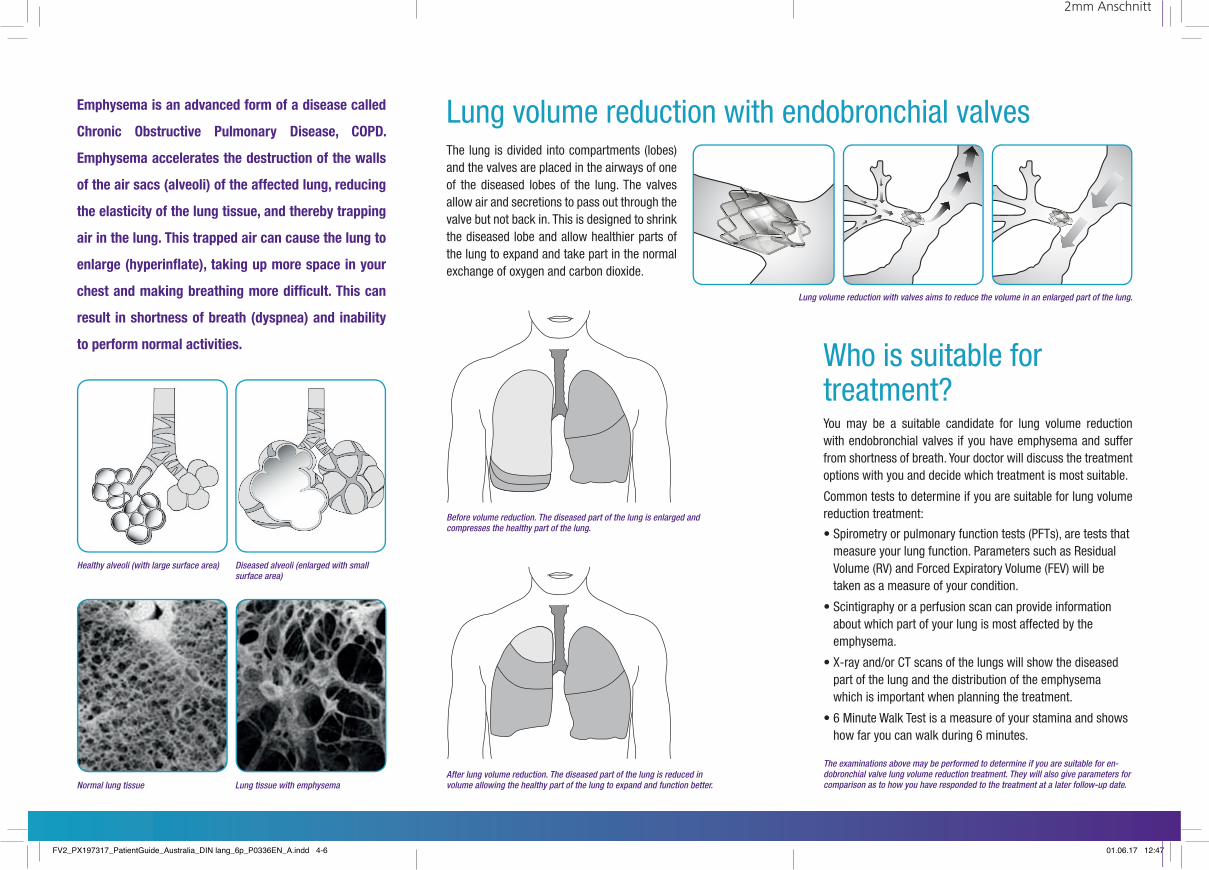
Healthy alveoli (with large surface area) Diseased alveoli (enlarged with small surface area)
Normal lung tissue Lung tissue with emphysema
Before volume reduction. The diseased part of the lung is enlarged and compresses the healthy part of the lung.
After lung volume reduction. The diseased part of the lung is reduced in volume allowing the healthy part of the lung to expand and function better.
Lung volume reduction with valves aims to reduce the volume in an enlarged part of the lung.
Who is suitable for treatment?
2mm Anschnitt
Lung volume reduction with endobronchial valvesEmphysema is an advanced form of a disease called
Chronic Obstructive Pulmonary Disease, COPD.
Emphysema accelerates the destruction of the walls
of the air sacs (alveoli) of the affected lung, reducing
the elasticity of the lung tissue, and thereby trapping
air in the lung. This trapped air can cause the lung to
enlarge (hyperinfl ate), taking up more space in your
chest and making breathing more diffi cult. This can
result in shortness of breath (dyspnea) and inability
to perform normal activities.
The lung is divided into compartments (lobes) and the valves are placed in the airways of one of the diseased lobes of the lung. The valves allow air and secretions to pass out through the valve but not back in. This is designed to shrink the diseased lobe and allow healthier parts of the lung to expand and take part in the normal exchange of oxygen and carbon dioxide.
• Spirometry or pulmonary function tests (PFTs), are tests that measure your lung function. Parameters such as Residual Volume (RV) and Forced Expiratory Volume (FEV) will be taken as a measure of your condition.
• Scintigraphy or a perfusion scan can provide information about which part of your lung is most affected by the emphysema.
• X-ray and/or CT scans of the lungs will show the diseased part of the lung and the distribution of the emphysema which is important when planning the treatment.
• 6 Minute Walk Test is a measure of your stamina and shows how far you can walk during 6 minutes.
You may be a suitable candidate for lung volume reduction with endobronchial valves if you have emphysema and suffer from shortness of breath. Your doctor will discuss the treatment options with you and decide which treatment is most suitable.
Common tests to determine if you are suitable for lung volume reduction treatment:
The examinations above may be performed to determine if you are suitable for en-dobronchial valve lung volume reduction treatment. They will also give parameters for comparison as to how you have responded to the treatment at a later follow-up date.
FV2_PX197317_PatientGuide_Australia_DIN lang_6p_P0336EN_A.indd 4-6 01.06.17 12:47