Ze Bra Fish Ba RU
40
Background Paper on FISH EMBRYO TOXICITY ASSAYS (UBA Contract Number 203 85 422) March 30, 2006 Prepared for GERMAN FEDERAL ENVIRONMENT AGENCY PO Box 1406 · D-06813 Dessau Thomas Braunbeck & Eva Lammer Aquatic Ecology & Toxicology Department of Zoology University of Heidelberg Im Neuenheimer Feld 230 D-69120 Heidelberg Germany
Transcript of Ze Bra Fish Ba RU
7/26/2019 Ze Bra Fish Ba RU
http://slidepdf.com/reader/full/ze-bra-fish-ba-ru 1/40
Background Paper
on
FISH EMBRYO TOXICITY ASSAYS
(UBA Contract Number 203 85 422)
March 30, 2006
Prepared for
GERMAN FEDERAL ENVIRONMENT AGENCY
PO Box 1406 · D-06813 Dessau
Thomas Braunbeck & Eva Lammer
Aquatic
Ecology & Toxicology
Department of Zoology
University of Heidelberg
Im Neuenheimer Feld 230
D-69120 Heidelberg
Germany

7/26/2019 Ze Bra Fish Ba RU
http://slidepdf.com/reader/full/ze-bra-fish-ba-ru 2/40
03/30/2006 2/298 UBA Contract No. 203 85 122

7/26/2019 Ze Bra Fish Ba RU
http://slidepdf.com/reader/full/ze-bra-fish-ba-ru 3/40
TABLE OF CONTENTS
Page
1.0 Executive summary 7
2.0 Introduction 92.1 Fish acute toxicity testing 9
2.2 Why tests on fish? 9
2.3 Fish in vitro tests 10
2.4 Fish embryo tests 12
2.5 Purpose of the present document 13
3.0 Methodological basis for fish embryo toxicity testing 14
3.1 Fish, fish maintenance and spawning procedure 14
3.1.1 Zebrafish 14
3.1.2 Fathead minnow 36
3.1.3 Japanese medaka 383.1.4 Other fish species used for embryo toxicity tests 45
3.2 General description of endpoints used for fish embryo toxicity tests 46
3.2.1 Endpoints used for the acute zebrafish embryo whole effluent toxicity test in
Germany (“fish egg test”)
46
3.2.2 Additional endpoints used for acute zebrafish embryo toxicity tests 48
3.2.3 Further potential endpoints in fish embryo toxicity endpoints 50
3.3 Validity criteria 50
4.0 Embryo toxicity tests with the zebrafish ( Danio rerio) 51
5.0 Embryo toxicity tests with the fathead minnow ( Pimephales promelas) 91
6.0 Embryo toxicity tests with the Japanese medaka (Oryzias latipes) 92
7.0 Embryo toxicity tests with other fish species 96
8.0 Correlation between embryo toxicity tests with different fish species 99
9.0 Statistical considerations 102
9.1 Inter-laboratory transferability of the zebrafish embryo toxicity test 102
9.2 Correlation of selected zebrafish embryo toxicity data to conventional acute
fish toxicity data according to a statistical analysis by Ratte and Hammers-
Wirtz (2003)
104
9.3 Correlation of all fish embryo toxicity data to conventional acute fish toxic-
ity data on the basis of data compiled in the present review
105
10.0 Scientific and ethical considerations as to the duration of exposure 116
11.0 Recommended protocol and testing strategy 119
12.0 Additional fields of application for the fish embryo test 119
13.0 References 123
ANNEX 135
03/30/2006 3/298 UBA Contract No. 203 85 122

7/26/2019 Ze Bra Fish Ba RU
http://slidepdf.com/reader/full/ze-bra-fish-ba-ru 4/40
List of tables
Page
Table 1 Maintenance, breeding and typical conditions for embryo toxicity tests
with the common OECD test fish species
15
Table 2 Stages of embryonic development of the zebrafish ( Danio rerio) at
26 ± 1 °C (Nagel, 2002)
17
Table 3 Endpoints of acute toxicity originally discussed by the German DIN
working group for whole effluent toxicity tests (DIN 38415-6; “fish egg
test”)
47
Table 4 Lethal and sublethal endpoints for evaluating the toxicity and teratogenic-
ity of chemicals on zebrafish embryos as listed by Nagel (2002, modi-
fied; according to Schulte and Nagel, 1994; Nagel, 1998; Bachmann,
2002)
48
Table 5 Embryo toxicity data from experiments with zebrafish ( Danio rerio) 53Table 6 Embryo toxicity data from experiments with fathead minnow (Pimepha-
les promelas)
89
Table 7 Embryo toxicity data from experiments with Japanese medaka (Oryzias
latipes)
93
Table 8 Embryo toxicity data from experiments with fish other than zebrafish,
medaka or fathead minnow
97
Table 9 EC50 data for selected reference compounds from embryo tests with ze-
brafish ( Danio rerio), Japanese medaka (Oryzias latipes) and fathead
minnow (Pimephales promelas). Toxicological endpoints as listed by
German DIN standards
99
Table 10 LC50 data (mg/L) for lethal effects by 20 selected substances in a German
national inter-laboratory calibration exercise.
102
Table 11 EC50 data (mg/L) for lethal and sublethal effects by 21 selected sub-
stances in a German national inter-laboratory calibration exercise.
103
Table 12 Comparison of critical parameters for the correlation between fish em-
bryo toxicity data and conventional acute fish toxicity (LC50)
108
Table 13 Comparison of critical parameters for the correlation between fish em-
bryo toxicity data and conventional acute fish toxicity (LC50) following
log-transformation
109
03/30/2006 4/298 UBA Contract No. 203 85 122

7/26/2019 Ze Bra Fish Ba RU
http://slidepdf.com/reader/full/ze-bra-fish-ba-ru 5/40
List of figures (incl. credits for figures)
Page
Fig. 1 Adult zebrafish ( Danio rerio) female (upper individual) and male
(lower individual)
14
Fig. 2a - d Selected stages of early zebrafish ( Danio rerio) development (from
Kimmel et al., 1995).
18
Fig. 3a - f Normal development of zebrafish ( Danio rerio) embryos 22
Fig. 3g Mortality in early zebrafish ( Danio rerio) eggs: coagulation (I),
heavy infestation with fungi (II) and invasion of Vorticella spec.
(III).
27
Fig. 4 Setup of the tanks used for breeding zebrafish in the Heidelberg labo-
ratory
28
Fig. 5 Setup of the tanks used for breeding zebrafish in the Berlin labora-
tory (courtesy of Dr. T. Meinelt, IGB)
29
Fig. 6 Oxygen consumption of control zebrafish ( Danio rerio) embryos in
relation to the amount of oxygen provided in the medium
30
Fig. 7 Oxygen consumption of control zebrafish ( Danio rerio) embryos in
dependence from the stocking density in given volume of medium
30
Fig. 8 Toxicity of 3,4-dichloroaniline to zebrafish ( Danio rerio) embryos in
various volumes of medium
31
Fig. 9 Toxicity of 2,4-dinitrophenol to zebrafish ( Danio rerio) embryos in
various volumes of medium
32
Fig. 10 Effect of the exposure start (time lapse after fertilization) on the cu-
mulative mortality of 2,4-dinitrophenol. After 24 and 48 h, it is evi-dent that mortalities are increased with early exposure start.
32
Fig. 11 Effect of the exposure start (time lapse after fertilization) on the cu-
mulative mortality of 3,4-dichloroaniline. After 24 and 48 h, it is
evident that mortalities are increased with early exposure start.
33
Fig. 12 Effects of potassium chromate to zebrafish ( Danio rerio) embryos w
and w/o dechorionation
34
Fig. 13 Toxicity of 4-chloroaniline to zebrafish ( Danio rerio) embryos w and
w/o dechorionation
35
Fig. 14 Fathead minnow (Pimephales promelas) 36
Fig. 15 Normal development of normal fathead minnow (Pimephales prome-las) embryos and embryos exposed to 15 mg/L 3,4-dichloroaniline 37
Fig. 16 Japanese medaka (Oryzias latipes) 39
Fig. 17 Normal development of Japanese medaka (Oryzias latipes) embryos
I - IV
40
Fig. 18 Development of control Japanese medaka (Oryzias latipes) controls
and after exposure to 9 mg/L 2,4-dinitrophenol
44
Fig. 19 Development of rosy barb (Puntius = Barbus conchonius) controls
and after exposure to 3,4-dichloroaniline
45
Fig. 20 Development of rosy barb (Puntius = Barbus conchonius) controls
and after exposure to 3,4-dichloroaniline
46
Figs. 21a, b Synchronicity of zebrafish ( Danio rerio) embryo development (cour-
tesy of Dr. T. Meinelt)
49
03/30/2006 5/298 UBA Contract No. 203 85 122
7/26/2019 Ze Bra Fish Ba RU
http://slidepdf.com/reader/full/ze-bra-fish-ba-ru 6/40
Page
Fig. 21c Dechorionated 48 h old zebrafish ( Danio rerio) embryo 49
Fig. 22 Sublethal endpoints (morphological malformation) versus acute le-
thality in zebrafish (Bachmann 2002)
50
Fig. 23 Acute toxicity of sodium dodecylsulphate (LC50) to selected fish spe-cies in vivo as well as to embryos of zebrafish, medaka and fathead
minnow in ovo
23
Fig. 24 Correlation between zebrafish embryo tests for 56 chemicals and the
corresponding 48 h acute fish LC50 test data (various species; n = 56;
Ratte & Hammers-Wirtz, 2003)
104
Fig. 25 Correlation between zebrafish ( Danio rerio) embryo toxicity LC50
and fish in vivo acute toxicity LC50 data from IUCLID (2000)
106
Fig. 26 Correlation between general fish embryo toxicity LC50 and fish in
vivo acute toxicity LC50 data from IUCLID (2000)
107
Fig. 27 Correlation between zebrafish ( Danio rerio) embryo toxicity LC50 and fish in vivo acute toxicity LC50 data from IUCLID (2000) plus
additional validated literature sources
107
Fig. 28 Correlation between general fish embryo toxicity LC50 and fish in
vivo acute toxicity LC50 data from IUCLID (2000) plus additional
validated literature sources
108
Fig. 29 Minimum and maximum acute fish LC50 data taken from IUCLID
(2000) versus acute zebrafish embryo data LC50
110
Fig. 30 Minimum and maximum acute fish LC50 data taken from various
validated literature sources other than IUCLID (2000) versus acute
zebrafish embryo data LC50
112
Fig. 31 Minimum and maximum acute fish LC50 data taken from IUCLID
(2000) versus acute fish embryo data LC50
113
Fig. 32 Minimum and maximum acute fish LC50 data taken from various
validated literature sources other than IUCLID (2000) versus acute
fish embryo data LC50
115
Fig. 33 Overview of different terminologies of a fish “embryo” and a fish
“larva”
118
Fig. 34 Effects of 48 h exposure to organic extracts (a, b) or native samples
(c, d) of Danube river sediments in zebrafish embryos (a, b) or
dechorionated zebrafish embryos (c, d).
119
Fig. 35 Embryos of zebrafish ( Danio rerio) after 24 and 48 h incubation with
native sediments from different locations in the Neckar river catch-
ment area
120
Fig. 36 Induction of micronuclei in the liver of a 48 h old zebrafish embryos
exposed in ovo to 190 μg/L 4-nitroquinoline- N -oxide
121
Fig. 37 Genotoxic effects of hydrogen peroxide in a heterogeneous cell sus-
pension obtained from zebrafish ( Danio rerio) embryos after 1 h of
incubation
122
03/30/2006 6/298 UBA Contract No. 203 85 122
7/26/2019 Ze Bra Fish Ba RU
http://slidepdf.com/reader/full/ze-bra-fish-ba-ru 7/40
1.0 Executive summary
This document provides information about embryo toxicity tests conducted in different
fish species. The review has been designed as a source of information and as an attachment to
a proposal for an OECD test guideline with embryos of zebrafish as an alternative for the
conventional acute fish toxicity test (OECD 203). Although most embryo toxicity tests have been conducted with zebrafish, other typical OECD fish species, namely fathead minnow and
medaka, were given particular emphasis; however, albeit scarce in the scientific literature,
information about other species was also included.
This document has been compiled after validation of approx. 200 reports in the peer-
reviewed scientific literature. Given the considerable body of information collected on espe-
cially zebrafish embryo toxicity in non-peer-reviewed sources, data from selected master and
PhD theses have also been included, provided they had been carried following a standardized
protocol.
The contents of this document can be subdivided into 6 major chapters:
(1) consideration of methodological aspects in fish embryo toxicity testing in-
cluding a description and illustration of the normal development of zebraf-
ish, fathead minnow and Japanese medaka;
(2) description of the toxicological endpoints used for the determination of tox-
icity in fish embryos;
(3)
in-depth analysis of embryo toxicity tests that have been conducted with ze-
brafish, fathead minnow and Japanese medaka as well as other fish species;
(4) statistical analyses of the correlation between fish embryo toxicity tests and
data from corresponding conventional in vivo acute fish toxicity tests;(5) considerations on both the scientific and ethical background of fish embryo
toxicity testing; and
(6)
the recommended OECD test guideline.
In the Annexes of this document, information can be found about:
(1)
the normal development of the medaka;
(2) physicochemical properties of substances tested in embryo toxicity tests
with zebrafish, fathead minnow and Japanese medaka as well as other fish
species;
(3)
the full version of an in-depth statistical evaluation of the existing data base
from fish embryo toxicity tests by Ratte & Hammers-Wirtz (2003); and
(4) a tabular compilation of acute fish toxicity data from conventional in vivo
fish tests for all substances that have been tested in fish embryo toxicity tests
so far.
Since 2005, fish embryo toxicity testing has been made mandatory for routine sewage
surveillance in Germany; since then, conventional fish tests are no longer accepted for routine
whole effluent testing. On the basis of the protocol originally designed for whole effluent test-
ing, modifications have been introduced to make the protocol fit for chemical testing. This
review provides a detailed illustration of the normal development of zebrafish (as well as theJapanese medaka in the Annex) and provides a detailed description of fish care and breeding
in zebrafish, fathead minnow and Japanese medaka.
03/30/2006 7/298 UBA Contract No. 203 85 122

7/26/2019 Ze Bra Fish Ba RU
http://slidepdf.com/reader/full/ze-bra-fish-ba-ru 8/40
The zebrafish embryo toxicity test is based on a 48 h exposure of newly fertilized eggs
in a static or semi-static system. As toxicological endpoints, coagulation of eggs and embryos,
failure to develop somites, lack of heart-beat as well as non-detachment of the tail from the
yolk are recorded after 24 and 48 h and used for the calculation of an LC 50 value. Additional
(mainly sublethal) endpoints may be recorded, but are not an integral part of the guideline
proposed.
By far, most embryo toxicity tests have been conducted with zebrafish in the laborato-
ries of Dr. Roland Nagel (Universities of Mainz and Dresden, Germany). Within an inter-
laboratory calibration exercise, the transferability has been demonstrated, and inter-laboratory
variability has been analyzed. According to an analysis of an independent biostatistician, there
is a reliable correlation between the fish embryo test and the acute fish test (R 2 = 0.854; α =
0.05). The confidence belt of the regression line was found to be relatively small, which was
regarded due to the large sample size of 56. In contrast, the prediction range was relatively
wide (2.36 to 2.5 logarithmic units), resulting in possible deviations by factors of 229 to 320.
As a consequence, the regression function was regarded to be appropriate to describe the av-
erage relationship between the acute fish test LC50 and the embryo test LC50 with good confi-dence, but to be less appropriate as a prediction model. The inter-laboratory comparison was
found to meet the requirements for a prevalidation study.
In an independent approach to analyze the correlation between fish embryo toxicity
data and acute fish data, the embryo data were not correlated to LC 50 data from specific fish
species, but were compared to the entire range of LC50 data available from certified sources
(IUCID, 2000). Only for 5 out of 100 substances, the fish embryo LC50 value was not within
the range documented for conventional acute fish LC50s; for none of these outliers, the maxi-
mum deviation from the mean did not exceed a factor of 10.
In a preliminary inter-species comparison of embryo toxicity data generated from ex-
periments with zebrafish, fathead minnow and the Japanese medaka using analogous proto-
cols, the transferability of the zebrafish protocol to the other OECD species could be demon-
strated. For 7 substances coming from different chemical classes and covering several orders
of magnitude of K OW, the differences in LC50 values between the three different species did
not extend a factor of 10.
Within the chorion, fish embryos are not subject to Directive 86/609/EEC, which regu-
lates the use of animals in scientific experiments. However, since there is no scientific con-
sensus as to where the transition from an embryo to a larva should be set, an extension of the
exposure period to the onset of external feeding (i.e., including the eleutheroembryo stage)has been proposed especially for the Japanese medaka, since the eleutheroembryo stage is
particularly long and well-defined for this species. Given the good correlation between em-
bryo and adult fish LC50 data, an extension does not seem necessary for the standard fish em-
bryo test protocol. However, should there be any evidence of delayed effects, exposure of,
e.g., zebrafish to up to 5 days seems possible without interfering with present animal welfare
legislation.
As a conclusion, the replacement of the acute fish toxicity test according to OECD TG
203 seems possible. For this end, a proposal for a future OECD test guideline with fish em-
bryos to assess the acute toxicity of chemicals is made.
03/30/2006 8/298 UBA Contract No. 203 85 122

7/26/2019 Ze Bra Fish Ba RU
http://slidepdf.com/reader/full/ze-bra-fish-ba-ru 9/40
INTRODUCTION
2.1 Fish acute toxicity testing
For individual substances, there are extensive regulatory requirements for fish acute
toxicity data in support of both environmental risk assessment and also hazard classification.For example, in the European Union, data requirements for the notification of new substances
are listed in the annexes to the Council Directive 67/548/EEC on the approximation of the
laws, regulations and administrative provisions relating to the classification, packaging and
labeling of dangerous substances (EC, 1967). The amount of data required increases accord-
ing to the quantity of substance put on the market (European Commission, 1992): The “base
set” data are stipulated for all substances for which the marketing quantity exceeds one ton
per year per manufacturer (Annex VII.A of the Directive). These requirements include acute
toxicity for freshwater fish (96h LC50), acute toxicity for daphnids (48h EC50) and growth
inhibition test on freshwater algae (growth rate: 72h Er C50 and/or biomass: 72h E bC50). After
submission by industry, these “base set” data contained in summary notification dossiers are
entered into the New Chemicals Database (NCD) hosted by the European Chemicals Bureauat Ispra, Italy. The reported data are used for deciding on the classification and labeling as
well as for hazard and risk assessment (calculation of Predicted No Effect Concentration –
PNEC) of the substance. Likewise, acute aquatic toxicity data are required for evaluation of
biocides and plant protection products (EC, 1998; EC, 1991).
Fish toxicity testing is also an important element of Whole Effluent Toxicity (WET)
testing in North American (Grothe et al., 1996) and its European counterpart, termed Whole
Effluent Assessment (WEA; OSPAR Commission, 2000) or Direct Toxicity Assessment
(DTA; Tinsley et al., 2004). Although differences between these concepts existed in particular
during the early days, each approach has benefited from the experience with the other one.
However, each of these included a recommendation to perform short-term toxicity testing
with fish (Wharfe & Heber, 1998). In an international review of this topic, Power and Boum-
phrey (2004) reported that many countries still do include fish testing in WEA; however,
some European countries are now moving away from fish LC50 testing and adopting alterna-
tive test methods such as the fish embryo test (Braunbeck et al., 2005; Nagel, 2002). Scien-
tific drivers to move towards the use of sublethal effects assessment in fish exposed to rivers
receiving effluents include concern over developmental and reproductive effects due to endo-
crine disrupters (Jobling et al., 1998; Vos et al., 2000). In summary, regulators in Europe and
many other regions may require the provision of aquatic ecotoxicity data for either environ-
mental risk assessment of single substances, for hazard classification of substances or, in
some cases, for the purpose of Whole Effluent Assessment. In technical terms, this requiresan assessment of acute and chronic toxicity to fish and also the potential of substances to bio-
concentrate in fish tissues. Increasingly, mechanistic data are seen as an important basis for
guiding and prioritizing fish testing (Escher and Hermans, 2002; Hutchinson et al., 2005).
2.2 Why tests on fish?
Fish as a taxonomic group are the only primarily aquatic vertebrate class and have,
thus, traditionally been regarded as an indispensable component of integrated toxicity testing
strategies. In fact, fish may differ not only from vertebrates, but also from most invertebrates
in terms of their metabolic capacities, e.g. in biotransformation competence for certain chemi-cal classes. Moreover, given the occasionally high pollution levels and frequencies of chemi-
cal spills, fish have frequently been the targets of overt chemical pollution. From both politi-
03/30/2006 9/298 UBA Contract No. 203 85 122

7/26/2019 Ze Bra Fish Ba RU
http://slidepdf.com/reader/full/ze-bra-fish-ba-ru 10/40
cal and emotional points of view, fish kills provided a straightforward tool to communicate
the need for reduction of chemical discharge via direct spills and by continuous release from
sewage treatment plants. Last, but not least, from an anthropogenic perspective, fish have tra-
ditionally been used as sentinels for the quality of waters that serve as sources for human
drinking water.
Given the importance of fish in aquatic pollution monitoring, both at the national and
international levels, fish have thus intensively been implemented in aquatic toxicity testing
regulations. At the OECD level, a whole set of test guidelines using fish as test organisms has
been established for the testing of acute toxicity (OECD 203), early life-stage toxicity (OECD
210), short term toxicity test on embryo and sac-fry stages (OECD 212), and juvenile growth
test (OECD 215). However, given the recent progress in the improvement of water quality in
numerous countries, focus has been redirected from the need to assess acute toxicity (mainly
of single compounds) to the identification of more subtle toxic effects (by complex mixtures
of compounds, each at much lower concentration levels), since – despite all efforts to reduce
chemical pollution – fish populations have not recovered in many regions. With respect to
potentially adverse effects following long-term exposure to sublethal concentration of chemi-cals (chemical mixtures), more emphasis has been given to the development of methodologies
to identify more specific modes of toxic action, e.g. endocrine disruption. Thus, OECD expert
groups are currently developing modified test guidelines, which incorporate such more so-
phisticated endpoints.
On the other hand, considerations of animal welfare have increasingly questioned
ecotoxicity testing with fish and stimulated efforts to develop various alternatives and/or re-
finements based on primary and permanent fish cell cultures as well as early developmental
stages of fish embryos. Another approach to reduce the number of fish used for toxicological
purposes is the use of Quantitative Structure-Activity Relationships (QSARs) especially for
the prediction of the inherent bioaccumulation potential of chemical substances.
2.3 Fish in vitro tests
In vitro assays with permanent fish cell lines have been used in ecotoxicology for
screening, for toxicity ranking of chemicals, chemical mixtures, environmental samples and in
Toxicity Identifications Evaluations (T.I.E.) during the last 30 years (for review, see Castaño
et al., 2003). A number of in vitro cytotoxicity assays using fish cells have been developed,
the majority of them employing cells derived from salmonid and cyprinid species. The end-
points used for in vitro cytotoxicity assays with fish cells included mainly measurements of basal cytotoxicity such as membrane integrity or energy metabolism (Segner, 2004). As a
general rule, good correlations were found among different cell lines, endpoints and among
different laboratories. The good correlation found between the in vitro data and the in vivo
fish data in ranking chemical acute toxicity over a large number of chemicals from a variety
of chemical classes, has lead to suggest them as an alternative to acute fish bioassays (Kile-
made & Quinn, 2003). Among the advantages count the fastness and the cost-effectiveness of
their use, on the limitations the lack of sensitivity. On average, the absolute sensitivity of in
vitro test is one or two orders of magnitudes less than that of the acute lethality of in vivo fish
bioassays (Castaño et al., 2003; Segner & Lenz, 1993). Therefore, in vitro tests do not have
the risk to indicate false positives, but include a certain risk of false negatives (Babich and
Borenfreund, 1987; Castaño et al., 2003).
03/30/2006 10/298 UBA Contract No. 203 85 122

7/26/2019 Ze Bra Fish Ba RU
http://slidepdf.com/reader/full/ze-bra-fish-ba-ru 11/40
Since basal cytotoxicity reflects adverse effects on cell structures and processes that
are intrinsic to virtually all cells, most cell systems should show similar reactions and also
respond similarly when toxicity is measured by various viability criteria (Babich and Boren-
freund 1991). It has been argued that mammalian cells, which are cultured at higher tempera-
tures and proliferate faster than fish cells, might be more sensitive and, therefore, should pro-
vide a better in vitro system to predict acute fish lethality, at least if cell growth is consideredas an endpoint (Segner 2004; Castaño et al. 2003). Recently, two studies have compared cyto-
toxicity data from fish and mammalian cell lines. Gülden and coworkers (2005) compared
fish and mammalian cell lines exposed to 26 reference chemicals within the Multicenter
Evaluation of In Vitro Cytotoxicity program (MEIC). They concluded that the cytotoxic po-
tencies of fish and mammalian cell lines were almost equally sensitive. The mammalian cell
line assay, however, becomes more sensitive than the fish cell line assays, if cell growth in-
stead of cell survival is used as an endpoint after prolonged exposure periods; nonetheless, the
increase of sensitivity did never exceed one order of magnitude.
In the other study, Castaño and Gómez-Lechón (2005) compared basal cytotoxicity as
evidenced by cell survival in mammalian and fish cell lines for a set of 51 chemicals. Linearcorrelation of IC50 values between fish cells and mammalian cells after 24 h of treatment was
good (r = 0.915), and both fish and mammalian cell lines showed similar sensitivities for most
chemicals after 24 h treatment.
In either study, however, there are remarkable exceptions: Paraquat was clearly more
toxic for mammalian cell lines than for fish cell lines (Castaño and Gómez-Lechón 2005;
Gülden et al. 2005), whereas sodium chloride, the mechanism of action of which is an in-
crease in osmotic pressure, was 200 times more toxic to fish cell lines than to mammalian cell
lines (Castaño and Gómez-Lechón 2005).
Increasing knowledge about fish cells has shown that there are many fundamental
similarities between fish and mammalian cells with respect to cellular mechanisms, but that
fish cells also reflect a number of fish-specific traits that cannot be assessed with mammalian
cells (Castaño et al. 2003, Segner, 2004; Wolf and Quimby 1969). Moreover, fish cells have
many practical advantages over mammalian cells: They can be incubated at room temperature
(20 °C) and in ambient atmosphere. Thus, sophisticated incubators are not needed. Fish cells
can be stored for long periods at 4 °C, circumventing the need for freezing/thawing the cul-
tures. They can be exposed to various aquatic environmental samples at varying osmolarities,
which, with mammalian cells, can only be done with renal cells. As a consequence, from a
practical point of view, fish cell lines seem a more promising alternative than mammalian
cells.
A major drawback of the studies cited above is the chemicals database used for the
comparisons: most chemicals were drugs for human use and only a few of them are of
ecotoxicological relevance. Likewise, the comparisons between fish in vivo data and in vitro
data from both fish and mammalian cell lines suffer from a severe lack of data for relevant
substances; in fact, most data that can be used come from the MEIC reference chemicals list.
Segner (2004) made such a comparison and confirmed that the in vitro cytotoxicity assay with
fish cells was less sensitive than the fish test in vivo; however, the mammalian cell lines re-
sponded fairly similarly as the fish cell line, i.e., the data set used did not provide evidence
that mammalian cytotoxicity assays are more sensitive or are better predictors of fish lethality
than fish cell cytotoxicity assays (Segner 2004).
03/30/2006 11/298 UBA Contract No. 203 85 122
7/26/2019 Ze Bra Fish Ba RU
http://slidepdf.com/reader/full/ze-bra-fish-ba-ru 12/40
In order to increase comparability of results and to go deeper into the detection of spe-
cies-specific differences, chemicals with higher aquatic (fish) toxicity relevance should be
tested. The comparison of more environmentally relevant data versus reliable in vivo fish
acute data would confirm whether or not fish cells are able to detect fish-specificity for differ-
ent chemical classes (Castaño and Gómez-Lechón 2005).
2.4 Fish embryo tests
From a review on approx. 150 toxicological studies using different life-stages of fish,
McKim (1977) arrived at the conclusion that in at least 80 % of the cases long-term toxicity
could be predicted by results from studies with early life-stages. This conclusion was later
corroborated by other studies, e.g. by Woltering (1984) or Chorus (1987). In a study compar-
ing the fish cytotoxicity test with the permanent fish cell line RTG-2 (Castaño et al. 1994,
1996) and an early version of the embryo toxicity test with zebrafish ( Danio rerio) as two
competing alternatives to the acute fish toxicity test, the embryo test proved to be more sensi-
tive (Lange et al., 1995), and Nagel (2002) documented that the zebrafish embryo assay is avery promising tool to replace the acute fish test. It should be noted, however, that for a small
set of particular substances, cytotoxicity tests may be more sensitive than fish embryo tests
(e.g., Zabel and Peterson, 1996).
Most importantly, in contrast to fish cytotoxicity tests, fish embryo tests have been
shown to detect not only samples characterized by strong fish toxicity, but also samples,
which induce only minor toxicity in conventional acute in vivo fish tests (Nagel, 2002, Braun-
beck et al., 2005). This is in line with conclusions drawn by Lange et al. (1995), who com-
pared fish cytotoxicity tests with the permanent rainbow trout cell line RTG-2 to the zebrafish
embryo test and found that the embryo test was more sensitive than the cell test.
In an independent statistical analysis, Ratte and Hammers-Wirtz (2003) analyzed ex-
isting data from zebrafish embryo tests with respect to the correlation to existing data from
acute in vivo fish tests. On the basis of tests with 56 substances, the authors arrived at the con-
clusion that there is a reliable correlation between the fish embryo test and the acute fish test
(R 2 = 0.854; α = 0.05). Due to the large sample size, the confidence belt of the regression line
was found to be relatively small, but the prediction range was relatively wide (2.36 to 2.5
logarithmic units) corresponding to possible deviations by a factor of 229 and 320. As a con-
sequence, the regression function seems appropriate to describe the average relationship be-
tween the acute fish test LC50 and the embryo test LC50 with good confidence, but less appro-
priate as a prediction model (Ratte and Hammers-Wirtz, 2003). The results of this statisticalreview will be reviewed in more detail later in this document.
In an attempt to establish an alternative method for the routine testing of whole efflu-
ent discharges, in 2002 Germany took the initiative and established a 48 h toxicity test starting
from fertilized zebrafish eggs as an alternative to the acute fish test. By 2005, this alternative
has become mandatory and thus replaced the acute fish test.
Most recently, Braunbeck et al. (2005) provided data substantiating that an optimized
test protocol can equally be applied to the early embryonic stages of other OECD species such
as the fathead minnow (Pimephales promelas) and the Japanese medaka (Oryzias latipes).
However, given its superior high number of eggs per spawning act, the rapid development,the perfect transparency of its eggs, and, last but not least, the immense body of already exist-
ing information on zebrafish development (Braunbeck et al., 2005; Nagel, 2002), the zebra-
03/30/2006 12/298 UBA Contract No. 203 85 122

7/26/2019 Ze Bra Fish Ba RU
http://slidepdf.com/reader/full/ze-bra-fish-ba-ru 13/40
fish seems to be first choice for routine embryo toxicity testing. Moreover, Braunbeck et al.
(2005) listed several modifications to the standard zebrafish embryo test protocol that extend
its use to fields other than acute toxicity testing, including in situ sediment toxicity evaluation,
genotoxicity and mutagenicity testing as well as histopathological studies within the frame-
work of laboratory and field studies.
2.5 Purpose of the present document
Given the lack of data for numerous (especially existing) industrial chemicals, in June
1999 the EC council adopted a new strategy on chemical testing in the European Community
known under the acronym of REACH (Registration, Evaluation and Authorization of Chemi-
cals). The future EU chemicals policy has been designed to ensure a high level of protection
of human health and the environment as enshrined in the Treaty both for the present and fu-
ture generations, while also ensuring the efficient functioning of the internal market and the
competitiveness of the chemical industry. Fundamental to achieving these objectives is the
Precautionary Principle. Whenever reliable scientific evidence is available that a substancemay have an adverse impact on human health and/or the environment, but there is still scien-
tific uncertainty about the precise nature or the magnitude of the potential damage, decision-
making must be based on precaution in order to prevent damage to human health and the en-
vironment. Another important objective is to encourage the substitution of dangerous by less
dangerous substances where suitable alternatives are available (White paper; EC, 2001).
Another important feature of the White Paper is that, whenever possible, toxicity and
ecotoxicity testing should be done on the basis of non-animal assays, thus giving credit to
growing concern by animal welfare organizations. Thus, in a further initiative, Germany has
taken the lead to submit a proposal for an OECD test guideline for a fish embryo test as an
alternative to the acute fish test in chemical registration (cf. Braunbeck et al., 2005). As a ba-
sis for future discussions at the OECD level, the present document has been prepared as a
review of the existing database on fish embryo toxicity tests. It not only includes data from
peer-reviewed scientific publications, but also incorporates data from master and PhD theses
that were regarded valid by the authors of this review.
This review also summarizes the results of an independent statistical analysis on be-
half of the German Federal Environmental Agency (UBA) by Ratte and Hammers-Wirtz
(2003), who analyzed the existing data from zebrafish embryo tests, most of which were car-
ried out within various diploma and PhD theses at Dresden University as well as within a
comparative laboratory study organized by Prof. Nagel (Dresden), with respect to their corre-lation to existing data from acute in vivo fish tests. In this context, embryo toxicity testing
approaches different to the German zebrafish embryo assay protocol will be discussed with
respect to both the correlation to in vivo data and animal welfare considerations.
Finally, an attempt will be made to provide a proposal for the integration of existing
embryo toxicity testing approaches into a strategy to maximally reduce the number of fish
required for acute toxicity testing of chemicals.
03/30/2006 13/298 UBA Contract No. 203 85 122
7/26/2019 Ze Bra Fish Ba RU
http://slidepdf.com/reader/full/ze-bra-fish-ba-ru 14/40
3.0 Methodological basis for fish embryo toxicity testing
3.1 Fish, fish maintenance and spawning procedure
It should be noted that – except for water quality criteria – maintenance conditions
(and partly also test conditions) can be modified within relatively wide ranges and are, thus,subject to discussion and refinement. Data given in Table 1 are those typically relevant for the
maintenance facilities of the authors of this document.
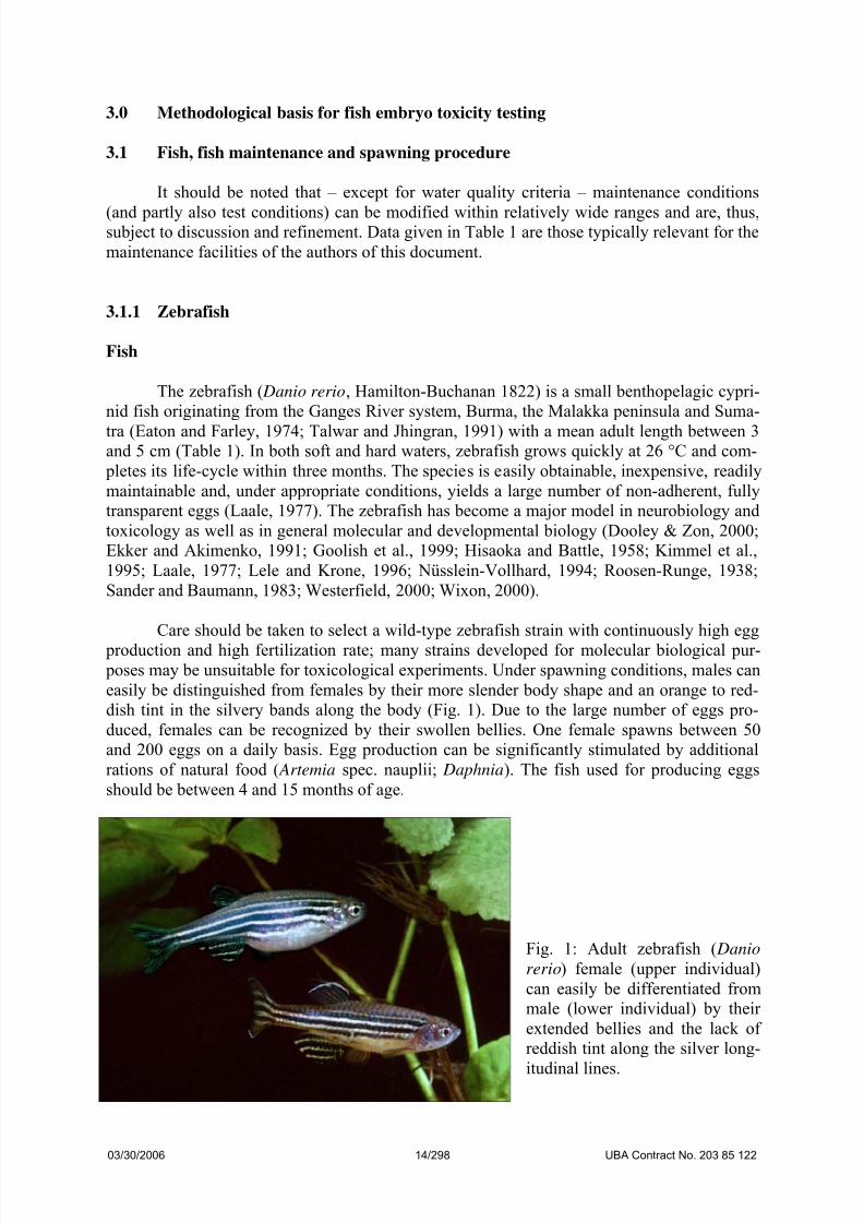
3.1.1 Zebrafish
Fish
The zebrafish ( Danio rerio, Hamilton-Buchanan 1822) is a small benthopelagic cypri-
nid fish originating from the Ganges River system, Burma, the Malakka peninsula and Suma-
tra (Eaton and Farley, 1974; Talwar and Jhingran, 1991) with a mean adult length between 3and 5 cm (Table 1). In both soft and hard waters, zebrafish grows quickly at 26 °C and com-
pletes its life-cycle within three months. The species is easily obtainable, inexpensive, readily
maintainable and, under appropriate conditions, yields a large number of non-adherent, fully
transparent eggs (Laale, 1977). The zebrafish has become a major model in neurobiology and
toxicology as well as in general molecular and developmental biology (Dooley & Zon, 2000;
Ekker and Akimenko, 1991; Goolish et al., 1999; Hisaoka and Battle, 1958; Kimmel et al.,
1995; Laale, 1977; Lele and Krone, 1996; Nüsslein-Vollhard, 1994; Roosen-Runge, 1938;
Sander and Baumann, 1983; Westerfield, 2000; Wixon, 2000).
Care should be taken to select a wild-type zebrafish strain with continuously high egg
production and high fertilization rate; many strains developed for molecular biological pur-
poses may be unsuitable for toxicological experiments. Under spawning conditions, males can
easily be distinguished from females by their more slender body shape and an orange to red-
dish tint in the silvery bands along the body (Fig. 1). Due to the large number of eggs pro-
duced, females can be recognized by their swollen bellies. One female spawns between 50
and 200 eggs on a daily basis. Egg production can be significantly stimulated by additional
rations of natural food ( Artemia spec. nauplii; Daphnia). The fish used for producing eggs
should be between 4 and 15 months of age.
Fig. 1: Adult zebrafish ( Danio
rerio) female (upper individual)
can easily be differentiated from
male (lower individual) by their
extended bellies and the lack of
reddish tint along the silver long-
itudinal lines.
03/30/2006 14/298 UBA Contract No. 203 85 122

7/26/2019 Ze Bra Fish Ba RU
http://slidepdf.com/reader/full/ze-bra-fish-ba-ru 15/40
7/26/2019 Ze Bra Fish Ba RU
http://slidepdf.com/reader/full/ze-bra-fish-ba-ru 16/40
T a b l e 1 : M
a i n t e n a n c e , b r e e d i n g a n d t y p i c a l c o n d i t i o n s f o r e m b r y o t o x i c
i t y t e s t s w i t h t h e c o m m o n O E C
D t e s t f i s h s p e c i e s ( c o n t ´ d . )
Z
e b r a f i s h ( D a n i o r e r i o )
F a t h e a d m i n n o w ( P i m e p h a l e s p r o m
e l a s )
J a p a n e s e m e d a k a ( O r y z i a s l a t i p e s )
M a l e t o f e m
a l e r a t i o f o r b r e e d i n g
2 : 1 ( 4 : 2 )
2 : 4
1 5 : 1 5
B r e e d i n g t a
n k s
4 L t a n k s e q u i p p e d w i t h s t e e l g r i d b o t t o m
a n d p l a n t d u m m y a s s p a w n i n g s t i m u l a n t ;
e x t e r n a l h e a t i n g m a t s ( c f . F i g . 4 )
3 0 L t a n k s w i t h b l a c k g l a s s w a l l s m a i n t a i n e d
a t 2 4 ° C a n d e q u i p p e d w i t h 2 c l a y t i l e s
d i v i d e d
i n
t o t w o h a l v e s a s s p a w n i n g s u b s t r a t e
3 0 L t a n k s w i t h b l a c k g l a s s
w a l l s e q u i p p e d
w i t h s e v e r a l p l a n t d u m m i e s o r C e r a t o p h y l -
l u m s p e c . a s s u b s t r a t e f o r s p a w n i n g
E g g s t r u c t u
r e a n d a p p e a r a n c e
S t a b l e c h o r i o n , h i g h l y t r a n s p a r e n t , n o n -
s t i c k y , d i a m
e t e r : ~ 0 . 8 m m
C h o r i o n o n l y h a r d e n s i n m u l t i c e l l u l a r s t a g e ,
t r a n s p a r e n t , s t i c k s t o s u r f a c e s , d i a m e t e r
<
1 m m
S t a b l e c h o r i o n w i t h s p i n y h
o o k s ( a d h e r e s t o
a n a l f i n o f f e m a l e ) , m o d e r a
t e l y t r a n s p a r e n t ,
d i a m e t e r < 1 m m
E m b r y o d e v e l o p m e n t a t 2 6 ° C
1 8 h : D e v e l o
p m e n t o f s o m i t e s
2 1 h : T a i l d e
t a c h m e n t
2 6 h : H e a r t - b e a t v i s i b l e
2 8 h : B l o o d
c i r c u l a t i o n
7 2 h : H a t c h i n g
2 2 h : D e v e l o p m e n t o f s o m i t e s
2 5 h : T a i l d e t a c h m e n t
2 7 h : H e a r t - b e a t v i s i b l e
3 0 h : B l o o d c i r c u l a t i o n
1 2 0 h : H a t c h i n g
3 0 h : D e v e l o p m e n t o f s o m
i t e s
5 4 h : H e a r t - b e a t v i s i b l e
7 8 h : B l o o d c i r c u l a t i o n
7 8 h : T a i l d e t a c h m e n t
1 6 0 h : H a t c h i n g
T e s t t y p e
S t a t i c o r s e m i - s t a t i c , 2 6 ° C , 2 4 - w e l l p l a t e s ( 2
m l p e r c a v i t y )
S t a t i c o r s e m i - s t a t i c , 2 5 ° C , 2 4 - w e l l p l a t e s ( 2
m
l p e r c a v i t y )
S t a t i c o r s e m i - s t a t i c , 2 6 ° C , 2 4 - w e l l p l a t e s ( 2
m l p e r c a v i t y )
M a j o r t o x i c
o l o g i c a l e n d p o i n t s a t
2 5 ° C
2 4 h : C o a g
u l a t i o n , t a i l d e t a c h m e n t , s o m i t e
d e v e l o p m e n t
4 8 h : H e a r t - b e a t v i s i b l e
2 8 h : C o a g u l a t i o n , t a i l d e t a c h m e n t , s o m i t e
d e v e l o p m e n t
3
d : B l o o d c i r c u l a t i o n
3
d : B l o o d c i r c u l a t i o n
4
d : B l o o d c i r c u l a t i o n
3 0 h : C o a g u l a t i o n , t a i l d e t a c h m e n t , s o m i t e
d e v e l o p m e n t
7 8 h : B l o o d c i r c u l a t i o n
4 d : P e c t o r a l f i n s ; 3 0 % t a i l d e t a c h m e n t
5 d : B l o o d c i r c u l a t i o n ; m
o v e m e n t o f p e c t o -
r a l f i n s ; t a i l d e t a c h e d
a n d p i g m e n t e d
7 d : H a t c h ; b l o o d c i r c u l a t i o n
03/30/2006 16/298 UBA Contract No. 203 85 122

7/26/2019 Ze Bra Fish Ba RU
http://slidepdf.com/reader/full/ze-bra-fish-ba-ru 17/40
Normal zebrafish development
The embryonic development of zebrafish has been described in most detail (Hisaoka
and Battle, 1958; Kimmel et al., 1988, 1995; Laale, 1977; Roosen-Runge, 1938; Thomas and
Waterman, 1978). The zebrafish egg is telolecithal, cleavage is meroblastic and discoidal.
Selected major stages of zebrafish development are given in Figs. 2 and 3; for more details,see Table 2 as well as Kimmel et al. (1995).
Table 2: Stages of embryonic development of the zebrafish ( Danio rerio) at 26 ± 1 °C (Nagel,
2002)
Time (h) Stage Characterization (after Kimmel et al., 1995)
0 Fertilization Zygote
0 Zygote period Cytoplasm accumulates at the animal pole, one-cell stage
0.75 Discoidal partial cleavage: 1. (median vertical) division: two-cell-stage
1 2. (vertical) division: four-cell-stage
1.25 3. (vertical and parallel to the plane of the first) division: 8-cell-stage
1.5
Cleavage period
4. (vertical and parallel to the second) division: 16-cell-stage
2 Start of blastula stage
3 Late cleavage; blastodisc contains approximately 256 blastomers
4
Blastula period
Flat interface between blastoderm and yolk
5.25 50 % of epibolic movements; blastoderm thins and interface between
periblast and blastoderm become curved
8 75 % of epibolic movement
10
Gastrula period
Epibolic movement ends, blastopore is nearly closed
10.5 First somite furrow
12 Somites are developed, undifferentiated mesodermal component of
the early trunk, tail segmented or metameric
20 Muscular twitches; sacculus; tail well extended
22
Segmentation
period
Side to side flexures; otoliths
24 Phylotypic stage, spontaneous movements, tail is detached from theyolk; early pigmentation
30 Reduced spontaneous movement; retina pigmented, cellular degen-
eration of the tail end; circulation in the aortic arch 1 visible
36
Pharyngula pe-riod
Tail pigmentation; strong circulation; single aortic arch pair, early
motility; heart beating
72 - 96 Hatching period Heart-beat regular; yolk extension beginning to taper; dorsal and
ventral pigmentation stripes meet at tail; segmental blood vessels
detectable: thickened sacculus with two chambers visible; foregut
development; neuromasts
03/30/2006 17/298 UBA Contract No. 203 85 122

7/26/2019 Ze Bra Fish Ba RU
http://slidepdf.com/reader/full/ze-bra-fish-ba-ru 18/40
Fig. 2a: Selected stages of early zebrafish ( Danio rerio) development: 0.2 - 3.3 h post-
fertilization (from Kimmel et al., 1995).
03/30/2006 18/298 UBA Contract No. 203 85 122
7/26/2019 Ze Bra Fish Ba RU
http://slidepdf.com/reader/full/ze-bra-fish-ba-ru 19/40
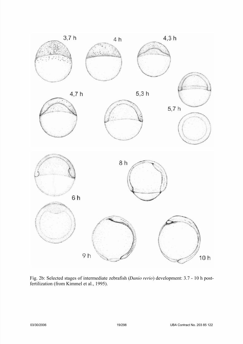
Fig. 2b: Selected stages of intermediate zebrafish ( Danio rerio) development: 3.7 - 10 h post-
fertilization (from Kimmel et al., 1995).
03/30/2006 19/298 UBA Contract No. 203 85 122

7/26/2019 Ze Bra Fish Ba RU
http://slidepdf.com/reader/full/ze-bra-fish-ba-ru 20/40
Fig. 2c: Selected stages of intermediate zebrafish ( Danio rerio) development: 11 – 19.5 h
post-fertilization (from Kimmel et al., 1995).
03/30/2006 20/298 UBA Contract No. 203 85 122
7/26/2019 Ze Bra Fish Ba RU
http://slidepdf.com/reader/full/ze-bra-fish-ba-ru 21/40
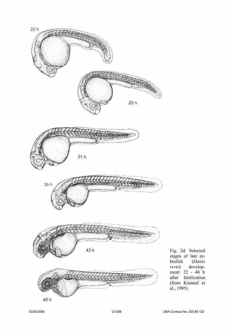
Fig. 2d: Selected
stages of late ze-
brafish ( Danio
rerio) develop-
ment: 22 - 48 h
after fertilization
(from Kimmel et
al., 1995).
03/30/2006 21/298 UBA Contract No. 203 85 122

7/26/2019 Ze Bra Fish Ba RU
http://slidepdf.com/reader/full/ze-bra-fish-ba-ru 22/40
Fig. 3a: Normal development of zebrafish ( Danio rerio) embryos I: (1) 0.75 h; (2) 1 h; (3)
1.2 h; (4) 1.5 h; (5) 4.7 h; (6) 5.3 h.
03/30/2006 22/298 UBA Contract No. 203 85 122

7/26/2019 Ze Bra Fish Ba RU
http://slidepdf.com/reader/full/ze-bra-fish-ba-ru 23/40
Fig. 3b: Normal development of zebrafish ( Danio rerio) embryos II: (1) 6 h; (2) 6 h; (3) 8 h;
(4) 9 h; (5) 12 h; (6) 14 h. A – eye anlage; Ch – chorion; O – ear bud; S – somites (muscle
segments).
03/30/2006 23/298 UBA Contract No. 203 85 122
7/26/2019 Ze Bra Fish Ba RU
http://slidepdf.com/reader/full/ze-bra-fish-ba-ru 24/40
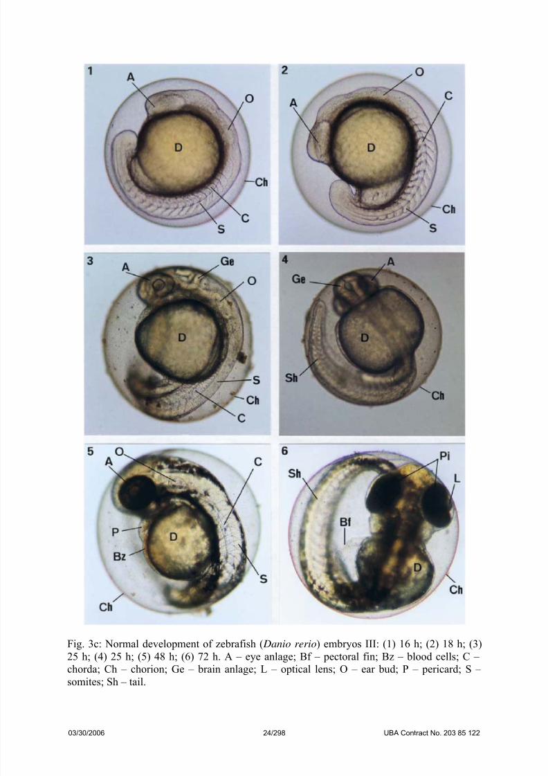
Fig. 3c: Normal development of zebrafish ( Danio rerio) embryos III: (1) 16 h; (2) 18 h; (3)
25 h; (4) 25 h; (5) 48 h; (6) 72 h. A – eye anlage; Bf – pectoral fin; Bz – blood cells; C –
chorda; Ch – chorion; Ge – brain anlage; L – optical lens; O – ear bud; P – pericard; S –
somites; Sh – tail.
03/30/2006 24/298 UBA Contract No. 203 85 122

7/26/2019 Ze Bra Fish Ba RU
http://slidepdf.com/reader/full/ze-bra-fish-ba-ru 25/40
Fig. 3d: Normal development of zebrafish ( Danio rerio) embryos IV: (1) 48 h; (2) 72 h; (3)
144 h; (4) 144 h. A – eye anlage; Bf – pectoral fin; Bz – blood cells; C – chorda; Ch –
chorion; D – yolk sac; E – gut; F – fin; Ge – brain; H – heart; K – gills; L – eye lens; M –melanophores; Ms – mouth slit; O – ear; P – pericard; Pi – ocular pigment layer; S – somites
(muscle segments); Sb – swimming bladder; Sh – tail.
03/30/2006 25/298 UBA Contract No. 203 85 122

7/26/2019 Ze Bra Fish Ba RU
http://slidepdf.com/reader/full/ze-bra-fish-ba-ru 26/40
Fig. 3e: Normal development of zebrafish ( Danio rerio) embryos V (following dechoriona-
tion): (1) 48 h, anal region; (2) 48 h, ear region. A – eye anlage; Af – position of the anus; Ba
– dorsal aorta; Bv – central ventral axial vein; D – yolk sac; E – peritoneum; F – fin; H –
heart; M – melanophores; P – pericard; O – ear; S – somites (muscle segments).
03/30/2006 26/298 UBA Contract No. 203 85 122

7/26/2019 Ze Bra Fish Ba RU
http://slidepdf.com/reader/full/ze-bra-fish-ba-ru 27/40
Fig. 3f: Selected stages of zebrafish ( Danio rerio) development: (a) 4-cell stage (approx. 1 h);
(b) 16-cell stage (approx. 1.3 h); (c) 64-cell stage (approx. 1.8 h); (d) detachment of tail
(approx. 17.5 h).
Fig. 3g: Mortality in early zebrafish ( Danio rerio) eggs: coagulation (I), heavy infestation
with fungi (II) and invasion of Vorticella spec. (V; III). Ch – chorion; Ek – coagulated egg;
Ph – fungi.
03/30/2006 27/298 UBA Contract No. 203 85 122
7/26/2019 Ze Bra Fish Ba RU
http://slidepdf.com/reader/full/ze-bra-fish-ba-ru 28/40
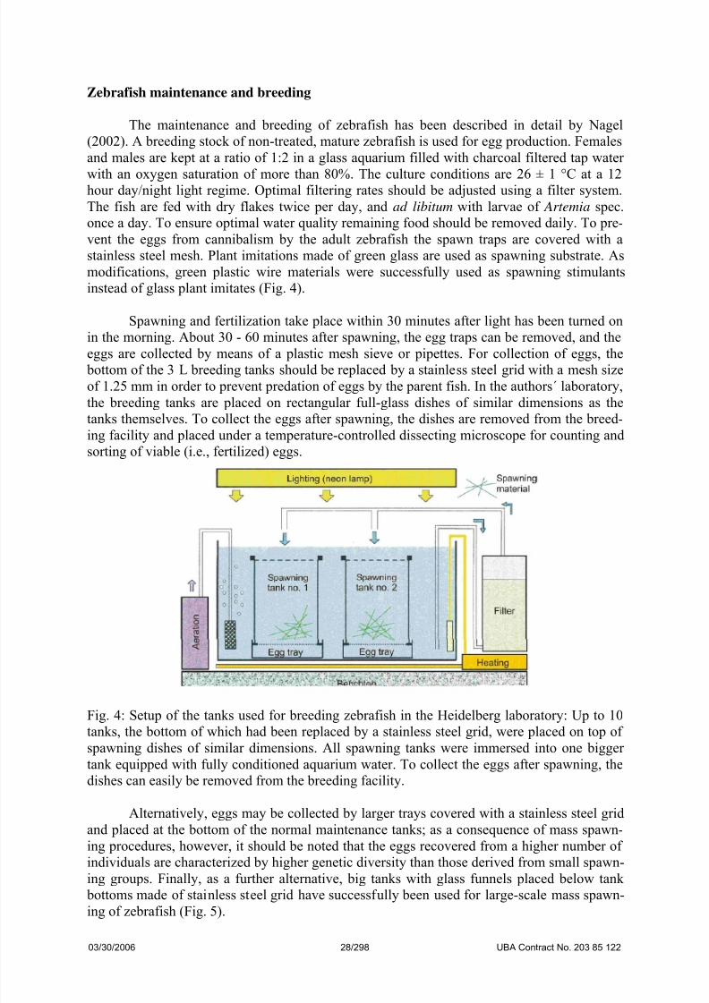
Zebrafish maintenance and breeding
The maintenance and breeding of zebrafish has been described in detail by Nagel
(2002). A breeding stock of non-treated, mature zebrafish is used for egg production. Females
and males are kept at a ratio of 1:2 in a glass aquarium filled with charcoal filtered tap water
with an oxygen saturation of more than 80%. The culture conditions are 26 ± 1 °C at a 12hour day/night light regime. Optimal filtering rates should be adjusted using a filter system.
The fish are fed with dry flakes twice per day, and ad libitum with larvae of Artemia spec.
once a day. To ensure optimal water quality remaining food should be removed daily. To pre-
vent the eggs from cannibalism by the adult zebrafish the spawn traps are covered with a
stainless steel mesh. Plant imitations made of green glass are used as spawning substrate. As
modifications, green plastic wire materials were successfully used as spawning stimulants
instead of glass plant imitates (Fig. 4).
Spawning and fertilization take place within 30 minutes after light has been turned on
in the morning. About 30 - 60 minutes after spawning, the egg traps can be removed, and the
eggs are collected by means of a plastic mesh sieve or pipettes. For collection of eggs, the bottom of the 3 L breeding tanks should be replaced by a stainless steel grid with a mesh size
of 1.25 mm in order to prevent predation of eggs by the parent fish. In the authors´ laboratory,
the breeding tanks are placed on rectangular full-glass dishes of similar dimensions as the
tanks themselves. To collect the eggs after spawning, the dishes are removed from the breed-
ing facility and placed under a temperature-controlled dissecting microscope for counting and
sorting of viable (i.e., fertilized) eggs.
Fig. 4: Setup of the tanks used for breeding zebrafish in the Heidelberg laboratory: Up to 10
tanks, the bottom of which had been replaced by a stainless steel grid, were placed on top of
spawning dishes of similar dimensions. All spawning tanks were immersed into one bigger
tank equipped with fully conditioned aquarium water. To collect the eggs after spawning, the
dishes can easily be removed from the breeding facility.
Alternatively, eggs may be collected by larger trays covered with a stainless steel grid
and placed at the bottom of the normal maintenance tanks; as a consequence of mass spawn-
ing procedures, however, it should be noted that the eggs recovered from a higher number of
individuals are characterized by higher genetic diversity than those derived from small spawn-
ing groups. Finally, as a further alternative, big tanks with glass funnels placed below tank bottoms made of stainless steel grid have successfully been used for large-scale mass spawn-
ing of zebrafish (Fig. 5).
03/30/2006 28/298 UBA Contract No. 203 85 122

7/26/2019 Ze Bra Fish Ba RU
http://slidepdf.com/reader/full/ze-bra-fish-ba-ru 29/40
Fig. 5: Setup of the tanks used for
breeding zebrafish in the Berlin labora-
tory (Leibniz-Institute of Freshwater
Ecology and Inland Fisheries): Largegroups of zebrafish are maintained in
tanks the bottom of which has been
replaced by a stainless steel grid to
avoid predation of the eggs by the par-
ent fish. The tanks are sitting on a big
funnel, which allows simple release of
the newly spawned eggs (courtesy of
Dr. T. Meinelt, IGB).
A single mature female lays 50-200 eggs per day. At the culture conditions describedabove, fertilized eggs undergo the first cleavage after approximately 15 min and consecutive
synchronous cleavages form 4, 8, 16, and 32 cell blastomeres. At these stages (4 - 32 cells),
eggs can be identified clearly as fertilized, and only these should be used for the experiments.
The principle of the zebrafish embryo toxicity test
The embryo test procedure itself has been described by Schulte and Nagel (1994) as
well as Nagel (1998) in detail. In brief, following initial range-finding experiments, the toxic-
ity of a chemical substance can be determined by using 24-well-plates. After preparing a
stock solution of the test substance, typically five concentrations are tested. Information about
the use of solvents can be found in Maiwald (1997) as well as in Nagel (1998).
In the original version of the zebrafish embryo test, 40 eggs were transferred to the test
solutions at latest 60 minutes after light had been turned on to initiate spawning. Fertilized
eggs were separated from the non-fertilized ones and placed in the 24-well-plates with a pi-
pette using a stereo microscope. 20 Fertilized eggs were placed individually in 2 ml of the
respective test solutions to exclude mutual influences. The remaining four wells of each plate
were used as internal control filled with dilution water amounting to a total of 20 controls per
test. The dilution water corresponded to the reconstituted water according to ISO-standard
7346/3, which was diluted 1:5 using deionized water. After this procedure, the 24-well-plateswere covered with a self-adhesive foil and incubated at 26°C ± 1°C. Lethal, sublethal and
teratogenic endpoints were recorded using a dissecting microscope within 48h. A test was
classified as valid, if 90% of the embryos in the control treatments showed neither sublethal
nor lethal effects. For a discussion of lethal and sublethal effects, see below.
03/30/2006 29/298 UBA Contract No. 203 85 122

7/26/2019 Ze Bra Fish Ba RU
http://slidepdf.com/reader/full/ze-bra-fish-ba-ru 30/40
Oxygen requirements of zebrafish ( Danio rerio) embryos
In order to determine the oxygen requirements and consumption by zebrafish embryos,
eggs were incubated and exposed to 3,4-dichloroaniline and 2,4-dinitrophenol under low oxy-
gen concentrations varying between 3 and 6 mg/l, which were prepared by selectively fumi-
gating the solutions with nitrogen (Braunbeck et al., 2005). For incubation, the 24-well micro-titer plates were kept within closed chambers under a pure nitrogen atmosphere. In a second
series of experiments, zebrafish eggs (embryos) were maintained at densities between 10 and
60 individuals per ml medium under similar conditions.
Fig. 6: Oxygen consumption of control zebrafish ( Danio rerio) embryos in relation to the
amount of oxygen provided in the medium. Apparently, zebrafish embryos are capable ofadapting to low oxygen levels without any symptoms of developmental effects. Data are
given as means from 4 independent experiments ± S.D. (Braunbeck et al., 2005).
Fig. 7: Oxygen consumption of control zebrafish ( Danio rerio) embryos in dependence from
the stocking density in given volume of medium. As could be expected from data presented inFig. 6, the embryos adapt to low oxygen levels by reducing their oxygen consumption. Data
are given as means from 5 independent experiments ± S.D. (Braunbeck et al., 2005).
03/30/2006 30/298 UBA Contract No. 203 85 122

7/26/2019 Ze Bra Fish Ba RU
http://slidepdf.com/reader/full/ze-bra-fish-ba-ru 31/40
Zebrafish embryos incubated at varying levels of oxygen in the medium were appar-
ently well capable of adapting to low oxygen tension. The more oxygen was provided in the
medium, the more oxygen the embryos consumed (Fig. 6). Even at oxygen concentrations as
low as 2 mg/l, which should be expected to be lethal to adults of most other cyprinid fish spe-
cies, zebrafish embryos did not show any symptom of malformation or even growth retarda-
tion (additional experiments; data not shown in Fig. 6). This observation is of particular im- portance for the routine testing of whole effluents, since in sewage there may be severe oxy-
gen depletion due to bacterial breakdown. As could be expected from the adaptive reduction
of oxygen consumption under conditions of low oxygen levels, zebrafish embryos are also
able to react to increasing stocking densities in a given volume of medium (Fig. 7).
Space requirements of zebrafish embryos
To elucidate the minimum space required by a zebrafish embryo, eggs were incubated
in 96-well microtiter plates filled with 100, 200 and 300 μl water or toxicant (3,4-
dichloroaniline, 2,4-dinitrophenol) concentrations and compared to eggs incubated in a totalvolume of 2 ml in 24-well microtiter plates (Braunbeck et al., 2005).
Fig. 8: Toxicity of 3,4-dichloroaniline to zebrafish ( Danio rerio) embryos in various volumes
of medium (2 ml in 24-well microtiter plates as well as 100, 200 and 300 μl in 96-well micro-
titer plates) after 24 h (left) and 48 h of exposure (right). Endpoints as listed by DIN stan-
dards. The incubation volume does not seem to take any influence on the toxicity of 3,4-
dichloroaniline (Braunbeck et al., 2005).
In fact, exposure experiments with 3,4-dichloroaniline and 2,4-dinitrophenol did not
show any change with respect to the exposure volume (Figs. 8, 9). As a consequence, zebra-
fish embryos cannot only be exposed in 24-well microtiter plates in a total volume of 2 ml
medium (as suggested in the OECD TG proposal), but also in even lower volumes of 300, 200
and even 100 μl within the cavities of 96-well microtiter plates. Thus, the fish embryo test
provides a tool to test even smallest volumes of test substances, which may be of particular
relevance to the testing of, for example, metabolites of pesticides. On the other hand, ex-
tremely lipophilic substances may require modification of the test protocol in that microtiter plates should be replaced by glassware. In such cases, the possibility to expose a higher num-
ber of embryos within one vessel to a small volume might also be a significant advantage.
03/30/2006 31/298 UBA Contract No. 203 85 122

7/26/2019 Ze Bra Fish Ba RU
http://slidepdf.com/reader/full/ze-bra-fish-ba-ru 32/40
Fig. 9: Toxicity of 2,4-dinitrophenol to zebrafish ( Danio rerio) embryos in various volumes
of medium (2 ml in 24-well microtiter plates as well as 100, 200 and 300 μl in 96-well micro-
titer plates) after 24 h (left) and 48 h of exposure (right). Endpoints as listed by DIN stan-
dards. The incubation volume does not seem to take any influence on the toxicity of 2,4-
dinitrophenol (Braunbeck et al., 2005).
Fig. 10: Effect of the exposure
start (time lapse after fertilization)
on the cumulative mortality of 2,4-
dinitrophenol. After 24 and 48 h, it
is evident that mortalities are in-
creased with early exposure start.
03/30/2006 32/298 UBA Contract No. 203 85 122

7/26/2019 Ze Bra Fish Ba RU
http://slidepdf.com/reader/full/ze-bra-fish-ba-ru 33/40
Importance of an early onset of exposure of zebrafish ( Danio rerio) eggs
In order to investigate the role of the start of the exposure, zebrafish eggs were ex-
posed to the model compounds 2,4-dinitrophenol and 3,4-dichloroaniline (Figs. 10, 11) with
different starting points from 1 to 9 h of egg age. For either substance, a clear relationship
between the age of the eggs at onset of exposure and the cumulative mortality after 24 and 48h of exposure was found. After 6 days of exposure, however, mortality had accumulated to
such a rate that the effect could no longer be seen (Figs. 10, 11). As a conclusion, the eggs
should be transferred to the test solutions at latest 1 h post-fertilization.
Fig. 11: Effect of the exposure start
(time lapse after fertilization) on
the cumulative mortality of 3,4-
dichloroaniline. After 24 and 48 h,
it is evident that mortalities are
increased with early exposure start.
The chorion of zebrafish ( Danio rerio) embryos as a barrier for the uptake of chemicals
In order to elucidate the effect of the chorion as a barrier, the chorion of 6 h old ze-
brafish embryos (pre-exposed from 30 min post fertilization) was softened by incubation in a
2 mg/l pronase solution (protease from Streptomyces griseus, Westerfield, 2000) with an ac-
tivity of 4 units per mg in dilution water for 1 ± 0.5 min at 28.5° C and mechanically dis-
rupted by means of two pairs of forceps or dissection needles. Control experiments were car-
ried out to ensure that pronase treatment did not have any effect on embryonic development.
Since the optimal incubation time depends on the developmental stage, the optimal durationshould be checked in range-finding experiments. Without enzymatic digestion, the mechani-
cal stress frequently resulted in destruction of the embryo. Disruption of the chorion without
03/30/2006 33/298 UBA Contract No. 203 85 122

7/26/2019 Ze Bra Fish Ba RU
http://slidepdf.com/reader/full/ze-bra-fish-ba-ru 34/40
enzymatic softening of the egg shell was only possible in embryos > 48 h. Dechorionated em-
bryos were incubated in 24-well microtiter plates and exposed to potassium chromate (hydro-
philic; Fig. 12), 4-chloroaniline (moderately lipophilic; Fig. 13) and lindane (lipophilic) and
compared to non-dechorionated embryos exposed under similar conditions. In order to avoid
excessive adsorption, the wells had been pre-incubated with toxicant 24 h prior to addition of
the eggs.
Fig. 12: Effects of potassium chromate to zebrafish ( Danio rerio) embryos depending on the
presence of a chorion (upper panel) or after dechorionation (lower panel) on the basis of dis-
turbances of the equilibrium after various periods of exposure. For potassium chromate,
dechorionation does not affect acute embryo toxicity, but clearly shows sublethal effects
(Braunbeck et al., 2005).
Whereas exposure to the relatively hydrophilic potassium chromate did not result in
any change of the core endpoints of the embryo toxicity test (details not shown), prolongedexposure of dechorionated embryos over 4 days (i.e., until hatching) produced severe distur-
bances to swimming equilibrium in hatched larvae (Fig. 12), thus indicating that the chorion
did act at least as some form of barrier, even for hydrophilic substances. In contrast, for 4-
chloroaniline, a significant increase in toxicity could already be recorded for the core end-
points of the fish embryo test as defined by the current DIN standards (Fig. 13). This increase
in toxicity is even more pronounced for more lipophilic substances such as lindane: Whereas
the EC50 value of lindane for normal embryos could be identified as 26.5 mg/l, the corre-
sponding value for dechorionated embryos is 11.3 mg/L. Albeit significant, this difference in
lindane embryo toxicity becomes relative when compared with the broad range of acute con-
ventional (in vivo) fish toxicity from 2 mg/L in bluegill sunfish over 12 mg/L in fathead min-
now, 14 mg/L in rainbow trout (Oncorhynchus mykiss) and 23 mg/L in golden ide ( Leuciscus
idus melanotus) to 26 mg/L in guppy (Poecilia reticulata; Verschueren, 1983). Nevertheless,
results indicate that the barrier function of the chorion may increase with lipophilicity, a fact
03/30/2006 34/298 UBA Contract No. 203 85 122

7/26/2019 Ze Bra Fish Ba RU
http://slidepdf.com/reader/full/ze-bra-fish-ba-ru 35/40
that should be taken into consideration in the interpretation of correlations between fish em-
bryo and conventional acute fish toxicity (Braunbeck et al., 2005).
Fig. 13: Toxicity of 4-chloroaniline to zebrafish ( Danio rerio) embryos depending on the
presence of a chorion (upper panel) or after dechorionation (lower panel) based on acutely
toxic effects as specified by German DIN (DIN, 2001) standards after various periods of ex-
posure. For 4-chloroaniline, dechorionation results in an increase of acute embryo toxicity
(Braunbeck et al., 2005)
03/30/2006 35/298 UBA Contract No. 203 85 122
7/26/2019 Ze Bra Fish Ba RU
http://slidepdf.com/reader/full/ze-bra-fish-ba-ru 36/40
3.1.2 Fathead minnow
The fathead minnow (Pimephales promelas, Rafinesque, 1820) is another demersal
cyprinid species originating from the temperate waters of central North America (Page and
Burr, 1991); it inhabits muddy pools of headwaters, creeks and small rivers. Among the three
primary OECD species, fathead minnow is most likely the one with the largest toxicologicaldatabase (Ankley et al., 2001; Gray et al., 2002; Keddy et al., 1995; Miracle et al., 2003;
Sinks and Schultz, 2001).
Whereas females grow up to 1.5 ± 0.3 g, males may reach 2.5 ± 0.5 g in weight. Water
quality parameters are essentially identical to those described for zebrafish; for water details
of maintenance, see Table 1. In order to prevent permanent stress, parental fathead minnow
can be maintained at temperatures of approx. 16.5 ± 1.5 °C (“winter conditions”); prior to
spawning, the temperature should then be gradually raised to 24 °C at a rate of 1 - 2 °C/day,
and fish should be given increasing ratios of live food (Table 1). During spawning, dominant
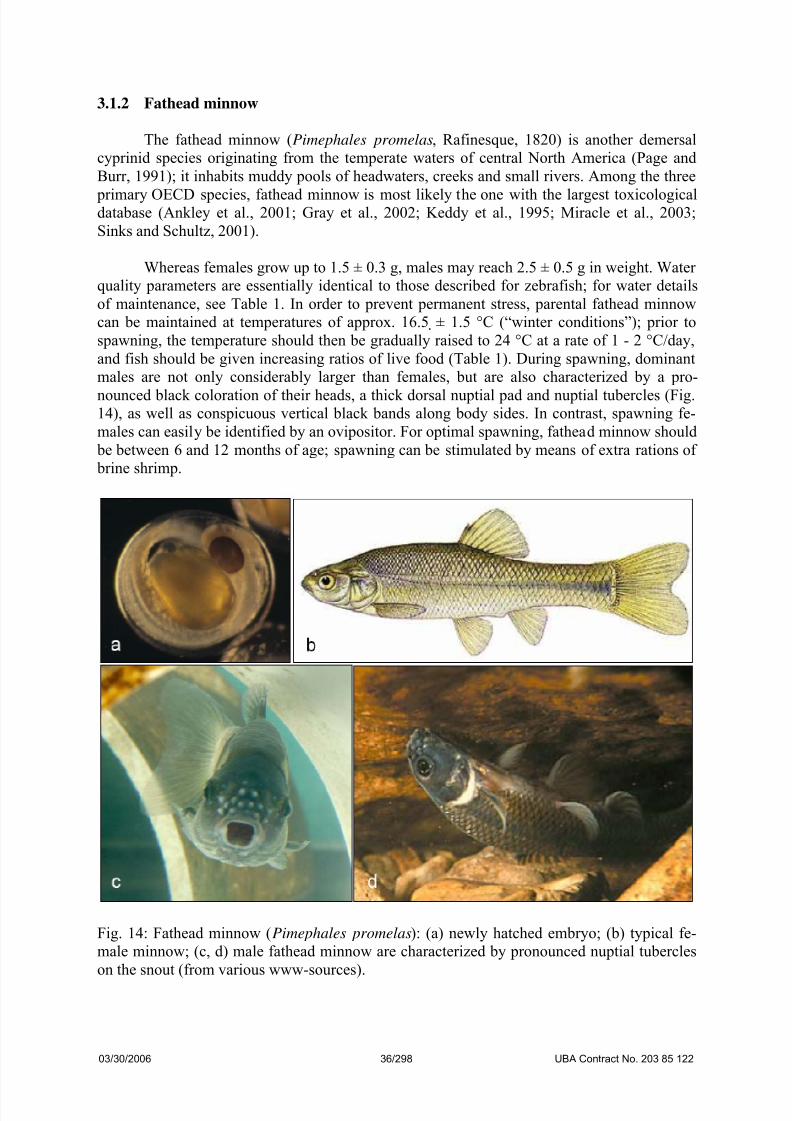
males are not only considerably larger than females, but are also characterized by a pro-
nounced black coloration of their heads, a thick dorsal nuptial pad and nuptial tubercles (Fig.14), as well as conspicuous vertical black bands along body sides. In contrast, spawning fe-
males can easily be identified by an ovipositor. For optimal spawning, fathead minnow should
be between 6 and 12 months of age; spawning can be stimulated by means of extra rations of
brine shrimp.
Fig. 14: Fathead minnow (Pimephales promelas): (a) newly hatched embryo; (b) typical fe-
male minnow; (c, d) male fathead minnow are characterized by pronounced nuptial tubercles
on the snout (from various www-sources).
03/30/2006 36/298 UBA Contract No. 203 85 122

7/26/2019 Ze Bra Fish Ba RU
http://slidepdf.com/reader/full/ze-bra-fish-ba-ru 37/40
For spawning, fathead minnow are normally kept in small groups of two males and
four females in tanks of about 30 L volumes. Since fathead minnow are quite sensitive to dis-
turbance during spawning, the spawning tanks should be kept under quiet conditions. As soon
as, e.g., a clay tile divided into two halves is added to each spawning group as a spawning
ground, male fathead minnow build up individual territories with the tiles as their centers.
During the spawning period, the tiles should be inspected for eggs at intervals of at latest 60minutes post fertilization. Usually, 100 - 250 eggs are laid per spawning act. Since the chorion
only hardens at the first multicellular stages, the transparent, sticky eggs should be given
about two hours, before they are removed and directly transferred to 6- or 24-well plates by
means of a flexible forceps.
The development of fathead minnow is basically similar to that of zebrafish (Manner
and Dewese, 1974); at least partly due to the lower temperature, however, the time schedule
of development is slightly slower (Fig. 15; cf. Table 1).
Fig. 15: Normal development of fathead minnow (Pimephales promelas) embryos (1 - 3) in
comparison to fathead minnow embryos exposed to 15 mg/L 3,4-dichloroaniline (4): (1, 2)
2 h post-fertilization, normal development; (3) 30 h post-fertilization, normal development:
tail has detached from yolk sac, and somites are clearly detectable; (4) following exposure to
15 mg/L 3,4-dichloroaniline, the entire embryo appears deformed, and the tail has not yet de-
tached from the yolk sac.
03/30/2006 37/298 UBA Contract No. 203 85 122

7/26/2019 Ze Bra Fish Ba RU
http://slidepdf.com/reader/full/ze-bra-fish-ba-ru 38/40
3.1.3 Japanese medaka
Similar to zebrafish, the Japanese medaka (Oryzias latipes, Temminck & Schlegel,
1846) has become a favorite model in developmental biology and molecular genetics (Furu-tani-Seikia and Wittbrod, 2004; Winn, 2002; Wittbrod et al., 2001) as well as in ecotoxicol-
ogy (Arcand-Hoy and Benson, 1998; De Koven et al., 1992; Hatanaka et al., 1982; Ishikawa
et al., 1984; Seki et al., 2002, 2003). The medaka belongs to the ricefishes (Adrianichthyidae,
Huber, 1996; Ishizaki, 1994) within the order of the Beloniformes and originates from Japan,
China, Vietnam and South Korea (Jordan and Snyder, 1906; Shima and Mitani, 2004). In its
original habitats, the medaka is severely threatened.
The biology of the medaka is well documented (Hyodo-Taguchi and Egami, 1985;
Kirchen and West, 1976; Yamamoto, 1975), and its requirements with respect to maintenance
are comparable to those of zebrafish; in fact, both species can easily be raised side by side in
one aquatic system (Furutani-Seikia and Wittbrod, 2004). Most of the standard experimental procedures can be applied to both species with slight modifications, including the observation
of embryos, gynogenesis, sperm freezing and in vitro fertilization, cell transplantation, as well
as RNA and DNA injection, in situ hybridization using riboprobes and immunohistochemis-
try. Once dechorionated, handling of the softer Medaka embryos requires some practice.
Medaka embryos tolerate a wide temperature range: 4 - 35 °C until the onset of heart beating
and 18 - 35 °C thereafter, compared to 25 - 33 °C in the case of zebrafish (Westerfield, 2000).
Since the original habitats of the Japanese medaka are still waters, for optimal mainte-
nance care should be taken to avoid excessive turbulence. Water characteristics are basically
identical to those described above for zebrafish. E.g., up to 60 individuals of either sex can be
kept in 50 L tanks at an ambient temperature of 24 ± 0.5 °C (Table 1). Whereas during the
spawning period females (0.35 ± 0.07 g) can readily be identified by a generally more plump
body shape and the sticky eggs attached to the anal fins (Fig. 16), male individuals (0.35 ±
0.07 g) are characterized by larger anal fins and the so-called papillary processes on posterior
dorsal fin rays. For optimal stimulation of spawning, 5 to 12 months old medaka should be at
best be kept under normal daylight conditions with extra ratios of frozen adult brine shrimp
( Artemia spec.). Moreover, spawning success can be further improved by means of natural
plants (Ceratophyllum spec.) in the aquaria. The female medaka spawns between 20 and 40
eggs every day within an hour after the onset of light. Again, care should be taken to select a
suitable strain (cf. Wakamatsu and Ozato, 2002).
03/30/2006 38/298 UBA Contract No. 203 85 122

7/26/2019 Ze Bra Fish Ba RU
http://slidepdf.com/reader/full/ze-bra-fish-ba-ru 39/40
Fig. 16: Japanese medaka (Oryzias latipes; from various www-sources).
Medaka eggs measure about 1 mm in diameter; they are transparent and characterized
by an orange color and conspicuous spiny hooks, which allow firm adhesion to the anal fin of
the female (Figs. 17, 18), but – to some degree – reduce the visibility of malformations during
development. On the other hand, the egg clutch attached allows instant identification of re-
productively active females. If required, attachment filaments on the chorion can be easily
removed by rolling eggs on a piece of Whatman filter paper or by proteinase K digestion.
Since the medaka chorion is quite stable, the eggs can be removed from the anal fin as soon as
30 minutes post fertilization and directly transferred to 6- or 24-well plates by means of thinmetal wire loop.
If compared to zebrafish, the development of medaka is slightly slower; normally de-
veloping embryos only hatch after approx. 7 days of incubation. Developmental stages and
corresponding morphological characteristics have been described in detail by Iwamatsu
(1994, 2004; see Annex 1); an in-depth comparison of developmental staging with zebrafish
has been published by Furutani-Seikia and Wittbrod (2004). Representative figures of devel-
opmental stages of Japanese medaka are given in Figs. 19a - d. As also outlined in Table 1,
the time schedule for the toxicological endpoints needs to be modified for the medaka:
(1)
After 30 h, the somites 3 to 10 have been developed. The optic vesicles are clearlydatable.
(2)
After 54 h, the eye development allows the differentiation of iris and pupils. Heart-
beat is visible, but blood circulation is not yet regular.
(3)
After 78 h, blood circulation is regular (can best be seen on yolk vessels). Occa-
sionally, spontaneous tail movements can be recorded. In the cranial area, some
pigment cells are visible. Tail tip has detached from the yolk.
(4)
After 4 d, pectoral fins are detectable. Tail tip reaches eyes and moves regularly;
about 30 % of the tail are detached from the yolk.
(5) After 5 d, a regular row of pigment cells along the tail are clearly discernable. Pec-
toral fins actively moving. Tail has detached completely.
(6)
After 6 d, when bent forward, the tail tip appears longer than head. Increasingnumber of pigment cells.
(7) After 7 d, the medaka embryo hatches.
03/30/2006 39/298 UBA Contract No. 203 85 122

7/26/2019 Ze Bra Fish Ba RU
http://slidepdf.com/reader/full/ze-bra-fish-ba-ru 40/40
Fig. 17a: Normal development of Japanese medaka (Oryzias latipes) embryos I: from unfertil-
ized eggs to the 4-cell stage (90 min).