Yellownails, lymphoedema, and pleural effusions -...
7
Thorax (1966), 21, 247. Yellow nails, lymphoedema, and pleural effusions PETER A. EMERSON From Westminster Hospital, London, S.W.1 The purpose of this paper is to draw attention to a new syndrome and to describe three examples of chronic pleural effusion due to chronic lymphoedema. Before encountering these patients I had never previously considered chronic lymph- oedema as a cause of chronic pleural effusion: it is certainly not described in any standard textbook, although it was tentatively referred to in one in 1963 (Robson and Emerson, 1963), and more recently Hurwitz and Pinnals (1964) have described two patients with pleural effusions and lymphoedema only. The thesis is that an otherwise unexplained pleural effusion may occur in patients with chronic lymphoedema and be due to some deficiency in the lymphatics draining the pleural space. These two conditions may be associated with yellow finger- and toe-nails, due also to impaired lymphatic drainage. CASE REPORTS CASE 1 Miss M. J., born in 1923, was known from infancy to have had swelling of the legs and to a lesser extent of the backs of the hands with puffiness of the face. There was no family history of lymph- oedema, and apart from the swelling she was well except for occasional attacks of bronchitis. These all cleared up completely until one attack in 1954, after which she became, and remained, short of breath, and was subsequently found to have a large right and a small left pleural effusion (Fig. 1). Over the subsequent years until her death in 1960 from an unrelated cause (malignant melanoma) the effusions persisted and recurred soon after each of the innumerable aspirations which were carried out. The pleural fluid was always clear, straw-coloured material; the predominant cell was the lymphocyte, with occasional histiocytes and mesothelial cells. The protein content was usually 4-5 to 5 g./100 ml., and malignant cells were never seen. The albumin turn- FIG. 1. Case 1. Chest radio- graph showing bilateral pkeural effusions. 247 on 21 May 2018 by guest. Protected by copyright. http://thorax.bmj.com/ Thorax: first published as 10.1136/thx.21.3.247 on 1 May 1966. Downloaded from
Transcript of Yellownails, lymphoedema, and pleural effusions -...

Thorax (1966), 21, 247.
Yellow nails, lymphoedema, and pleural effusionsPETER A. EMERSON
From Westminster Hospital, London, S.W.1
The purpose of this paper is to draw attention toa new syndrome and to describe three examplesof chronic pleural effusion due to chroniclymphoedema. Before encountering these patientsI had never previously considered chronic lymph-oedema as a cause of chronic pleural effusion: itis certainly not described in any standard textbook,although it was tentatively referred to in one in1963 (Robson and Emerson, 1963), and morerecently Hurwitz and Pinnals (1964) havedescribed two patients with pleural effusions andlymphoedema only.The thesis is that an otherwise unexplained
pleural effusion may occur in patients with chroniclymphoedema and be due to some deficiency inthe lymphatics draining the pleural space. Thesetwo conditions may be associated with yellowfinger- and toe-nails, due also to impairedlymphatic drainage.
CASE REPORTS
CASE 1 Miss M. J., born in 1923, was known frominfancy to have had swelling of the legs and to alesser extent of the backs of the hands with puffinessof the face. There was no family history of lymph-oedema, and apart from the swelling she was wellexcept for occasional attacks of bronchitis. These allcleared up completely until one attack in 1954, afterwhich she became, and remained, short of breath,and was subsequently found to have a large rightand a small left pleural effusion (Fig. 1). Over thesubsequent years until her death in 1960 from anunrelated cause (malignant melanoma) the effusionspersisted and recurred soon after each of theinnumerable aspirations which were carried out.The pleural fluid was always clear, straw-coloured
material; the predominant cell was the lymphocyte,with occasional histiocytes and mesothelial cells. Theprotein content was usually 4-5 to 5 g./100 ml., andmalignant cells were never seen. The albumin turn-
FIG. 1. Case 1. Chest radio-graph showing bilateral pkeuraleffusions.
247
on 21 May 2018 by guest. P
rotected by copyright.http://thorax.bm
j.com/
Thorax: first published as 10.1136/thx.21.3.247 on 1 M
ay 1966. Dow
nloaded from
Peter A. Emerson
FIG. 2
FIo. 4
FIG. 3
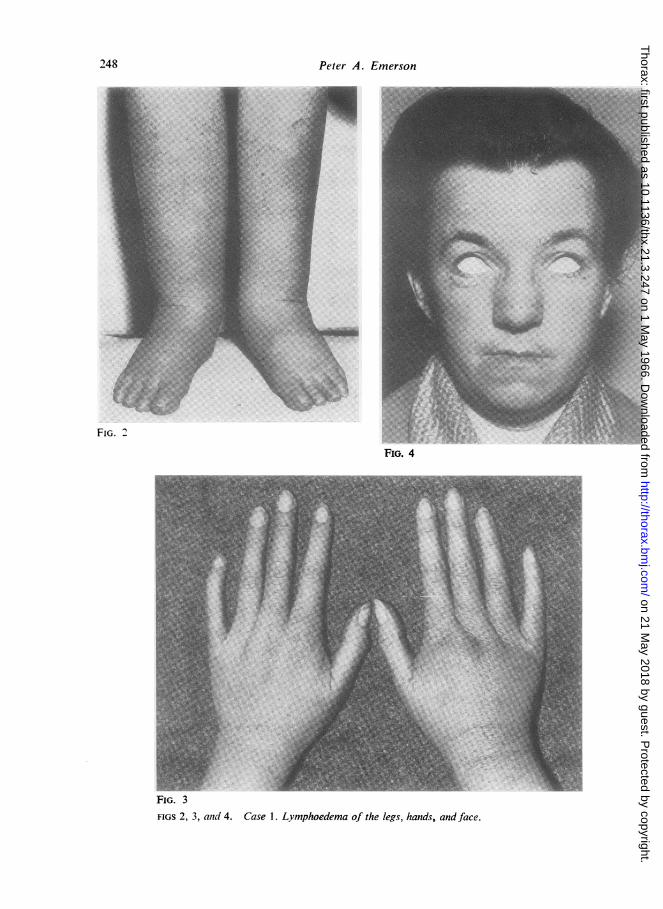
FIGS 2, 3, and 4. Case 1. Lymphoedema of the legs, hands, and face.
2)48
on 21 May 2018 by guest. P
rotected by copyright.http://thorax.bm
j.com/
Thorax: first published as 10.1136/thx.21.3.247 on 1 M
ay 1966. Dow
nloaded from

Yellow nails, lymphoedema, and pleural eJfusions
over was investigated using radioactive iodinatedserum albumin injected into the pleural effusion, butthese studies showed only that the exchange ofalbumin was very slow, as would be expected in anychronic effusion.
In addition to many biochemical investigations, toonumerous to detail here, she was studied by broncho-scopy, thoracoscopy, and bronchography, and hadseveral pleural biopsies. None of these revealed anysignificant abnormality to account for the effusions.
Figures 2, 3, and 4 show the patient at the age of 33when she was admitted to the Brompton Hospital in1956. At that time she was not inconvenienced bythe swelling of the legs (Fig. 2) and hands (Fig. 3) orthe puffiness of the face (Fig. 4), but she was shortof breath due to bilateral pleural effusions.
Various diagnoses, including myxoedema, wereconsidered and excluded. In spite of the negativebacteriological and pleural biopsy studies, she wasgiven a therapeutic trial of antimycobacterial therapyon the outside chance that the effusions might betuberculous in origin. She was treated with strepto-mycin and isoniazid for three months, but the sub-sequent behaviour of the effusions was so unlike thatseen in cases of tuberculosis of the pleura that thistreatment was abandoned. However, in the followingyear (1957) one out of seven specimens of pleural
fluid examined after seven different aspirations wasreported positive for acid-fast bacilli on direct smear.Since the organism did not grow on culture, and aguinea-pig inoculation was negative, this finding wasdiscounted.
Subsequently, once in 1958 and again in 1959, posi-tive cultures for tubercle bacilli were returned fromamong seven separate specimens of pleural fluidexamined in those two years. After the second posi-tive culture she was restarted on antimycobacterialdrugs in the form of streptomycin and isoniazid,but this made no difference to the reaccumulationof the fluid nor to the clinical condition, and sherequired repeated aspirations of the effusions. In Janu-ary 1959 a malignant melanoma appeared on the leg,and, although this was excised, she subsequentlydeveloped generalized melanomatosis and died inJune 1960; there was no necropsy.
CASE 2 Mr. C. E., born in 1913, was well untilFebruary 1960, when at the age of 47 he was foundto have a right pleural effusion at mass miniatureradiography (Fig. 5). As in the first patient the sub-sequent investigations, which included bronchoscopy,bronchograms, and several pleural biopsies, showedno significant abnormalities to explain the effusion.The pleural fluid was clear yellow material, with a
FIG. 5. Case 2. Chest radiographs showing moderate-sized right and very small leftpleural effuisions.
249
on 21 May 2018 by guest. P
rotected by copyright.http://thorax.bm
j.com/
Thorax: first published as 10.1136/thx.21.3.247 on 1 M
ay 1966. Dow
nloaded from

Peter A. Emerson
FIG. 6. Case 2. The deformed finger-nails.
protein content of 9 g./100 ml.: the predominantcell was the lymphocyte. The fluid was sterile onculture for both pyogenic organisms and tuberclebacilli. Nevertheless, again on the outside chancethat the effusion might be tuberculous in origin, hewas treated with para-aminosalicylicacidandisoniazidfor two years: this had no effect on the pleuraleffusion, which remained unchanged in size.
In June 1964, at the age of 51, he was admitted toWestminster Hospital on account of yellow finger-(Fig. 6) and toe-nails, when lymphoedema of the legs(Fig. 7) and of the hands was seen. The face showeda certain puffiness (Fig. 8) reminiscent of the firstpatient (Fig. 4). Lymphangiography showed that thelymphatics in the legs were grossly deficient; therewere no megalymphatics. Since that time the condi-tion of the nails, the lymphoedema, and pleural effu-sion have remained unchanged. There was no familyhistory of lymphoedema.
CASE 3 Miss A. M.. born in 1898, first developedlymphoedema of the left leg at the age of 21 years.Her mother, aunt, three cousins, and sister all hadMilroy's disease of the late onset type. At the age of37 the patient's right leg also became swollen, andfrom this time onwards she usually took diuretics,mostly mercurials. At the age of 54 she was notedto have swelling of the dorsum of both hands, puffi-ness of the face, and 'very brittle nails'.
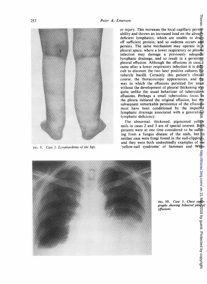
In January 1960, at the age of 62, she had an attackof 'bronchitis with asthma' and developed breath-lessness on exertion. In September 1960 there waspain in the left chest with increased shortness ofbreath: she was admitted to Epping Hospital. Inaddition to gross lymphoedema (Fig. 9) she was found
to have bilateral pleural effusions (Fig. 10). Therewere no clinical signs of cardiac failure, but theE.C.G. showed evidence of an anterior myocardialinfarction with a B.P. of 175/95 mm. Hg.Her recovery from this episode was uneventful,
but over the subsequent years she required regularand repeated aspirations of one or two pints ofpleural fluid, which was clear, straw-coloured materialwith a protein content of 4 g./100 ml., and the pre-dominant cell was the lymphocyte. All other bio-chemical and haematological tests failed to revealany significant abnormality to account for theeffusions.
In May 1961, at the age of 63, she was admitted toGuy's Hospital on account of increased breathless-ness. The clinical condition, the bilateral lymph-oedema, the pleural effusions, and hypertension wereall unchanged. On this admission it was again notedthat the nails were abnormal and showed 'an over-growth of the nail-bed and pigmentation of one year'sduration'. Nail-clippings were investigated for fungi,but none was demonstrated. At this time the bilateraleffusions were thought to be in some way related tothe lymphoedema.Over the following years she had numerous other
admissions to Guy's and Epping Hospitals forrepeated aspirations. In 1962 she became unconsciousand died after a period of severe diarrhoea: herplasma electrolytes taken before death suggested ametabolic acidosis.Necropsy showed bilateral straw-coloured pleural
effusions, more on the right than on the left, wherethe lung was firmly attached to the chest wall bydense adhesions. The left kidney was small andcontracted, the right being twice the normal size.
250
on 21 May 2018 by guest. P
rotected by copyright.http://thorax.bm
j.com/
Thorax: first published as 10.1136/thx.21.3.247 on 1 M
ay 1966. Dow
nloaded from

FIG. 7FIGS 7 and 8. Case 2. Lymphoedema of the legs and face.
No other significant abnormalities were found andthe brain was normal. It was assumed that death wasdue to a metabolic acidosis related to the period ofdiarrhoea. No studies of the lymphatics were made,because at this time lymphoedema as a cause for apleural effusion had not, so far as I am aware, beendescribed.
DISCUSSION
The association of lymphoedema with serouseffusions in the pleural and peritoneal spaces wasdescribed by Morris, Blood, Sidman, Steel, andWhittem (1954) in newborn Australian Ayrshirecalves, and the three present patients probablyrepresent a related condition. All three were suffer-ing from lymphoedema not only of the legs butalso of the hands and face. The puffy appearanceof the face (Figs 4 and 8) was surprisingly similar,and in both the female patients it led to the seriousconsideration of a diagnosis of myxoedema. Case3 was in fact treated for a period with thyroidextract without improvement: objective thyroidfunction tests in both the patients were normal.The effusions, at least in cases 1 and 3, apparentlyappeared after a lower respiratory infectiondescribed as 'bronchitis' in case 1 and as'bronchitis and asthma' in case 3.
It is well recognized that patients with a con-genital deficiency of the lymphatics often do notdevelop persistent lymphoedema until after someinfection or other episode such as an insect biteFIG. 8
UI
on 21 May 2018 by guest. P
rotected by copyright.http://thorax.bm
j.com/
Thorax: first published as 10.1136/thx.21.3.247 on 1 M
ay 1966. Dow
nloaded from
Peter A. Emerson
Nll.F Fror injury. This increases the local capillary perme-ability and throws an increased load on the alreadydeficient lymphatics, which are unable to drainoff sufficient protein, and so oedema occurs andpersists. The same mechanism may operate in apleural space, where a lower respiratory or pleural
.;.vSStinfection may damage a previously adequate* ~~lymphatic drainage, and so result in a persistent
a pleural effusion. Although the effusions in case 1came after a lower respiratory infection it is diffi-
M X cult to discount the two later positive cultures for' tubercle bacilli. Certainly this patient's clinical
course, the thoracoscopic appearances, and theway in which the effusions persisted for yearswithout the development of pleural thickening was
quite unlike the usual behaviour of tuberculouseffusions. Perhaps a small tuberculous focus inthe pleura initiated the original effusion, but thesubsequent remarkable persistence of the effusionsmust have been conditioned by the impairedlymphatic drainage associated with a generalizedlymphatic deficiency.The abnormal, thickened, pigmented yellow
Nfi:f.\-,jh . nails in cases 2 and 3 are of special interest. Bothpatients were at one time considered to be suffer-ing from a fungus disease of the nails, but inneither case were fungi found in the nail-clippings,and they were both undoubtedly examples of the
9. Case 3. Lvinphoedema of the legs. 'yellow-nail syndrome' of Samman and White
FIG. 10 . Case 3. Chest radio-graphs showing bilateral pleuraleffusions.
FIG. S
252
on 21 May 2018 by guest. P
rotected by copyright.http://thorax.bm
j.com/
Thorax: first published as 10.1136/thx.21.3.247 on 1 M
ay 1966. Dow
nloaded from

Yellow nails, lymphoedema, and pleural eftusions
(1964). These authors first described the associa-tion of slow-growing discoloured yellow nails withlymphoedema. The nails are usually smooth, butmay show transverse ridging and are usuallyexcessively curved from side to side, so that cover-age of the lateral ridges by the surrounding softtissue is less than normal and the cuticles aredeficient. The colour is usually pale yellow, butmay be slightly greenish. Nail separation mayoccur and some partially separated nails have adistinct hump. Although the nail changes arebelieved to be the result of defective lymphaticdrainage, evidence of lymphoedema may notappear until years after the finger-nails or toe-nails have become abnormal.Minor and even moderate degrees of lymph-
oedema are easily overlooked. The patient mayhave had the condition for so long that he hascome to regard it as almost normal, so that nospecific complaint is made, and the evidence mayremain hidden under the bed-clothes when thechest or nails are examined. It is important, there-fore, in chronic unexplained pleural effusion tolook for evidence of impaired lymphatic drainage;this may be obvious in the legs, hands, and evenface, but it is also important to look for the slowlygrowing deformed yellow nails which may indicateimpaired lymphatic drainage even in the absenceof obvious lymphoedema. Although such caseshave not been encountered, if a patient with achronic unexplained pleural effusion were foundto have deformed yellow nails without obviousevidence of lymphoedema it would be wise tocarry out lymphangiography of the legs to seewhether there was any evidence of lymphaticdeficiency or megalymphatics.The three present patients all had a great deal
of investigation and treatment which proved tobe irrelevant when the relationship of the pleuraleffusion to the lymphoedema was eventuallyrealized. None of them had any specific treatment
to prevent the recurrence of pleural effusions. Case2 had only a small effusion which did not requireaspiration, but cases 1 and 3 were both severelyhandicapped and distressed by bilateral effusions,which required the frequent removal of largequantities of pleural fluid.
In case 3 necropsy showed that the left pleuralspace was largely obliterated by adhesions, pre-sumably explaining why the effusion was small andnot troublesome on that side. The rational treat-ment of future cases would appear to bepleurodesis by thoracotomy and parietalpleurectomy.
SUMMARY
Three patients with chronic and otherwiseunexplained pleural effusions are described and thecondition is attributed to a deficiency of thelymphatics draining the pleural space. All threehad chronic lymphoedema of the legs, hands, andface: two of them had abnormal yellow nailswhich are also believed to be due to deficientlymphatic drainage.
I am most grateful to Dr. K. Robson and Dr.C. G. Baker for permission to report cases 1 and 3;to Dr. R. Grenville Mathers and Dr. P. Samman forpermission to report case 2, and particularly to Dr.P. Samman and the Institute of Dermatology forallowing me to reproduce photographs of case 2 andof his nails.
REFERENCES
Hurwitz, P. A., and Pinnals, D. J. (1964). Pleural effusion in chronichereditary lymphedema. (Nonne, Milroy, Meige's disease.)Radiology, 82, 246.
Morris, B., Blood, D. C., Sidman, W. R., Steel, J. D., and Whittem,J. H. (1954). Congenital lymphatic oedema in Ayrshire calves.Aust. J. exp. Biol. med. Sci., 32, 265.
Robson, K., and Emerson, P. A. (1963). In Chest Diseases, ed.K. M. A. Perry and T. Holmes Sellors, Vol. I, p. 194. Butter-worths, London.
Samman, P. D., and White, W. F. (1964). The 'Yellow Nail' syndrome.Brit. J. Derm., 76, 153.
253
on 21 May 2018 by guest. P
rotected by copyright.http://thorax.bm
j.com/
Thorax: first published as 10.1136/thx.21.3.247 on 1 M
ay 1966. Dow
nloaded from