Year-round algal toxin exposure in free-ranging sea lionsmyweb.facstaff.wwu.edu/aceveda/PDFs/lab...
16
MARINE ECOLOGY PROGRESS SERIES Mar Ecol Prog Ser Vol. 583: 243–258, 2017 https://doi.org/10.3354/meps12345 Published November 16 INTRODUCTION Harmful algal blooms (HABs) are increasing world- wide and have led to mortalities of both humans and marine wildlife, including marine mammals (Fire & Van Dolah 2012). This trend is particularly concerning for declining and depleted wildlife populations (Durbin et al. 2002), providing the need to understand what factors most influence exposure to these toxins. Mar- ine mammal exposure to algal toxins has been exam- ined through sampling prey and fecal remains of ac- tively foraging or live-captured animals (Doucette et al. 2006, Lefebvre et al. 2016), plankton and nearshore bivalves in areas near mortality events (Scholin et al. 2000), and feces or stomach contents of ill or deceased animals (Lefebvre et al. 1999, 2010). However, the po- tential for year-round exposure through the food web has not been examined. Two of the phytoplankton taxa implicated in mar- ine mammal mortalities are Pseudo-nitzschia spp. diatoms and Alexandrium spp. dinoflagellates, which produce domoic acid and paralytic shellfish toxins (PSTs; saxitoxin and a suite of congeners of which saxitoxin is the most toxic), respectively. The north- west coast of Washington State, USA, is a unique marine environment for studying domoic acid and PSTs, both of which impact state shellfish fisheries (Trainer et al. 2002, 2003). Situated at the northern- most extent of the California Current, the region is characterized by high phytoplankton biomass and primary productivity, driven by wind and topo- graphic induced upwelling, the seasonally formed Juan de Fuca Eddy, and nutrient inputs from the Fraser and Columbia River outflows (MacFadyen et al. 2005). The Juan de Fuca Eddy, a cold-water gyre located offshore of the mouth of the Strait of Juan de Fuca, is a hotspot for Pseudo-nitzschia blooms (Trainer et al. 2002). The eddy forms in the spring and dissipates in the fall, and some waters escape from the eddy bringing toxins to the nearshore en- *Corresponding author: [email protected] Year-round algal toxin exposure in free-ranging sea lions Adrianne M. Akmajian 1,2, *, Jonathan J. Scordino 1 , Alejandro Acevedo-Gutiérrez 2 1 Makah Fisheries Management, Neah Bay, WA 98357, USA 2 Department of Biology, Western Washington University, Bellingham, WA 98225, USA ABSTRACT: Harmful algal bloom toxins cause illness and mortality in marine mammals world- wide, yet the potential for year-round exposure to these toxins has not previously been studied. We measured concentrations of domoic acid and saxitoxin in scats from Steller sea lions Eume- topias jubatus (n = 383 scats) and California sea lions Zalophus californianus (n = 125 scats) over a 2 yr period. Toxin concentrations in the scats were compared to the prey remains in the scats and to concentrations in nearshore bivalves. Saxitoxin was detected in 45% and domoic acid was detected in 17% of all scats tested, and both toxins were detected in all seasons and months of the year. A variety of benthic and pelagic fish were significantly associated with toxins in sea lion scats, including prey with low occurrence in the sea lions’ diet. Toxins detected in winter scats confirm that US West Coast marine mammals are exposed to domoic acid and saxitoxin through their prey outside of the expected algal bloom seasons. KEY WORDS: Year-round · Harmful algal blooms · HABs · Saxitoxin · Domoic acid · Eumetopias jubatus · Zalophus californianus · Diet analysis OPEN PEN ACCESS CCESS © The authors 2017. Open Access under Creative Commons by Attribution Licence. Use, distribution and reproduction are un- restricted. Authors and original publication must be credited. Publisher: Inter-Research · www.int-res.com
Transcript of Year-round algal toxin exposure in free-ranging sea lionsmyweb.facstaff.wwu.edu/aceveda/PDFs/lab...
MARINE ECOLOGY PROGRESS SERIESMar Ecol Prog Ser
Vol. 583: 243–258, 2017https://doi.org/10.3354/meps12345
Published November 16
INTRODUCTION
Harmful algal blooms (HABs) are increasing world-wide and have led to mortalities of both humans andmarine wildlife, including marine mammals (Fire &Van Dolah 2012). This trend is particularly concerningfor declining and depleted wildlife populations (Durbinet al. 2002), providing the need to understand whatfactors most influence exposure to these toxins. Mar-ine mammal exposure to algal toxins has been exam-ined through sampling prey and fecal remains of ac-tively foraging or live-captured animals (Doucette etal. 2006, Lefebvre et al. 2016), plankton and nearshorebivalves in areas near mortality events (Scholin et al.2000), and feces or stomach contents of ill or deceasedanimals (Lefebvre et al. 1999, 2010). However, the po-tential for year-round exposure through the food webhas not been examined.
Two of the phytoplankton taxa implicated in mar-ine mammal mortalities are Pseudo-nitzschia spp.
diatoms and Alexandrium spp. dinoflagellates, whichproduce domoic acid and paralytic shellfish toxins(PSTs; saxitoxin and a suite of congeners of whichsaxitoxin is the most toxic), respectively. The north-west coast of Washington State, USA, is a uniquemarine environment for studying domoic acid andPSTs, both of which impact state shellfish fisheries(Trainer et al. 2002, 2003). Situated at the northern-most extent of the California Current, the regionis characterized by high phytoplankton biomassand primary productivity, driven by wind and topo-graphic induced upwelling, the seasonally formedJuan de Fuca Eddy, and nutrient inputs from theFraser and Columbia River outflows (MacFadyen etal. 2005). The Juan de Fuca Eddy, a cold-water gyrelocated offshore of the mouth of the Strait of Juande Fuca, is a hotspot for Pseudo-nitzschia blooms(Trainer et al. 2002). The eddy forms in the springand dissipates in the fall, and some waters escapefrom the eddy bringing toxins to the nearshore en -
*Corresponding author: [email protected]
Year-round algal toxin exposure in free-rangingsea lions
Adrianne M. Akmajian1,2,*, Jonathan J. Scordino1, Alejandro Acevedo-Gutiérrez2
1Makah Fisheries Management, Neah Bay, WA 98357, USA2Department of Biology, Western Washington University, Bellingham, WA 98225, USA
ABSTRACT: Harmful algal bloom toxins cause illness and mortality in marine mammals world-wide, yet the potential for year-round exposure to these toxins has not previously been studied.We measured concentrations of domoic acid and saxitoxin in scats from Steller sea lions Eume-topias jubatus (n = 383 scats) and California sea lions Zalophus californianus (n = 125 scats) over a2 yr period. Toxin concentrations in the scats were compared to the prey remains in the scats andto concentrations in nearshore bivalves. Saxitoxin was detected in 45% and domoic acid wasdetected in 17% of all scats tested, and both toxins were detected in all seasons and months of theyear. A variety of benthic and pelagic fish were significantly associated with toxins in sea lionscats, including prey with low occurrence in the sea lions’ diet. Toxins detected in winter scatsconfirm that US West Coast marine mammals are exposed to domoic acid and saxitoxin throughtheir prey outside of the expected algal bloom seasons.
KEY WORDS: Year-round · Harmful algal blooms · HABs · Saxitoxin · Domoic acid · Eumetopiasjubatus · Zalophus californianus · Diet analysis
OPENPEN ACCESSCCESS
© The authors 2017. Open Access under Creative Commons byAttribution Licence. Use, distribution and reproduction are un -restricted. Authors and original publication must be credited.
Publisher: Inter-Research · www.int-res.com

Mar Ecol Prog Ser 583: 243–258, 2017
vironment (MacFadyen et al. 2005). In contrast,Alexandrium blooms occur nearshore, typically inembayments with highly stratified waters, afterwater temperatures begin warming in the spring(Trainer et al. 2003). These differences in bloomdynamics likely result in different pathways of toxinexposure to top predators such as marine mammals.Toxin screening of marine mammals in Washingtonhas detected sublethal concentrations of domoic acidin stranded cetacean and pinniped species (McCabeet al. 2016) and of domoic acid and saxitoxin in livesea otters (White et al. 2013). The first confirmed caseof acute HAB toxicity in Washington was docu-mented in May 2015 in a California sea lion Zalophuscalifornianus (McCabe et al. 2016).
Given the presence of both toxins on the Washing-ton coast, the overlapping range of the eastern distinct population segment (DPS) Steller sea lionsEumetopias jubatus and California sea lions suggeststhat both species could be exposed to algal toxinsthrough contaminated prey. Steller sea lions from alldemographic groups live and forage on the Washing-ton coast year-round, while primarily male Californiasea lions migrate into Washington waters seasonally(NMFS 2013, Gearin et al. 2017). While the US popu-lation of California sea lions is estimated at a healthy300 000 individuals (Gearin et al. 2017), the Stellersea lion experienced an 80% reduction in abundancefrom the 1970s through the 1990s in US waters (Milleret al. 2005). This reduction was driven by declines inthe western DPS (Gulf of Alaska through Russia;west of 144° W), which is currently listed as endan-gered under the US Endangered Species Act whilethe eastern DPS was recently de listed (NMFS 2013).Although no mortalities related to algal toxins havebeen reported in Steller sea lions, monitoring thethreat of biotoxins is a priority in the recovery plan ofthe western DPS and post-delisting monitoring of theeastern DPS (NMFS 2013).
Sea lions change their diet based on seasonally andlocally abundant prey and may migrate in response toseasonal prey fluctuations (Sigler et al. 2009). The di-ets of both Steller and California sea lions, composedof a variety of bony and cartilaginous fishes andcephalopods, are often dominated by only a few spe-cies (Sinclair & Zeppelin 2002, Orr et al. 2011), al-though as many as 25 different prey items have beenfound in a single scat (Riemer et al. 2011). Domoicacid and PSTs have been detected in both benthic andpelagic fish (Lefebvre et al. 2002, Vigilant & Silver2007, Jester et al. 2009, Jensen et al. 2015), suggestingthat while a more diverse diet could lessen the effectsof acute toxicity experienced by ingesting a single
contaminated prey species, generalist predators areat risk of exposure from multiple prey items.
In this study, we assessed the year-round exposureof free-ranging Steller and California sea lions spe-cies in Washington State to domoic acid and saxitoxinby comparing the toxin concentrations measured infeces to the prey remains in the scat and to toxin con-centrations measured in nearshore bivalves. Thegoals of this study were to (1) determine year-roundbaseline levels of HAB toxins in the feces of both sealion species and to identify factors associated withtoxin exposure, including season, year, and hauloutlocation; (2) de termine if there was a difference indiet between sea lions with and without measureablelevels of toxins and whether particular prey itemswere more associated with toxin ex posure; and (3)compare the presence and con centration of toxins inthe scat to that in nearshore bivalves to determinehow toxin levels in nearshore bivalves predictedtoxin exposure of top predators.
MATERIALS AND METHODS
Study area and sample collection
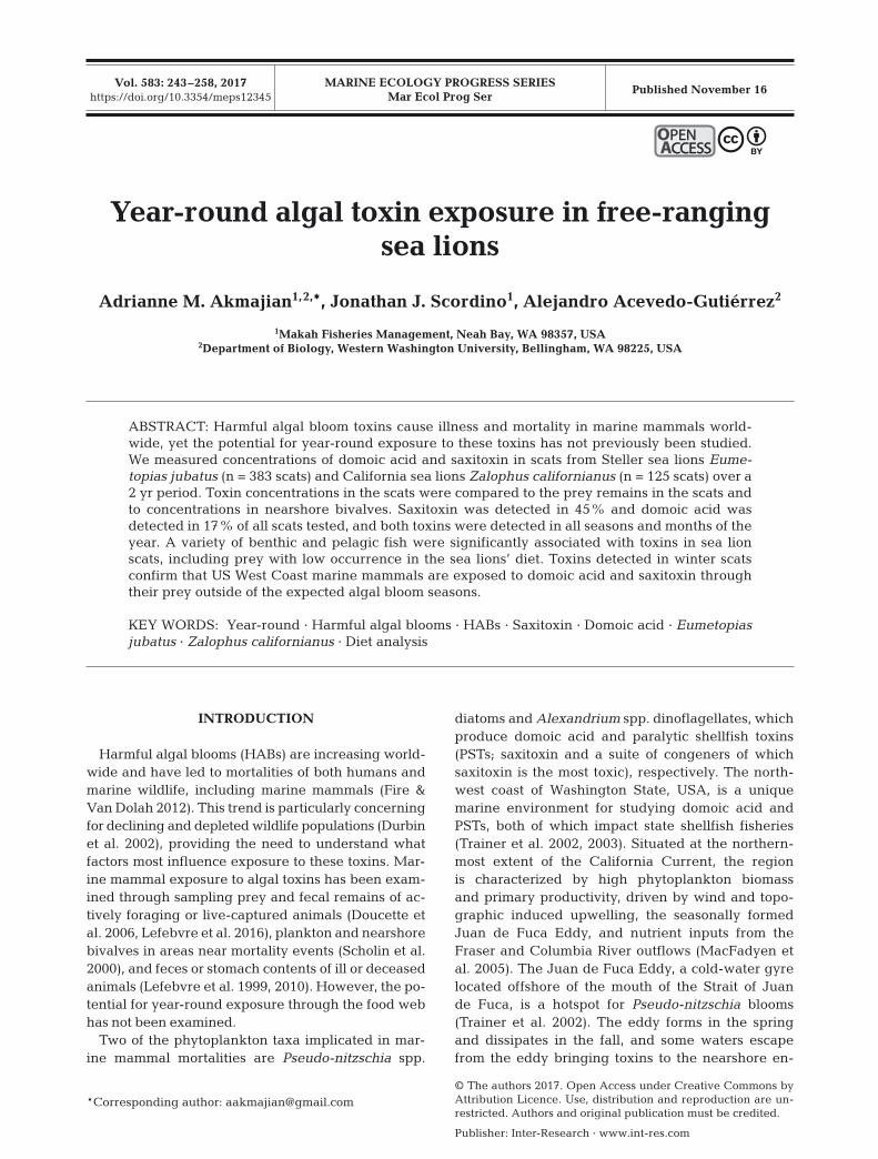
Steller and California sea lion scats were collectedat haulout sites on the northern Washington coast(Fig. 1). Scats were collected seasonally from March2011 through February 2013. Seasons were de -fined as spring: March−May; summer: June−August;fall: September−November; and winter: December−February. Scats were only collected from sites where≥95% of sea lions at the collection site belonged to asingle species. Feces were scooped into individualplastic Whirl-Pak™ bags for storage. Because domoicacid and saxitoxin are water-soluble, only <48 h oldscats (moist, not weathered or dried out) were ana -lyzed. For collection and analysis, we grouped haul -outs in close proximity to each other (between 0.5−1.3 km) into 3 complexes: Tatoosh Island, BodeltehIslands, and Carroll Island/Sea Lion Rock (Fig. 1).
Toxin analysis
We collected approximately 4 g of fecal materialfrom each scat, stored each in a 15 ml centrifugetube, and froze all samples at −20°C. To ensure thatno prey remains were lost during subsampling, the4 g of fresh scat were pushed through either a nylon,fine mesh, paint strainer bag (0.25 mm), or through a0.5 mm brass sieve to be collected for toxin analysis.
244
Akmajian et al.: Year-round algal toxins in sea lions
When scats were small and had insufficient materialfor analyzing both toxins (i.e. <4 g), we prioritizedthe analysis of domoic acid because the toxin hascaused strandings and mortalities of sea lions on theUS West Coast (Fire &Van Dolah 2012).
Scat subsamples were analyzed using direct com-petitive enzyme-linked immunosorbent assays (ELISA)following methods described by Lefebvre et al.(2016). Domoic acid was analyzed using ASP directcELISA kits (Biosense Laboratories). Saxitoxin wasanalyzed using saxitoxin (paralytic shellfish poison-ing [PSP]) ELISA kits (Abraxis). The lower quantifi-cation limits were 3 ng g−1 for saxitoxin and 4 ng g−1
for domoic acid. Due to a sample-processing error,the sample with the highest concentration of domoicacid measured (672.2 ng g−1 detected in a Californiasea lion scat) was erroneously processed twice andrecorded with 2 different identifications. Because wewere unable to identify the sample, it was discardedfrom statistical analyses, but was included in therange of concentrations detected.
ELISA analysis of domoic acid in marine mammalfeces and fluids has been validated by comparison totraditional methodologies including high-performanceliquid chromatography (HPLC) and liquid chromato -graphy tandem mass spectrometry (LC-MS/MS)(Lefebvre et al. 2010, Frame & Lefebvre 2013). ELISA
results from marine mammal tissues generally agreewith those of HPLC and LC-MS/MS (Frame & Lefeb-vre 2013). ELISA is also able to detect domoic acid atconcentrations below the quantification limit for thesetraditional methods, particularly in marine mammalfeces (Lefebvre et al. 2010, Frame & Lefebvre 2013).
Previous studies of saxitoxin in marine mammalfeces and fluids have also used the Abraxis SaxitoxinELISA (Lefebvre et al. 2016). The Abraxis ELISA de -tects saxitoxin 100%, but has limited cross-reactivity(<30%) of other PST congeners as specified by themanufacturer. We report our results as saxitoxin onlybecause we cannot detect the presence or total con-centration of all PST congeners. Other toxic congers,such as the gonyautoxins, may be more prevalent inthe environment than saxitoxin itself, and this limitedcross-reactivity could under estimate the actual toxic-ity of the sample (Turner et al. 2014). While HPLC orLC-MS/MS would provide a more extensive toxinprofile and the concentrations of other congeners,these analyses for PSTs are costly, time-consuming,and limited to the congeners for which commercialstandards are available (Costa et al. 2009, Humpageet al. 2010). In seawater and shellfish, the AbraxisELISA detects saxitoxin at lower concentrations com-pared to traditional methods such as HPLC (Costa etal. 2009).
245
Fig. 1. Sample locations of bivalves and sea lion scat along the western Strait of Juan de Fuca and northwest Washington coast, USA. Parentheses refer to sites having more than 1 sample location. Bathymetry is shaded according to depth (m)
Mar Ecol Prog Ser 583: 243–258, 2017
Analysis of food habits
The remaining fecal material was processed foranalysis of food habits. For the majority of samples,we used a washing machine to clean the fresh fecalmaterial from the prey remains (Orr et al. 2003).Samples with gravel were washed by hand throughnested sieves of 2, 1, and 0.5 mm. Prey remainswere identified to the lowest taxonomic group pos-sible using a comparative reference collection offish from the northeast Pacific Ocean (Riemer et al.2011). Prey remains included bones (e.g. otoliths,vertebrae, teeth, gill rakers, etc.), cartilaginousstructures, and cephalopod beaks. Salmonids (Onco-rhynchus spp.) were identified by size and groupedas ‘juvenile’ (smolts) and ‘non-juvenile’ (all otherage classes). Salmonid bones were classified asjuveniles based on time of year and juvenile salmongrowth rates in the study area (Duffy & Beauchamp2011) and comparison to reference collections ofjuvenile and non-juvenile salmonid bones. Remainsthat could not be identified to a more specific taxo-nomic group were recorded as unidentified bonyfish (class Osteichthyes) or unidentified cartilagi-nous fish (subclass Elasmobranchii). Prey itemswere recorded as present/absent for each sampleand converted to percent frequency of occurrence(FO) in sea lion diet, where FO of a particular taxonis equal to the number of scats having that taxondivided by the total number of scats with any identi-fiable prey.
Bivalve samples
Biotoxin results measured in nearshore bivalvesfrom January 2011 through March 2013 were pro-vided by the Washington Department of Health(WDOH) Biotoxin Monitoring Program. PSTs werede tected using the standardized mouse bioassay,with a detection limit of approximately 40 µg 100g−1 shellfish (400 ng g−1) (AOAC 1965, APHA 1970).Domoic acid was detected using HPLC, with adetection limit of approximately 0.5 ppm (500 ngg−1) (Quilliam et al. 1995). Bivalves were collectedfrom 8 locations in the western Strait of Juan deFuca and northern Washington coast (Fig. 1). Con-tributors to this dataset included the Makah andQuileute Tribes and the Washington Department ofFish and Wildlife, who perform monitoring for sub-sistence and recreational harvest. Although a vari-ety of bivalve species are collected for monitoring,only the 2 most common species are included here:
California mussel Mytilus californianus and Pacificrazor clam Siliqua patula. California mussels werethe dominant species collected at all sites exceptfor 2 locations at Kalaloch Beach (Fig. 1), whererazor clams were dominant.
Nearshore bivalve toxin concentrations were plot-ted against the concentrations in sea lion scats toexamine the relationship between presence andconcentration of toxin in bivalves to that in thescats. For both toxins, samples reported by WDOHas ‘not detected’ are reported here as ‘0.’ Due tomethod-detection limits and reporting criteria,WDOH re ports concentrations of saxitoxin consid-ered below the quantification limit as <380 ng g−1,which we plotted as 380 ng g−1. Similarly, fordomoic acid, WDOH reports concentrations below1000 ng g−1 as <1000 ng g−1. We substituted re -ported values of <1000 ng g−1 with 500 ng g−1 in ourplots to dif ferentiate between a true measured con-centration of 1000 ng g−1 and <1000 ng g−1 becausethe true concentration between the detection limit(500 ng g−1) and reporting limit (1000 ng g−1) wasnot available.
Statistical analysis
We used binary logistic regression models andAkaike’s information criterion (AIC) in the statisticalprogram R (R Core Team 2015) to examine which fac-tors best predicted the presence or absence of toxinin sea lion scats. The dependent variable was thepresence/absence of either domoic acid or saxitoxinin the scat. All explanatory variables were modeledas categorical and included haulout complex, season,and year, which was defined as ‘Year 1,’ representingsamples from March 2011 through February 2012,and ‘Year 2,’ representing samples from March 2012through February 2013.
For analyzing the influence of diet on toxinexposure, we compared the prey remains in thescat to the presence/absence or categorical level oftoxin. We included only prey taxa that had a totalFO of ≥5% averaged over all seasons and dis-carded scats containing unidentified bony or carti-laginous fish only. Prey taxa were included at thelowest taxonomic classification identified, bothindividual species and unidentified species groupedby a higher taxonomic level. For both toxins, cate-gorical levels were defined as ‘no’ representingsamples below the detection limit, ‘low’ between 0and 20 ng g−1, ‘med’ between >20 and 50 ng g−1,and ‘high’ representing concentrations >50 ng g−1.
246
Akmajian et al.: Year-round algal toxins in sea lions
Designation of toxin levels was based on naturalbreaks in the concentrations measured in scatsand is not intended to be indicative of toxicologicaleffects.
We used nonmetric multi-dimensional scaling(NMDS) in the Community Ecology Package (vegan)of R (Oksanen et al. 2017) to investigate the influ-ence of overall diet on toxin exposure and usedPearson’s chi-squared and Fisher’s exact tests tocompare the presence of individual prey items topresence/ absence or categorical toxin levels. Forthe 2 sea lion species, we ran NMDS for each toxinseparately using the metaMDS function, which canhandle zero distance where 2 points are identical(Oksanen et al. 2017), and a binary Jaccard distancematrix. For ana lysis of individual prey items, weused chi-squared tests when expected values of atleast 80% of the cells were ≥5 and Fisher’s exacttests when expected values were <5 (McHugh2013). Standardized residuals from chi-squared testswere evaluated to determine whether toxin was sig-nificantly higher or lower than expected valuesbased on presence or absence of each prey itemwhere residual values >|1.96| were considered sig-nificant at p < 0.05 (Agresti 2007). For Fisher’s exacttests, we used odds ratios and confidence intervalsto evaluate positive or negative relationships of sig-nificant p-values and performed post hoc testingusing Bonferroni-corrected p-values where the cor-rected alpha was calculated as 0.05 divided by thenumber of pairwise comparisons.
RESULTS
Toxin detection
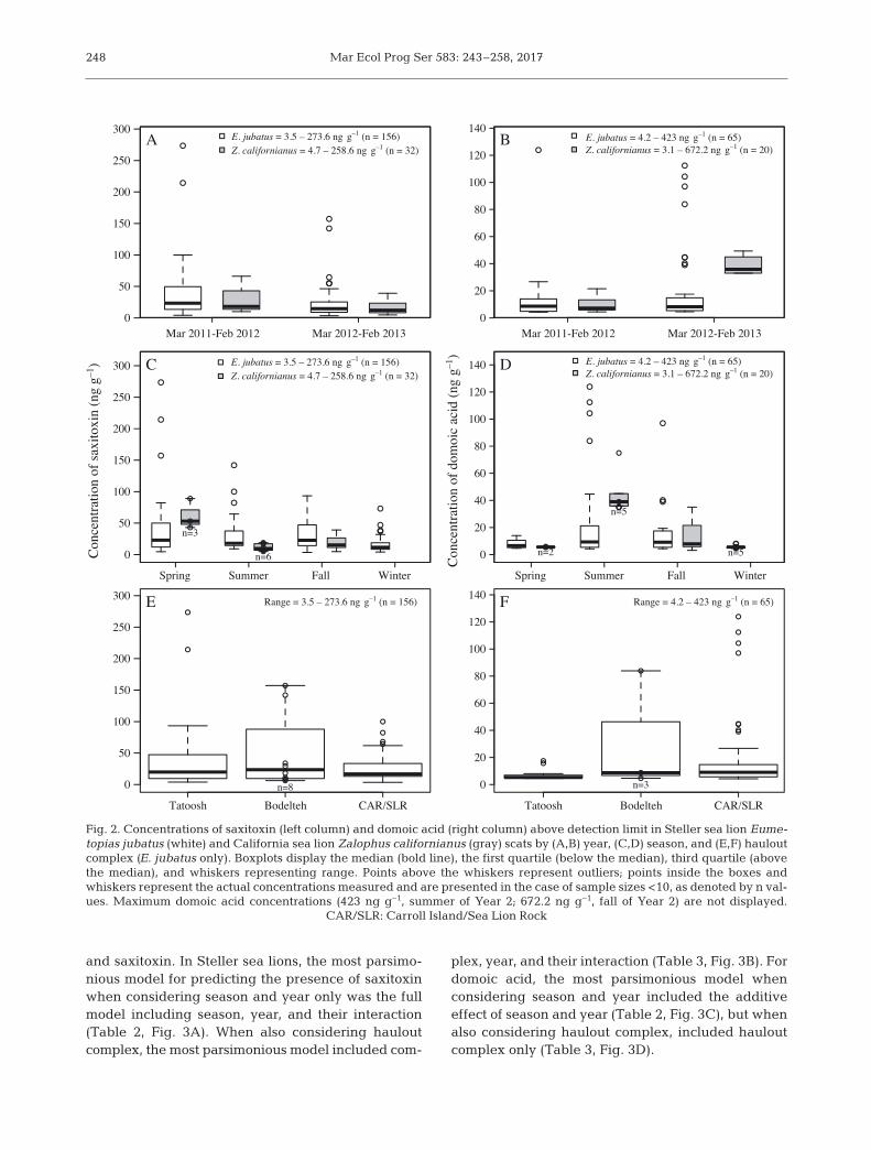
We collected a total of 383 scats from Steller sealions and 125 scats from California sea lions (Table 1).Of the 508 total scats, 14 samples did not have suffi-cient fecal material to analyze both toxins and wereonly analyzed for domoic acid (Table 1). Saxitoxinwas detected in 45% of all scat samples analyzed anddomoic acid was detected in 17% of samples ana-lyzed. The toxins were detected concurrently in 26scats from Steller sea lions and 6 scats from Californiasea lions. The highest concentrations of saxitoxin inboth sea lion species were detected in Year 1 (Fig. 2A)and in spring (Fig. 2C), while domoic acid was higherin Year 2 for both species (Fig. 2B) and in summer forSteller sea lions and fall for California sea lions(Fig. 2D). In Steller sea lions, the highest concentra-tions of saxitoxin were from the Tatoosh Island com-plex (Fig. 2E), while the highest concentrations ofdomoic acid were from the Carroll Island/Sea LionRock complex (Fig. 2F).
Toxin prevalence by season, year, and location
Logistic regression analysis identified several ex -planatory models for the presence of toxins in Stellersea lions, but for California sea lions, the intercept-only model had the lowest AIC for both domoic acid
247
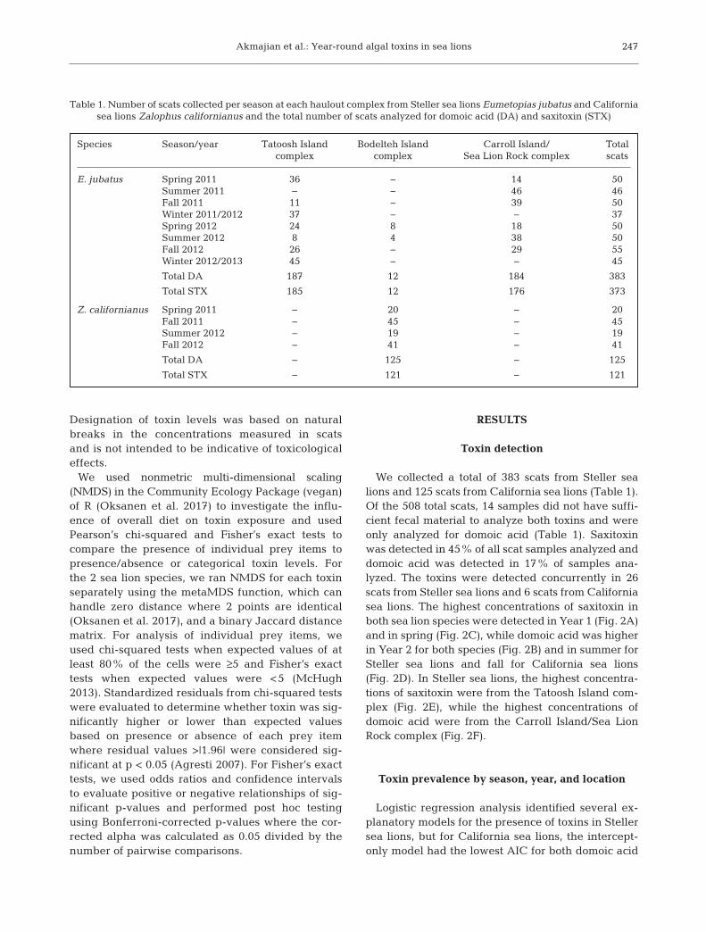
Species Season/year Tatoosh Island Bodelteh Island Carroll Island/ Total complex complex Sea Lion Rock complex scats
E. jubatus Spring 2011 36 − 14 50Summer 2011 − − 46 46Fall 2011 11 − 39 50Winter 2011/2012 37 − − 37Spring 2012 24 8 18 50Summer 2012 8 4 38 50Fall 2012 26 − 29 55Winter 2012/2013 45 − − 45
Total DA 187 12 184 383
Total STX 185 12 176 373
Z. californianus Spring 2011 − 20 − 20Fall 2011 − 45 − 45Summer 2012 − 19 − 19Fall 2012 − 41 − 41
Total DA − 125 − 125
Total STX − 121 − 121
Table 1. Number of scats collected per season at each haulout complex from Steller sea lions Eumetopias jubatus and Californiasea lions Zalophus californianus and the total number of scats analyzed for domoic acid (DA) and saxitoxin (STX)
Mar Ecol Prog Ser 583: 243–258, 2017
and saxitoxin. In Steller sea lions, the most parsimo-nious model for predicting the presence of saxitoxinwhen considering season and year only was the fullmodel including season, year, and their interaction(Table 2, Fig. 3A). When also considering haul outcomplex, the most parsimonious model included com -
plex, year, and their interaction (Table 3, Fig. 3B). Fordomoic acid, the most parsimonious model whenconsidering season and year included the additiveeffect of season and year (Table 2, Fig. 3C), but whenalso considering haulout complex, included hauloutcomplex only (Table 3, Fig. 3D).
248
0
50
100
150
200
250
300
Mar 2011-Feb 2012 Mar 2012-Feb 2013
A
0
20
40
60
80
100
120
140
Mar 2011-Feb 2012 Mar 2012-Feb 2013
B
0
50
100
150
200
250
300
Con
cent
ratio
n of
sax
itoxi
n (n
g g–1
)
n=3
n=6
Spring Summer Fall Winter
C E. jubatus = 3.5 – 273.6 ng g–1 (n = 156)Z. californianus = 4.7 – 258.6 ng g–1 (n = 32)
0
20
40
60
80
100
120
140
Con
cent
ratio
n of
dom
oic
acid
(ng
g–1
)
n=5n=2
n=5
Spring Summer Fall Winter
D E. jubatus = 4.2 – 423 ng g–1 (n = 65)Z. californianus = 3.1 – 672.2 ng g–1 (n = 20)
Tatoosh Bodelteh CAR/SLR
0
50
100
150
200
250
300 Range = 3.5 – 273.6 ng g–1 (n = 156)
n=8
E
Tatoosh Bodelteh CAR/SLR
0
20
40
60
80
100
120
140Range = 4.2 – 423 ng g–1 (n = 65)
n=3
F
E. jubatus = 3.5 – 273.6 ng g–1 (n = 156)Z. californianus = 4.7 – 258.6 ng g–1 (n = 32)
E. jubatus = 4.2 – 423 ng g–1 (n = 65)Z. californianus = 3.1 – 672.2 ng g–1 (n = 20)
Fig. 2. Concentrations of saxitoxin (left column) and domoic acid (right column) above detection limit in Steller sea lion Eume-topias jubatus (white) and California sea lion Zalophus californianus (gray) scats by (A,B) year, (C,D) season, and (E,F) hauloutcomplex (E. jubatus only). Boxplots display the median (bold line), the first quartile (below the median), third quartile (abovethe median), and whiskers representing range. Points above the whiskers represent outliers; points inside the boxes andwhiskers represent the actual concentrations measured and are presented in the case of sample sizes <10, as denoted by n val-ues. Maximum domoic acid concentrations (423 ng g−1, summer of Year 2; 672.2 ng g−1, fall of Year 2) are not displayed.
CAR/SLR: Carroll Island/Sea Lion Rock
Akmajian et al.: Year-round algal toxins in sea lions
Sea lion diet
A total of 39 prey taxa (lowest taxonomic group)were identified in Steller sea lion scats (see Table S1in the Supplement at www.int-res.com/articles/suppl/m583 p243 _ supp. pdf) and 30 prey taxa were identifiedin California sea lion scats (Table S2). Eleven scatsfrom Steller sea lions and 6 scats from California sealions contained no identifiable prey remains (uniden-tified bony or cartilaginous fish) and were removedfrom the diet analyses. For Steller sea lions, the mostcommon prey items (>20% FO in the diet) were clu-peids (family Clupeidae, 56%), salmonids (Onco-rhynchus spp., 40%), skates (family Rajidae, 40%),rockfish (Sebastes spp., 36%), Pacific spiny dogfishSqualus acanthias (28%), and flatfish (order Pleu-ronectiformes, 21%) (Table S1). For California sealions, the most common prey items were clupeids(79%), salmonids (38%), Pacific hake Merlucciusproductus (32%), and dogfish (30%) (Table S2). The2 sea lion species had several major differences indiet including skate consumption (40% in Stellercompared to 5% in California sea lions), flatfishes(21% in Steller compared to 8% in California sealions), and codfishes (family Gadidae) (18% in Stellerand 6% in California sea lions).
Sea lion diet varied by season, collection year, andhaulout complex. For Steller sea lions, consumption ofskates, rockfish, and Pacific herring Clupea pallasiiwas noticeably higher in spring of Year 2 (68, 60, and22% FO, respectively) compared to Year 1 (34, 30, and6%), whereas dogfish was more common in spring ofYear 1 (42%) compared to Year 2 (22%). Walleye pol-lock Gadus chalco gramma had a spike in occurrencein spring of Year 1 (30% FO), but was present in <5%of samples in all other seasons of both collection years.Northern anchovy Engraulis mordax was present inspring, summer, and fall of Year 1 (14, 13, and 18%
FO), but was virtually absent in Year 2 (0, 2,and 4%). Juvenile salmonids and Pacificsand lance Ammodytes hexapterus werepresent in winter of Year 2 (38 and 13%, re-spectively), but absent in Year 1 (0 and 1%).For California sea lions, hake consumptiondropped dramatically from fall of Year 1(53%) to Year 2 (8%). For Steller sea lions,salmonids and codfishes were more com-mon in samples from Tatoosh Island (57 and29%, respectively) compared to Carroll Is-land/Sea Lion Rock (22 and 12%), whereasthe reverse was true for flatfishes (40% atCarroll Island/Sea Lion Rock compared to7% at Tatoosh).
Diet influences on toxin prevalence
There were no convergent solutions using NMDSfor analyzing diet of either Steller or California sealions to look for differences in toxin presence. Chi-squared and Fisher’s exact analyses identified sev-eral prey items significantly associated with the pres-ence or concentration level of toxins found in the scat(Tables 4−6). In Steller sea lion scats, saxitoxin pres-ence was significantly higher than ex pected whenAmerican shad Alosa sapidissima and walleye pol-lock were present in the scat (Table 4). Saxitoxin wasdetected in every scat with pollock prey remains (n =18 scats), and scats with pollock were significantlymore likely to have medium and high levels of saxi-toxin compared to low concentrations (Table 6),including 2 of the highest concentrations measured(214.4 and 273.6 ng g−1). Domoic acid presence wassignificantly higher than expected when Pacific sar-dine Sardinops sagax and starry flounder Platichthysstellatus were present (Table 4). Scats with sardinewere significantly more likely to have high con -centrations of domoic acid (Table 6). In Californiasea lions, scats with anchovy were about 5 timesmore likely to have domoic acid compared to thosewithout an chovy (Table 4).
Comparison to nearshore bivalves
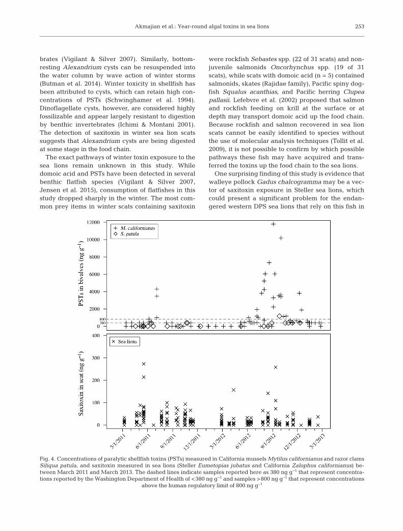
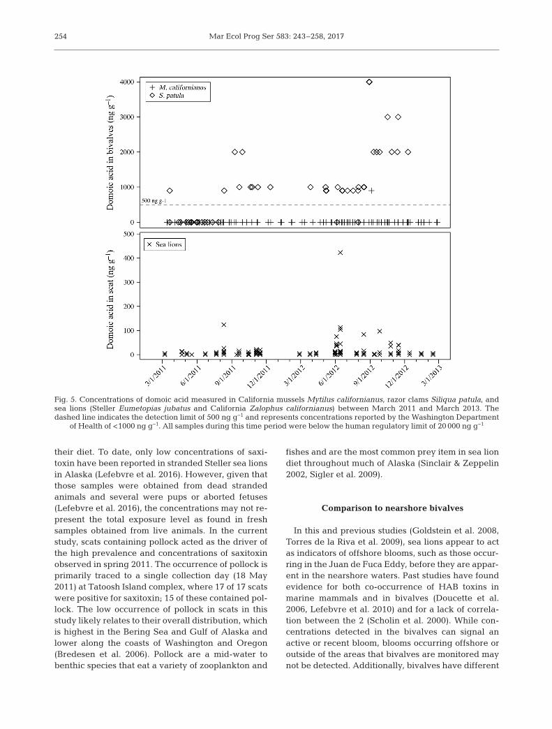
Between January 2011 and March 2013, PSTs weredetected in concentrations >380 ng g−1 in 58 samplesof California mussels Mytilus californianus and 4samples of razor clams Siliqua patula (Fig. 4). Domoicacid was almost exclusively detected in razor clams(Fig. 5). PST concentrations peaked in the summer of
249
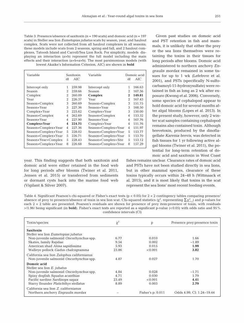
Variable Saxitoxin Variable Domoic aciddf AIC df AIC
Intercept only 1 509.07 Intercept only 1 350.86Season 4 511.67 Season 4 336.77Year 2 484.84 Year 2 346.66Season+Year 5 488.15 Season+Year 5 331.99Season×Year 8 482.46 Season×Year 8 333.87
Table 2. Presence/absence of saxitoxin (n = 373 scats) and domoic acid(n = 383 scats) in Steller sea lion Eumetopias jubatus scats compared toseason and year. For simplicity, models displaying an interaction (a×b)signify that the full model was tested including the main effects and theirinteraction (a+b+a×b). The most parsimonious models (with lowest
Akaike’s information criterion, AIC) are shown in bold
Mar Ecol Prog Ser 583: 243–258, 2017
2011 and the summer/fall of 2012 (Fig. 4). Domoicacid peaked in the fall of both years, detected in con-centrations above 1000 ng g−1 in 24 razor clam sam-ples and at a concentration <1000 ng g−1 in a singlesample of California mussel (Fig. 5). In periods ofhigh PST concentrations in bivalves (e.g. July andAugust 2012) the majority of sea lion scats did nothave detectable levels of saxitoxin (Fig. 4). Con-versely, time periods when most sea lion scats haddetectable levels of saxitoxins (e.g. April and May2011) bi valves had no to very low (<380 ng g−1) con-centrations of PSTs (Fig. 4). In several time periods(e.g. February and March 2012), sea lions had de -tectable levels of saxitoxin while no toxins were de -tected in nearshore bivalves (Fig. 4). Domoic acid in
sea lion scats appeared to peak slightly before peaksob served in bivalves in the fall of both years (Fig. 5).
DISCUSSION
Toxin retention in the food web
This is the first study to systematically documentthat West Coast marine mammals are exposed todomoic acid and saxitoxin year-round. AlthoughSteller sea lions and California sea lions on theouter coast of Washington were more often exposedto saxitoxin than to domoic acid, both toxins weredetected in scats in every season and month of the
250
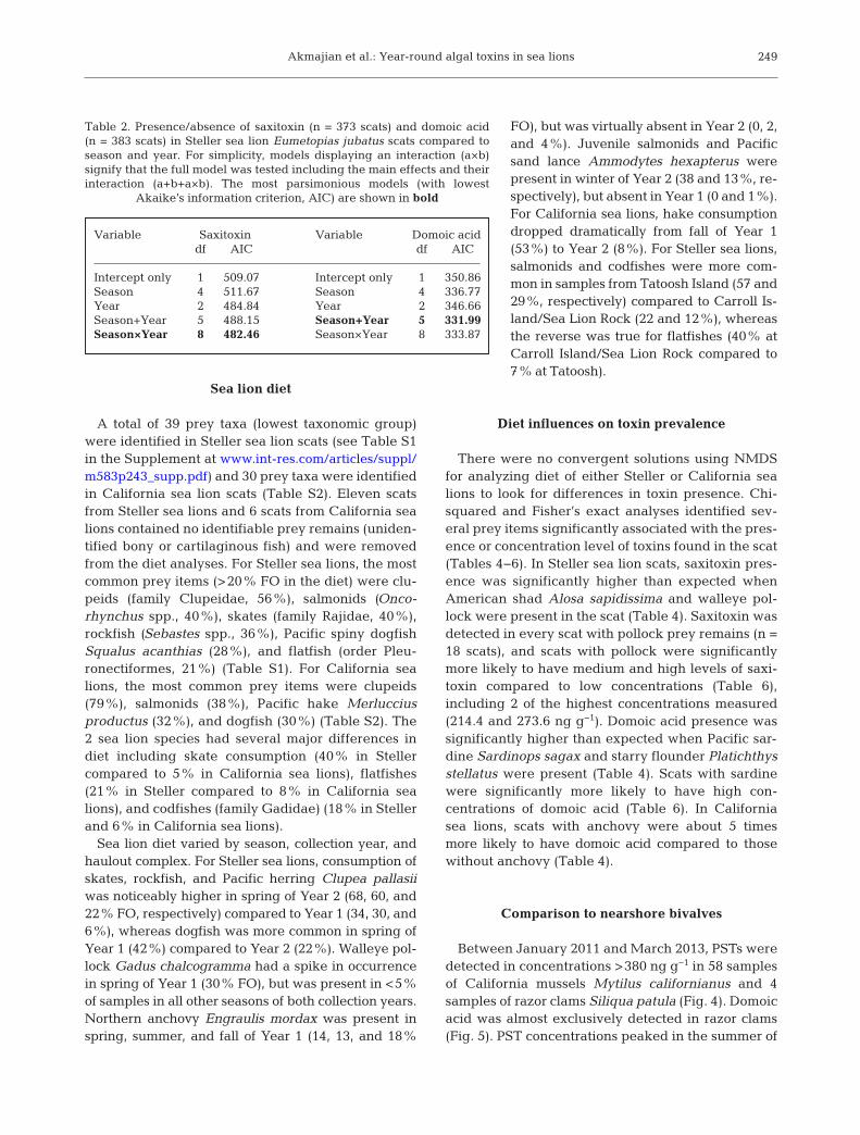
Fig. 3. Percent of scats with (A,B) saxitoxin and (C,D) domoic acid detected for Steller sea lions Eumetopias jubatus (A,C) by season and year and (B,D) by year and haulout complex (see Fig. 1)
Akmajian et al.: Year-round algal toxins in sea lions
year. This finding suggests that both saxitoxin anddomoic acid were either retained in the food webfor long periods after blooms (Twiner et al. 2011,Jensen et al. 2015) or transferred from sedimentsor dormant cysts back into the marine food web(Vigilant & Silver 2007).
Given past studies on domoic acidand PST retention in fish and mam-mals, it is unlikely that either the preyor the sea lions themselves were re-taining the toxins in their tissues forlong periods after blooms. Domoic acidadministered to northern anchovy En -graulis mordax remained in some tis-sues for up to 1 wk (Lefebvre et al.2001), and PSTs (specifically N-sulfo-carbamoyl-11-hydroxysulfate) were re -tained in fish as long as 2 wk after ex-posure (Kwong et al. 2006). Conversely,some species of cephalopod appear tohold domoic acid for several months af-ter algal blooms (Lopes et al. 2013). Inthe present study, however, only 2 win-ter scat samples containing cephalopodremains also contained toxin. Althoughbrevetoxin, produced by the dinofla-gellate Karenia brevis, was detected infish tissues for 1 yr following active al-gal blooms (Twiner et al. 2011), the po-tential for long-term retention of do-moic acid and saxitoxin in West Coast
fishes re mains unclear. Clearance rates of domoic acidand PSTs have not been studied di rectly in sea lions,but in other mammal species, clearance of these toxins typically occurs within 24−48 h (Wittmaack etal. 2015), and it is most likely that toxins in the scatrepresent the sea lions’ most recent feeding events.
251
Variable Saxitoxin Variable Domoic aciddf AIC df AIC
Intercept only 1 259.98 Intercept only 1 166.63Season 2 259.66 Season 2 167.56Complex 2 260.09 Complex 2 149.81Year 2 226.37 Year 2 167.43Season+Complex 3 260.69 Season+Complex 3 151.75Season+Year 3 227.36 Season+Year 3 168.50Complex+Year 3 225.62 Complex+Year 3 150.00Season×Complex 4 262.69 Season×Complex 4 153.32Season×Year 4 227.90 Season×Year 4 167.76Complex×Year 4 224.75 Complex×Year 4 151.76Season+Complex+Year 4 227.36 Season+Complex+Year 4 151.97Season×Complex+Year 5 228.02 Season×Complex+Year 5 153.77Season+Complex×Year 5 226.71 Season+Complex×Year 5 153.70Season×Year+Complex 5 228.43 Season×Complex+Site 5 153.12Season×Complex×Year 8 226.68 Season×Complex×Year 8 157.29
Table 3. Presence/absence of saxitoxin (n = 190 scats) and domoic acid (n = 197scats) in Steller sea lion Eumetopias jubatus scats by season, year, and hauloutcomplex. Scats were not collected from all haulout complexes in all seasons;these models include scats from 2 seasons, spring and fall, and 2 haulout com-plexes, Tatoosh Island and Carroll/Sea Lion Rock. For simplicity, models dis-playing an interaction (a×b) represent the full model including the maineffects and their interaction (a+b+a×b). The most parsimonious models (with
lowest Akaike’s Information Criterion, AIC) are shown in bold
Toxin/species χ2 p Presence prey:presence toxin
SaxitoxinSteller sea lion Eumetopias jubatusNon-juvenile salmonid Oncorhynchus spp. 6.77 0.010 1.66Skates, family Rajidae 9.54 0.002 −1.89American shad Alosa sapidissima 5.93 0.015 1.99Walleye pollock Gadus chalcogramma 23.86 <0.001 3.82
California sea lion Zalophus californianusNon-juvenile salmonid Oncorhynchus spp. 4.87 0.027 1.70
Domoic acidSteller sea lion E. jubatusNon-juvenile salmonid Oncorhynchus spp. 4.84 0.028 −1.71Spiny dogfish Squalus acanthias 4.71 0.030 1.79Pacific sardine Sardinops sagax 23.49 <0.001 4.41Starry flounder Platichthys stellatus 8.89 0.003 2.79
California sea lion Z. californianusNorthern anchovy Engraulis mordax – Fisher’s p: 0.011 Odds 4.99, CI: 1.24−19.44
Table 4. Significant Pearson’s chi-squared or Fisher’s exact tests (p < 0.05) for 2 × 2 contingency tables comparing presence/absence of prey to presence/absence of toxin in sea lion scat. Chi-squared statistics (χ2, representing Σχ2
i–j ) and p-values foreach 2 × 2 table are presented. Pearson’s residuals are shown for presence of prey item:presence of toxin, with residuals> |1.96| being significant (in bold). Fisher’s exact tests are reported as a significant p-value (<0.05) with odds ratio and 95%
confidence intervals (CI)
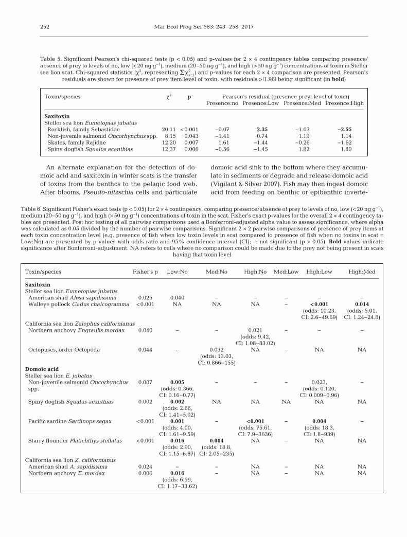
Mar Ecol Prog Ser 583: 243–258, 2017
An alternate explanation for the detection of do -moic acid and saxitoxin in winter scats is the transferof toxins from the benthos to the pelagic food web.After blooms, Pseudo-nitzschia cells and particulate
domoic acid sink to the bottom where they accumu-late in sediments or degrade and release domoic acid(Vigilant & Silver 2007). Fish may then ingest domoicacid from feeding on benthic or epi benthic inverte-
252
Toxin/species χ2 p Pearson’s residual (presence prey: level of toxin)Presence:no Presence:Low Presence:Med Presence:High
SaxitoxinSteller sea lion Eumetopias jubatusRockfish, family Sebastidae 20.11 <0.001 −0.07 2.35 −1.03 −2.55Non-juvenile salmonid Oncorhynchus spp. 8.15 0.043 −1.41 0.74 1.19 1.14Skates, family Rajidae 12.20 0.007 1.61 −1.44 −0.26 −1.62Spiny dogfish Squalus acanthias 12.37 0.006 −0.56 −1.45 1.82 1.80
Table 5. Significant Pearson’s chi-squared tests (p < 0.05) and p-values for 2 × 4 contingency tables comparing presence/absence of prey to levels of no, low (<20 ng g−1), medium (20−50 ng g−1), and high (>50 ng g−1) concentrations of toxin in Stellersea lion scat. Chi-squared statistics (χ2, representing Σχ2
i–j ) and p-values for each 2 × 4 comparison are presented. Pearson’s residuals are shown for presence of prey item:level of toxin, with residuals >|1.96| being significant (in bold)
Toxin/species Fisher’s p Low:No Med:No High:No Med:Low High:Low High:Med
SaxitoxinSteller sea lion Eumetopias jubatusAmerican shad Alosa sapidissima 0.025 0.040 − − − − −Walleye pollock Gadus chalcogramma <0.001 NA NA NA − <0.001 0.014
(odds: 10.23, (odds: 5.01,CI: 2.6−49.69) CI: 1.24−24.8)
California sea lion Zalophus californianusNorthern anchovy Engraulis mordax 0.040 − − 0.021 − − −
(odds: 9.42,CI: 1.08−83.02)
Octopuses, order Octopoda 0.044 − 0.032 NA − NA NA(odds: 13.03,
CI: 0.866−155)Domoic acidSteller sea lion E. jubatusNon-juvenile salmonid Oncorhynchus 0.007 0.005 − − − 0.023, −spp. (odds: 0.366, (odds: 0.120,
CI: 0.16−0.77) CI: 0.009−0.96)Spiny dogfish Squalus acanthias 0.002 0.002 NA NA NA NA NA
(odds: 2.66,CI: 1.41−5.02)
Pacific sardine Sardinops sagax <0.001 0.001 − <0.001 − 0.004 −(odds: 4.00, (odds: 75.61, (odds: 18.3,
CI: 1.61−9.59) CI: 7.9−3636) CI: 1.8−939)Starry flounder Platichthys stellatus <0.001 0.016 0.004 NA − NA NA
(odds: 2.90, (odds: 18.8,CI: 1.15−6.87) CI: 2.05−235)
California sea lion Z. californianusAmerican shad A. sapidissima 0.024 − − NA − NA NANorthern anchovy E. mordax 0.006 0.016 − NA − NA NA
(odds: 6.59,CI: 1.17−33.62)
Table 6. Significant Fisher’s exact tests (p < 0.05) for 2 × 4 contingency, comparing presence/absence of prey to levels of no, low (<20 ng g−1),medium (20−50 ng g−1), and high (>50 ng g−1) concentrations of toxin in the scat. Fisher’s exact p-values for the overall 2 × 4 contingency ta-bles are presented. Post hoc testing of all pairwise comparisons used a Bonferroni-adjusted alpha value to assess significance, where alphawas calculated as 0.05 divided by the number of pairwise comparisons. Significant 2 × 2 pairwise comparisons of presence of prey items ateach toxin concentration level (e.g. presence of fish when low toxin levels in scat compared to presence of fish when no toxins in scat =Low:No) are presented by p-values with odds ratio and 95% confidence interval (CI); –: not significant (p > 0.05). Bold values indicate significance after Bonferroni-adjustment. NA refers to cells where no comparison could be made due to the prey not being present in scats
having that toxin level
Akmajian et al.: Year-round algal toxins in sea lions
brates (Vigilant & Silver 2007). Similarly, bottom-resting Alexandrium cysts can be resuspended intothe water column by wave action of winter storms(Butman et al. 2014). Winter toxicity in shellfish hasbeen attributed to cysts, which can retain high con-centrations of PSTs (Schwinghamer et al. 1994).Dinoflagellate cysts, however, are considered highlyfossilizable and appear largely re sistant to digestionby benthic invertebrates (Ichimi & Montani 2001).The detection of saxitoxin in winter sea lion scatssuggests that Alexandrium cysts are being digestedat some stage in the food chain.
The exact pathways of winter toxin exposure to thesea lions remain unknown in this study. Whiledomoic acid and PSTs have been detected in severalbenthic flatfish species (Vigilant & Silver 2007,Jensen et al. 2015), consumption of flatfishes in thisstudy dropped sharply in the winter. The most com-mon prey items in winter scats containing saxitoxin
were rockfish Sebastes spp. (22 of 31 scats) and non-juvenile sal monids Oncorhynchus spp. (19 of 31scats), while scats with domoic acid (n = 5) containedsalmonids, skates (Rajidae family), Pacific spiny dog-fish Squalus acanthias, and Pacific herring Clupeapallasii. Lefebvre et al. (2002) proposed that salmonand rockfish feeding on krill at the surface or atdepth may transport domoic acid up the food chain.Be cause rockfish and salmon recovered in sea lionscats cannot be easily identified to species withoutthe use of molecular analysis techniques (Tollit et al.2009), it is not possible to confirm by which possiblepathways these fish may have acquired and trans-ferred the toxins up the food chain to the sea lions.
One surprising finding of this study is evidence thatwalleye pollock Gadus chalcogramma may be a vec-tor of saxitoxin exposure in Steller sea lions, whichcould present a significant problem for the endan-gered western DPS sea lions that rely on this fish in
253
Fig. 4. Concentrations of paralytic shellfish toxins (PSTs) measured in California mussels Mytilus californianus and razor clamsSiliqua patula, and saxitoxin measured in sea lions (Steller Eumetopias jubatus and California Zalophus californianus) be-tween March 2011 and March 2013. The dashed lines indicate samples reported here as 380 ng g−1 that represent concentra-tions reported by the Washington Department of Health of <380 ng g−1 and samples >800 ng g−1 that represent concentrations
above the human regulatory limit of 800 ng g–1
Mar Ecol Prog Ser 583: 243–258, 2017
their diet. To date, only low concentrations of saxi-toxin have been reported in stranded Steller sea lionsin Alaska (Lefebvre et al. 2016). However, given thatthose samples were obtained from dead strandedanimals and several were pups or aborted fetuses(Lefebvre et al. 2016), the concentrations may not re -present the total exposure level as found in freshsamples obtained from live animals. In the currentstudy, scats containing pollock acted as the driver ofthe high prevalence and concentrations of saxitoxinobserved in spring 2011. The occurrence of pollock isprimarily traced to a single collection day (18 May2011) at Tatoosh Island complex, where 17 of 17 scatswere positive for saxitoxin; 15 of these contained pol-lock. The low occurrence of pollock in scats in thisstudy likely relates to their overall distribution, whichis highest in the Bering Sea and Gulf of Alaska andlower along the coasts of Washington and Oregon(Bredesen et al. 2006). Pollock are a mid-water tobenthic species that eat a variety of zooplankton and
fishes and are the most common prey item in sea liondiet throughout much of Alaska (Sinclair & Zeppelin2002, Sigler et al. 2009).
Comparison to nearshore bivalves
In this and previous studies (Goldstein et al. 2008,Torres de la Riva et al. 2009), sea lions appear to actas indicators of offshore blooms, such as those occur-ring in the Juan de Fuca Eddy, before they are appar-ent in the nearshore waters. Past studies have foundevidence for both co-occurrence of HAB toxins inmarine mammals and in bivalves (Doucette et al.2006, Lefebvre et al. 2010) and for a lack of correla-tion between the 2 (Scholin et al. 2000). While con-centrations detected in the bivalves can signal anactive or recent bloom, blooms occurring offshore oroutside of the areas that bivalves are monitored maynot be detected. Additionally, bivalves have different
254
Fig. 5. Concentrations of domoic acid measured in California mussels Mytilus californianus, razor clams Siliqua patula, andsea lions (Steller Eumetopias jubatus and California Zalophus californianus) between March 2011 and March 2013. Thedashed line indicates the detection limit of 500 ng g−1 and represents concentrations reported by the Washington Department
of Health of <1000 ng g−1. All samples during this time period were below the human regulatory limit of 20 000 ng g−1
Akmajian et al.: Year-round algal toxins in sea lions
rates of toxin depuration, and depending on sam-pling frequency, there may not be apparent overlapin the bivalves and sea lion scats. For example, Cali-fornia mussels depurate PSTs in a span of 3 to >12 wk(Bricelj & Shumway 1998), but depurate domoic acidin as little as 2 wk (Whyte et al. 1995). Razor clamstypically accumulate only low levels of PSTs (Traineret al. 2003), whereas they hold domoic acid in theirtissues for as long as 6 to 18 mo (Wekell et al. 1994).In our study, domoic acid was detected in sea lionscats somewhat prior to the spikes seen in nearshoreshellfish, perhaps indicating that the sea lions wereeither foraging in areas of blooms prior to thosewaters making it nearshore or that their prey speciestraveled through or foraged in these areas. Similarly,high concentrations of saxitoxin (>200 ng g−1) weredetected in sea lion scat prior to PST concentrationsin bivalves reaching levels above the regulatorylimit. Of particular interest is the occurrence of saxi-toxin in sea lion scat during winter and early springwhen low (<380 ng g−1) to no PSTs were detected inbivalves and when blooms typically do not occur. Ifthis exposure resulted from Alexandrium cysts beingresuspended into the water column, it is possiblethat this occurred outside of areas that are presently sampled for bivalves or that the depuration periodhad already passed, and thus was not detected in thebivalves. A more comprehensive analysis of planktonand toxin movement through the food web is neededto fully understand this dynamic system.
Study limitations
Despite many compelling findings, there are somelimitations to this study’s interpretation. There is noliterature on what effects weathering might have onthe toxin content in scats deposited on haulout sites,and the toxin concentrations in this study are as -sumed to represent the full amount at the time ofdeposition. Because both saxitoxin and domoic acidare flushed from the digestive system quickly, it islikely that toxins in scats were from a recent feedingevent (Jensen et al. 2015); however, hard parts in asingle scat may not represent all fish eaten or all fishfrom the most recent meal. Biases in hard-part scatanalysis, such as identifying prey structures, degra-dation of bones in digestion, and different passageand recovery rates of fish ingested (Tollit et al. 2015),could also have affected our results. Although weidentified all prey structures to improve documenta-tion of consumed prey (Tollit et al. 2015), it is still possible that prey species that had very friable bones
or that were not wholly consumed (e.g. depredatedsalmon from troll fisheries) were not documented. FOdescribes the presence or absence of a prey itemwithout enumerating the number of fish eaten or esti-mating volume or biomass of eaten prey, which likelylimited our ability to evaluate the contribution ofeach prey species in the diet. Future studies on expo-sure of sea lions to HAB toxins would benefit fromemploying newly developed molecular techniquesthat allow quantifying diet and prey biomass (Deagleet al. 2013, Thomas et al. 2016).
In addition, given the previously discussed limita-tions of the saxitoxin ELISA, our saxitoxin resultsshould be interpreted with caution. Few publicationshave attempted to compare or validate the saxitoxinELISA in marine mammal feces or tissues with othermethods, although some authors have performedmatrix testing to avoid false positives or negatives(e.g. Lefebvre et al. 2016). Due to the limited-crossreactivity with other PST toxins, we must assumethat the saxitoxin ELISA potentially underestimatedthe full sample toxicity. For example, in Sequim Bay,Washington, the gonyautoxins GTX1,4, GTX2,3, andneosaxitoxin were more prevalent in Alexandriumspp. isolates compared to saxitoxin itself (Lefebvreet al. 2008). Further, detection of algal toxins in thescats remains an underestimate of the full amount oftoxins the sea lions were exposed to because it onlyreflects the toxin that was passed through digestionand does not account for the amount initially ingested.
Consequences of toxin exposure
Toxin concentrations measured in this study arenot likely to have caused acute illness and likely indicate very low concentrations metabolized intotissues. Domoic acid concentrations in this studywere well below those measured in scat from harborseals Phoca vitulina and northern fur seals Callorhi-nus ursinus exhibiting acute domoic acid toxicity(Lefebvre et al. 2010, McHuron et al. 2013), but werecomparable to concentrations in California sea lionswith acute and chronic symptoms (Lefebvre et al.1999, Goldstein et al. 2008). The concentrations werehigher than those measured in stranded, harvested,and live pinnipeds in Alaska (Lefebvre et al. 2016),but lower than maximum concentrations reported instranded northern fur seals in Alaska (Lefebvre et al.2010) and free-ranging harbor seals in Scotland(Jensen et al. 2015). Both acute and chronic patholo-gies of domoic acid toxicity have been described inmarine mammals (Goldstein et al. 2008). Symptoms
255
Mar Ecol Prog Ser 583: 243–258, 2017
of chronic domoic acid exposure include epilepsy,compromised foraging behavior and navigation, andreproductive failure (Brodie et al. 2006, Goldstein etal. 2008, Thomas et al. 2010).
It is not known what concentrations of saxitoxincause mortality in marine mammals, but the concen-trations measured in this study fall below the lethaltotal dose in humans of 1−4 mg (James et al. 2010).PSTs have been suspected in mass mortalities ofhumpback whales Megaptera novaeangliae, Medi-terranean monk seals Monachus monachus, and bot-tlenose dolphins Tursiops truncatus, although con-centrations in feces and other body fluids were notavailable for comparison to this study (Fire & VanDolah 2012). Saxitoxin concentrations in this studywere higher than those reported in 13 marine mam-mal species sampled in Alaska (Lefebvre et al. 2016),but within the range of PSTs detected in free-rangingand stranded harbor seals in Scotland (Jensen et al.2015) and North Atlantic right whales Eubalaenaglacialis in the Bay of Fundy, Canada (Doucette et al.2006). Durbin et al. (2002) suggested that repeatedexposure to PSTs could affect diving capabilities,such as causing longer surfacing time after a dive asobserved in right whales. Bogomolni et al. (2016)found that repeated exposure to low concentrationsof saxitoxin increased phocine distemper virus in har-bor seal lymphocytes and suggested that monitoringlow levels of saxitoxin is important for understandingproliferation of diseases such as morbillivirus infection.
While concentrations in feces represent exposure,domoic acid measured in feces is frequently orders ofmagnitude higher than concentrations in the urine orserum, which are better indicators of the amountsmetabolized (Goldstein et al. 2008, Lefebvre et al.2010). Although sea lions have a high digestion effi-ciency of fish (>90%, Rosen & Trites 2000), the assim-ilation efficiency of these HAB toxins is not known. Interrestrial mammal species, only about 4−7% ofingested domoic acid is absorbed across the digestivetract (Truelove et al. 1997). The amount of saxitoxinabsorbed by the digestive tract has not been reportedin mammals; however, there is evidence in bothhumans (García et al. 2004) and whales (Doucette etal. 2006) of biotransformation of saxitoxin into otherderivatives.
CONCLUSIONS
In conclusion, this study presents evidence of low-level exposure to saxitoxin and domoic acid in bothSteller and California sea lions on the Washington
coast and suggests that prey with relatively low oc-currence in the diet may lead to significant algal toxintransfer up the food chain. While chronic ex posure todomoic acid has been well documented in Californiasea lions (Goldstein et al. 2008), it is unclear what con-centrations induce toxicity and how those compare tothe concentrations measured in this study. Low level,chronic PST toxicity in wildlife has not been docu-mented, although several authors have proposed pos-sible effects, including immunomodulatory effects inharbor seals (Durbin et al. 2002, Bogomolni et al.2016). What effects either chronic or acute algal toxinexposure may have at the population level remainsunknown. Our findings suggest that generalist preda-tors with a more diverse diet may not have any respitefrom exposure to, or the effects of, marine algal toxinsas compared to predators that specialize in specificprey items. Sampling at a finer timescale (e.g. weeklyor monthly) may be necessary for understanding therole of infrequent prey species in transfer of algal tox-ins to top predators. This study reaffirms previousstudies documenting that marine mammals can be ex-posed to algal toxins through their prey outside of theexpected algal bloom seasons (Twiner et al. 2011) andsuggests further investigation into food web transferof precipitated cells and dormant cysts or long-termretention of algal toxins in the West Coast food web.
Acknowledgements. This study was conducted as part of aMaster’s thesis at Western Washington University com-pleted by A.M.A. in June 2016. Research was funded by theMakah Tribe through the Olympic Coast National MarineSanctuary and a NOAA Fisheries’ Species Recovery GrantProgram and conducted under NMFS research permit nos.14326 and 13430 awarded to NOAA’s National MarineMammal Laboratory (NMML), Seattle, WA. Special thanksto Russell Svec at Makah Fisheries Management and to theMakah Tribal Council for their support. This project waspossible thanks to the work of Elizabeth Frame and SusanRiemer. Thank you to Adi Hanein, Shelley Lankford, andstaff at the Washington Department of Health for providingcoastal bivalve data. We thank the staff from the MakahTribe, Quileute Tribe, and Washington Department of Fishand Wildlife for collecting the bivalve samples. Thank you toPat Gearin and Merrill Gosho at NMML for assisting in sam-ple collection. We thank Brian Bingham and Suzanne Stromfor input and assistance in data analysis and manuscriptediting.
LITERATURE CITED
Agresti A (2007) An introduction to categorical data analy-sis, 2nd edn. John Wiley & Sons, Hoboken, NJ
AOAC (Association of Official Agricultural Chemists) (1965)Biological method. In: Horowitz W (ed) Official methodsof analysis of the Association of Official AgriculturalChemists, 10th edn. AOAC, Washington, DC, p 282−284
256
Akmajian et al.: Year-round algal toxins in sea lions
APHA (American Public Health Association) (1970) Recom-mended procedures for the examination of water andshellfish, 4th edn. APHA, New York, NY
Bogomolni AL, Bass AL, Fire S, Jasperse L and others (2016)Saxitoxin increases phocine distemper virus replicationupon in-vitro infection in harbor seal immune cells.Harmful Algae 51: 89−96
Bredesen EL, Coombs AP, Trites AW (2006) Relationshipbetween Steller sea lion diets and fish distributions in theeastern North Pacific. In: Trites A, Atkinson S, DeMasterD, Fritz L, Gelatt T, Rea L, Wynne K (eds) Sea lions of theworld. Alaska Sea Grant College Program, University ofAlaska Fairbanks, Fairbanks, AK, p 131−140
Bricelj VM, Shumway SE (1998) Paralytic shellfish toxins inbivalve molluscs: occurrence, transfer kinetics, and bio-transformation. Rev Fish Sci 6: 315−383
Brodie EC, Gulland FMD, Greig DJ, Hunter M and others(2006) Domoic acid causes reproductive failure in Cali-fornia sea lions (Zalophus californianus). Mar Mamm Sci22: 700−707
Butman B, Aretxabaleta AL, Dickhudt PJ, Dalyander PS andothers (2014) Investigating the importance of sedimentresuspension in Alexandrium fundyense cyst populationdynamics in the Gulf of Maine. Deep Sea Res Part II TopStud Oceanogr 103: 79−95
Costa PR, Baugh KA, Wright B, RaLonde R and others (2009)Comparative determination of paralytic shellfish toxins(PSTs) using five different toxin detection methods inshellfish species collected in the Aleutian Islands,Alaska. Toxicon 54: 313−320
Deagle BE, Thomas AC, Shaffer AK, Trites AW, Jarmon SN(2013) Quantifying sequence proportions in a DNA-based diet study using Ion Torrent amplicon sequencing: Which counts count? Mol Ecol Resour 13: 620−633
Doucette GJ, Cembella AD, Martin JL, Michaud J, ColeTVN, Rolland RM (2006) Paralytic shellfish poisoning(PSP) toxins in North Atlantic right whales Eubalaenaglacialis and their zooplankton prey in the Bay of Fundy,Canada. Mar Ecol Prog Ser 306: 303−313
Duffy EJ, Beauchamp DA (2011) Rapid growth in the earlymarine period improves the marine survival of Chinooksalmon (Oncorhynchus tshawytscha) in Puget Sound,Washington. Can J Fish Aquat Sci 68: 232−240
Durbin E, Teegarden G, Campbell R, Cembella A, Baum-gartner MF, Mate BR (2002) North Atlantic right whales,Eubalaena glacialis, exposed to paralytic shellfish poi-soning (PSP) toxins via a zooplankton vector, Calanusfinmarchicus. Harmful Algae 1: 243−251
Fire SE, Van Dolah FM (2012) Marine biotoxins: emergenceof harmful algal blooms as health threats to marinewildlife In: Aguirre AA, Ostfeld R, Daszak P (eds) Newdirections in conservation medicine: applied cases ofecological health. Oxford University Press, New York,NY, p 374−389
Frame E, Lefebvre K (2013) ELISA methods for domoic acidquantification in multiple marine mammal species andsample matrices. Tech Memo NMFS-NWFSC-122. USDepartment of Commerce, NOAA, Seattle, WA
García C, del Bravo MC, Lagos M, Néstor L (2004) Paralyticshellfish poisoning: post-mortem analysis of tissue andbody fluid samples from human victims in the Patagoniafjords. Toxicon 43: 149−158
Gearin PJ, Melin SR, DeLong RL, Gosho ME, Jeffries SJ(2017) Migration patterns of adult male Californiasea lions (Zalophus californianus). Tech Memo NMFS-
AFSC-346. US Department of Commerce, NOAA,Seattle, WA
Goldstein T, Mazet JAK, Zabka TS, Langlois G and others(2008) Novel symptomatology and changing epidemiol-ogy of domoic acid toxicosis in California sea lions (Zalo-phus californianus): an increasing risk to marine mam-mal health. Proc R Soc Lond B Biol Sci 275: 267−276
Humpage AR, Magalhaes VF, Froscio SM (2010) Compari-son of analytical tools and biological assays for detectionof paralytic shellfish poisoning toxins. Anal BioanalChem 397: 1655−1671
Ichimi K, Montani S (2001) Effects of deposit feeder inges-tion on the survival and germination of marine flagellatecysts. Fish Sci 67: 1178−1180
James KJ, Carey B, O’Halloran J, van Pelt FNAM,Škrabáková Z (2010) Shellfish toxicity: human healthimplications of marine algal toxins. Epidemiol Infect 138: 927−940
Jensen SK, Lacaze JP, Hermann G, Kershaw J, Brownlow A,Turner A, Hall A (2015) Detection and effects of harmfulalgal toxins in Scottish harbour seals and potential linksto population decline. Toxicon 97: 1−14
Jester RJ, Baugh KA, Lefebvre KA (2009) Presence ofAlexandrium catenella and paralytic shellfish toxins infinfish, shellfish and rock crabs in Monterey Bay, Cali -fornia, USA. Mar Biol 156: 493−504
Kwong RWM, Wang WX, Lam PKS, Yu PKN (2006) Theuptake, distribution and elimination of paralytic shellfishtoxins in mussels and fish exposed to toxic dinoflagel-lates. Aquat Toxicol 80: 82−91
Lefebvre KA, Powell CL, Busman M, Doucette GJ and others (1999) Detection of domoic acid in northernanchovies and California sea lions associated with anunusual mortality event. Nat Toxins 7: 85−92
Lefebvre KA, Dovel SL, Silver MW (2001) Tissue distributionand neurotoxic effects of domoic acid in a prominent vector species, the northern anchovy Engraulis mordax.Mar Biol 138: 693−700
Lefebvre KA, Bargu S, Kieckhefer T, Silver MW (2002) Fromsandabs to blue whales: the pervasiveness of domoicacid. Toxicon 40: 971−977
Lefebvre KA, Bill BD, Erickson A, Baugh KA and others(2008) Characterization of intracellular and extracellularsaxitoxin levels in both field and cultured Alexandriumspp. samples from Sequim Bay, Washington. Mar Drugs6: 103−116
Lefebvre KA, Robertson A, Frame ER, Colegrove KM andothers (2010) Clinical signs and histopathology associ-ated with domoic acid poisoning in northern fur seals(Callorhinus ursinus) and comparison of toxin detectionmethods. Harmful Algae 9: 374−383
Lefebvre KA, Quakenbush L, Frame E, Huntington KB andothers (2016) Prevalence of algal toxins in Alaskan mar-ine mammals foraging in a changing arctic and subarcticenvironment. Harmful Algae 55: 13−24
Lopes VM, Lopes AR, Costa P, Rosa R (2013) Cephalopodsas vectors of harmful algal bloom toxins in marine foodwebs. Mar Drugs 11: 3381−3409
MacFadyen A, Hickey BM, Foreman MGG (2005) Transportof surface waters from the Juan de Fuca eddy region tothe Washington coast. Cont Shelf Res 25: 2008−2021
McCabe RM, Hickey BM, Kudela RM, Lefebvre KA and others (2016) An unprecedented coastwide toxic algalbloom linked to anomalous ocean conditions. GeophysRes Lett 43: 10366−10376
257
Mar Ecol Prog Ser 583: 243–258, 2017
McHugh ML (2013) The chi-square test of independence.Biochem Med (Zagreb) 23: 143−149
McHuron E, Greig DJ, Colegrove KM, Fleetwood M andothers (2013) Domoic acid exposure and associate clini-cal signs and histopathology in Pacific harbor seals(Phoca vitulina richardii). Harmful Algae 23: 28−33
Miller A, Trites AW, Maschner DG (2005) Ocean climatechanges and the Steller sea lion decline. Arct Res UnitedStates 19: 54−63
NMFS (National Marine Fisheries Service) (2013) Statusreview of the eastern distinct population segment ofSteller sea lion (Eumetopias jubatus). Protected Re -sources Division, Alaska Region, National Marine Fish-eries Service, Juneau, AK
Oksanen J, Blanchet FG, Friendly M, Kindt R and others(2017) vegan: Community Ecology Package. R packageversion 2.4-4. https:// CRAN.R-project. org/ package =vegan
Orr AJ, Laake JL, Dhruv MI, Banks AS, DeLong RL, HuberHR (2003) Comparison of processing pinniped scat sam-ples using a washing machine and nested sieves. WildlSoc Bull 31: 253−257
Orr AJ, Van Blaricom GR, DeLong RL, Cruz-Escalona VH,Newsome SD (2011) Intraspecific comparison of diet ofCalifornia sea lions (Zalophus californianus) assessedusing fecal and stable isotope analyses. Can J Zool 89: 109−122
Quilliam MA, Xie M, Hardstaff WR (1995) Rapid extractionand cleanup for liquid chromatography determination ofdomoic acid in unsalted seafood. J AOAC Int 78: 543−554
R Core Team (2015) R: a language and environment for sta-tistical computing. R Foundation for Statistical Comput-ing, Vienna. www.r-project.org/
Riemer S, Wright BE, Brown RF (2011) Food habits of Stellersea lions (Eumetopias jubatus) off Oregon and northernCalifornia, 1986-2007. Fish Bull 108: 369−381
Rosen DAS, Trites AW (2000) Digestive efficiency and dry-matter digestibility in Steller sea lions fed herring, pol-lock, squid, and salmon. Can J Zool 78: 234−239
Scholin CA, Gulland F, Doucette GJ, Benson S and others(2000) Mortality of sea lions along the central Californiacoast linked to a toxic diatom bloom. Nature 403: 80−84
Schwinghamer P, Hawryluk M, Powell C, MacKenzie CH(1994) Resuspended hypnozygotes of Alexandrium fund -yense associated with winter occurrence of PSP ininshore Newfoundland waters. Aquaculture 122: 171−179
Sigler MF, Tollit DJ, Vollenweider JJ, Thedinga JF and oth-ers (2009) Steller sea lion foraging response to seasonalchanges in prey availability. Mar Ecol Prog Ser 388: 243−261
Sinclair EH, Zeppelin TK (2002) Seasonal and spatial differ-ences in diet in the western stock of Steller sea lions(Eumetopias jubatus). J Mammal 83: 973−990
Thomas K, Harvey JT, Goldstein T, Barakos J, Gulland F(2010) Movement, dive behavior, and survival of Califor-nia sea lions (Zalophus californianus) posttreatment fordomoic acid toxicosis. Mar Mamm Sci 26: 36−52
Thomas AC, Deagle BE, Eveson JP, Harsch CH, Trites AW(2016) Quantitative DNA metabarcoding: improved esti-
mates of species proportional biomass using correctionfactors derived from control material. Mol Ecol Resour16: 714−726
Tollit DJ, Schulze AD, Trites AW, Olesiuk PF and others(2009) Development and application of DNA techniquesfor validating and improving pinniped diet estimates.Ecol Appl 19: 889−905
Tollit DJ, Wong MA, Trites AW (2015) Diet composition ofSteller sea lions (Eumetopias jubatus) in FrederickSound, southeast Alaska: a comparison of quantificationmethods using scats to describe temporal and spatialvariabilities. Can J Zool 93: 361−376
Torres de la Riva G, Johnson CK, Gulland FMD, LangloisGW, Heyning JE, Rowles TK, Mazet JAK (2009) Associa-tion of an unusual marine mammal mortality event withPseudo-nitzschia spp. blooms along the southern Cali -fornia coastline. J Wildl Dis 45: 109−121
Trainer VL, Hickey BM, Horner RA (2002) Biological andphysical dynamics of domoic acid production off theWashington coast. Limnol Oceanogr 47: 1438−1446
Trainer VL, Eberhart BTL, Wekell JC, Adams NG, HansonL, Cox F, Dowell J (2003) Paralytic shellfish toxins inPuget Sound, Washington State. J Shellfish Res 22: 213−223
Truelove J, Mueller R, Pulido O, Martin L, Fernie S, IversonF (1997) 30-day oral toxicity study of domoic acid incynomolgus monkeys: lack of overt toxicity at dosesapproaching the acute toxic dose. Nat Toxins 5: 111−114
Turner AD, Stubbs B, Coates L, Dhanji-Rapkova M and oth-ers (2014) Variability of paralytic shellfish toxin occur-rence and profiles in bivalve molluscs from Great Britainfrom official control monitoring as determined by pre-column oxidation liquid chromatography and implica-tions for applying immunochemical tests. Harmful Algae31: 87−99
Twiner MJ, Fire S, Schwacke L, Davidson L and others(2011) Concurrent exposure of bottlenose dolphins (Tur-siops truncatus) to multiple algal toxins in Sarasota Bay,Florida, USA. PLOS ONE 6: e17394
Vigilant VL, Silver MW (2007) Domoic acid in benthic flat-fish on the continental shelf of Monterey Bay, California,USA. Mar Biol 151: 2053−2062
Wekell JC, Gauglitz EJ Jr, Barnett HJ, Hatfield CL, SimonsD, Ayres D (1994) Occurrence of domoic acid in Wash-ington state razor clams (Siliqua patula) during 1991-1993. Nat Toxins 2: 197−205
White CL, Schuler KL, Thomas NJ, Webb JL and others(2013) Pathogen exposure and blood chemistry in theWashington, USA population of northern sea otters(Enhydra lutris kenyoni). J Wildl Dis 49: 887−899
Wittmaack C, Lahvis GP, Keith EO, Self-Sullivan C (2015)Diagnosing domoic acid toxicosis in the California sealion (Zalophus californianus) using behavioral criteria: anovel approach. Zoo Biol 34: 314–320
Whyte JNC, Ginther NG, Townsend LD (1995) Accumula-tion and depuration of domoic acid by the mussel Mytiluscalifornianus. In: Lassus P, Arzul G, Erard E, GentienP, Marcaillou C (eds) Harmful marine algal blooms.Lavoisier Intercept, Paris, p 531−537
258
Editorial responsibility: Antonio Bode, A Coruña, Spain
Submitted: November 3, 2016; Accepted: September 21, 2017Proofs received from author(s): November 9, 2017


![[Adrian Akmajian] Linguistics, An Introduction to (BookFi.org)](https://static.fdocuments.net/doc/165x107/55349a7c4a79599f5e8b4bd0/adrian-akmajian-linguistics-an-introduction-to-bookfiorg.jpg)