X-Ray Diffraction and Crystal StructureX-Ray Diffraction and Crystal...
15
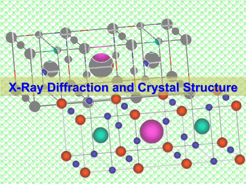
X-Ray Diffraction and Crystal Structure
Transcript of X-Ray Diffraction and Crystal StructureX-Ray Diffraction and Crystal...
X-Ray Diffraction and Crystal StructureX-Ray Diffraction and Crystal Structure
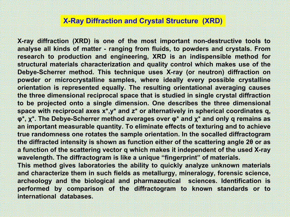
X-Ray Diffraction and Crystal Structure (XRD)
X-ray diffraction (XRD) is one of the most important non-destructive tools to analyse all kinds of matter - ranging from fluids, to powders and crystals. From research to production and engineering, XRD is an indispensible method for structural materials characterization and quality control which makes use of the Debye-Scherrer method. This technique uses X-ray (or neutron) diffraction on powder or microcrystalline samples, where ideally every possible crystalline orientation is represented equally. The resulting orientational averaging causes the three dimensional reciprocal space that is studied in single crystal diffraction to be projected onto a single dimension. One describes the three dimensional space with reciprocal axes x*,y* and z* or alternatively in spherical coordinates q, φ*, χ*. The Debye-Scherrer method averages over φ* and χ* and only q remains as an important measurable quantity. To eliminate effects of texturing and to achieve true randomness one rotates the sample orientation. In the socalled diffractogramthe diffracted intensity is shown as function either of the scattering angle 2θ or as a function of the scattering vector q which makes it independent of the used X-ray wavelength. The diffractogram is like a unique “fingerprint” of materials.This method gives laboratories the ability to quickly analyze unknown materials and characterize them in such fields as metallurgy, mineralogy, forensic science, archeology and the biological and pharmazeutical sciences. Identification is performed by comparison of the diffractogram to known standards or to international databases.
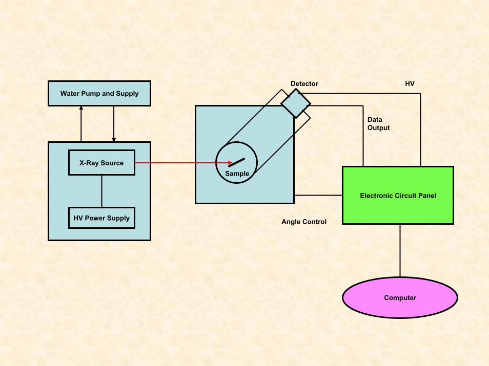
X-Ray Source
HV Power Supply
Water Pump and Supply
Electronic Circuit Panel
Computer
Sample
Detector HV
DataOutput
Angle Control
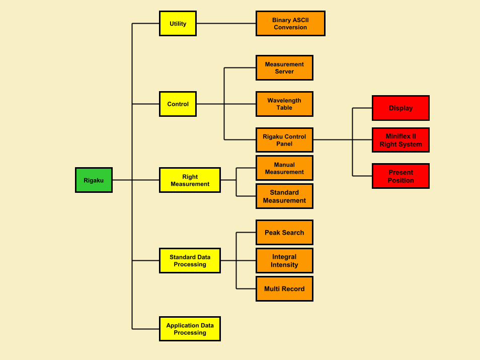
Rigaku
Utility
Control
Standard Data Processing
Right Measurement
Application Data Processing
Binary ASCII Conversion
Measurement Server
Wavelength Table
Rigaku Control Panel
Manual Measurement
Standard Measurement
Peak Search
Integral Intensity
Multi Record
Display
Miniflex II Right System
Present Position
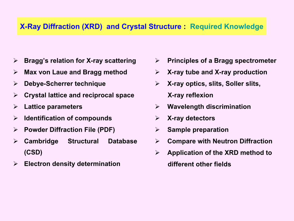
X-Ray Diffraction (XRD) and Crystal Structure : Required Knowledge
Bragg’s relation for X-ray scattering
Max von Laue and Bragg method
Debye-Scherrer technique
Crystal lattice and reciprocal space
Lattice parameters
Identification of compounds
Powder Diffraction File (PDF)
Cambridge Structural Database (CSD)
Electron density determination
Principles of a Bragg spectrometer
X-ray tube and X-ray production
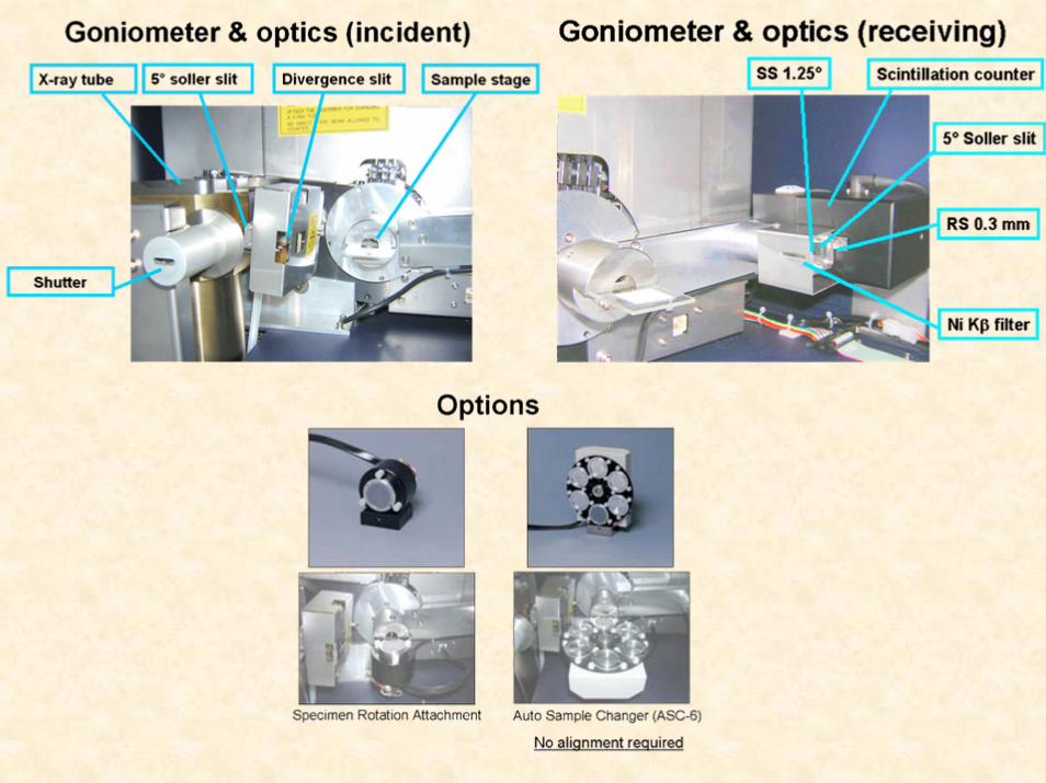
X-ray optics, slits, Soller slits,
X-ray reflexion
Wavelength discrimination
X-ray detectors
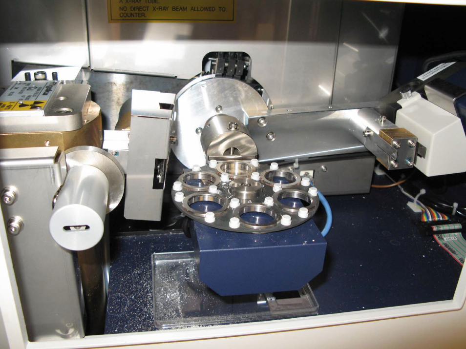
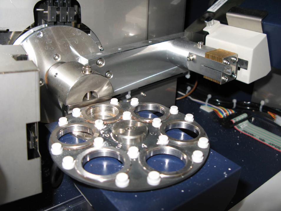
Sample preparation
Compare with Neutron Diffraction
Application of the XRD method to
different other fields
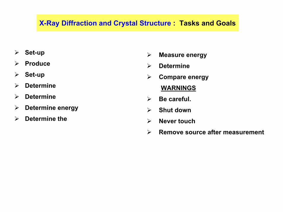
X-Ray Diffraction and Crystal Structure : Tasks and Goals
Set-up
Produce
Set-up
Determine
Determine
Determine energy
Determine the
Measure energy
Determine
Compare energy
WARNINGS
Be careful.
Shut down
Never touch
Remove source after measurement
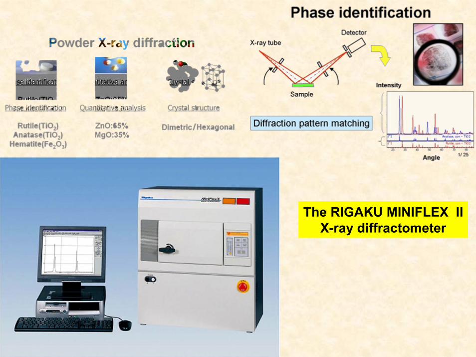
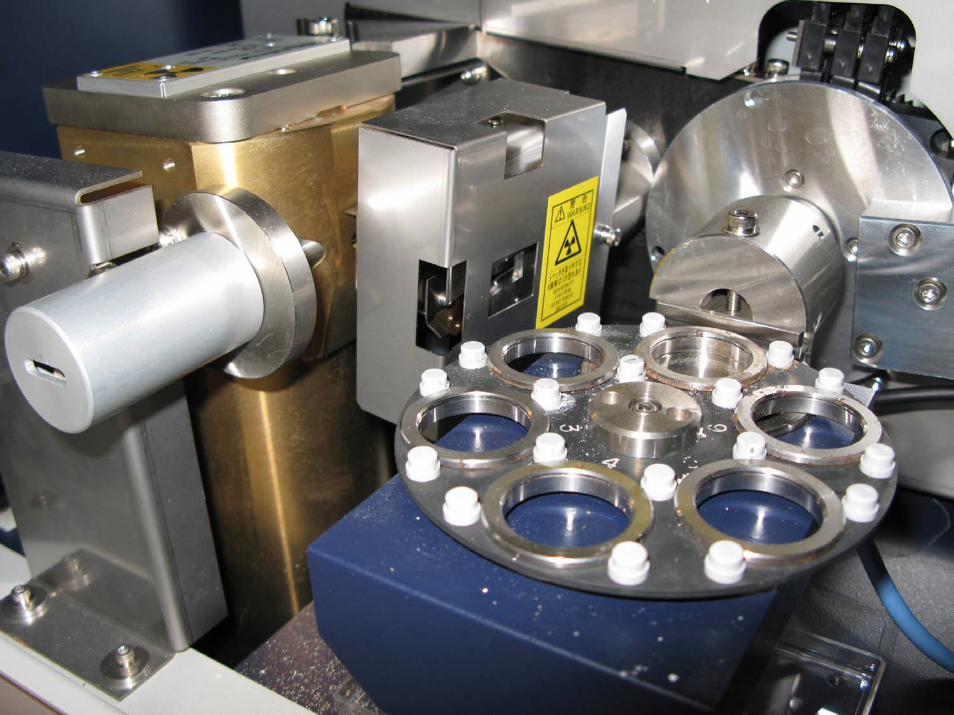
The RIGAKU MINIFLEX IIX-ray diffractometer
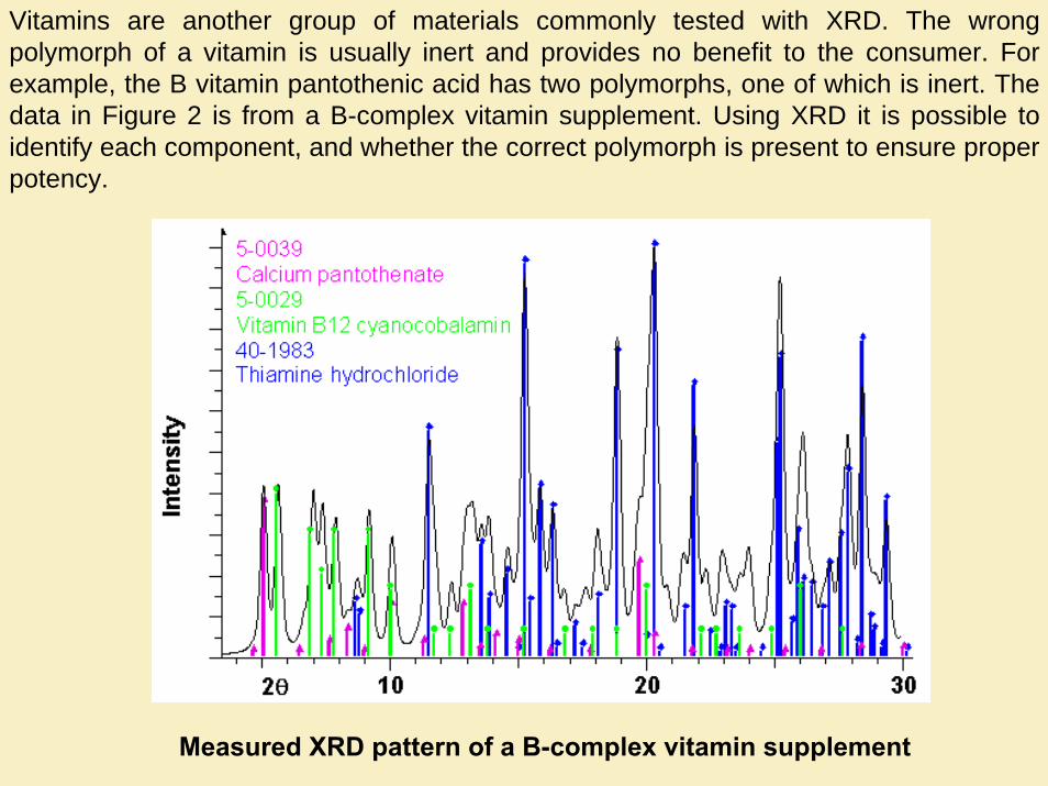
Vitamins are another group of materials commonly tested with XRD. The wrong polymorph of a vitamin is usually inert and provides no benefit to the consumer. For example, the B vitamin pantothenic acid has two polymorphs, one of which is inert. The data in Figure 2 is from a B-complex vitamin supplement. Using XRD it is possible to identify each component, and whether the correct polymorph is present to ensure proper potency.
Measured XRD pattern of a B-complex vitamin supplement
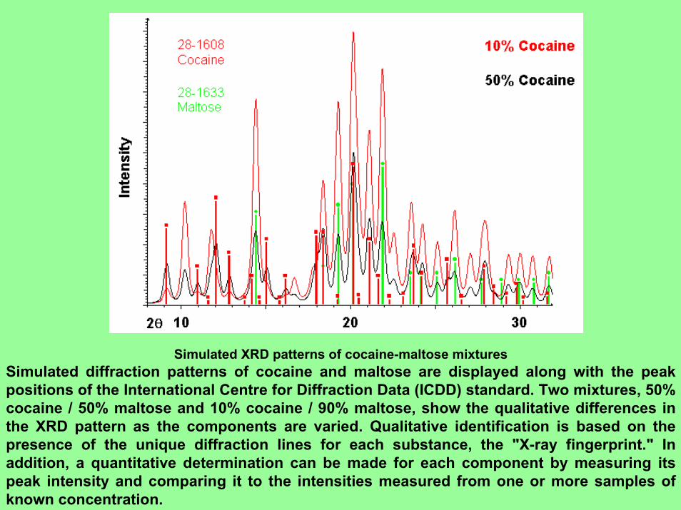
Simulated XRD patterns of cocaine-maltose mixturesSimulated diffraction patterns of cocaine and maltose are displayed along with the peak positions of the International Centre for Diffraction Data (ICDD) standard. Two mixtures, 50% cocaine / 50% maltose and 10% cocaine / 90% maltose, show the qualitative differences in the XRD pattern as the components are varied. Qualitative identification is based on the presence of the unique diffraction lines for each substance, the "X-ray fingerprint." In addition, a quantitative determination can be made for each component by measuring its peak intensity and comparing it to the intensities measured from one or more samples of known concentration.
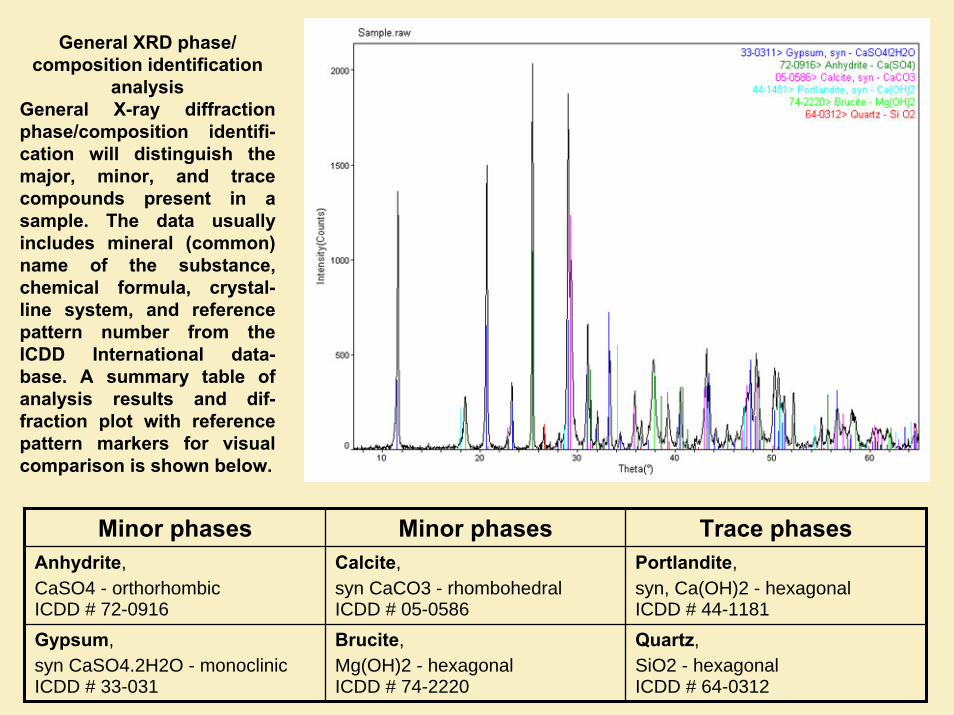
General XRD phase/ composition identification
analysisGeneral X-ray diffraction phase/composition identifi-cation will distinguish the major, minor, and trace compounds present in a sample. The data usually includes mineral (common) name of the substance, chemical formula, crystal-line system, and reference pattern number from the ICDD International data-base. A summary table of analysis results and dif-fraction plot with reference pattern markers for visual comparison is shown below.
Minor phases Minor phases Trace phasesAnhydrite, CaSO4 - orthorhombicICDD # 72-0916
Calcite, syn CaCO3 - rhombohedralICDD # 05-0586
Portlandite, syn, Ca(OH)2 - hexagonalICDD # 44-1181
Gypsum, syn CaSO4.2H2O - monoclinicICDD # 33-031
Brucite, Mg(OH)2 - hexagonalICDD # 74-2220
Quartz, SiO2 - hexagonalICDD # 64-0312


