Wound healing of apical tissues after root canal therapy: a long-term ...
13
Wound healing of apical tissues after root canal therapy: a long-term clinical, radiographic, and histopathologic observation study Domenico Ricucci, MD, DDS, a Louis M. Lin, BDS, DMD, PhD, b and Larz S. W. Spångberg, DDS, PhD, c Rome, Italy; New York, New York; and Farmington, Connecticut NEW YORK UNIVERSITY AND UNIVERSITY OF CONNECTICUT Objective. The purpose of this study was to evaluate the pulp healing process and the dentin-cementum complex in 51 endodontically treated human teeth after long observation periods and to correlate histologic observations with conventional periapical radiographic findings. Study design. Specimens were obtained from the extraction of 77 treated teeth, which were deemed to be unrestorable, with no evidence of periapical bone lesion at the follow-up. After stringent evaluation of the radiographs, 51 cases that 3 independent evaluators assessed as having normal periapical conditions were selected. The specimens were histologically evaluated using serial sections. Results. In the majority of the cases, complete healing was observed, with no signs of acute or chronic inflammatory processes in the remaining apical tissue or periodontal tissue fragments. Some cases showed moderate inflammation in the root canal tissue. Narrowing of the apical root canal by cementum was a common finding in most cases, but total closure was not observed. Debris intermixed with necrotic tissue and sealer particles was a common finding in the pulp stump. Bacteria were present in the coronal portion of the root in almost all cases, but in only 1 case could bacteria be demonstrated in the coronal and apical portions of the root. Conclusions. Apical tissue of properly treated teeth with no signs of periapical changes is only rarely significantly inflamed. When the tissue is inflamed, microbial causes can always be demonstrated. Despite the presence of microorganisms coronally in nearly all cases, apical tissue is seldom affected. (Oral Surg Oral Med Oral Pathol Oral Radiol Endod 2009;108:609-621) Traditionally, diagnosis of disease of tissues or organs is dependent on clinical and histologic findings. Clinical diagnosis is a provisional diagnosis and uses objective and subjective signs and/or symptoms and all available tests in clinical dental medicine as well as imaging systems, such as conventional radiography, digital radiography, and computerized tomography. Radiography is designed to detect pathologic changes at the tissue or organ level and not at the cellular level. Histologic examination provides information that makes a definitive and final diagnosis possible at a cellular level. To increase the sensitivity and optimize the chances of detecting the presence of a few inflammatory cells in the periapical tissues, meticulous techniques must be applied. This includes serial sectioning or step-serial sectioning of the entire specimen in an optimal direction. Random and/or selective sections are never sufficient for optimal histologic diagnosis. Further- more, tissue fixation and preparation must be optimal. Substandard histologic information also makes attempts to correlate histologic and radiographic information ques- tionable. 1-4 Because histologic examination cannot be routinely used in clinical practice to diagnose pathologic changes of tissues or organs at the cellular level, radio- graphic examination is indispensable as a screening tool to diagnose pathologic changes of tissues or organs. There- fore it is desirable to increase the sensitivity and specific- ity of radiographic examinations of the periapical region of teeth. 4 Much reliance has been placed on results from a Private practice, Rome, Italy. b Professor, Department of Endodontics, New York University. c Professor, Division of Endodontology, University of Connecticut. Received for publication May 7, 2009; accepted for publication May 11, 2009. 1079-2104/$ - see front matter © 2009 Mosby, Inc. All rights reserved. doi:10.1016/j.tripleo.2009.05.028 609 Vol. 108 No. 4 October 2009 ENDODONTOLOGY Editor: Larz S.W. Spångberg
Transcript of Wound healing of apical tissues after root canal therapy: a long-term ...
Vol. 108 No. 4 October 2009
ENDODONTOLOGY Editor: Larz S.W. Spångberg
Wound healing of apical tissues after root canal therapy: a long-termclinical, radiographic, and histopathologic observation studyDomenico Ricucci, MD, DDS,a Louis M. Lin, BDS, DMD, PhD,b andLarz S. W. Spångberg, DDS, PhD,c Rome, Italy; New York, New York; and Farmington,ConnecticutNEW YORK UNIVERSITY AND UNIVERSITY OF CONNECTICUT
Objective. The purpose of this study was to evaluate the pulp healing process and the dentin-cementum complex in51 endodontically treated human teeth after long observation periods and to correlate histologic observations withconventional periapical radiographic findings.Study design. Specimens were obtained from the extraction of 77 treated teeth, which were deemed to beunrestorable, with no evidence of periapical bone lesion at the follow-up. After stringent evaluation of the radiographs,51 cases that 3 independent evaluators assessed as having normal periapical conditions were selected. The specimenswere histologically evaluated using serial sections.Results. In the majority of the cases, complete healing was observed, with no signs of acute or chronic inflammatoryprocesses in the remaining apical tissue or periodontal tissue fragments. Some cases showed moderate inflammation inthe root canal tissue. Narrowing of the apical root canal by cementum was a common finding in most cases, but totalclosure was not observed. Debris intermixed with necrotic tissue and sealer particles was a common finding in thepulp stump. Bacteria were present in the coronal portion of the root in almost all cases, but in only 1 case couldbacteria be demonstrated in the coronal and apical portions of the root.Conclusions. Apical tissue of properly treated teeth with no signs of periapical changes is only rarely significantlyinflamed. When the tissue is inflamed, microbial causes can always be demonstrated. Despite the presence ofmicroorganisms coronally in nearly all cases, apical tissue is seldom affected. (Oral Surg Oral Med Oral Pathol Oral
Radiol Endod 2009;108:609-621)Traditionally, diagnosis of disease of tissues or organs isdependent on clinical and histologic findings. Clinicaldiagnosis is a provisional diagnosis and uses objective andsubjective signs and/or symptoms and all available tests inclinical dental medicine as well as imaging systems, suchas conventional radiography, digital radiography, andcomputerized tomography. Radiography is designed todetect pathologic changes at the tissue or organ level andnot at the cellular level. Histologic examination providesinformation that makes a definitive and final diagnosis
aPrivate practice, Rome, Italy.bProfessor, Department of Endodontics, New York University.cProfessor, Division of Endodontology, University of Connecticut.Received for publication May 7, 2009; accepted for publication May11, 2009.1079-2104/$ - see front matter© 2009 Mosby, Inc. All rights reserved.
doi:10.1016/j.tripleo.2009.05.028possible at a cellular level. To increase the sensitivity andoptimize the chances of detecting the presence of a fewinflammatory cells in the periapical tissues, meticuloustechniques must be applied. This includes serial sectioningor step-serial sectioning of the entire specimen in anoptimal direction. Random and/or selective sections arenever sufficient for optimal histologic diagnosis. Further-more, tissue fixation and preparation must be optimal.Substandard histologic information also makes attempts tocorrelate histologic and radiographic information ques-tionable.1-4 Because histologic examination cannot beroutinely used in clinical practice to diagnose pathologicchanges of tissues or organs at the cellular level, radio-graphic examination is indispensable as a screening tool todiagnose pathologic changes of tissues or organs. There-fore it is desirable to increase the sensitivity and specific-ity of radiographic examinations of the periapical region
of teeth.4 Much reliance has been placed on results from609
OOOOE610 Ricucci et al. October 2009
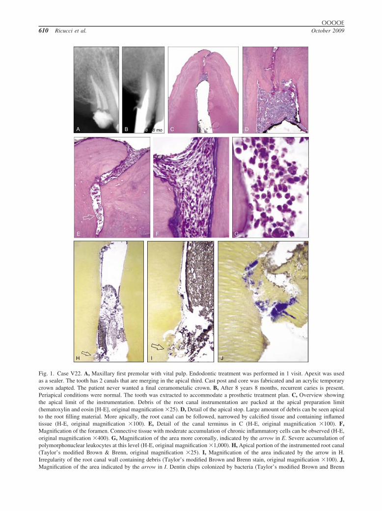
Fig. 1. Case V22. A, Maxillary first premolar with vital pulp. Endodontic treatment was performed in 1 visit. Apexit was usedas a sealer. The tooth has 2 canals that are merging in the apical third. Cast post and core was fabricated and an acrylic temporarycrown adapted. The patient never wanted a final ceramometalic crown. B, After 8 years 8 months, recurrent caries is present.Periapical conditions were normal. The tooth was extracted to accommodate a prosthetic treatment plan. C, Overview showingthe apical limit of the instrumentation. Debris of the root canal instrumentation are packed at the apical preparation limit(hematoxylin and eosin [H-E], original magnification �25). D, Detail of the apical stop. Large amount of debris can be seen apicalto the root filling material. More apically, the root canal can be followed, narrowed by calcified tissue and containing inflamedtissue (H-E, original magnification �100). E, Detail of the canal terminus in C (H-E, original magnification �100). F,Magnification of the foramen. Connective tissue with moderate accumulation of chronic inflammatory cells can be observed (H-E,original magnification �400). G, Magnification of the area more coronally, indicated by the arrow in E. Severe accumulation ofpolymorphonuclear leukocytes at this level (H-E, original magnification �1,000). H, Apical portion of the instrumented root canal(Taylor’s modified Brown & Brenn, original magnification �25). I, Magnification of the area indicated by the arrow in H.Irregularity of the root canal wall containing debris (Taylor’s modified Brown and Brenn stain, original magnification �100). J,
Magnification of the area indicated by the arrow in I. Dentin chips colonized by bacteria (Taylor’s modified Brown and Brenn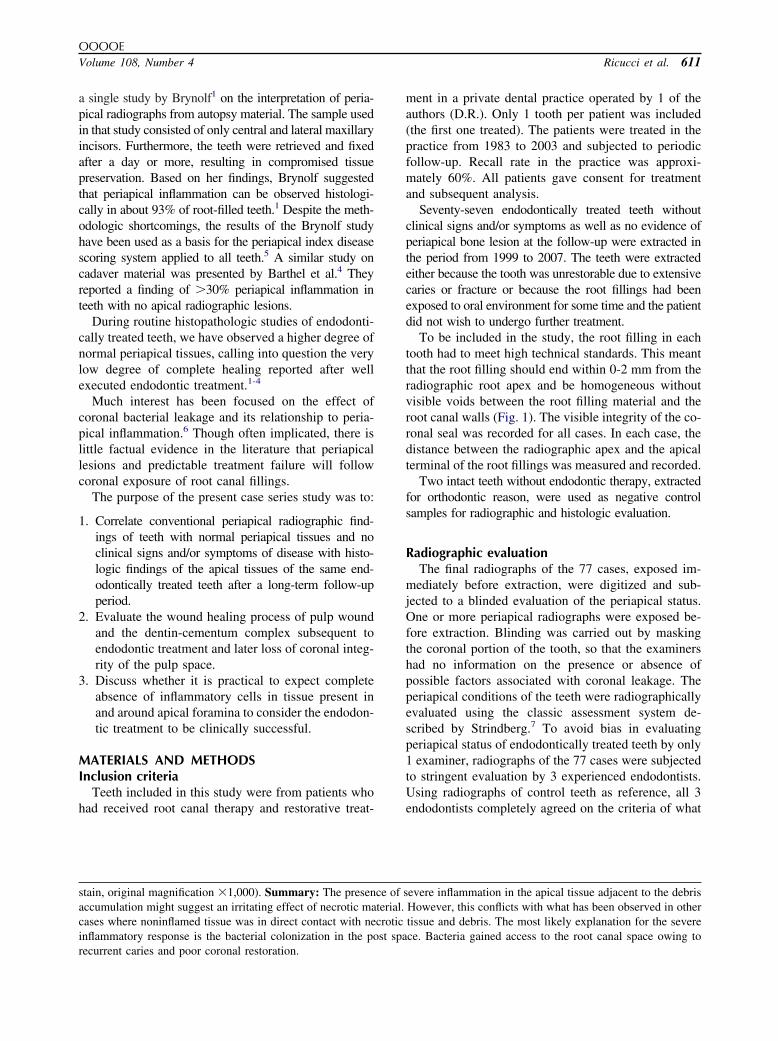
OOOOEVolume 108, Number 4 Ricucci et al. 611
a single study by Brynolf1 on the interpretation of peria-pical radiographs from autopsy material. The sample usedin that study consisted of only central and lateral maxillaryincisors. Furthermore, the teeth were retrieved and fixedafter a day or more, resulting in compromised tissuepreservation. Based on her findings, Brynolf suggestedthat periapical inflammation can be observed histologi-cally in about 93% of root-filled teeth.1 Despite the meth-odologic shortcomings, the results of the Brynolf studyhave been used as a basis for the periapical index diseasescoring system applied to all teeth.5 A similar study oncadaver material was presented by Barthel et al.4 Theyreported a finding of �30% periapical inflammation inteeth with no apical radiographic lesions.
During routine histopathologic studies of endodonti-cally treated teeth, we have observed a higher degree ofnormal periapical tissues, calling into question the verylow degree of complete healing reported after wellexecuted endodontic treatment.1-4
Much interest has been focused on the effect ofcoronal bacterial leakage and its relationship to peria-pical inflammation.6 Though often implicated, there islittle factual evidence in the literature that periapicallesions and predictable treatment failure will followcoronal exposure of root canal fillings.
The purpose of the present case series study was to:
1. Correlate conventional periapical radiographic find-ings of teeth with normal periapical tissues and noclinical signs and/or symptoms of disease with histo-logic findings of the apical tissues of the same end-odontically treated teeth after a long-term follow-upperiod.
2. Evaluate the wound healing process of pulp woundand the dentin-cementum complex subsequent toendodontic treatment and later loss of coronal integ-rity of the pulp space.
3. Discuss whether it is practical to expect completeabsence of inflammatory cells in tissue present inand around apical foramina to consider the endodon-tic treatment to be clinically successful.
MATERIALS AND METHODSInclusion criteria
Teeth included in this study were from patients whohad received root canal therapy and restorative treat-
stain, original magnification �1,000). Summary: The presenaccumulation might suggest an irritating effect of necrotic macases where noninflamed tissue was in direct contact with neinflammatory response is the bacterial colonization in the po
recurrent caries and poor coronal restoration.ment in a private dental practice operated by 1 of theauthors (D.R.). Only 1 tooth per patient was included(the first one treated). The patients were treated in thepractice from 1983 to 2003 and subjected to periodicfollow-up. Recall rate in the practice was approxi-mately 60%. All patients gave consent for treatmentand subsequent analysis.
Seventy-seven endodontically treated teeth withoutclinical signs and/or symptoms as well as no evidence ofperiapical bone lesion at the follow-up were extracted inthe period from 1999 to 2007. The teeth were extractedeither because the tooth was unrestorable due to extensivecaries or fracture or because the root fillings had beenexposed to oral environment for some time and the patientdid not wish to undergo further treatment.
To be included in the study, the root filling in eachtooth had to meet high technical standards. This meantthat the root filling should end within 0-2 mm from theradiographic root apex and be homogeneous withoutvisible voids between the root filling material and theroot canal walls (Fig. 1). The visible integrity of the co-ronal seal was recorded for all cases. In each case, thedistance between the radiographic apex and the apicalterminal of the root fillings was measured and recorded.
Two intact teeth without endodontic therapy, extractedfor orthodontic reason, were used as negative controlsamples for radiographic and histologic evaluation.
Radiographic evaluationThe final radiographs of the 77 cases, exposed im-
mediately before extraction, were digitized and sub-jected to a blinded evaluation of the periapical status.One or more periapical radiographs were exposed be-fore extraction. Blinding was carried out by maskingthe coronal portion of the tooth, so that the examinershad no information on the presence or absence ofpossible factors associated with coronal leakage. Theperiapical conditions of the teeth were radiographicallyevaluated using the classic assessment system de-scribed by Strindberg.7 To avoid bias in evaluatingperiapical status of endodontically treated teeth by only1 examiner, radiographs of the 77 cases were subjectedto stringent evaluation by 3 experienced endodontists.Using radiographs of control teeth as reference, all 3endodontists completely agreed on the criteria of what
evere inflammation in the apical tissue adjacent to the debrisHowever, this conflicts with what has been observed in othertissue and debris. The most likely explanation for the severece. Bacteria gained access to the root canal space owing to
ce of sterial.croticst spa
OOOOE612 Ricucci et al. October 2009
normal periapical tissues should look like before eval-uating experimental teeth. Radiographically, normalperiapical tissues require: 1) uniform width of the peri-odontal ligament space; 2) no obvious breakdown oflamina dura; and 3) well organized bone trabeculae.7
The radiographs were projected on a screen in a darkroom without any other light interference. The radio-logic examination was performed without prior knowl-edge of the results of the histologic evaluation. Threeexaminers evaluated the radiographs independently. Incase of disagreement, no attempts were made to cometo a consensus and the case was excluded. Only caseswhere all 3 evaluators completely agreed were in-cluded. Out of 77 cases, 51 cases without clinical signsand/or symptoms, as well as normal radiographic ap-pearance of the periapical tissue, were included.7
Case historyComplete records of initial diagnosis, endodontic
procedures used, and follow-up examinations wereavailable for all 51 cases. At the beginning of theendodontic treatment, 27 teeth were diagnosed as hav-ing irreversible pulpitis, 10 teeth had a necrotic pulpwithout apical periodontitis, 12 teeth presented withpulp necrosis and radiographic evidence of apical pe-riodontitis, and 2 teeth had earlier root fillings of which1 showed apical periodontitis. The majority of the teethwere premolars (n � 28). Six teeth were molars, 11were incisors, and 6 were canines. Patients were be-tween 18 and 69 years of age (mean 42 years). Thegender distribution was 30 women and 21 men.
Observation periods ranged from 2 years to 22 years4 months (mean 10 years 3 months).
Endodontic treatment proceduresAll endodontic treatments were performed following
a standardized protocol. Before the initiation of treat-ment at least 1 diagnostic radiograph was exposed. Thisradiograph and subsequent radiographs were obtainedusing a film holder (Rinn Corp., Elgin, IL) to permitparalleling technique projection. A long cone (Ex-plor-X 65 KV; Fiad, Trezzano, Italy) and Kodak Ul-traspeed film 31 � 41 (DF58) or 22 � 35 (DF54;Eastman Kodak Co., Rochester, NY) were used. Ra-diographs were processed manually in a dark roomfollowing the recommendation of the manufacturer.
A pulpal and periapical diagnosis was establishedafter obtaining objective and subjective symptoms,pulp tests, percussion, and palpation as well as ra-diographic findings. Endodontic treatments were per-formed using a strict aseptic technique. Plaque andcalculus were removed from the tooth surfaces usingultrasound scaling and/or curettes followed by polish-
ing with a prophylaxis paste in a rubber cup. Afterrubber dam isolation, the field (tooth, rubber dam, andclamp) was disinfected with 30% H2O2 and 5% tinctureof iodine.8 In case of substantial loss of tooth substance,build-ups were carried out with either a copper ring,composite material, or reinforced glass ionomer cementto exclude bacterial contamination during the opera-tion. After the working length was established, an effortwas undertaken to limit the instrumentation to the con-fines of the root canals. Abundant amounts of 1%sodium hypochlorite solution were used for irrigation.After adequate preflaring of the coronal two-thirds ofthe root canals and apical instrumentation, the rootcanals were filled with cold laterally compacted gutta-percha and a sealer. Different sealers were used overthe years, including AH 26 (De Trey Frères, Zurich,Switzerland), Bio Seal (Ogna, Milano, Italy), Endo-methasone (Septodont, Saint-Maur-des-Fossés, France),Pulp Canal Sealer (Kerr Manufacturing Co., Romulus,MI), Tubli-Seal (Kerr Manufacturing Co.), Apexit (Vi-vadent, Schaan, Liechtenstein), and AH Plus (De Trey,Konstanz, Germany). The treatments were completedin either 1 visit (usually teeth with vital pulps) or �2visits (teeth with necrotic pulp) after placement ofCa(OH)2 or metacresylacetate (Cresatina; Ogna, Mi-lano, Italy) as an intracanal dressing.
Tissue processingImmediately after extraction, the teeth were fixed in
10% neutral buffered formalin for at least 48 hours.Demineralization was carried out in an aqueous solu-tion consisting of a mixture of 22.5% (v/v) formic acidand 10% (w/v) sodium citrate for 3-4 weeks with theend point being determined radiographically. All spec-imens were then washed in running tap water for 24-48hours, dehydrated in ascending grades of ethanol,cleared in xylene, and infiltrated and embedded inparaffin (melting point 56°C) according to standardprocedures. To produce sections parallel to the longaxis of the root canal, special precautions were under-taken. Roots in multirooted teeth were dissected freeand processed separately. If curved, roots were sepa-rated into 2 pieces, 1 encompassing the coronal two-thirds and 1 including the apical third. These 2 pieceswere embedded separately. With the microtome set at4-5 �m, meticulous longitudinal serial sections weretaken until each specimen was exhausted. Particularcare was taken in sectioning the apical third to obtainsections passing through the apical foramen in directcontinuity with the periapical tissues. Longitudinal orcross-cut sections were obtained from the coronal two-thirds. Every fifth section was stained with hematoxylinand eosin for screening purposes and for assessment of
inflammation. A modified Brown and Brenn technique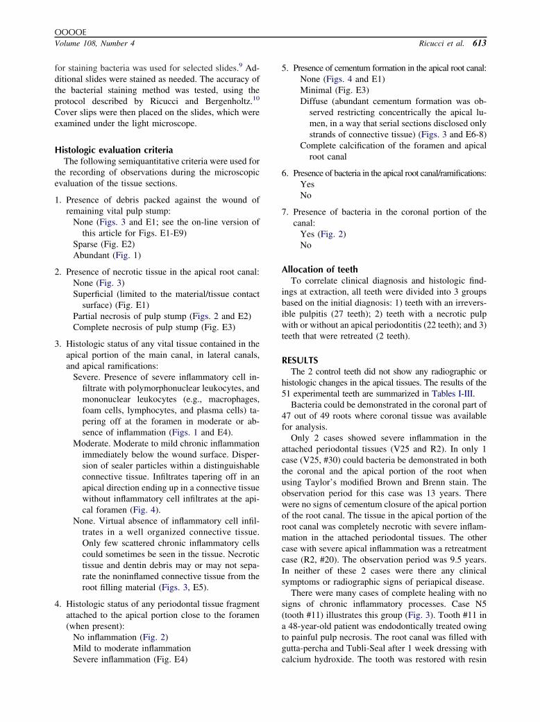
OOOOEVolume 108, Number 4 Ricucci et al. 613
for staining bacteria was used for selected slides.9 Ad-ditional slides were stained as needed. The accuracy ofthe bacterial staining method was tested, using theprotocol described by Ricucci and Bergenholtz.10
Cover slips were then placed on the slides, which wereexamined under the light microscope.
Histologic evaluation criteriaThe following semiquantitative criteria were used for
the recording of observations during the microscopicevaluation of the tissue sections.
1. Presence of debris packed against the wound ofremaining vital pulp stump:
None (Figs. 3 and E1; see the on-line version ofthis article for Figs. E1-E9)
Sparse (Fig. E2)Abundant (Fig. 1)
2. Presence of necrotic tissue in the apical root canal:None (Fig. 3)Superficial (limited to the material/tissue contact
surface) (Fig. E1)Partial necrosis of pulp stump (Figs. 2 and E2)Complete necrosis of pulp stump (Fig. E3)
3. Histologic status of any vital tissue contained in theapical portion of the main canal, in lateral canals,and apical ramifications:
Severe. Presence of severe inflammatory cell in-filtrate with polymorphonuclear leukocytes, andmononuclear leukocytes (e.g., macrophages,foam cells, lymphocytes, and plasma cells) ta-pering off at the foramen in moderate or ab-sence of inflammation (Figs. 1 and E4).
Moderate. Moderate to mild chronic inflammationimmediately below the wound surface. Disper-sion of sealer particles within a distinguishableconnective tissue. Infiltrates tapering off in anapical direction ending up in a connective tissuewithout inflammatory cell infiltrates at the api-cal foramen (Fig. 4).
None. Virtual absence of inflammatory cell infil-trates in a well organized connective tissue.Only few scattered chronic inflammatory cellscould sometimes be seen in the tissue. Necrotictissue and dentin debris may or may not sepa-rate the noninflamed connective tissue from theroot filling material (Figs. 3, E5).
4. Histologic status of any periodontal tissue fragmentattached to the apical portion close to the foramen(when present):
No inflammation (Fig. 2)Mild to moderate inflammation
Severe inflammation (Fig. E4)5. Presence of cementum formation in the apical root canal:None (Figs. 4 and E1)Minimal (Fig. E3)Diffuse (abundant cementum formation was ob-
served restricting concentrically the apical lu-men, in a way that serial sections disclosed onlystrands of connective tissue) (Figs. 3 and E6-8)
Complete calcification of the foramen and apicalroot canal
6. Presence of bacteria in the apical root canal/ramifications:YesNo
7. Presence of bacteria in the coronal portion of thecanal:
Yes (Fig. 2)No
Allocation of teethTo correlate clinical diagnosis and histologic find-
ings at extraction, all teeth were divided into 3 groupsbased on the initial diagnosis: 1) teeth with an irrevers-ible pulpitis (27 teeth); 2) teeth with a necrotic pulpwith or without an apical periodontitis (22 teeth); and 3)teeth that were retreated (2 teeth).
RESULTSThe 2 control teeth did not show any radiographic or
histologic changes in the apical tissues. The results of the51 experimental teeth are summarized in Tables I-III.
Bacteria could be demonstrated in the coronal part of47 out of 49 roots where coronal tissue was availablefor analysis.
Only 2 cases showed severe inflammation in theattached periodontal tissues (V25 and R2). In only 1case (V25, #30) could bacteria be demonstrated in boththe coronal and the apical portion of the root whenusing Taylor’s modified Brown and Brenn stain. Theobservation period for this case was 13 years. Therewere no signs of cementum closure of the apical portionof the root canal. The tissue in the apical portion of theroot canal was completely necrotic with severe inflam-mation in the attached periodontal tissues. The othercase with severe apical inflammation was a retreatmentcase (R2, #20). The observation period was 9.5 years.In neither of these 2 cases were there any clinicalsymptoms or radiographic signs of periapical disease.
There were many cases of complete healing with nosigns of chronic inflammatory processes. Case N5(tooth #11) illustrates this group (Fig. 3). Tooth #11 ina 48-year-old patient was endodontically treated owingto painful pulp necrosis. The root canal was filled withgutta-percha and Tubli-Seal after 1 week dressing with
calcium hydroxide. The tooth was restored with resin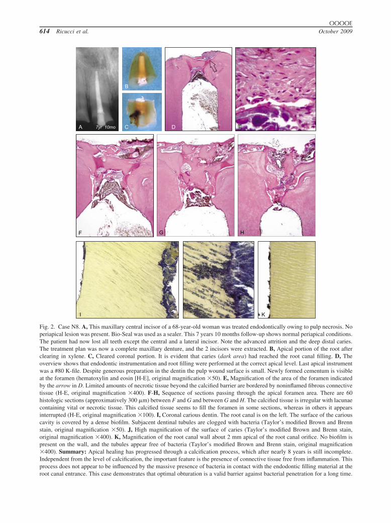
OOOOE614 Ricucci et al. October 2009
A
B
C7yr 10mo
F G H
D E
SI J K
Fig. 2. Case N8. A, This maxillary central incisor of a 68-year-old woman was treated endodontically owing to pulp necrosis. Noperiapical lesion was present. Bio-Seal was used as a sealer. This 7 years 10 months follow-up shows normal periapical conditions.The patient had now lost all teeth except the central and a lateral incisor. Note the advanced attrition and the deep distal caries.The treatment plan was now a complete maxillary denture, and the 2 incisors were extracted. B, Apical portion of the root afterclearing in xylene. C, Cleared coronal portion. It is evident that caries (dark area) had reached the root canal filling. D, Theoverview shows that endodontic instrumentation and root filling were performed at the correct apical level. Last apical instrumentwas a #80 K-file. Despite generous preparation in the dentin the pulp wound surface is small. Newly formed cementum is visibleat the foramen (hematoxylin and eosin [H-E], original magnification �50). E, Magnification of the area of the foramen indicatedby the arrow in D. Limited amounts of necrotic tissue beyond the calcified barrier are bordered by noninflamed fibrous connectivetissue (H-E, original magnification �400). F-H, Sequence of sections passing through the apical foramen area. There are 60histologic sections (approximatively 300 �m) between F and G and between G and H. The calcified tissue is irregular with lacunaecontaining vital or necrotic tissue. This calcified tissue seems to fill the foramen in some sections, whereas in others it appearsinterrupted (H-E, original magnification �100). I, Coronal carious dentin. The root canal is on the left. The surface of the cariouscavity is covered by a dense biofilm. Subjacent dentinal tubules are clogged with bacteria (Taylor’s modified Brown and Brennstain, original magnification �50). J, High magnification of the surface of caries (Taylor’s modified Brown and Brenn stain,original magnification �400). K, Magnification of the root canal wall about 2 mm apical of the root canal orifice. No biofilm ispresent on the wall, and the tubules appear free of bacteria (Taylor’s modified Brown and Brenn stain, original magnification�400). Summary: Apical healing has progressed through a calcification process, which after nearly 8 years is still incomplete.Independent from the level of calcification, the important feature is the presence of connective tissue free from inflammation. Thisprocess does not appear to be influenced by the massive presence of bacteria in contact with the endodontic filling material at the
root canal entrance. This case demonstrates that optimal obturation is a valid barrier against bacterial penetration for a long time.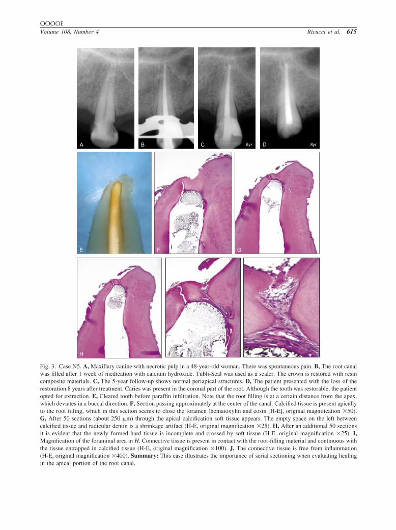
OOOOEVolume 108, Number 4 Ricucci et al. 615
G
H I J
A B 5yrC D
E F
8yr
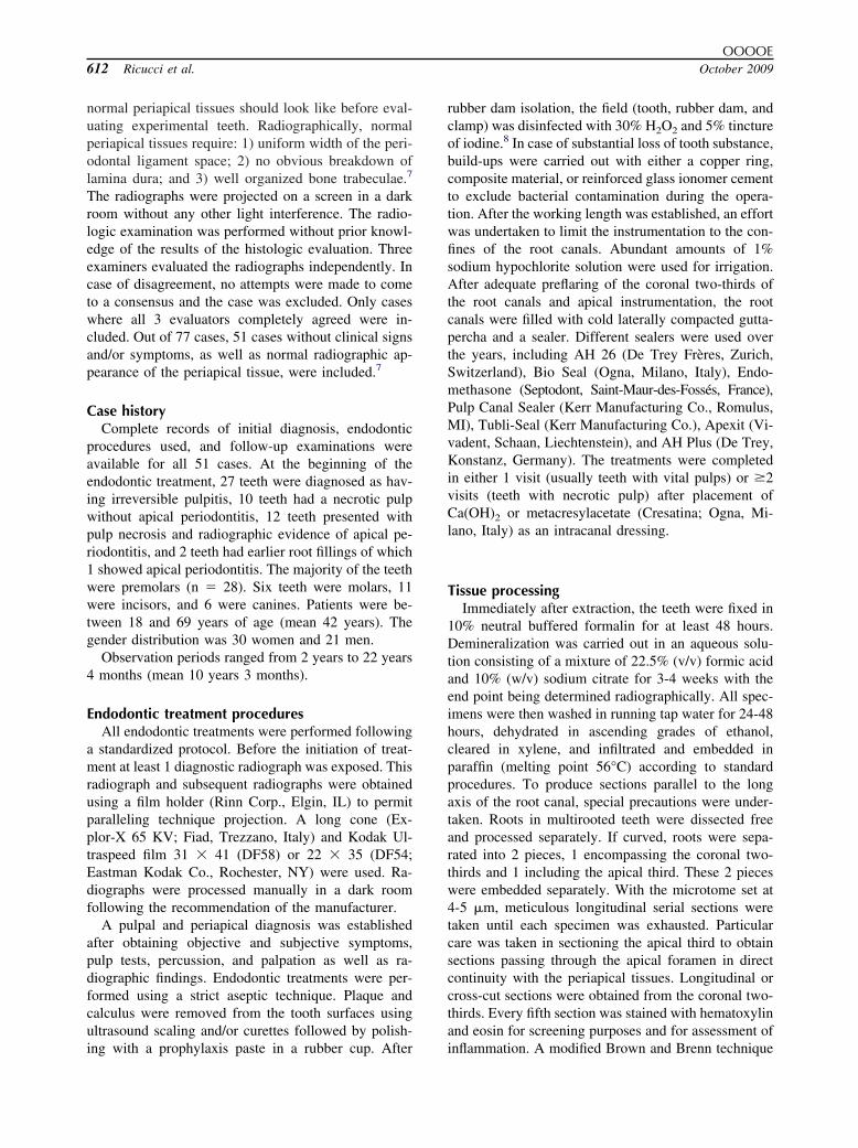
Fig. 3. Case N5. A, Maxillary canine with necrotic pulp in a 48-year-old woman. There was spontaneous pain. B, The root canalwas filled after 1 week of medication with calcium hydroxide. Tubli-Seal was used as a sealer. The crown is restored with resincomposite materials. C, The 5-year follow-up shows normal periapical structures. D, The patient presented with the loss of therestoration 8 years after treatment. Caries was present in the coronal part of the root. Although the tooth was restorable, the patientopted for extraction. E, Cleared tooth before paraffin infiltration. Note that the root filling is at a certain distance from the apex,which deviates in a buccal direction. F, Section passing approximately at the center of the canal. Calcified tissue is present apicallyto the root filling, which in this section seems to close the foramen (hematoxylin and eosin [H-E], original magnification �50).G, After 50 sections (about 250 �m) through the apical calcification soft tissue appears. The empty space on the left betweencalcified tissue and radicular dentin is a shrinkage artifact (H-E, original magnification �25). H, After an additional 50 sectionsit is evident that the newly formed hard tissue is incomplete and crossed by soft tissue (H-E, original magnification �25). I,Magnification of the foraminal area in H. Connective tissue is present in contact with the root-filling material and continuous withthe tissue entrapped in calcified tissue (H-E, original magnification �100). J, The connective tissue is free from inflammation(H-E, original magnification �400). Summary: This case illustrates the importance of serial sectioning when evaluating healing
in the apical portion of the root canal.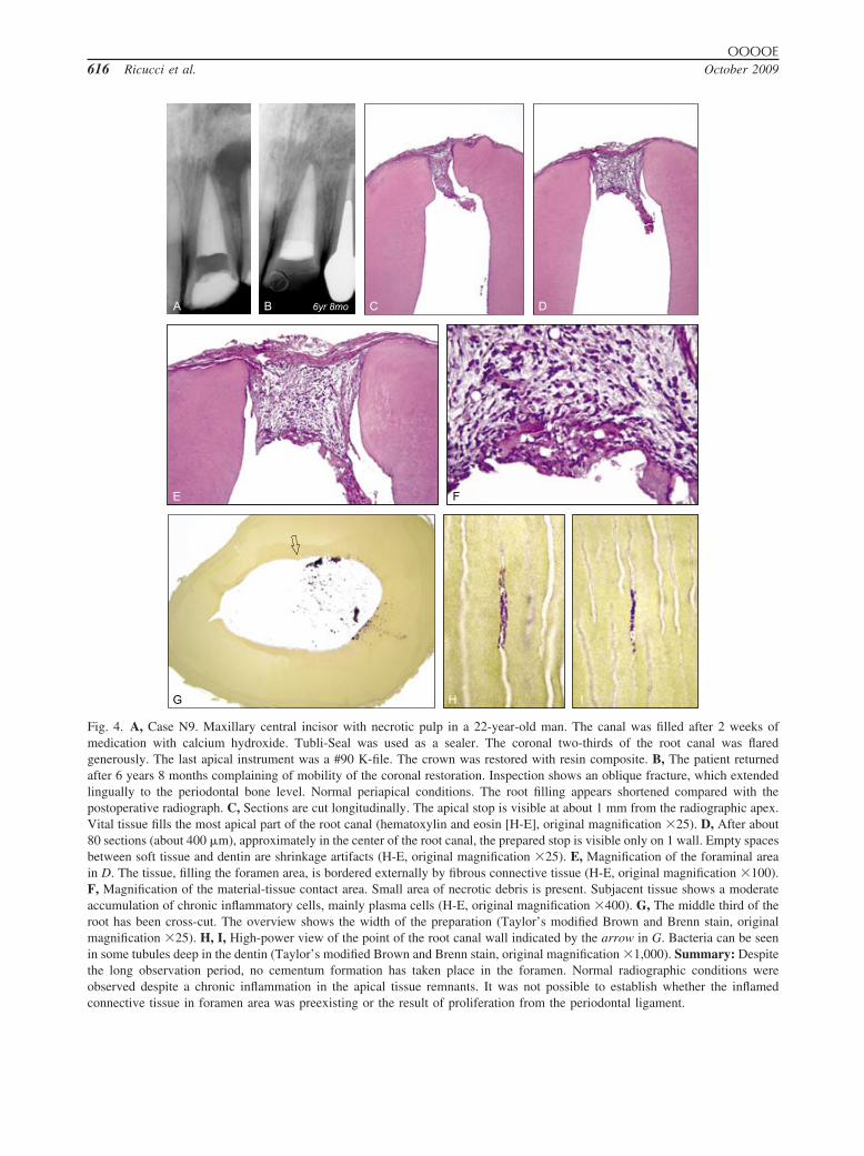
OOOOE616 Ricucci et al. October 2009
H I
A B C
G
D
E F
6yr 8mo
Fig. 4. A, Case N9. Maxillary central incisor with necrotic pulp in a 22-year-old man. The canal was filled after 2 weeks ofmedication with calcium hydroxide. Tubli-Seal was used as a sealer. The coronal two-thirds of the root canal was flaredgenerously. The last apical instrument was a #90 K-file. The crown was restored with resin composite. B, The patient returnedafter 6 years 8 months complaining of mobility of the coronal restoration. Inspection shows an oblique fracture, which extendedlingually to the periodontal bone level. Normal periapical conditions. The root filling appears shortened compared with thepostoperative radiograph. C, Sections are cut longitudinally. The apical stop is visible at about 1 mm from the radiographic apex.Vital tissue fills the most apical part of the root canal (hematoxylin and eosin [H-E], original magnification �25). D, After about80 sections (about 400 �m), approximately in the center of the root canal, the prepared stop is visible only on 1 wall. Empty spacesbetween soft tissue and dentin are shrinkage artifacts (H-E, original magnification �25). E, Magnification of the foraminal areain D. The tissue, filling the foramen area, is bordered externally by fibrous connective tissue (H-E, original magnification �100).F, Magnification of the material-tissue contact area. Small area of necrotic debris is present. Subjacent tissue shows a moderateaccumulation of chronic inflammatory cells, mainly plasma cells (H-E, original magnification �400). G, The middle third of theroot has been cross-cut. The overview shows the width of the preparation (Taylor’s modified Brown and Brenn stain, originalmagnification �25). H, I, High-power view of the point of the root canal wall indicated by the arrow in G. Bacteria can be seenin some tubules deep in the dentin (Taylor’s modified Brown and Brenn stain, original magnification �1,000). Summary: Despitethe long observation period, no cementum formation has taken place in the foramen. Normal radiographic conditions wereobserved despite a chronic inflammation in the apical tissue remnants. It was not possible to establish whether the inflamed
connective tissue in foramen area was preexisting or the result of proliferation from the periodontal ligament.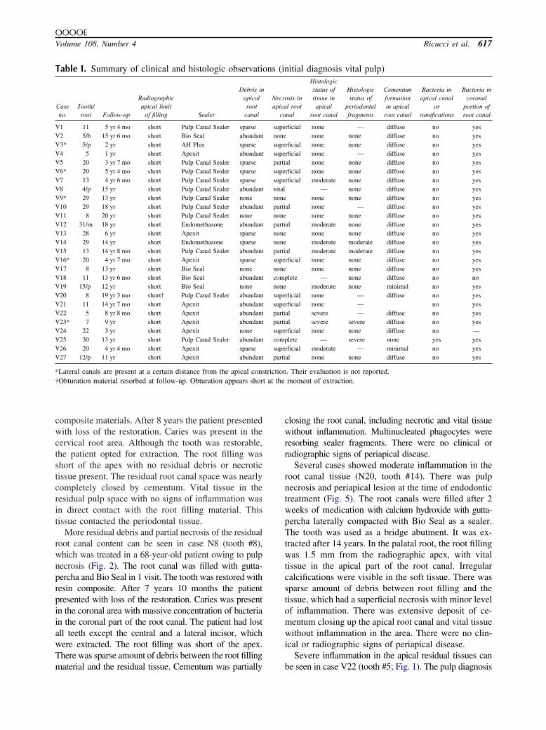
t at the
OOOOEVolume 108, Number 4 Ricucci et al. 617
composite materials. After 8 years the patient presentedwith loss of the restoration. Caries was present in thecervical root area. Although the tooth was restorable,the patient opted for extraction. The root filling wasshort of the apex with no residual debris or necrotictissue present. The residual root canal space was nearlycompletely closed by cementum. Vital tissue in theresidual pulp space with no signs of inflammation wasin direct contact with the root filling material. Thistissue contacted the periodontal tissue.
More residual debris and partial necrosis of the residualroot canal content can be seen in case N8 (tooth #8),which was treated in a 68-year-old patient owing to pulpnecrosis (Fig. 2). The root canal was filled with gutta-percha and Bio Seal in 1 visit. The tooth was restored withresin composite. After 7 years 10 months the patientpresented with loss of the restoration. Caries was presentin the coronal area with massive concentration of bacteriain the coronal part of the root canal. The patient had lostall teeth except the central and a lateral incisor, whichwere extracted. The root filling was short of the apex.There was sparse amount of debris between the root filling
Table I. Summary of clinical and histologic observati
Caseno.
Tooth/root Follow-up
Radiographicapical limit
of filling Sealer
Debris inapicalroot
canal
V1 11 5 yr 4 mo short Pulp Canal Sealer sparseV2 5/b 15 yr 6 mo short Bio Seal abundantV3* 5/p 2 yr short AH Plus sparseV4 5 1 yr short Apexit abundantV5 20 3 yr 7 mo short Pulp Canal Sealer sparseV6* 20 5 yr 4 mo short Pulp Canal Sealer sparseV7 13 4 yr 6 mo short Pulp Canal Sealer sparseV8 4/p 15 yr short Pulp Canal Sealer abundantV9* 29 13 yr short Pulp Canal Sealer noneV10 29 18 yr short Pulp Canal Sealer abundantV11 8 20 yr short Pulp Canal Sealer noneV12 31/m 18 yr short Endomethasone abundantV13 28 6 yr short Apexit sparseV14 29 14 yr short Endomethasone sparseV15 13 14 yr 8 mo short Pulp Canal Sealer abundantV16* 20 4 yr 7 mo short Apexit sparseV17 8 13 yr short Bio Seal noneV18 11 13 yr 6 mo short Bio Seal abundantV19 15/p 12 yr short Bio Seal noneV20 8 19 yr 3 mo short† Pulp Canal Sealer abundantV21 11 14 yr 7 mo short Apexit abundantV22 5 8 yr 8 mo short Apexit abundantV23* 7 9 yr short Apexit abundantV24 22 3 yr short Apexit noneV25 30 13 yr short Pulp Canal Sealer abundantV26 20 4 yr 4 mo short Apexit sparseV27 12/p 11 yr short Apexit abundant
*Lateral canals are present at a certain distance from the apical cons†Obturation material resorbed at follow-up. Obturation appears shor
material and the residual tissue. Cementum was partially
closing the root canal, including necrotic and vital tissuewithout inflammation. Multinucleated phagocytes wereresorbing sealer fragments. There were no clinical orradiographic signs of periapical disease.
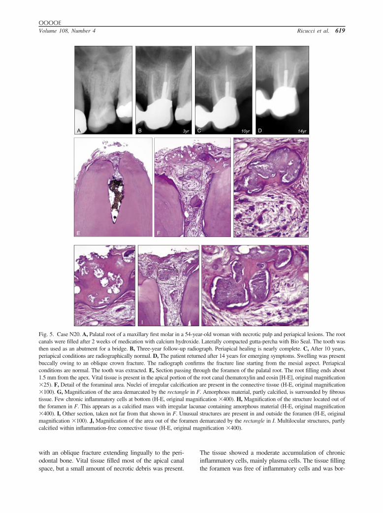
Several cases showed moderate inflammation in theroot canal tissue (N20, tooth #14). There was pulpnecrosis and periapical lesion at the time of endodontictreatment (Fig. 5). The root canals were filled after 2weeks of medication with calcium hydroxide with gutta-percha laterally compacted with Bio Seal as a sealer.The tooth was used as a bridge abutment. It was ex-tracted after 14 years. In the palatal root, the root fillingwas 1.5 mm from the radiographic apex, with vitaltissue in the apical part of the root canal. Irregularcalcifications were visible in the soft tissue. There wassparse amount of debris between root filling and thetissue, which had a superficial necrosis with minor levelof inflammation. There was extensive deposit of ce-mentum closing up the apical root canal and vital tissuewithout inflammation in the area. There were no clin-ical or radiographic signs of periapical disease.
Severe inflammation in the apical residual tissues can
itial diagnosis vital pulp)
osis inl rootnal
Histologicstatus oftissue inapical
root canal
Histologicstatus of
periodontalfragments
Cementumformationin apical
root canal
Bacteria inapical canal
orramifications
Bacteria incoronal
portion ofroot canal
rficial none — diffuse no yesnone none diffuse no yes
rficial none none diffuse no yesrficial none — diffuse no yesal none none diffuse no yesrficial none none diffuse no yesrficial moderate none diffuse no yes
— none diffuse no yesnone none diffuse no yes
al none — diffuse no yesnone none diffuse no yes
al moderate none diffuse no yesnone none diffuse no yesmoderate moderate diffuse no yes
al moderate moderate diffuse no yesrficial none none diffuse no yes
none none diffuse no yeslete — none diffuse no no
moderate none minimal no yesrficial none — diffuse no yesrficial none — no yesal severe — diffuse no yesal severe severe diffuse no yesrficial none none diffuse no —lete — severe none yes yes
rficial moderate — minimal no yesal none none diffuse no yes
. Their evaluation is not reported.moment of extraction.
ons (in
Necrapica
ca
supenonesupesupepartisupesupetotalnonepartinonepartinonenonepartisupenonecompnonesupesupepartipartisupecompsupeparti
triction
be seen in case V22 (tooth #5; Fig. 1). The pulp diagnosis
t at the
superfici
OOOOE618 Ricucci et al. October 2009
at treatment was pulpitis and the tooth was treated in 1visit with gutta-percha and Apexit. Cast post and core wasplaced with a temporary crown that was never replaced.The tooth was extracted after 8 years 8 months owing tocaries. The pulp space along the post was heavily infectedbut could not be followed in the apical part of the rootcanal. Large amount of debris could be observed apical ofthe root filling materials. The root canal, which was nar-rowed by calcified tissue, contained connective tissue withmoderate accumulation of chronic inflammatory cells.Some areas contained severe accumulation of polymor-phonuclear leukocytes. There were no clinical or radio-graphic signs of periapical disease.
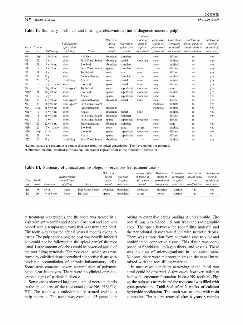
Some cases showed large amounts of necrotic debrisin the apical area of the root canal (case N6, #18; Fig.E3). The tooth was endodontically treated owing to
Table II. Summary of clinical and histologic observat
Caseno.
Tooth/root Follow-up
Radiographicapical limit
of filling Sealer
Debris inapicalroot
canal
N1 5/p 2 yr 2 mo short AH Plus abundantN2 22 5 yr short Pulp Canal Sealer abundantN3 29 8 yr 9 mo short Bio Seal abundantN4* 8 8 yr 5 mo short Pulp Canal Sealer sparseN5 11 8 yr short Tubli-Seal noneN6 18 15 yr short Endomethasone noneN7 20 5 yr overfilling Apexit noneN8 8 7 yr 10 mo short Bio Seal sparseN9 9 6 yr 8 mo Rad. Apex† Tubli-Seal noneN10* 21 10 yr 9 mo short Bio Seal sparseN11 7 2 yr short Apexit sparseN12 13 17 yr 8 mo Rad. Apex† Endomethasone abundantN13 29 6 yr 6 mo Rad. Apex† Pulp Canal Sealer —N14 30/d 18 yr 9 mo short Endomethasone abundantN15* 9 1 yr 7 mo short Apexit abundantN16 5 18 yr 8 mo short Pulp Canal Sealer abundantN17 9 3 yr short Pulp Canal Sealer sparseN18* 20 22 yr 4 mo short Endomethasone abundantN19 20 7 yr 10 mo short Bio Seal noneN20 14/p 14 yr short Bio Seal sparseN21 21 9 yr short Apexit sparseN22 20 17 yr overfilling Pulp Canal Sealer abundant
*Lateral canals are present at a certain distance from the apical cons†Obturation material resorbed at follow-up. Obturation appears shor
Table III. Summary of clinical and histologic observa
Caseno.
Tooth/root Follow-up
Radiographicapical limit
of filling Sealer
Debris inapicalroot
canal
R1 9 19 yr short Pulp Canal Sealer abundantR2 20 9 yr 5 mo short Bio Seal sparse
pulp necrosis. The tooth was extracted 15 years later
owing to extensive caries making it unrestorable. Theroot filling was placed 1.5 mm from the radiographicapex. The space between the root filling material andthe periodontal tissues was filled with necrotic debris.There was a transition from necrotic tissue to vital andnoninflamed connective tissue. This tissue was com-posed of fibroblasts, collagen fibers, and vessels. Therewas no sign of microorganisms in the apical area.Midroot, there were microorganisms in the canal inter-mixed with the root filling material.
In most cases significant narrowing of the apical rootcanal could be observed. A few cases, however, failed toheal with cementum formation. In case N9, tooth #9 (Fig.4), the pulp was necrotic and the root canal was filled withgutta-percha and Tubli-Seal after 2 weeks of calciumhydroxide medication. The tooth was restored with resin
initial diagnosis necrotic pulp)
sis inroot
al
Histologicstatus oftissue inapical
root canal
Histologicstatus of
periodontalfragments
Cementumformationin apical
root canal
Bacteria inapical canal orramifications ordentinal tubules
Bacteria incoronal
portion ofroot canal
lete — none diffuse no yesl moderate none minimal no yeslete — none minimal no yeslete none — diffuse no yes
none none diffuse no yeslete — none minimal no yesl none none minimal no yesl none none diffuse no yesficial moderate none none no yesficial moderate none minimal no yesficial moderate none none no yesl none none diffuse no yes
— moderate minimal no yes— moderate minimal no yes
l none — minimal no yeslete — — diffuse no yesficial moderate none diffuse no —lete — none none no yes
severe none minimal no yesficial moderate none diffuse no yesficial none none diffuse no no
— none minimal no yes
. Their evaluation not reported.moment of extraction.
(retreatment cases)
slal
Histologic statusof tissue inapical root
canal
Histologicstatus of
periodontalfragments
Cementumformationin apical
root canal
Bacteria inapical canal
orramifications
Bacteria incoronal
portion ofroot canal
al moderate moderate diffuse no yesal severe severe diffuse no yes
ions (
Necroapical
can
comppartiacompcompnonecomppartiapartiasupersupersuperpartia
——
partiacompsupercompnonesupersuper
—
triction
tions
Necrosiin apica
root can
superfici
composite. The patient returned after 6 years 8 months
amen dnal ma
OOOOEVolume 108, Number 4 Ricucci et al. 619
with an oblique fracture extending lingually to the peri-odontal bone. Vital tissue filled most of the apical canal
H I
A B 3
E F
Fig. 5. Case N20. A, Palatal root of a maxillary first molar in acanals were filled after 2 weeks of medication with calcium hydthen used as an abutment for a bridge. B, Three-year follow-upperiapical conditions are radiographically normal. D, The patienbuccally owing to an oblique crown fracture. The radiographconditions are normal. The tooth was extracted. E, Section pass1.5 mm from the apex. Vital tissue is present in the apical portion�25). F, Detail of the foraminal area. Nuclei of irregular calcifi�100). G, Magnification of the area demarcated by the rectangtissue. Few chronic inflammatory cells at bottom (H-E, originalthe foramen in F. This appears as a calcified mass with irregula�400). I, Other section, taken not far from that shown in F. Unmagnification �100). J, Magnification of the area out of the forcalcified within inflammation-free connective tissue (H-E, origi
space, but a small amount of necrotic debris was present.
The tissue showed a moderate accumulation of chronicinflammatory cells, mainly plasma cells. The tissue filling
J
D10yr 14yr
G
r-old woman with necrotic pulp and periapical lesions. The rootLaterally compacted gutta-percha with Bio Seal. The tooth wasraph. Periapical healing is nearly complete. C, After 10 years,ed after 14 years for emerging symptoms. Swelling was presents the fracture line starting from the mesial aspect. Periapicalugh the foramen of the palatal root. The root filling ends aboutroot canal (hematoxylin and eosin [H-E], original magnificationre present in the connective tissue (H-E, original magnification
. Amorphous material, partly calcified, is surrounded by fibrousfication �400). H, Magnification of the structure located out ofae containing amorphous material (H-E, original magnificationstructures are present in and outside the foramen (H-E, originalemarcated by the rectangle in I. Multilocular structures, partly
gnification �400).
Cyr
54-yearoxide.
radiogt returnconfirming thro
of thecation ale in Fmagnir lacunusual
the foramen was free of inflammatory cells and was bor-
OOOOE620 Ricucci et al. October 2009
dered externally by a fibrous connective tissue. No bac-teria could be seen in the apical region, but in the midrootarea bacteria could be seen in the dentin tubules. Despitenormal radiographic conditions, chronic inflammationwas present in the pulp stump.
It was not unusual to find reactive calcified tissuedeposits in the apical tissues surrounded by normalconnective tissue (N1, tooth #5).
DISCUSSIONUnlike the examination materials in the studies by
Brynolf, Green et al., and Barthel et al.,1,3,4 the teeth inthe present study were collected from living patients,which allowed optimal tissue handling, fixation, andpreservation. All endodontic treatment was performedby 1 operator during a 20-year period, and the qualityof all root canal therapy was carefully controlled.Therefore, the complete case history, such as clinicalsigns and/or symptoms, endodontic and restorativetreatment details, status of coronal restorations, as wellas period of follow-up time, was available. This is incontrast to cadaver material, where most such informa-tion, including the operator, is unavailable.
Much reliance has been afforded the results from thestudy by Brynolf1 on cadaver teeth and the proposedtrue status of periapical tissues associated with end-odontically treated teeth. Her study inferred the notionthat in most cases of endodontic treatment there isresidual inflammation in the apical/periapical tissueswhich can be visualized radiographically.1,5
Previously, the descriptions of the periapical tissuesafter endodontic treatment was often known as de-scribed by Kronfeld11: “Most roots are slightly under-filled, the length of the unfilled portion of the maincanal varying between 0.5 and 2 mm [and] containingfibrous connective tissue, which may be either a rem-nant of the original pulp tissue or periodontal connec-tive tissue that proliferated into the open apical portionof the root canal. The connective tissue has a tendencyto form cementum, which is deposited in layers on thewall of the pulp canal.” Then he describes several caseswhere “The apical portion of the root canal containswell vascularized connective tissue that is entirely freefrom inflammation; cementum has been deposited overall of the walls of the root canal.”
Nygaard-Østby12 stated: “To leave the apical andforaminal part of the pulp tissue and to retain its vitalitywill play a decisive role for the success in the treatmentof the vital pulp . . . . In cases where the pulp is vitalbefore treatment, no matter whether the diagnosis isclinically intact pulp [or] acute or chronic pulpitis,partial extirpation seems to give the most favorableprognosis. By appropriate treatment, in the majority of
cases, the vitality of the residual pulp may be con-served, the result being a normal apical periodontalligament and fibrous connective tissue in the apicalportion of the root canal.”
In random observations from extracted root filledteeth, our observations have been more in agreementwith the description by Kronfeld11 than with the de-scription by Brynolf.1
Another issue of great interest has been the conceptof coronal leakage, which was emphasized by Ray andTrope.6 Careful study of this issue, however, suggestedthat coronal microbial ingress throughout the root canalof root-filled teeth, although possible, may be a randomprocess.10 In optimally endodontically treated teeth,prolonged exposure of the endodontic filling to the oralenvironment may evoke a localized histologic inflam-mation in the apical connective tissue, but developmentof a frank periapical lesion was not a common occur-rence.
Histologic observation of the healing after endodon-tic treatment requires a biopsy of the tissue to bestudied. In some earlier studies on human clinical cases,such biopsy involved the surgical removal of a substan-tial amount of periapical bone together with the apicalroot portion.13,14 This invasive methodology restrictsthe number of cases available to study owing to ethicallimitations. However, when the endodontic treatment issystematically confined within the root canal system itis possible to obtain substantial and valuable informa-tion on extraction material by observing the tissueremaining on the root surface and within the pulpspace.15 The endodontic technique used in the presentstudy always limited the root canal preparation andfilling to be short of the apex.
The material presented here suggests that the apicaltissue of carefully root-filled teeth, with no conven-tional radiographic signs of periapical changes, such asthickening of periodontal ligament space, loss of lam-ina dura, or breakdown of apical alveolar bone, is onlyrarely significantly inflamed. When the tissue is in-flamed, microbial causes can always be demonstrated.These findings suggest that moderate inflammation inthe apical connective tissue is compatible with normalconventional periapical radiography.
In this case series study, we were able to demonstratethe presence of microorganisms in the coronal portionof the root canal in 47 of the 49 cases with coronaltissue. This is compatible with the fact that all rootcanals were lacking coronal closure. The coronal pres-ence of bacteria, however, did not necessarily result inthe presence of microorganisms in the apical portionof the root canal. Only 1 case (V25) had bacteria in boththe coronal and the apical portions of the root canal.That case was originally diagnosed with pulpitis. At the
time of extraction, 13 years after original treatment, the
OOOOEVolume 108, Number 4 Ricucci et al. 621
residual tissue in the apical pulp space was necroticwith severe inflammation in the apical tissues. Despitethis, there were no clinical symptoms or periapicalpathologic disease visible on the radiograph.
The presence of debris and partially necrotic tissue wasa common observation in the apical pulp space. In mostcases this was not associated with significant inflamma-tion in the tissues. We conclude from these observationsthat unless infected, this necrotic tissue is not able by itselfto sustain tissue inflammation, because uninfected autol-ogous necrotic tissue cannot indefinitely release inflam-matory mediators and proinflammatory cytokines.16
Various sealers were used in a random fashion duringthis study (Tables I-III). Most commercially availablesealers become practically inert after some time. Becausethe observation periods in this study were very long, it isvery unlikely that any material toxicity would be discern-ible as tissue changes. No clinical association with out-come could be observed when comparing the long-termtreatment results of the sealers used here (Fig. E9).17
The root fillings, which were well performed, appear tohave effectively prevented the complete penetration ofbacteria to the apical foramen. All root canals included inthis study were severely infected at the coronal end, be-cause the teeth were not protected by permanent restora-tion and often had catastrophic caries lesions (Fig. 2).Despite these conditions, bacteria could be seen in theapical part of the root canal in only 1 case. However, inone-third of the cases we could observe a mild inflamma-tory response in the apical tissues remnants. This is mostlikely the result of the penetration of nonantigenic and anti-genic active bacterial byproducts inducing a mild inflamma-tory response. This microleakage, however, appeared to beunable to sustain a frank apical bone resorption.
It was common to find significant but incompleteapical closures of the apical part of the root canal withcementum. Despite the long observation time, no casewith complete cementum closure could be observed.
All teeth in the present study were clinically free ofsymptoms and were radiographically without any discern-ible disease. This suggests that good diagnostic tools toassess the state of health of endodontically treated teethare lacking. The ultimate goal of endodontic treatmentmust be not only radiographically normal periapical con-ditions, but also absence of inflammation in the periodon-tal ligament space and in remaining connective tissue inthe apical foramen and apical pulp tissue ramifications.
REFERENCES1. Brynolf I. A histological and roentgenological study of the peri-
apical region of human upper incisors. Odont Revy 1967;18(Suppl 11):1-176.
2. Rowe AHR, Binnie WH. Correlation between radiological andhistological inflammatory changes following root canal treat-
ment. Int Endod J 1974;7:57-63.3. Green TL, Walton RE, Taylor JK, Merrel P. Radiographic andhistologic periapical findings of root canal treated teeth in ca-daver. Oral Surg Oral Med Oral Pathol Oral Radiol Endod1997;83:707-11.
4. Barthel CR, Zimmer S, Trope M. Relationship of radiologic andhistologic signs of inflammation in human root-filled teeth. JEndod 2004;30:75-9.
5. Ørstavik D, Kerekes K, Eriksen HM. The periapical index: ascoring system for radiographic assessment of apical periodon-titis. Endod Dent Traumatol 1986;2:20-34.
6. Ray HA, Trope M. Periapical status of endodontically treatedteeth in relation to the technical quality of the root filling and thecoronal restoration. 1995;28:12-8.
7. Strindberg LZ. The dependence of the results of pulp therapy oncertain factors. An analytic study based on radiographic andclinical follow-up examinations. Acta Odont Scand 1956;14(Suppl 21):1-175.
8. Möller ÅJR. Microbiological examination of root canals andperiapical tissues of human teeth. Odont Tidskr 1966;74(SpecIss):1-380.
9. Taylor RD. Modification of the Brown and Brenn Gram stain forthe differential staining of gram-positive and gram-negative bac-teria in tissue sections. Am J Clin Pathol 1966;46:472-6.
10. Ricucci D, Bergenholtz G. Bacterial status in root-filled teethexposed to the oral environment by loss of restoration andfracture or caries—a histobacteriological study of treated cases.Int Endod J 2003;36:787-802.
11. Kronfeld R. Histopathology of the teeth and their surroundingstructures. 2nd ed. Philadelphia: Lea and Febiger; 1943; p. 228-9.
12. Nygaard-Østby B. Om vevsforandringer i det apikale paraden-tium hos mennesket ved rotbehandling. Nye kliniske, røntgen-ologiske og histopatologiske studier. Oslo: Norske Videnskaps-Akademi; 1944.
13. Nair PNR, Sjögren U, Kahnberg KE, Krey G, Sundqvist G. Intrara-dicular bacteria and fungi in root-filled, asymptomatic human teethwith therapy-resistant periapical lesions: a long term light and elec-tron microscopic follow-up study. J Endod 1990;16:580-8.
14. Nair PNR, Sjögren U, Figdor D, Sundqvist G. Persistent peria-pical radiolucencies of root-filled human teeth, failed endodontictreatment, and periapical scars. Oral Surg Oral Med Oral PatholOral Radiol Endod 1999;87:617-27.
15. Engström B, Spangberg L. Wound healing after partial pulpec-tomy. A histological study performed on contralateral toothpairs. Odontol Tidskr 1967;75:1-18.
16. Lin LM, Di Fiore PM, Lin J, Rosenberg PA. Histological studyof periapical tissue responses to uninfected and infected devital-ized pulps in dogs. J Endod 2006;32:34-8.
17. Ng V-L, Mann V, Rahbaran S, Lewsey J, Gulabivala K. Out-come of primary root canal treatment: systematic review of theliterature—part 2. Influence of clinical factors. Int Endod J2008;41:6-31.
Reprint requests:
Dr. Domenico RicucciPiazza Calvario 787022 Cetraro (CS)[email protected]
SUPPLEMENTARY DATASupplementary data associated with this article can
be found in the online version, at doi: 10.1016/j.tripleo.
2009.05.028.