Work-Related Foot and Ankle Conditions
54
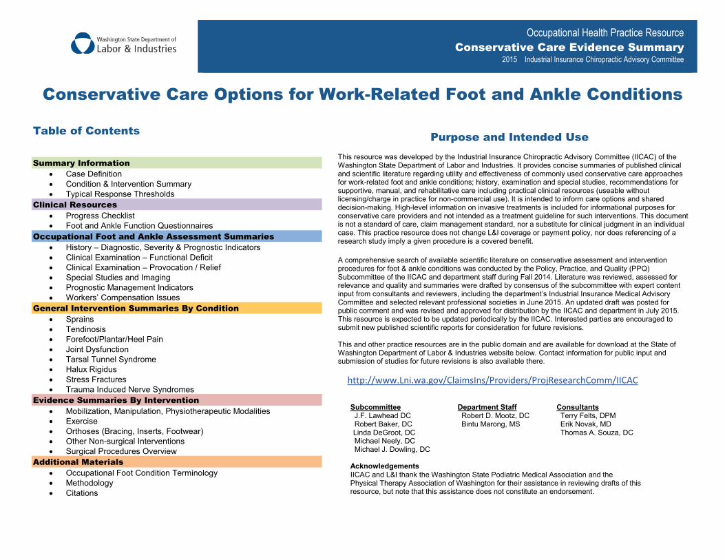
Occupational Health Practice Resource Conservative Care Evidence Summary 2015 Industrial Insurance Chiropractic Advisory Committee Conservative Care Options for Work-Related Foot and Ankle Conditions Table of Contents Summary Information • Case Definition • Condition & Intervention Summary • Typical Response Thresholds Clinical Resources • Progress Checklist • Foot and Ankle Function Questionnaires Occupational Foot and Ankle Assessment Summaries • History – Diagnostic, Severity & Prognostic Indicators • Clinical Examination – Functional Deficit • Clinical Examination – Provocation / Relief • Special Studies and Imaging • Prognostic Management Indicators • Workers’ Compensation Issues General Intervention Summaries By Condition • Sprains • Tendinosis • Forefoot/Plantar/Heel Pain • Joint Dysfunction • Tarsal Tunnel Syndrome • Halux Rigidus • Stress Fractures • Trauma Induced Nerve Syndromes Evidence Summaries By Intervention • Mobilization, Manipulation, Physiotherapeutic Modalities • Exercise • Orthoses (Bracing, Inserts, Footwear) • Other Non-surgical Interventions • Surgical Procedures Overview Additional Materials • Occupational Foot Condition Terminology • Methodology • Citations Purpose and Intended Use This resource was developed by the Industrial Insurance Chiropractic Advisory Committee (IICAC) of the Washington State Department of Labor and Industries. It provides concise summaries of published clinical and scientific literature regarding utility and effectiveness of commonly used conservative care approaches for work-related foot and ankle conditions; history, examination and special studies, recommendations for supportive, manual, and rehabilitative care including practical clinical resources (useable without licensing/charge in practice for non-commercial use). It is intended to inform care options and shared decision-making. High-level information on invasive treatments is included for informational purposes for conservative care providers and not intended as a treatment guideline for such interventions. This document is not a standard of care, claim management standard, nor a substitute for clinical judgment in an individual case. This practice resource does not change L&I coverage or payment policy, nor does referencing of a research study imply a given procedure is a covered benefit. A comprehensive search of available scientific literature on conservative assessment and intervention procedures for foot & ankle conditions was conducted by the Policy, Practice, and Quality (PPQ) Subcommittee of the IICAC and department staff during Fall 2014. Literature was reviewed, assessed for relevance and quality and summaries were drafted by consensus of the subcommittee with expert content input from consultants and reviewers, including the department’s Industrial Insurance Medical Advisory Committee and selected relevant professional societies in June 2015. An updated draft was posted for public comment and was revised and approved for distribution by the IICAC and department in July 2015. This resource is expected to be updated periodically by the IICAC. Interested parties are encouraged to submit new published scientific reports for consideration for future revisions. This and other practice resources are in the public domain and are available for download at the State of Washington Department of Labor & Industries website below. Contact information for public input and submission of studies for future revisions is also available there. http://www.Lni.wa.gov/ClaimsIns/Providers/ProjResearchComm/IICAC Subcommittee J.F. Lawhead DC Robert Baker, DC Linda DeGroot, DC Michael Neely, DC Michael J. Dowling, DC Department Staff Robert D. Mootz, DC Bintu Marong, MS Consultants Terry Felts, DPM Erik Novak, MD Thomas A. Souza, DC Acknowledgements IICAC and L&I thank the Washington State Podiatric Medical Association and the Physical Therapy Association of Washington for their assistance in reviewing drafts of this resource, but note that this assistance does not constitute an endorsement.
Transcript of Work-Related Foot and Ankle Conditions
Occupational Health Practice Resource Conservative Care Evidence Summary
2015 Industrial Insurance Chiropractic Advisory Committee
Conservative Care Options for Work-Related Foot and Ankle Conditions
Table of Contents
Summary Information • Case Definition• Condition & Intervention Summary• Typical Response Thresholds
Clinical Resources • Progress Checklist• Foot and Ankle Function Questionnaires
Occupational Foot and Ankle Assessment Summaries • History – Diagnostic, Severity & Prognostic Indicators• Clinical Examination – Functional Deficit• Clinical Examination – Provocation / Relief• Special Studies and Imaging• Prognostic Management Indicators• Workers’ Compensation Issues
General Intervention Summaries By Condition • Sprains• Tendinosis• Forefoot/Plantar/Heel Pain• Joint Dysfunction• Tarsal Tunnel Syndrome• Halux Rigidus• Stress Fractures• Trauma Induced Nerve Syndromes
Evidence Summaries By Intervention • Mobilization, Manipulation, Physiotherapeutic Modalities• Exercise• Orthoses (Bracing, Inserts, Footwear)• Other Non-surgical Interventions• Surgical Procedures Overview
Additional Materials • Occupational Foot Condition Terminology• Methodology• Citations
Purpose and Intended Use This resource was developed by the Industrial Insurance Chiropractic Advisory Committee (IICAC) of the Washington State Department of Labor and Industries. It provides concise summaries of published clinical and scientific literature regarding utility and effectiveness of commonly used conservative care approaches for work-related foot and ankle conditions; history, examination and special studies, recommendations for supportive, manual, and rehabilitative care including practical clinical resources (useable without licensing/charge in practice for non-commercial use). It is intended to inform care options and shared decision-making. High-level information on invasive treatments is included for informational purposes for conservative care providers and not intended as a treatment guideline for such interventions. This document is not a standard of care, claim management standard, nor a substitute for clinical judgment in an individual case. This practice resource does not change L&I coverage or payment policy, nor does referencing of a research study imply a given procedure is a covered benefit.
A comprehensive search of available scientific literature on conservative assessment and intervention procedures for foot & ankle conditions was conducted by the Policy, Practice, and Quality (PPQ) Subcommittee of the IICAC and department staff during Fall 2014. Literature was reviewed, assessed for relevance and quality and summaries were drafted by consensus of the subcommittee with expert content input from consultants and reviewers, including the department’s Industrial Insurance Medical Advisory Committee and selected relevant professional societies in June 2015. An updated draft was posted for public comment and was revised and approved for distribution by the IICAC and department in July 2015. This resource is expected to be updated periodically by the IICAC. Interested parties are encouraged to submit new published scientific reports for consideration for future revisions.
This and other practice resources are in the public domain and are available for download at the State of Washington Department of Labor & Industries website below. Contact information for public input and submission of studies for future revisions is also available there.
http://www.Lni.wa.gov/ClaimsIns/Providers/ProjResearchComm/IICAC
Subcommittee J.F. Lawhead DC Robert Baker, DC Linda DeGroot, DC Michael Neely, DC Michael J. Dowling, DC
Department Staff Robert D. Mootz, DC Bintu Marong, MS
Consultants Terry Felts, DPM Erik Novak, MD Thomas A. Souza, DC
Acknowledgements IICAC and L&I thank the Washington State Podiatric Medical Association and the Physical Therapy Association of Washington for their assistance in reviewing drafts of this resource, but note that this assistance does not constitute an endorsement.

2
PRACTICAL APPLICATION POINTS
• Work-related foot and ankle conditions result from an identifiable injury. Withthe possible exceptions of metatarsal stress fracture and fat pad syndrome,conditions related to repetitive stress are unlikely to ever be occupational.
• Using the Ottawa or Bernese rules to determine indications for x-ray to rule inankle fractures significantly reduces unnecessary (negative) films.
• Stability tests may have limited utility due to inadequate evidence of reliabilityand validity. However, expert opinion encourages stress testing for ligamentdamage.
• Achilles tendon rupture may typically be determined clinically with calfsqueeze and palpation without need for MRI.
• Functional improvement should be determined using validated functionaltracking instruments at baseline and follow up.
• Early mobility and weight bearing to tolerance facilitates a better and fasterresponse for lower grade sprains. However a short period of immobilizationyields faster and more sustained recovery from higher grade sprains.
• Generally, manual techniques (mobilization, manipulation, soft tissue work)plus exercise (eccentric stretching) offer better outcomes than exercise aloneor electrophysiological modalities for sprains and tendinosis.
• Low grade sprains, acute tendinosis, and forefoot pain, typically have a rapidinitial response to conservative care and resolve within a few weeks. Highergrade sprains, high ankle sprains, and chronic conditions such as chronicplantar pain may take substantially longer to resolve.
• Eccentric exercise facilitates recovery for tendinosis. Neuromuscular exercisemay reduce recurrence of ankle sprains. Supervised exercise may offermarginal benefit to home programs for higher grade ankle sprain recovery.
• Physiotherapeutic modalities do not add benefit for recovery from most footand ankle conditions. Microcurrent may be helpful in chronic tendinopathy.
• Shoe inserts in general may assist in comfort and recovery for foot and ankleinjuries but there do not appear to be advantages for custom made productsover off-the-shelf versions.
Work-Related Foot and Ankle Conditions
Ankle sprains are a common work related injury. Fractures, Achilles tendon rupture, hallux rigidus, and some tendinopathies (with onset closely following a work trauma) may also result from occupational exposures. Plantar and heel pain are common complaints, however, causation has rarely been associated with work exposure. Pre-existing conditions unlikely to be caused by workplace exposure include biomechanical problems (e.g., pronation/supination), chronic ankle instability, and some pain conditions associated with peripheral neuropathies. Although interventions are individualized for patients, all treatments and support for injured workers need to be directly related to the accepted work-related condition.
Evaluation Summary • Determination and thorough documentation of work-relatedness of foot and ankle
conditions is crucial for acceptance of an occupational foot or ankle condition,particularly where onset is not a direct result of an identifiable work injury.
• Rule-out potential urgent conditions requiring specialist attention (e.g., fracture,dislocation, tendon rupture, syndesmosis injury, 3rd degree sprains).
• Rule out infection, vascular compromise, neoplasms, metabolic red flags• Rule-in mechanical components prior to initiating manual care.• Document lower extremity function (e.g., validated instruments) at baseline and at
regular follow-up (e.g., 2-3 week intervals).
Intervention Summary • Evidence supports ‘low tech’ approaches such as early mobilization, eccentric
exercise, manual therapies, and NSAIDs for most straightforward foot and ankleconditions (sprains, tendinopathy, forefoot pain).
• Recovery is typically rapid from sprains (other than high ankle) and most forefootinjuries. Tendinosis and plantar pain tend to respond slower.
• Severe injuries should be managed initially by specialists due to potential difficultyto identify complications and complexities.
• Consider reassessment and specialist consult if there is inadequate responsewithin 3-4 weeks of conservative care.
Typical Interventions and Approximate Response Thresholds
• Initially: Patients with red flags orpersistent severe pain should be referred toa specialist for urgent evaluation.
• Uncertain mechanical etiology, severepain/restriction: rule out fracture anddislocation; expect some early measurableimprovement w/ combined active exerciseand manual work within patient tolerance.
• Known mechanical etiology: expect earlysignificant improvement for low gradesprains, tendinosis, etc, however recoverymay be delayed in chronic and more severeconditions.
• Early: Re-assess pain/function within 2-3weeks of beginning care.
• Good improvement: Function and weighttolerance improves measurably andperceptively. Continue, emphasize self-care.
• Limited improvement: Conditions likely torespond slower include Grade III sprains,Achilles tendon rupture, hallux rigidus, highankle sprain. Measureable change should bedocumented.
• Inadequate improvement: Worsening or nochange in function (e.g., higher score on FAAMor SEFAS). Consider additional diagnostics,specialist consultation. If only smallimprovement, consider change in intervention(e.g., supervised exercise, more intensemanual work).
• Demonstrable improvement should beevident. Inadequate response warrantsconsideration for evaluation by foot andankle specialist.
• Good improvement: At or near pain free,nearly full function. Transition to self-care,periodic follow-up assessment.
• Inadequate improvement: Pain & functionlimitations persist, minimal improvement.Consider specialist referral.
• Resolution: Most foot & ankle injuriesgenerally should achieve tolerance ofweight bearing and normal walking.
• Good improvement: Most acutemechanical foot and ankle problemsshould resolve fully. Improvement infunction should be significant andmeasurable in severe sprains.
• Inadequate improvement: Consideradditional diagnostics, specialistconsultation.
1-2 wks 3-6 wks 7-8 wks Beyond 8 wks

3
FOOT & ANKLE PROGRESS CHECKLIST Voluntary educational / practice aid – Not an L&I documentation requirementA
sses
smen
t / P
rogr
ess
Date:
Work limitation: Off work Weight restriction:_______ Activity limits: __________ Weight-bearing work tolerance:
___________ hrs
Function Score (e.g., FAAM, SEFAS) Baseline: ___________
Pain Interference w/ activity: None Total
0 1 2 3 4 5 6 7 8 9 10
Baseline (check all that apply): Difficult weight bearing Unable to walk normally Activity limited by pain ______________________
Date:
Work limitation improvement: Off work Weight restriction:_______ Activity limits: __________ Weight-bearing work tolerance:
___________ hrs
Function Score ___________
Pain Interference w/ activity: None Total
0 1 2 3 4 5 6 7 8 9 10
Percent Improvement (pt. perception): ___ Weight bearing ___ Walking ___ Activity limitation ___ ______________________
Inte
rven
tion
Opt
ions
Manual • Combined mobilization, initial active
and passive exercise, and softtissue work typically reduce painand improve function formechanical foot/ankle problems.Treatment frequency reported intrials typically 2-3 times per week.
Modalities/Self Care • Full immobilization for severe
conditions and fracture:.R/MICE* totolerance initially for most other footand ankle conditions.
• Consider home exercise totolerance.
• Physiotherapeutic modalities maynot be particularly helpful.
• NSAIDs and analgesics may behelpful for initial pain control.
Manual • Incrementally increasing intensity of
manual techniques within patienttolerance is advisable. In absence ofmeaningful functional improvement,consider modification of methods.
• Consider incorporation of exercises (e.g.,range of motion, eccentric and balanceapproaches).
• Supervised exercise may be beneficialwith slower responding conditions (e.g.,Grade III sprains, Achilles tendon rupture,hallux rigidus, high ankle spraincapsulitis).
Response • 30-50% improvement in function scores is
considered meaningful clinical change.• Lower grade sprains typically attain this
rapidly. Tendinoses usually experienceslower response
Good Improvement • Progression of uncomplicated foot/ankle problems (e.g., Grade 1sprains) is typically ~50%
improvement in pain and function within first 2 weeks and fully resolved within 8 weeks.• For tendinosis 30-50% improvement in pain and function scores within first month can be
expected.• Low grade sprains respond very quickly to conservative intervention. Grade III sprains, Achilles
tendon rupture, hallux rigidus, and high ankle sprain may have significantly delayed response.
Inadequate improvement • Reassessment for red flags, further diagnostics, and specialist consultation is warranted in non-
responding cases.• Consider specialist consult for apparent low grade traumatic injuries if only minimal improvement
is seen within first month.
* R/MICE = Rest/Modified pain-free activity, Ice, Compression, Elevation
Baseline 1-2 wks 3-6 wks 7-8 wks Beyond 8 wks

4
Foot & Ankle Ability Measure (FAAM) Voluntary educational / practice aid – Not an L&I documentation requirement Please answer every question by circling one response that most closely describes your condition within the past week. If the activity in question is limited by something other than your foot or ankle, check N/A (Not Applicable)
Activity: N
o Di
ffic
ulty
Slig
ht
Diff
icul
ty
Mod
erat
e Di
ffic
ulty
Extr
eme
Diff
icul
ty
Una
ble
To
Do
N/A
Name _____________________ Date ____________
Office Use Only: Scoring: Each item is scored on a five point scale with 4 being “No Difficulty” and 0 being “Unable To Do.” The lowest potential score of the Activities of Daily Living (ADL) subscale of the FAAM is 0 points, the highest 84 points. Total score is converted into percentage. Higher percentage indicates higher level of physical function
Source: Martin R, Irrang J, Conti S, vanSwearingen J. Evidence of validity for the foot and ankle Ability Measure. Foot Ankle Intern 2005; 26(11):968-983.
Standing 4 3 2 1 0 □ Walking on even ground 4 3 2 1 0 □ Walking on even ground without shoes 4 3 2 1 0 □ Walking up hills 4 3 2 1 0 □ Walking down hills 4 3 2 1 0 □ Going up stairs 4 3 2 1 0 □ Going down stairs 4 3 2 1 0 □ Walking on uneven ground 4 3 2 1 0 □ Stepping up and down curbs 4 3 2 1 0 □ Squatting 4 3 2 1 0 □ Coming up on your toes 4 3 2 1 0 □ Initiating walking 4 3 2 1 0 □ Walking 5 minutes or less 4 3 2 1 0 □ Walking approximately 10 minutes 4 3 2 1 0 □ Walking 15 minutes or greater 4 3 2 1 0 □ Because of your foot and ankle, how much difficulty do you have with:
Home responsibilities 4 3 2 1 0 □ Activities of daily living (eg, around the house) 4 3 2 1 0 □ Personal care (eg, bathing, shaving) 4 3 2 1 0 □ Light to moderate work (standing, walking) 4 3 2 1 0 □ Heavy work (pushing/pulling, climbing, carrying)
4 3 2 1 0 □
Recreational activities 4 3 2 1 0 □
Column Totals:
SCORE ______ / 84
5
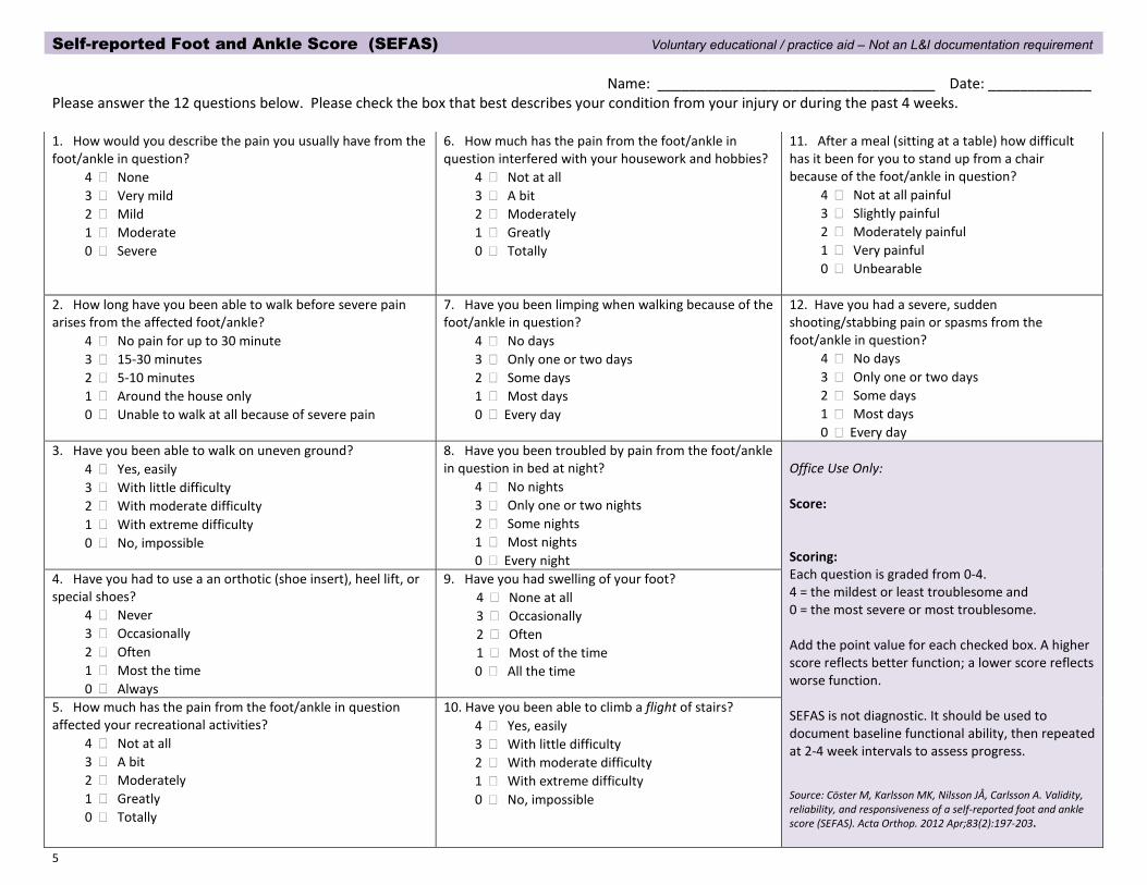
Self-reported Foot and Ankle Score (SEFAS) Voluntary educational / practice aid – Not an L&I documentation requirement
Name: ___________________________________ Date: _____________ Please answer the 12 questions below. Please check the box that best describes your condition from your injury or during the past 4 weeks.
1. How would you describe the pain you usually have from thefoot/ankle in question?
4 � None 3 � Very mild 2 � Mild 1 � Moderate 0 � Severe
6. How much has the pain from the foot/ankle inquestion interfered with your housework and hobbies?
4 � Not at all 3 � A bit 2 � Moderately 1 � Greatly 0 � Totally
11. After a meal (sitting at a table) how difficulthas it been for you to stand up from a chair because of the foot/ankle in question?
4 � Not at all painful 3 � Slightly painful 2 � Moderately painful 1 � Very painful 0 � Unbearable
2. How long have you been able to walk before severe painarises from the affected foot/ankle?
4 � No pain for up to 30 minute 3 � 15-30 minutes 2 � 5-10 minutes 1 � Around the house only 0 � Unable to walk at all because of severe pain
7. Have you been limping when walking because of thefoot/ankle in question?
4 � No days 3 � Only one or two days 2 � Some days 1 � Most days 0 � Every day
12. Have you had a severe, suddenshooting/stabbing pain or spasms from the foot/ankle in question?
4 � No days 3 � Only one or two days 2 � Some days 1 � Most days 0 � Every day
3. Have you been able to walk on uneven ground?4 � Yes, easily 3 � With little difficulty 2 � With moderate difficulty 1 � With extreme difficulty 0 � No, impossible
8. Have you been troubled by pain from the foot/anklein question in bed at night?
4 � No nights 3 � Only one or two nights 2 � Some nights 1 � Most nights 0 � Every night
Office Use Only:
Score:
Scoring: Each question is graded from 0-4. 4 = the mildest or least troublesome and 0 = the most severe or most troublesome.
Add the point value for each checked box. A higher score reflects better function; a lower score reflects worse function.
SEFAS is not diagnostic. It should be used to document baseline functional ability, then repeated at 2-4 week intervals to assess progress.
Source: Cöster M, Karlsson MK, Nilsson JÅ, Carlsson A. Validity, reliability, and responsiveness of a self-reported foot and ankle score (SEFAS). Acta Orthop. 2012 Apr;83(2):197-203.
4. Have you had to use a an orthotic (shoe insert), heel lift, orspecial shoes?
4 � Never 3 � Occasionally 2 � Often 1 � Most the time 0 � Always
9. Have you had swelling of your foot?4 � None at all 3 � Occasionally 2 � Often 1 � Most of the time 0 � All the time
5. How much has the pain from the foot/ankle in questionaffected your recreational activities?
4 � Not at all 3 � A bit 2 � Moderately 1 � Greatly 0 � Totally
10. Have you been able to climb a flight of stairs?4 � Yes, easily 3 � With little difficulty 2 � With moderate difficulty 1 � With extreme difficulty 0 � No, impossible

6
OCCUPATIONAL FOOT AND ANKLE ASSESSMENT SUMMARY
Occupational Foot and Ankle Conditions
Nature of foot and ankle disorders • Urgent and serious medical conditions – infection, vascular compromise, neoplasms, metabolic conditions (e.g., gout, diabetes)• Urgent mechanical conditions – fractures, third degree ankle sprains, syndesmosis injury, tendon ruptures (Achilles, tibialis
anterior or posterior, peroneal), dislocations• Mechanical conditions – ligamentous strains, subluxation, soft tissue disorders• Neurological conditions – peripheral neuropathy, radicular pain, sclerotomal radiation, paresthesia (Note: trauma and fracture
may also involve significant neurological compromise)
Clinical presentation 1-3 • The most common foot and ankle injuries include inversion sprains, stress fractures, and lateral foot trauma leading to peroneal
tendinosis, fifth metatarsal fracture, or cuboid subluxation. • Simple sprains may be associated with various ligament ruptures and/or fractures, thus careful evaluation of the mechanism of
injury, follow-up, and reassessment and special studies may be needed with inadequate or sluggish recovery. • Foot and ankle conditions may present with a number of signs and symptoms including pain, swelling, stiffness,
weakness/sensation of “giving out”, discoloration, popping/crepitus, locking, paresthesias, and/or numbness. • Most foot conditions are biomechanical in nature and nearly all foot and ankle conditions have biomechanical impacts. Footwear,
work surfaces, postural adaptations, and concurrent biomechanical problems in the knee, hip or back may impact foot function, stability, and/or symptoms.
• Vascular compromise, peripheral and radicular neuropathies of the back and lower extremities may manifest as foot complaints.Diabetes, myelopathy (usually canal stenosis), proximal trauma and other factors can contribute to sensory deficits with long term consequences that can contribute to, or exacerbate, injury.
Work place exposure: work injury types • Direct trauma (e.g., blunt force; crush injuries, stubbing toes – 5th toe most common; sudden first toe dorsiflexion / plantarflexion)• Plantarflexion/inversion injury – most common ankle sprain typically impacts anterior talofibular and calcaneofibular areas• Dorsiflexion/eversion injury – typically impacts deltoid ligament area (medial); talar dislocation may result when severe• Calcaneal injury/heel pain associated with landing from a jump• Twisting injury (e.g., “ski-boot” injury) frequently associated with distal tibia and fibula fractures and/or diastasis
Work place exposure: occupational disease • When activities outside of work may also contribute to foot and ankle conditions, case law requires establishing that the workplace
activities contributed to the development or worsening of the condition on a more-probable-than-not basis compared to the risks in everyday life. (Dennis V. Dept. of Labor & Industries, 1987) This can be particularly relevant when considering repetitive stress (e.g., prolonged standing, working on a hard surface) as a potential contributor to a foot and ankle condition.
Diagnostic corroboration • History (e.g., mechanics of exposure - trauma, assessment of contributing factors, concurrent conditions).• Pain localization – symptomatic area typically identifies affected structures and should correlate with exposure onset• Plain film imaging may be helpful to assess for:
o Osseous damage/fracture with substantial trauma and when swelling and tenderness immediately follow an injury.o Instability with special bilateral stress views (under anesthesia) assessing the inter-tibiofibular talar space.o Non-mechanical etiology such as tumor or infection.
• More severe sprains are likely to result in instability that over time may damage joint surfaces and lead to degeneration.
HISTORY – Diagnostic/Severity Indicators
7
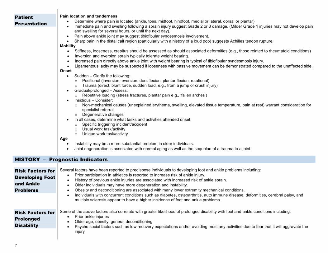
Patient Presentation
Pain location and tenderness • Determine where pain is located (ankle, toes, midfoot, hindfoot, medial or lateral, dorsal or plantar)• Immediate pain and swelling following a sprain injury suggest Grade 2 or 3 damage. (Milder Grade 1 injuries may not develop pain
and swelling for several hours, or until the next day).• Pain above ankle joint may suggest tibiofibular syndesmosis involvement.• Sharp pain in the distal calf region (particularly with a history of a loud pop) suggests Achilles tendon rupture.
Mobility • Stiffness, looseness, crepitus should be assessed as should associated deformities (e.g., those related to rheumatoid conditions)• Inversion and eversion sprain typically tolerate weight bearing.• Increased pain directly above ankle joint with weight bearing is typical of tibiofibular syndesmosis injury.• Ligamentous laxity may be suspected if looseness with passive movement can be demonstrated compared to the unaffected side.
Onset • Sudden – Clarify the following:
o Positional (inversion, eversion, dorsiflexion, plantar flexion, rotational)o Trauma (direct, blunt force, sudden load, e.g., from a jump or crush injury)
• Gradual/prolonged – Assess:o Repetitive loading (stress fractures, plantar pain e.g., ‘fallen arches’)
• Insidious – Consider:o Non-mechanical causes (unexplained erythema, swelling, elevated tissue temperature, pain at rest) warrant consideration for
specialist referral.o Degenerative changes
• In all cases, determine what tasks and activities attended onset:o Specific triggering incident/accidento Usual work task/activityo Unique work task/activity
Age • Instability may be a more substantial problem in older individuals.• Joint degeneration is associated with normal aging as well as the sequelae of a trauma to a joint.
HISTORY – Prognostic Indicators
Risk Factors for Developing Foot and Ankle Problems
Several factors have been reported to predispose individuals to developing foot and ankle problems including: • Prior participation in athletics is reported to increase risk of ankle injury.• History of previous ankle injuries are associated with increased risk of ankle sprain.• Older individuals may have more degeneration and instability.• Obesity and deconditioning are associated with many lower extremity mechanical conditions.• Individuals with concurrent conditions such as diabetes, osteoarthritis, auto immune disease, deformities, cerebral palsy, and
multiple sclerosis appear to have a higher incidence of foot and ankle problems.
Risk Factors for Prolonged Disability
Some of the above factors also correlate with greater likelihood of prolonged disability with foot and ankle conditions including: • Prior ankle injuries• Older age, obesity, general deconditioning• Psycho social factors such as low recovery expectations and/or avoiding most any activities due to fear that it will aggravate the
injury

8
CLINICAL EXAMINATION – Inspection
Observation Skin changes (e.g., erythema), temperature, and deformity should be noted and quantified where possible. Detailed attention to location and extent of size differences should be given with circumference measurements, photographing of bruising, use of skin-marking, etc. Such baseline information can inform progress as well as consistency of patient’s subjective complaints. Objective findings include: 1, 4
• Swelling• Atrophy• Deformity
Palpation Tissue consistency, specific location of tenderness, and temperature should be assessed and ideally compared to the unaffected side. This baseline should be carefully assessed to serve as a comparison at follow-up. Palpation of the Achilles tendon may be particularly helpful in identifying full rupture, but less so for partial tear. 5
Neuro-vascular Assessment Peripheral pulses, temperature, trophic skin changes, sensation along peripheral and radicular nerve distributions, reflex symmetry, and
strength symmetry should be documented.
CLINICAL EXAMINATION – Functional Deficit
Range of Motion Active range of ankle motion including dorsi-flexion, plantar-flexion, inversion, and eversion may be observed for symmetry with unaffected foot and pain on movement can help localize affected structures. Ankle dorsiflexion is clinically important for assessing and monitoring ankle sprains and fractures. Taking the mean of three lunge tests has been reported to be a reproducible method for quantifying weight bearing dorsiflexion. 6, 7 The test is performed by aligning the big toe and the calcaneus on a tape measure on the floor adjacent to a wall. The patient lunges forward to contact the knee to the wall. The distance between the wall and big toe where both the big toe and calcaneus maintain contact with the floor represents the measure. Execution is iterative to find the distance at which this can be done within patient tolerance. This process is repeated three times and the mean of the three measurements is used to establish baseline and progress over time.
Also of clinical utility is metatarsal-phalangeal (MTP) flexion and dorsiflexion, particularly with pain and stiffness at the big toe suggestive of hallux rigidus.
Qualitatively, passive movement that is pain free compared to active movement suggests contractile tissue involvement. Stability and laxity is typically compared qualitatively to the unaffected side and adjacent MTPs. Utility and evidence regarding systematic laxity tests are described in the section below on provocation tests.
Strength Careful muscle strength testing can be particularly helpful in identifying nerve damage that could result from an occupational injury. • Painful resisted contraction typically suggests irritation or damage to the muscles and/or tendons involved.• Asymptomatic weakness compared to an unaffected side suggests a neurological etiology and is more likely useful as
differentiation for foot and ankle origins. Inability to dorsiflex the ankle (or sustain dorsiflexion against resistance) implicates tibialisanterior muscle weakness that may be associated with L4/5 motor innervation. Extensor digitorum muscles are also reflective ofL4/5 supply. Weakness associated with big toe dorsiflexion may implicate the extensor hallices longus primarily attributed toL5/S1 distribution. Inability or weakness to stand on the toes implicates an S1 distribution.
• Peroneal nerve palsy may manifest as mild to complete dorsiflexion/eversion weakness. A history of contusion along the peronealtrajectory (including twisting injuries involving stretching and/or direct trauma to the outer leg) should flag careful consideration of

9
this possibility. • An acute foot drop following injury may be important to address early (e.g., with application of an ankle and foot orthosis until
function returns) as it can result in equinus contracture.• Peripheral nerve damage from injury or diabetes, rheumatoid arthritis, and muscle/tendon ruptures of the Achilles tendon or rarely
the tibialis posterior may also be associated with weakness.
Functional Disability Questionnaire
There are a number of validated foot and ankle function questionnaires that may be used to establish baseline functional status and progress with treatment over time. • Self-reported Foot and Ankle Score (SEFAS) – A 12-item questionnaire based on the New Zealand Total Ankle Questionnaire
(NZTAQ) that has been validated against other instruments (FAOS, SF-36, and EQ-5D) for responsiveness in forefoot, midfoot,hindfoot, and ankle disorders. 8, 9
• Foot and Ankle Ability Measure (FAAM) – A revised version of the FADI, including a sports subscale, with a few questionsmodified or removed to improve the survey’s psychometric properties. 10-12
• Foot and Ankle Disability Index (FADI) – A scale with 26 elements of routine daily activities, each rated on a 5-point difficulty orpain level scale. In addition, an optional sport module addresses 8 elements associated with common athletic activities. The scalehas been validated and appears especially useful for ankle instability. 10, 11, 13 http://www.middleburg-pt.com/pdfs/fadi.pdf
• Foot Function Index (FFI) - Developed to measure the impact of foot pathology on function in terms of pain, disability and activityrestriction. 14
• Victorian Institute of Sport Assessment - Achilles Questionnaire (VISA-A) – An 8-question scale covering domains of pain,function, and activity validated for severity against two other clinical severity measures and reported reliable in a well donesystematic review. 15, 16 http://bjsm.bmj.com/content/35/5/335.full
• Total Ankle Replacement Questionnaire (TARQ) – A simple 12-question scale directed at assessing total ankle replacementoutcomes has been validated as a predictor of longer term success and failure rates from the procedure. 17
http://www.nzoa.org.nz/total-ankle-replacement-questionnaire• American Orthopedic Foot and Ankle Society (AOFAS) scales and sub-scales – AOFAS ankle scales have been popular since
the 1990 due in part to their promulgation by the society but have not been as well validated or as straightforward to use asalternatives. 18 The subjective portions of the scale have been shown to be comparable to other quality of life (QoL) measures. 19
• Foot and Ankle Outcome Score – A 42-question scale focusing on foot and ankle disability assessing pain, related symptoms,quality of life, function in recreation, and activities of daily living. 20
• Cumberland Ankle Instability Tool (CAIT) – A 9-question self-report questionnaire that focuses on symptoms of instability duringseveral physical tasks. Some studies have validated as a tool to discriminate individuals with or without chronic ankle instability. 21, 22
However, it does appear to predict likelihood of re-sprain. 23
Pain Interference
Specific attention to how a patients’ pain interferes with their ability to perform usual activities has been shown to be useful in predicting chronicity for low back and other musculoskeletal problems, particularly in injured worker populations. A fast and simple approach to track the impact of the patient’s pain on their function could be a simple anchored 0-10 scale such as: 24, 25
In the last month, how much has your ankle pain/problem interfered with your daily activities? (Use a scale from 0 to 10, where 0 is "no interference" and 10 is "unable to carry on any activities" )
CLINICAL EXAMINATION – Provocation - Relief
Achilles Tendon Rupture Tests
Calf Squeeze Test (Thompson, Simmonds-Thompson Test): With the patient prone and the affected leg bent 90°, or seated with the knees flexed and feet hanging free, the calf is squeezed to assess if the foot plantar flexes. The absence of any flexion indicates Achilles tendon rupture, however, plantar flexion does not rule out partial ruptures. Sensitivity (96%) and specificity (93) have been reported as high. 5

10
Knee Flexion Test (Matles Test): In the prone position, the patient actively flexes the knee of the affected side through 90°. If the foot dorsiflexes or remains neutral throughout the range, rupture is suspected. Sensitivity (88%) and specificity (85%) are good. 5
Palpation Test: Simply palpating the Achilles tendon along its entire course can determine if the tendon is intact. However, sensitivity reduces the older the rupture is. 5
Stability Tests Anterior Drawer Test: The drawer test appears to have some limited ability to identify significant ligamentous laxity. Manually stabilizing the lower leg with the knee slightly flexed, the calcaneus is cupped and pulled forward. Laxity compared to the opposite foot is thought to be indicative of tearing or loosening to the anterior talofibular ligament. A blinded prospective study of diagnostic accuracy examined 66 adults with a history of inversion ankle sprain. 26 The test was compared to digitally measure ultrasound images of the talofibular interval during test performance. An additional 20 control subjects were imaged to establish a reference standard. The sprained group had laxity of 3.36 + 3.25 mm compared to 0.17 + 1/87 mm in controls. Sensitivity was 0.74 for a 2.3 mm reference and 0.83 at 3.7mm (95% confidence).Specificity was 0.38 and 0.40 respectively with likelihood ratios of 1.2 and 1.4. Negative likelihood ratios were 0.66 and 0.41. Another recent study also reported concurrence of the drawer test with stress radiography and stress ultrasound. 27 A slight modification of the test allowing some internal rotation of the foot before performing the anterior glide may have slightly better intra and inter rater agreements between more and less experienced examiners as well as direct anatomic measurements in cadaver specimens. 28
Talar Tilt Test: Calcaneofibular ligament stability is usually assessed by stressing the ankle into inversion. One study was found that attempted to assess diagnostic accuracy by comparing manual talar tilt testing with arthrometer measurement against the Cumberland Ankle Instability Tool (self-report questionnaire). 29 88 subjects included 39 with chronic ankle instability; 17 with ankle sprains, and 32 healthy controls. Sensitivity of both the arthrometer and manual test to the instrument scores was low (0.36 for arthrometer; 0.49 for manual testing) however specificity was fairly good (0.72-0.94 for the arthrometer and 0.78.-0.88 for manual testing). Both clinical and arthrometer laxity testing appear to have poor overall diagnostic value for evaluating chronic ankle instability as stand-alone measures. Laxity testing to assess chronic ankle instability may only be useful to rule in the condition.
Directional Stress Tests: The foot may be stressed in several additional directions to assess stability including side to side (Transverse), inversion, eversion, and posterior directions. The assumption is that laxity of ligaments can be assessed, however diagnostic accuracy studies are sparse and of low quality.
Vertical Stress (Lachman) Test: Stabilizing the proximal metatarsal and elevating the related digit dorsally may help assess the integrity of the plantar plate ligaments. Translocation of the digit greater than two millimeters is thought to be suggestive of plantar plate rupture.
Tibialis Posterior Integrity
The tibialis posterior muscle inserts on the navicular and is involved in supporting the arch as well as contributing to standing and walking on the forefoot. Acute falls involving external rotation and eversion of the ankle may induce tibialis posterior muscle or tendon damage. Rupture or dysfunction is usually associated with pain in the medial foot area and may result in flattening of the arch and foot deformity.
Heel Rise Test: Heel rise involves most of the calf muscles. When the patient raises their heels (standing on toes) the heels should invert symmetrically. Lack of heel inversion may be a flag for tibialis posterior rupture.
Syndesmosis Tests
Syndesmosis Squeeze Test: At the mid-shaft region of the lower leg, the tibia and fibula are gently squeezed together. If this produces pain at the ankle, the possibility of a high ankle sprain increases, particularly with a history of onset such as landing on the feet. Pain production at the proximal fibular during this maneuver is suggestive of a proximal fibular (Maisonneuve) fracture which may be associated with substantial ankle injury.
External Rotation Test (Kleiger's Test): Pain produced in the distal leg while dorsiflexing and externally rotating the foot (while stabilizing the lower leg) is also indicative of high ankle sprain. A 2013 systematic review of the literature on the value of these tests concluded that an inability to hop, syndesmosis ligament tenderness and the dorsiflexion-external rotation stress test (sensitive) may be combined with pain out of proportion to the injury and the squeeze test (specific) to arrive at a high-level of suspicion. 30
11
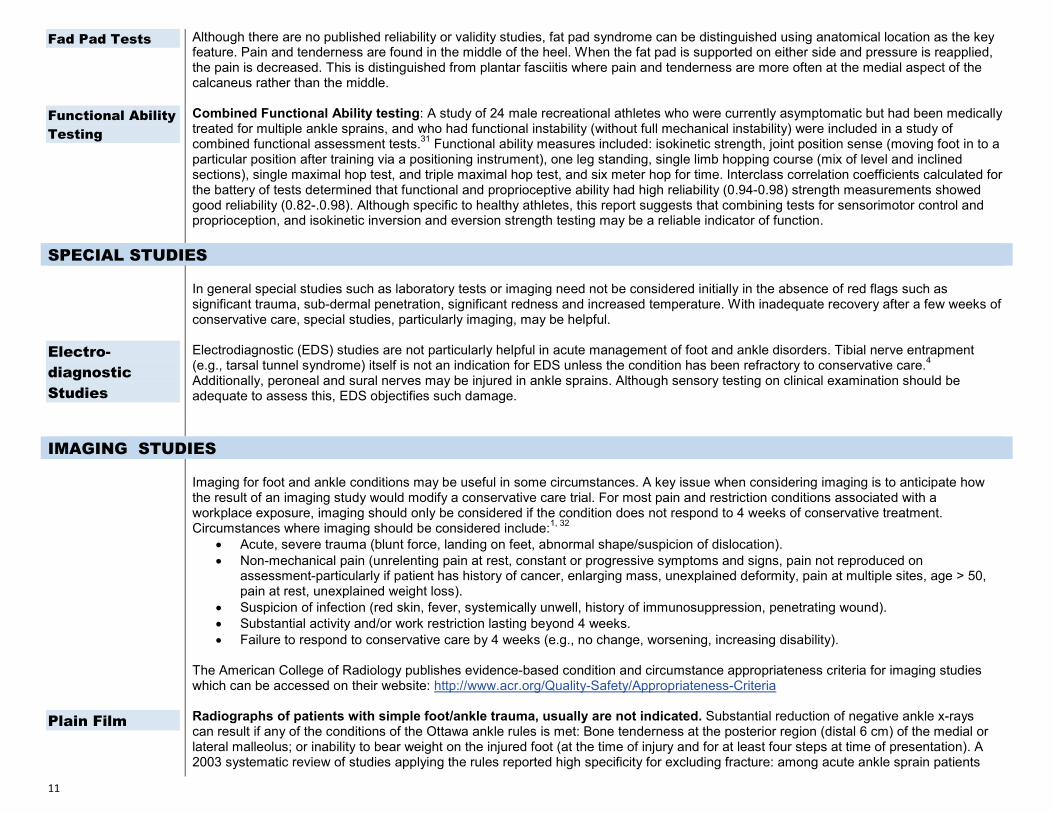
Fad Pad Tests Although there are no published reliability or validity studies, fat pad syndrome can be distinguished using anatomical location as the key feature. Pain and tenderness are found in the middle of the heel. When the fat pad is supported on either side and pressure is reapplied, the pain is decreased. This is distinguished from plantar fasciitis where pain and tenderness are more often at the medial aspect of the calcaneus rather than the middle.
Functional Ability Testing
Combined Functional Ability testing: A study of 24 male recreational athletes who were currently asymptomatic but had been medically treated for multiple ankle sprains, and who had functional instability (without full mechanical instability) were included in a study of combined functional assessment tests.31 Functional ability measures included: isokinetic strength, joint position sense (moving foot in to a particular position after training via a positioning instrument), one leg standing, single limb hopping course (mix of level and inclined sections), single maximal hop test, and triple maximal hop test, and six meter hop for time. Interclass correlation coefficients calculated for the battery of tests determined that functional and proprioceptive ability had high reliability (0.94-0.98) strength measurements showed good reliability (0.82-.0.98). Although specific to healthy athletes, this report suggests that combining tests for sensorimotor control and proprioception, and isokinetic inversion and eversion strength testing may be a reliable indicator of function.
SPECIAL STUDIES
Electro-diagnostic Studies
In general special studies such as laboratory tests or imaging need not be considered initially in the absence of red flags such as significant trauma, sub-dermal penetration, significant redness and increased temperature. With inadequate recovery after a few weeks of conservative care, special studies, particularly imaging, may be helpful.
Electrodiagnostic (EDS) studies are not particularly helpful in acute management of foot and ankle disorders. Tibial nerve entrapment (e.g., tarsal tunnel syndrome) itself is not an indication for EDS unless the condition has been refractory to conservative care.4 Additionally, peroneal and sural nerves may be injured in ankle sprains. Although sensory testing on clinical examination should be adequate to assess this, EDS objectifies such damage.
IMAGING STUDIES
Plain Film
Imaging for foot and ankle conditions may be useful in some circumstances. A key issue when considering imaging is to anticipate how the result of an imaging study would modify a conservative care trial. For most pain and restriction conditions associated with a workplace exposure, imaging should only be considered if the condition does not respond to 4 weeks of conservative treatment. Circumstances where imaging should be considered include:1, 32
• Acute, severe trauma (blunt force, landing on feet, abnormal shape/suspicion of dislocation).• Non-mechanical pain (unrelenting pain at rest, constant or progressive symptoms and signs, pain not reproduced on
assessment-particularly if patient has history of cancer, enlarging mass, unexplained deformity, pain at multiple sites, age > 50,pain at rest, unexplained weight loss).
• Suspicion of infection (red skin, fever, systemically unwell, history of immunosuppression, penetrating wound).• Substantial activity and/or work restriction lasting beyond 4 weeks.• Failure to respond to conservative care by 4 weeks (e.g., no change, worsening, increasing disability).
The American College of Radiology publishes evidence-based condition and circumstance appropriateness criteria for imaging studies which can be accessed on their website: http://www.acr.org/Quality-Safety/Appropriateness-Criteria
Radiographs of patients with simple foot/ankle trauma, usually are not indicated. Substantial reduction of negative ankle x-rays can result if any of the conditions of the Ottawa ankle rules is met: Bone tenderness at the posterior region (distal 6 cm) of the medial or lateral malleolus; or inability to bear weight on the injured foot (at the time of injury and for at least four steps at time of presentation). A 2003 systematic review of studies applying the rules reported high specificity for excluding fracture: among acute ankle sprain patients

12
testing negative using Ottawa, less than 2% actually had a fracture. 33 Production of pain with the application of indirect fibular stress, medial malleolar stress, or compression stress of the mid and hind foot (Bernese Ankle Rules) has also been reported to predict fracture. 34
The decision to obtain x-rays can also be informed by the mechanism and severity of injury and how it relates to the specific location of the problem the patient presents with. The Ottawa and Bernese Rules may be particularly helpful in lower grade sprains and trauma, and even when negative, a foot injury that is not showing any improving within a week or so may warrant reconsideration for imaging. The rules are not useful for individuals with diminished sensation. Patients with a lot of lateral swelling and bruising following an ankle sprain may be at increased risk of lateral talus process fracture, and significant tenderness at the proximal fibular head may raise suspicion of fracture in that region.
Plain film radiography may be useful for assessing: • Achilles tendon insertion problems• Symptoms caused by blunt trauma• Retro-calcaneal bursitis• Suspected fractures (e.g., malleolar, distal fibula and talar dome)• Syndesmosis separation (high ankle sprain; requires comparison with unaffected side)
Plain film radiography is not useful for assessing any of the following unless fracture is suspected: • Plantar conditions or heel pain• Ankle sprains
Usual plain film series include • Ankle pathology – 3 view series (AP, mortise and lateral views)• Foot pathology – 3 view series (AP, oblique, and lateral views)• Calcaneal pathology – 2 view series (axial and lateral)
Advanced Imaging
Advanced imaging includes magnetic resonance imaging (MRI), computed tomography (CT), diagnostic ultrasonography (US) and scintography (bone scans). These should typically be reserved for cases where conservative care has failed to resolve the problem. Generally, plain film and MRI are preferable to CT scans for most non-responsive foot and ankle problems.
MRI is generally considered useful for evaluating • Achilles tendon involvement (suspected rupture, tendinosis, retrocalcaneal bursitis, paratendon tissues). However, for rupture,
basing surgical decisions of clinical findings alone have been reported to be more sensitive than MRI findings. 35 Additionally, reduced wait time for surgery and fewer additional procedures based on false positive MRI findings were reported.
• Refractory tarsal tunnel syndrome• Articular cartilage damage (persistent ankle pain, locking, clicking, swelling increases suspicion)• Some suspected occult or stress fractures particularly of the talus, calcaneus and metatarsals• Differentiating tissues and degree of damage (e.g. metatarsophalangeal sprain versus plantar plate rupture)
CT is helpful in visualizing • Distal leg and ankle fracture• Midfoot fracture or dislocation (Lisfranc injury)
US may be helpful for: • Assessing fluid accumulation in the retrocalcaneal bursa• Distinguishing paratendon disorders from tendinosis
13
• Ultrasound may detect tendon rupture and ligament damage and has the advantages of assessing structures dynamically.Similar points outlined for MRI would apply. Although it is lower cost than MRI, it is highly operator and anatomy dependent, thuscan be highly variable.
Scintography may be helpful for • Identification of occult or stress fractures but involves significant total body radiation exposure
DIAGNOSTIC CATEGORIZATION
General Diagnostic Classification
Diagnostic conclusions for occupational foot and ankle conditions require elements of workplace exposure related to condition onset, presentation, and clinical findings. There are numerous foot and ankle conditions that manifest in pain and discomfort that result from normal weight bearing and other pre-existing conditions which confound adjudication in workers’ compensation claims. Quality population-based epidemiological studies identifying work-relatedness of most foot and ankle conditions other than sprain are lacking in the literature. It is important to carefully document any workplace exposures that are believed to directly cause or contribute to the foot and ankle condition.
General Categorization for Care Triage
• Urgent and serious medical conditions – infection, vascular compromise, neoplasms, metabolic conditions (e.g., gout,diabetes) warrant consideration for specialty referral
• Urgent mechanical conditions – fractures, tendon ruptures, dislocations, severe sprains, and compartment syndrome warrantconsideration for specialty management
• Mechanical conditions – sprains, strains, subluxation, and soft tissue disorders are typical examples warranting considerationfor conservative management
• Neurological conditions – peripheral neuropathy, radicular pain, sclerotomal radiation, and paresthesias warrant closemonitoring under conservative care and may warrant consideration for specialty co-management.
Ankle Sprain Grading (by degree of swelling, pain and bruising)
• Grade 1 (1st Degree) – Overstretching with some microscopic damage to ligament fibers. Pain and swelling may arise after a fewhours. Weight bearing is tolerated; Splinting/casting not indicated; rehab exercise to tolerance indicated.
• Grade 2 (2nd Degree) – Partial tearing of ligament tissue. Pain and swelling typical soon after injury. Loosening of affected jointmay be demonstrable compared to contralateral ankle. Ecchymosis possible. Temporary splint (e.g., air splint) immobilizationusually appropriate; incrementally increasing mobilization, range of motion, stretching and strengthening exercise indicated.
• Grade 3 (3rd Degree) – Complete/large ligament tear. Significant pain, swelling and instability evident following injury.Ecchymosis typical. Immobilization appropriate; incrementally increasing rehabilitation work indicated. Depending on extent andseverity of tear, surgical reconstruction may be needed. May involve dislocation.
Note: Weight bearing is tolerable in most inversion and eversion sprains, but may be more problematic in high ankle sprains.
Categorization by Likelihood of Occupational Exposure
Potentially Occupationally-Related Conditions • Ankle sprains – May result from inversion, eversion, rotational trauma and be of varying grades. Compression trauma (e.g., a
jump) may induce a distal tibia-fibula syndesmosis injury (high ankle sprain).• Achilles tendinosis, tendinopathy, and retrocalcaneal bursitis – All three terms refer to painful conditions in the region of the
Achilles tendon and heel. 5, 36 Tendinopathy is a general term to characterize general pain and/or swelling of a tendon. Tendinosishas replaced the term “tendonitis” due to the lack of histological signs of inflammation. Because these conditions are also
14
frequently related to chronic vascular and degenerative changes, it is important to document a clear linkage of a workplace exposure to the onset of the condition. Standardized terminology and definitions have been proposed for Achilles tendinopathies. 37, 38
o Mid-portion Achilles tendinopathy: a clinical syndrome characterized by a combination of pain (2-7cm proximal to thecalcaneal insertion), swelling and impaired performance. It includes, but is not limited to, the histopathological diagnosis oftendinosis.
o Achilles paratendinopathy: an acute or chronic inflammation and/or degeneration of the thin membrane around theAchilles tendon. There are clear distinctions between acute paratendinopathy and chronic paratendinopathy, both insymptoms as in histopathology.
o Insertional Achilles tendinopathy: located at the insertion of the Achilles tendon onto the calcaneus, bone spurs andcalcifications in the tendon proper at the insertion site may exist.
o Retrocalcaneal bursitis: an inflammation of the bursa in the recess between the anterior inferior side of the Achillestendon and the posterosuperior aspect of the calcaneus (retrocalcaneal recess).
o Superficial calcaneal bursitis: inflammation of the bursa located between a calcaneal prominence or the Achilles tendonand the skin.
• Achilles tendon rupture – Identifiable work trauma with significant load to tendon can induce an Achilles tendon rupture which istypically accompanied by audible pop and sudden loss of plantar flexion. Causation is poorly understood; tendon degenerationfrom repetitive micro trauma and limited vascular supply has been postulated as contributing factors.
• Peroneal and posterior tibial tendinosis, tendinopathies - Like the Achilles tendon, the lateral (peroneus brevis and longus)and medial (posterior tibialis) tendons from lower leg muscles may also become injured and painful as a result of acute traumasuch as a fall or jump and may be associated with calf muscle strain or other ankle ligament sprain. Tendinosis in these structurestends to be less common in typical acute work injuries; they may be more closely associated with extended ankle use such as inmarathon running. Careful history of the mechanics of the injury along with palpation for tenderness along the tendons’ coursehelps differentiate which tendons are involved.
• Plantar pain (arch pain, heel pain, plantar fasciitis, fat pad syndrome, high arches) – There are a number of causes of posteriorand plantar foot pain. Diagnoses such as plantar fasciitis have poorly understood etiologies. Because of this, and due to theabsence of medical literature linking the onset of chronic plantar pain to specific work activities, it can be a challenge to make acase for work-relatedness. However, direct trauma to the hindfoot such as a sudden heel strike on a sharp object can traumatizesoft tissue in the arch, or under the calcaneus (fat pad).
• Forefoot pain (metatarsophalangeal sprain, metatarsalgia, sesamoid injury) Metatarsophalangeal joint sprains are gradedsimilarly to ankle sprains above (Grade 1, 2, 3) and diagnosed by history (flexion or extension trauma) and presentation (localizedpain exacerbated by movement). Plantar plate rupture is a specialized case involving hyper dorsiflexion of ligaments under themetatarsophalangeal joints that may result in instability and longer term hammer toe deformity. Sesamoids are small accessorybones that help anchor the flexor hallucis tendons. Loading that stresses these tendons (e.g., pushing off with the big toe, anextensive increase in loading/amount of walking or running with unsupportive footwear) may irritate the attachments to thesesamoids. Foot mechanics and structural factors unrelated to a work exposure (e.g., cavus, plantar flexed 1st ray) may alsoaggravate sesamoids.
• Trauma-induced degenerative joint disease (hallux rigidus, turf toe, traumatic arthritis) – Traumatic arthritis is common withjamming of the first toe usually into dorsiflexion traumatizing the first metatarsal or metatarsal-talar joints. Characterized bylocalized pain in the affected joint, it is typically provoked with dorsiflexion of the big toe. A joint so traumatized may experienceaccelerated degeneration but this may appear as a longer term sequelae of recovery from injury. When hallux rigidus is believedto be directly caused by a previous work exposure, it would be expected that a previous workers’ compensation claim for an injuryto the affected big toe would have been accepted. Rather than a new claim, the degenerative condition is best addressed as areopening of the original claim. Post-traumatic arthritis can occur following significant ankle, hindfoot and midfoot trauma. Thismay present a number of years after the initial injury. Because the initial workplace injury is generally more significant, the linkbetween the DJD and the initial injury is relatively easy to establish. The relationship should be evident when taking the patient’shistory. Although uncommon compared to ankle sprains, displaced tibial plafond (pilon) fractures, calcaneus fractures, talar bodyor neck fractures, midfoot fracture/dislocations (Lisfranc injuries) frequently result in post-traumatic arthropathy years later .
15
Typically, such fractures require a higher energy injury mechanism such as a fall from a height, car accident, or crush injury. • Tarsal tunnel syndrome – An uncommon condition involving entrapment or stretching of the tibial nerve or one of its branches.
The tarsal tunnel is formed by the distal tibial malleolus, calcaneus, and flexor retinaculum ligament. In addition to the tibial nerve,the posterior tibial artery and vein traverse it as well as posterior tibialis, flexor digitorum longus, and flexor hallucis tendons.Trauma to the area may induce edema, however structural/functional conditions, particularly hyperpronation, may stressstructures in the tunnel which may confound the work-relatedness of the condition.
• Foot and ankle subluxations (e.g. cuboid, navicular, talus, metatarsals) – Typically attributable to an identifiable mechanicalexposure, subluxations of tarsal joints and metatarsals are characterized by discrete pain/discomfort and limited motions in thefoot, usually without swelling.
• Stress fractures (e.g., March fracture) – The best-known etiology of stress fractures of the foot relates to extensive, prolongedwalking or running in unconditioned individuals (such as from marching in new military recruits and long distance runners).Fractures are typically tiny partial cracks in weight bearing bones of the foot that are difficult to visualize radiographically.Diagnosis is usually based on the presentation of localized pain that worsens following an identifiable exposure history. Thesecond metatarsal is frequently involved due to its longer length (leverage) and its central role in absorbing impact to the ball ofthe foot during foot strike. Plantar plate tearing (fibrocartilage under the metatarsal-phalangeal area) may also result from suddenhyperextension, but is usually progressive due to pre-existing foot mechanics such as congenitally short 2nd or 3rd metatarsals.Although bone scans are definitive for stress fracture diagnosis, they are not recommended unless conservative managementfails. It may be important to differentiate stress fractures due to work exposures from those related to pre-existing mechanicalstress from deformities such as a bunion.
• Trauma-induced nerve syndromes (Morton’s neuroma, metatarsalgia, complex regional pain syndrome) – Many nervesyndromes of the foot are insidious in nature and any suspected rationale for work-relatedness should be carefully documented.However scarring and post-traumatic degenerative change may lead to peripheral nerve entrapment or inflammation. Mostcommon are Morton’s neuroma and metatarsalgia. Morton’s neuroma is thought to stem from irritation of nerve fibers on theplantar surface of intermetatarsal ligaments and has been associated with palpable painful nodules in the region. Metatarsalgiahas also been attributed to irritation or trauma to ligaments under the plantar surface, particularly transverse ligaments. Much lesscommon, and of varying degrees of controversy, are complex regional pain syndromes (CRPS). CRPS may be a rare, insidious,chronic pain condition that affects a limb (CRPS I, reflex sympathetic dystrophy), or, more commonly, develops subsequent to anidentifiable trauma (CRPS II, causalgia).39 The condition is associated with severe pain, joint stiffness, hypersensitive skin(allodynia), skin-color changes (ranging from redness to bluish or white), temperature changes and limb swelling. Most cases aremild and self-limiting, but a small number may become severe and chronic. Central and peripheral nervous system anomalies arebelieved to be a primary mediator for the condition; however genetic predisposition and autoimmune conditions may contribute orinfluence the condition. L&I’s Work-related CRPS guideline 40 delineates diagnostic criteria regarding when the condition may beconsidered as occupational, including that another work-related condition for the same foot/ankle has been previously accepted.
Potentially Pre-existing (confounding/complicating/non-occupational) Conditions • Pronation (pes planus) and supination (pes cavus) – Propensity to pronation or supination may be functional or structural and is
associated with variant foot mechanics that predispose one to mechanical stresses that may contribute to foot complaints. Theseconditions are typically hereditary or pre-existing conditions that would not be considered as occupationally related. However theymay influence some treatment and rehabilitation decisions (e.g., braces or supports) for occupational injuries. Additionally, in moresubstantial trauma and fracture, resultant deformity may induce these or other mechanical states where the condition itself couldbecome a sequela of an accepted occupational condition.
• Achilles tendinosis, tendinopathy, retrocalcaneal bursitis and other tendinosis – Unless onset is closely correlated with aspecific occupational injury, these conditions are most likely pre-existing to occupational conditions (e.g., resulting from obesity oranatomic anomalies that impact biomechanics).
• Associated systemic conditions – Diabetes is an increasingly common affliction associated with peripheral neuropathies in thefoot related to vascular deficiencies. Diabetics may have slower healing times associated with wounds and tissue injury. Othersystemic conditions that may manifest in the distal lower extremities include thrombolytic vascular conditions, auto-immunedisorders, and arthritis such as gout and rheumatoid arthritis.

16
• Associated neurological conditions (e.g., radiculopathy, peripheral neuropathy) – These conditions may manifest with foot andankle symptoms and may or may not be concurrent with an occupational foot condition.
• Chronic ankle instability – Ankle instability is associated with histories of pre-existing exposure (such as previous sports injury).Differentiating contributions from a current work-related exposure from those associated with the pre-existing condition can bechallenging. Careful and thorough history taking as well as assessment of the unaffected side and review of available prior clinicalrecords can be helpful.
• Osteochondritis dissecans – An uncommon condition involving damage (desiccation) to articular cartilage associated withmicrofracture of subchondral bone resulting in loss of blood supply leading to necrotic bone formation. It is more common injuveniles and adolescents and may be associated with repetitive trauma (e.g., sports) as vulnerable cartilage and bone matures.Avascular necrosis may be associated with growth plates in children (e.g. Kohler’s disease of the navicular tarsal). Damage to thearticular cartilage of the superior surface of the talus may occur in ankle trauma (e.g., more severe sprains). Cracks in thecartilage itself, or in the subchondral bone may contribute to delayed recovery or show up as an intra-articular fragments overtime.
WORKERS’ COMPENSATION ASSESSMENT ISSUES
Causation & Work Relatedness
Exceptionally clear clinical justification for specific work exposure(s) is essential for fair and timely decisions in nearly all workers compensation claims. Typically, an identifiable incident or incidents on the job shortly before the conditions onset would be expected. The concept of cumulative industrial trauma (such as prolonged standing) as an etiology of foot and ankle conditions does not generally have support in the medical literature. 4, 41 To be accepted by the department as a cumulative trauma leading to an occupational disease, specific additional legal requirements must be met (RCW 51.08.100). Generally, pain and other manifestations of both industrial injuries and occupational diseases become evident within 3 months of an inciting event. In a situation where a foot and ankle condition reported for the first time more than 3 months after a patient is first seen by a provider, it is important that the clinical rationale for its relationship to work be very well documented.
To establish a diagnosis of an occupational disease, all of the following are required:
1. Exposure: Workplace activities that contribute to or cause the specific foot and/or ankle condition(s), and2. Outcome: A diagnosis of a foot and ankle condition that meets reasonable diagnostic criteria such as those delineated in this
resource, and3. Relationship: For a foot and ankle condition to be allowed as an occupational disease, the provider must document that, based
on generally accepted scientific evidence, the work exposures created a risk of contracting or worsening the condition relative tothe risks in everyday life, on a more-probable-than-not basis (Dennis v. Dept. of Labor and Industries, 1987). In epidemiologicalstudies, this will usually translate to an odds ratio (OR) ≥ 2.
More information on filing a claim for an occupational disease, including billing information, can be found in the Attending Provider’s Handbook: http://www.Lni.wa.gov/FormPub/Detail.asp?DocID=1669
• Acute workplace trauma has been linked to tendinosis, tenosynovitis, fractures, and ligament strains. Stress fractures have beenreported with substantial increases in walking and weight-bearing activities (for example, a worker who normally has a sedentaryjob that is required to spend a day moving heavy equipment over long distances, or engage in tasks that required prolongedrunning for which they were not conditioned).4
• No well-designed studies have documented a relationship between work activities, other than a specific trauma, anddegenerative joint disease.4
• Most of the literature regarding causation of foot and ankle problems relates to sports activities, not occupational exposure.Further, non-occupational factors are strongly associated with foot and ankle problems including a person’s weight, recreationalactivities, gender, age, foot mechanics and shape, footwear, and concurrent disease status (e.g., diabetes) confoundingdelineation of occupational contributors. 3, 42
17
• An observational study of cold weather training among military recruits reported an increased risk of Achilles tendinopathy. 43
• The etiology of seven foot and ankle disorders commonly involved in compensation litigation (hallux valgus, interdigital neuroma,tarsal tunnel syndrome, lesser toe deformity, heel pain, adult acquired flatfoot, and foot and ankle osteoarthritis) was reviewed inone study using a logistic framework based on Koch’s postulates to analyze the potential for cumulative industrial trauma tocause foot pathology. 41 In none of the disorders that were analyzed, could cumulative industrial trauma reasonably beconsidered a distinctive factor in a large proportion of cases, or be a condition that consistently occurs in particular occupations(as is the case in a condition such as hypothenar hammer syndrome). These conditions often occur from intrinsic footmechanics, regardless of exposure factors. The evolutionary adaptation of the foot to prolonged ambulation and the absence ofindustrial demands that significantly alter such mechanics on feet likely account for reduced vulnerability of the foot to industrialrepetitive motion disorders compared to the upper extremity.
• A systematic review of prevalence studies reported that 24% of older and middle age adults in the general population report footpain problems and another 15% report ankle pain. Prevalence increased with advancing age and among women. 44
Assessment of Re-exposure on Return to Work
No studies were identified with current search strategies.
Physical Capacity & Work Restrictions
No studies were identified with current search strategies.
GENERAL INTERVENTION SUMMARIES BY CONDITION
Conditions General Approaches for Common Work Related Syndromes
Ankle Sprains (inversion, eversion, rotational, syndesmosis)
First week post-injury typically includes protecting the ankle and controlling any swelling, but current thought and research supports rapid incorporation of pain-free activity and motion of the affected ankle.
• Grade 1: Modified pain-free activity, Ice, Compression, Elevation (MICE)o Avoid walking or weight bearing on affected ankle initiallyo Apply ice immediately to reduce swelling. Apply for 20-30 minutes, 3-4 times daily.o Compression dressings, bandages or ace-wraps may be considered to help support the injured ankle.o Regular elevation of the affected foot during the first 48 hours can help reduce swelling.
• Grade 2: MICE plus an immobilization brace/splint• Grade 3: MICE and significant immobilization (e.g., short leg cast or brace) may be indicated for 2-3 weeks. Short periods of
non-weight bearing (e.g., crutches) may be appropriate particularly if significant pain occurs with weight bearing. Permanentinstability may be a residual from a Grade 3 sprain. Surgery is rarely indicated.
Second through third weeks should focus on restoring range of motion, strength and flexibility within patient tolerance Thereafter gradually returning to regular activities that do not involve twisting the ankle can begin along with more specific rehabilitation and strengthening exercise. Activities that require sharp, sudden cuts and turns (e.g., sports like tennis or basketball) may require several weeks to months for full recovery. The degree of injury guides specific treatment approach. Remain attentive for bone tenderness at the posterior aspect of medial or lateral malleolus, or inability to bear weight on the injured foot (Ottawa ankle rules) which can be indicative of fracture as can pain with indirect fibular stress, medial malleolar stress, or compression stress of the mid and hind foot (Bernese ankle rules). If any of these are present, imaging may be warranted.

18
Rehabilitation is used to help to decrease pain and swelling and to prevent chronic ankle problems. Ultrasound and electrical stimulation may also be used as needed to help with pain and swelling. At first, rehabilitation exercises may involve active range of motion or controlled movements of the ankle joint without resistance. Water exercises may be used if land-based strengthening exercises, such as toe-raising, are too painful. Lower extremity exercises and endurance activities are added as tolerated. Proprioception training is very important, as poor proprioception is a major cause of repeat sprain and an unstable ankle joint. Once pain-free, other exercises may be added, such as agility drills. The goal is to increase strength and range of motion as balance improves over time.
Achilles tendinopathies, tendinosis, retrocalcaneal bursitis
Achilles tendinosis and retrocalcaneal bursitis are characterized by pain during rest or activity at, or above, the heel posteriorly. Either can result from direct trauma or stress to the ankle from sudden increased activity such as jumping or running with inadequate conditioning. In general, acute, inflammatory tendonitis may be considered to exist in someone without a history of pain in the region when pain in the insertion area follows a work exposure. Tendinosis is the term characterizing subacute, chronic, or episodic insertional pain. Effective management of the inflammatory phase is believed to help prevent progression to tendinosis. 45 Management can be organized as follows:
• Initially: Conservative treatment would be aimed at alleviating symptoms, loosening muscle belly and associated musclegroups, graded return to activity involving loading of the tendon, possibly including supportive bracing during the healing processand while returning to tendon loading activities. Gradual progressive stretching of the calf muscle groups and tendon reducetension to the tendon. Eccentric calf muscle training appears to have the most consistent literature support. 46 Typicalconventional management often includes frequent icing, heel lift to reduce stress on tendon, a trial of oral NSAIDs or analgesics(acetaminophen) in the first few weeks following onset. Steroids and opioids should be avoided.
• Post inflammation: Rehabilitation interventions should include eccentric loading exercise and stretching of the gastrocnemiusand soleus muscles 1-2 times per day. Alternating heat and ice application may increase microcirculation in the area. The role oflifts or orthotics is unclear. Extracorporeal shockwave therapy has been reported to be of benefit in refractory chronic AT, but notin acute/sub-acute cases. 47, 48 Resolution typically occurs within 4-12 weeks, depending on the nature, severity and degree ofinjury. In refractory cases recovery can take up to six months.
• Onset over existing tendinosis: Acute onset of consistent pain at work in someone with a history of short or long termintermittent pain in the Achilles tendon region may occur with or without visible inflammation and may be managed and expectedto respond similarly to a new episode.
Retrocalcaneal bursitis may be concomitant with Achilles tendinosis and exhibits a similar presentation of posterior heel pain exacerbated by standing on tiptoes, typically accompanied by redness and tenderness over the back of the heel. Treatment is essentially the same as for Achilles tendinosis, and the bursitis should resolve within a few weeks. Because glucocorticosteroid weakens connective tissue, and the significant amount of stress on the ankle can put the Achilles tendon at greater risk of rupture, injection in the region of the Achilles tendon should be avoided. 49 If employed as a last resort, it should only be attempted once and any stretching on the tendon (e.g., calf stretching exercise) should be avoided for about two weeks. For refractory cases, low level evidence suggests endoscopic surgical techniques meet with higher patient satisfaction than open surgical approaches, however well-done effectiveness studies have not been done. 50
Achilles tendon rupture
There is moderate quality evidence for both surgical repair and non-operative management with casting and functional bracing for Achilles tendon rupture. Generally, an early surgical consultation should be obtained for all cases of complete Achilles tendon rupture and a treatment plan should be initiated as soon after the injury as possible, particularly with non-operative management.
• Surgical repair with immobilization (casting or functional splinting) followed by rehabilitation is the typical approach for Achillestendon ruptures and appears to be associated with a slightly lower re-rupture rate than functional splinting alone. 51 Howeverfunctional splinting may be used for individuals with contra-indications for surgery and who may have minimal physical demands.

19
52, 53
• Overall, there appear to be no differences in function between surgical and non-surgical approaches, with non-surgicalapproaches avoiding potential surgical complications. 54
• In both cases, tendon healing can be slow and scar tissue is of poorer quality and strength than undamaged tendon. Bothapproaches are associated with lower high-demand performance long term.
• The use of surgical approaches appears to be declining as a result of better quality comparative trials.55
Posterior tibial, peroneal tendinosis or tendinopathy
Peroneal and posterior tibial tendinosis usually heal well (albeit slowly) with conservative management including MICE, myofascial work, and gentle stretching in associated muscles. Limiting walking until pain subsides (usually a few weeks) is useful and ankle bracing may be helpful. Especially severe or painful cases may benefit from a more rigid support such as a walking boot. Gradually increasing muscle training should be deferred until pain is reduced and should be done only to tolerance. Cross training approaches for mobility and exercise allow affected tendons rest time which may assist recovery. Severe and refractory cases may warrant imaging (ultrasound, or MRI) to assess tearing and need for surgical consultation. As with other tendinosis, corticosteroids should be avoided.
Plantar pain (arch pain, heel pain, “plantar fasciitis/fasciosis”, fat pad syndrome, high arches)
Pain in the heel region is common and may follow many different exposures, or arise idiopathically. Arthritic, neurologic, traumatic, or systemic conditions may account for symptoms, however biomechanical foot considerations are thought to account for the majority of complaints. 56 Significant pain when walking after waking up or being sedentary is characteristic of plantar and heel pain. It is typically self-limiting. If presenting concurrently with unexplained lower back pain, consideration of spondyloarthropathy may be warranted, particularly in younger individuals. Persistent or recurrent plantar heel pain has many names including plantar fasciitis, runner’s heel, calcaneodynia, calcaneal periostosis, and heel spur syndrome. Symptomatically it typically refers to pain on the bottom of the arch or hind foot region. Tissue thickening of the plantar fascia is a principle criterion. The diagnosis and etiology of plantar fascitis remain controversial, particularly as related to occupational causation. Attribution has been made to excessive running, obesity, pronation, and prolonged standing, but data are insufficient to definitively determine risk factors. 57, 58
• Common conservative interventions include: Ativity modification, rest, ice, deep tissue work in the plantar fascia, stretchingexercise, shock absorbing orthotics or shoes, night splints, and oral NSAIDs.
• Corticosteroid injections are frequently employed with plantar pain. However, it is important to distinguish fat pad syndrome fromother plantar pain syndromes due to the concerns that injecting corticosteroid can diminish any remaining fat pad. 49 Heel cushioning is the preferred method of treatment for fat pad syndrome.
Tarsal tunnel syndrome
Tarsal tunnel syndrome is a very rare condition as indicated previously. Severe pronation trauma or significant direct trauma would be expected modes of onset.
• R/MICE is the typical initial approach when trauma acutely triggers symptoms of tibial nerve entrapment.• Manipulation, mobilization, nerve glide exercise, and corrective footwear or orthotics are commonly employed interventions. The
condition typically resolves without problems.1, 59
• For cases that do not resolve within a few weeks, nerve conduction studies and/or MRI can be considered to determine if theposterior tibial nerve is compromised.
• Oral NSAIDs or glucocorticosteroid injections are commonly used for entrapment neuropathies. However, use of injectedsteroids has long been discouraged due to potential for nerve and connective tissue damage. 60 No studies specific to steroidinjection for tibial nerve entrapment were identified with our search strategy.
• Surgical treatment for tarsal tunnel appears variable, with open tibial nerve decompression approaches increasingly beingreplaced by endoscopic approaches. Endoscopic tarsal tunnel decompression has been reported to be a safe procedure with alow rate of recurrence or failure and allows for near-immediate ambulation. 61
20
Foot and ankle joint dysfunction/subluxation (e.g. cuboid, navicular, talus, metatarsals) Frequently described in manual medicine, osteopathic, and chiropractic literature, primarily as case reports and series, foot and ankle joint dysfunction/subluxations might be best characterized as findings associated with various other conditions and precipitating injuries such as ankle and hind foot sprains. 62-65 However, although a standardized case definition is not well developed, the frequency of diagnostic attribution, its mechanical nature, and association with other common work-related foot and ankle conditions warrants its mention.
• Extremity manipulation is the most described treatment and has been shown to be effective for such inciting events and conditions as ankle inversion sprain (Grade B evidence short term and Grade C long term), plantar fasciitis (Grade B evidence short term and Grade C long term), metatarsalgia, hallux limitus, and foot/ankle proprioception/balance problems. 1, 66
Forefoot pain (e.g., metatarsalgia, metatarsophalangeal sprain, plantar plate rupture, sesamoid injury) Pain in the forefoot region can be associated with a number of structures and differentiation can often be challenging. They generally can be grouped as acute (e.g., sprains, contusions) and chronic (e.g., overload, callusing), the latter situation requiring careful documentation of the clinical rationale for being the result of a workplace exposure. Additionally, regardless of structure involved, there is typically great similarity in conservative management. Metatarsophalangeal (MTP) joint sprains are graded similarly to ankle sprains above (Grade 1, 2, 3) and diagnosed by history of joint trauma typically in flexion or extension. Presentation involves localized pain, usually exacerbated with passive or active movement and swelling may be evident. Plantar plate (ligaments on the plantar aspect of the metatarsal phalangeal joints) disruption may result from substantial dorsiflexion trauma (commonly from tripping) and may result in instability and longer term hammer toe deformity. It is most common in the second metatarsophalangeal joint. 67
• Initial management typically includes MICE, NSAIDs, and mobilization. • Taping may be used to limit motion after acute inflammation is controlled, with splinting or casting reserved for higher severity
sprains. • Increasing passive and active mobilization and joint manipulation may be helpful in speeding full recovery The big toe is the
most commonly affected (turf toe). • Lower grade sprains usually resolve within a week and require little to no activity modification. Grade 2 injuries may need to
avoid sustained weight bearing for up to two weeks (crutches or walker), and several weeks of restriction may be appropriate for severe sprains. A general guide of 50-60° pain-free dorsiflexion is often used to recommend unrestricted loading/activity. Non-weight bearing with crutches is typically needed with severe sprains or plantar plate ruptures.
• Conservative measures for suspected plantar plate rupture include 4-6 weeks of cross-over taping to plantarflex the affected digit (to reduce stress on the ruptured plate) along with a cushioning pad and compressive strapping to improve alignment. Particular orthoses/orthotics are sometimes used to reduce stress on the plate to dorsiflex the metatarsals while allowing plantar flexion of the digits. The aim of conservative care is to realign the affected digit and plantar plate preventing or slowing progressive deformity.
• Surgical repair may be considered based on the severity of acute injury (with extremely loose vertical stress test) or signs of hammer deformity. MRI arthrogram may be needed to assess degree of rupture as the ligaments approximate when relaxed and regular MRIs are negative.
Trauma-induced Sesamoiditis: Sesamoids are accessory bones that variably occur as anchoring attachments for the flexor, adductor, and abductor muscles of the big toe. They may become inflamed or irritated with direct trauma to the ball of the foot (e.g., landing from a big jump, extensive loading when pushing off with the big toe) and may be concurrent with stress fractures and sprains of the metatarsals.68 Presentation usually involves pain and tenderness on the plantar surface of the first metatarsophalangeal joint which is exacerbated on dorsiflexion of the big toe.
• Acute management may include MICE and NSAIDs. Sustained management is usually focused on reducing direct irritation of the

21
region with strategies such as taping, in-shoe padding, sturdier footwear, and/or modifying hard work surfaces. Trauma-induced degenerative joint disease The first toe is the most common arthritic joint in the foot, and hallux rigidus is the second most common foot condition after bunion. It is usually associated with hallux valgus, and has its highest prevalence in the 30-70 year age range. It is often of unknown etiology, however, may be associated with prior traumatic workplace injury to the big toe. In such instances, documented attribution of a specific work exposure is important. For example, a previous acute injury (such as at least a Grade 2 metatarsophalangeal sprain) might be expected to have been accepted as a workers’ compensation claim, and would be expected to present unilaterally. The presentation involves dorsal pain upon loading, especially pushing off for walking. Presentation should also include, stiffness, palpable exostosis, pain with axial compression and rotation (axial grind test), occasional synovitis, and decreased motion on motion palpation (particularly dorsiflexion). It can be expected to be accompanied by positive X-ray findings (spurring and joint narrowing). At least four of the previous findings should be present to make the diagnosis. Higher energy work injuries (e.g., fall from a height, car accident, crush injuries) that resulted in displaced tibial fracture, calcaneus fractures, talar body or neck fractures, midfoot fracture/dislocations (Lisfranc injuries) may result in delayed-onset, post-traumatic arthropathy and should be readily liked to original injury.
• Non-surgical treatment typically includes cryotherapy, contrast baths, mobilization, manipulation, NSAIDs, and/or footwear modification (or orthoses with a Morton’s extension) that minimizes 1st MTP joint motion. Corticosteroid injections are also used. Surgical removal of bone spurs (cheilectomy) may be considered in refractory cases and joint fusion (arthrodesis) is sometime recommended for severe cases. Joint replacement (arthrodesis) is also used, but usually only in older individuals without high functional demands on their feet.
Stress fractures (“march” fracture) Metatarsal stress fractures tend to occur after an identifiable period of increased higher impact activity (e.g., prolonged walking or running). They typically present with continuous achy, forefoot pain that exacerbates on weight bearing and is relieved by rest. Tenderness, bruising and/or swelling may also be present.
• Stress fractures of the middle metatarsals are usually self-limiting within 6-8 weeks using pain control including ice and elevation early on along with rigid footwear. However, if refractory, or with an occupation placing high demand on the feet, the use of crutches or a walking cast may be necessary.
• Outer metatarsals, particularly the 5th are more likely to be acute fractures from a more direct trauma (such as kicking, the foot getting stepped on or twisting while landing from a jump). Avulsion of the styloid process at the proximal base of the 5th metatarsal is the more likely with a twisting or inversion sprain while a transverse fracture near its base (Jones fracture) is usually associated with repeated stress exposures. 5th metatarsal fractures can be managed similarly to other stress fractures, however some transverse fractures become non-unions due to blood supply limitations and stresses from where muscles attach. 69, 70
• There can be many different locations and types of stress fractures with subtle nuances in management to ensure optimal healing. Foot and ankle specialists such as podiatrists, orthopedists, sports practitioners may be important resources in evaluation and management of foot and ankle stress fractures.
Trauma-induced nerve syndromes (Morton’s neuroma, complex regional pain syndromes) Work-relatedness can be difficult to establish for nerve syndromes due to multivariate in etiologies and various anatomic structures that may become associated with persistent or recurrent nerve pain.
• Morton’s Neuroma: Presentation is typically pain on the plantar surface of the foot in the region of a middle inter-tarsal space, exacerbated by direct pressure (e.g., palpatory, shoes that are too small). Histologically it has been linked to scarring or other amorphous deposits entrapping nerve fibers coursing along intertarsal ligaments. 71 In some instances a small tender nodule may be palpated. The condition may be associated with an altered gait due to increased lateral stress on the foot to avoid painful pressure on the affected area. A previous work injury (e.g., stress fracture to an adjacent metatarsal, landing or crush injury) to

22
the affected area would be expected to have been previously accepted for a claim. • Complex regional pain syndrome (CRPS): Typically affecting a particular limb (arms, legs, hands, or feet), CRPS is a chronic
pain condition that would need to be causally associated with an occupational injury or trauma to that limb. CRPSpathophysiology is poorly understood, however one form, called CRPS II, has been attributed to damage of peripheral nervesand or the central nervous system. It is characterized by persistent mild to severe pain and is associated with changes in skincolor, temperature, sweating, and/or edema in the affected region thus implicating the sympathetic as well as the somato-sensory nervous system. Symptoms and findings may be highly variable. This, along with poorly understood mechanisms,contributes to diagnostic uncertainty and resultant controversy about the condition. Vitamin C (500mg per day) administered afterextremity trauma or surgery has been shown to prevent development of CRPS 72 and may be useful if begun early in onset ofsymptoms. Treatment approaches with evidence of benefit include CRPS-focused physical/occupational therapy includingdesensitization & neuromuscular re-education to improve neuromuscular function, progressive active exercise to improve bloodsupply and flexibility, functional goal development including weight bearing and gait training, and training in self-managementincluding home exercise. Medications for symptom management include NSAIDs for pain control and others linked to individualpresentation. Cognitive behavioral therapy may be considered for individuals with fear avoidance or psychological barriers tousing the affected limb. Some cases have been documented to respond to lumbar sympathetic blocks. In refractory cases amultidisciplinary pain management program may be helpful. Early referral should be made to specialists in management ofCRPS. L&I has a medical treatment guideline addressing diagnostic criteria and other issues for CRPS as an acceptedoccupational condition. 40 http://www.Lni.wa.gov/ClaimsIns/Files/OMD/MedTreat/ComplexRegionalPain2011.pdf
EVIDENCE SUMMARIES BY INTERVENTION
Early Mobilization
Ankle sprains
Early mobilization may be considered a functional intervention that includes flexible or semi-rigid ankle support and combinations of incrementally increasing active movement and weight bearing. There are a number of studies and reviews which support this approach for all grades of sprains and following surgical stabilization. 73-79 However, functional braces have been associated with higher rates of post-operative complications with wound healing in surgical treatment of fractures compared to rigid casts. 80 Generally, more rapid and aggressive return to normal activity can be implemented with lesser grade sprains. There are a variety of braces, supports, boots, tapes/wraps utilizing pneumatics, gels, fabrics and plastics on the market. Head-to-head comparative studies of styles and brands of different products were not identified using the current search strategy. Most studies compared rigid casting to semi-rigid support. Utility of many studies was confounded by small samples, access to multiple co-interventions, and questionable comparison groups among other limitations.
• In a prospective cohort study of 60 Grade 3 lateral ligament sprain patients, surgical treatment (primary repair plus early controlledmobilization) was compared to functional intervention (early controlled mobilization alone). 81 Thirty patients in each group werematched by age, height, weight, gender, and sports activities. One patient in each group did not have a stable ankle at 9 months.Ankle range of motion was restricted in the surgical treatment group at 6 weeks and did not normalize while the functional groupshowed no restrictions. Self-reported subjective pain and function was reported to be good in 87% of the functionally managedsubject and 60% of the surgically treated subjects.
Manipulation and Mobilization
Ankle sprains
Overall, passive and active mobilization has been shown to immediately improve ankle dorsiflexion in acute and subacute ankle sprains. Manipulation in addition to RICE may improve other functional measures including stride speed within a shorter time frame and may be associated with a slightly sooner return-to-work. Manipulation combined with exercise has small advantages over either alone. The addition of soft tissue work may enhance the effectiveness of combined mobilization/manipulation and exercise. 66, 82, 83 Generally, mobilization and manipulation are often provided in combination with exercise and soft tissue work. Reasonable evidence suggests utility with plantar pain. Some low quality evidence also suggests helpfulness of these approaches for milder hallux rigidus, and tarsal tunnel

23
syndrome, however comparative and experimental studies are lacking.
• A systematic review identified 8 studies meeting quality criteria assessing manual joint mobilization and manipulation for ankle sprain- 3 for acute, 5 for subacute or chronic. 84 Outcome measures included pain level, range of motion, swelling, functional scores,stabilometry and gait parameters. Most studies only assessed outcome measures immediately after treatment. No detrimental effectsfrom the joint techniques were revealed in any of the studies reviewed. For acute ankle sprains, manual mobilization/manipulationwas reported to diminish pain and increase dorsiflexion range of motion. For subacute/chronic sprains, mobilization and manipulationtechniques improved ankle range-of-motion, pain levels, and function scores were reported.
• In a small crossover design study of 16 recurrent ankle sprain subjects with no long term follow-up, passive mobilization with weightbearing vs. no weight bearing vs. no treatment were compared. Improved posterior talar glide and dorsiflexion was reported posttreatment with both mobilization groups compared to no treatment. 85
• 16 subacute Grade 2 ankle sprain subjects were evaluated in a crossover design study without long term follow-up receiving passivemobilization with movement vs. placebo mobilization vs. no treatment. 86 Improved dorsiflexion immediately post-treatment wasreported.
• RICE + manipulation vs RICE alone was compared in a study of 41 acute inversion sprains referred from an emergency room.Subjects were seen within 72 hours of injury and although no grading was reported, all subjects required assistance for ambulation.RICE + manipulation achieved increased dorsiflexion after the first treatment which was sustained on follow up. Full, pain free ROMand improved stride speed after 4 sessions was reported as was a small, but statistically insignificant reduction in lost work days. 83
• A ninety subject randomized trial assessed six sessions of joint mobilization combined with active movement over a 3-week periodwas compared to sham mobilization sessions of the same duration. 82 The treatment group reported improved dorsiflexion, posturalcontrol and self-reported instability at 6-month follow-up. Subjects were had chronic ankle instability with a history of multiple anklesprains.
• A small randomized trial compared 33 patients with recurrent ankle sprain and instability assigned to chiropractic manipulation andrehabilitation (6 sessions over 4 weeks) or rehabilitation alone. 87 At 5 weeks, the group receiving manipulation reported lower painscores and had increased range of motion; however no differences in the Foot and Ankle Disability Index (FADI) scores wereidentified.
• Fifty post-acute inversion ankle sprain patients were randomized to 2 groups: 1) an experimental group that received thrust and nothrust manipulation, exercise intervention, and soft tissue myofascial work; or 2) a comparison group that received just thrust and nothrust manipulation and exercise intervention. Primary outcomes of ankle pain at rest and functional ability with secondary outcomesincluding ankle mobility and pressure pain threshold over the affected ankle were reported. All outcomes were blindly assessed atbaseline, immediately after the treatment period, and at a 1-month follow-up. Group by time analyses indicated a significant butminimally clinically important difference favoring the addition of soft tissue work in all outcomes. 88
• A convenience sample of acute adult ankle sprain patients (n=55) presenting to an emergency department were randomized to ausual care (RICE and analgesics) or usual care plus a single osteopathic manipulation session (foot and ankle jointmobilization/manipulation and soft tissue work). 89 Patients in the manipulation group had statistically significant improvement inedema and pain compared to the usual care group immediately following the visit. At one week follow up both groups’ pain andswelling improved comparably, with the manipulation group showing greater range of motion on dorsiflexion.
• A single rear foot distraction manipulation improved dynamic standing balance immediately after the procedure in 20 healthy subjectsunder 30 years old. 90 Although not a clinical study, this does illustrate a proprioceptive component to passive distractionmanipulation which is commonly utilized by manipulation practitioners in the management of ankle sprains.
• A 43-subject physiologic trial of chronic ankle instability patients randomized them to 1) proximal tibiofibular manipulation, 2) distaltibiofibular manipulation, or 3) no manipulation control groups. Changes in the ratio of the maximum H-reflex and maximum M-wavemeasurements (H/M ratio) of the fibularis longus and soleus were compared between groups over time (pre, post 0, 10, 20, 30 min).The distal tibiofibular joint manipulation group demonstrated a significant increase (P<.05) in soleus H/M ratio at all post-interventiontime periods except 20 min post-intervention (P=.48). The proximal tibiofibular joint manipulation and control groups did notdemonstrate a change in soleus H/M ratios. All groups demonstrated a decrease (P<.05) from baseline values in fibularis longus (10-30 min post-intervention) and soleus (30 min post-intervention) H/M ratios. This study indicates that distal tibiofibular jointmanipulation acutely increases soleus muscle activation. 91

24
• Neither 3 weeks of proximal nor distal tibiofibular manipulation improved ankle dorsiflexion compared to controls in a 43-subjectthree-group chronic ankle instability randomized trial. All three groups improved in ankle dorsiflexion and functional instrumentscores. The authors point out that repeated range of motion assessment on the controls may have the effect of increasing mobility. Itshould be noted that the manipulation did not include talofibular or other tarsal manipulation. 92
• A systematic review of randomized trials on exercise and manual mobilization identified four studies that evaluated mobilization inacute ankle sprains (from1966-2005). 93 Although mobilization was shown to have an initial effect on improving dorsiflexion, theauthors expressed caution regarding the clinical relevance of the effect.
• A potential clinical prediction rule for circumstances where joint manipulation may not be helpful in treating acute ankle sprains wasdeveloped by following 85 consecutive inversion ankle sprain patients. 94 While 75% of the patients had dramatic and rapidsuccessful outcome, the remaining 25% did not. Four variables were associated with poor responders: symptoms worse whenstanding; symptoms worse in the evening; navicular drop greater than or equal to 5.0 mm; distal tibiofibular joint hypomobility. Usingthe presence of 3 out of 4 of the variables as a clinical prediction rule for unsuccessful outcome yielded a post-test probability ofsuccess of 95%.
• A single-blind randomized trial of 30 Grade 1 or 2 ankle sprain patients allocated subjects to up to 8 sessions of talocrural jointmanipulation treatments or sham (five minutes of detuned ultrasound) within a 4-week period. 95 The intervention group improved inpain (McGill) scores, goniometric dorsiflexion, and a combined 11-test functional battery (Kaikkonen) score.
• A study of 52 adults randomized lateral ankle sprain subjects to a single talocrural joint manipulation group or a no treatment controlto assess post-session dorsiflexion range of motion. 96 No statistically significant differences were found, however joints thatexperienced a cavitation during the thrust trended toward greater increases in ROM.
• A well-done multicenter randomized trial of 74 patients randomized them to a manual therapy and exercise group or a home exercisegroup. 97 Ankle function (FAAM and LEFS scales) and a numeric pain scale outcomes were collected at baseline, 4 weeks and 6months. Overall group-by-time interaction (mixed-model analysis of variance) was statistically significant for the FAAM activities ofdaily living subscale (P<.001), FAAM sports subscale (P<.001), Lower Extremity Functional Scale (P<.001), and pain (P ≤.001).Improvements in all functional outcome measures and pain were significantly greater at both the 4-week and 6-month follow-upperiods in favor of the manual therapy and exercise group.
• A study of 33 subjects with chronic instability associated with recurrent ankle sprains were randomized to 4 weeks of eitherrehabilitation (up to 29 sessions over 5 weeks of daily balance training and progressive strength training) plus ankle manipulation (upto 6 treatments of high velocity mortise, subtalar, and or tarsal joint), or rehabilitation only (up to 35 sessions in 5 weeks). 87 Bothgroups showed similar improvements in ankle function scores (FADI) with the manipulation-added group reporting significant shortterm reduction in pain level (visual analog) compared to rehabilitation alone.
• A comparative trial randomized 52 chronic ankle instability patients to a mobilization with movement group (therapist applied glidewith a belt and active dorsiflexion), high velocity talocrural manipulation group (axial), or placebo (therapist position belt without glideor active dorsiflexion). Weight bearing dorsiflexion was measured at baseline, immediately after, 10 minutes, 24 hours, and 48 hoursafter treatment. 98 A single application of either manual technique improved dorsiflexion with effects that persisted for at least twodays. Both techniques have similar effectiveness for improving ankle dorsiflexion compared to placebo.
Achilles tendinosis, tendinopathy, and retrocalcaneal bursitis
No high quality studies specifically addressing manipulation with Achilles tendinosis or retrocalcaneal bursitis were found using the employed search strategy. The rationale for joint mobilization or manipulation regarding Achilles tendinosis or bursitis would be to address mechanical restrictions in the ankle or foot that may be associated with facilitating muscle spasm/tension/dysfunction that could irritate these structures (e.g., guarding and limping contributing to soleus or gastrocnemius tightness). Some evidence is available for other regions such as epicondylosis of the elbow and rotator cuff tendinosis. 99, 100 Clinically, joint manipulation and mobilization would be used in combination with soft tissue work and exercise for which some evidence is available as noted in other sections.
Plantar pain
A large variety of conditions may contribute to or cause plantar and heel pain. A rationale similar to manipulation for Achilles tendosis

25
applies here with facilitating of unrestricted foot and ankle mechanical function reducing sources of irritation to soft tissue and tendon insertions. Likewise, joint manipulation is likely to be combined with mobilization, soft tissue work and exercise. • A comparative trial of 60 plantar pain patients treated with manual methods (six sessions of manipulation, mobilization and exercise)
or six sessions of extraphysiologic modalities were followed and analyzed to assess if age and body mass index (BMI) negatively impact care response. Better function and pain scores were reported with the manual methods group. 101 Patients with less than 7 months of symptoms may have a better response to manual interventions for plantar heel pain. Age and BMI did not appear to be associated with worse outcomes with manual approaches.
• Combined manual approaches (manipulation, mobilization) and exercise have been reported to result in lower disability scores (Lower Extremity Function Scale, Foot and Ankle Ability Measure) at 4 weeks and 6 months than electrophysiological modalities and exercise in a randomized trial of 60 chronic plantar pain patients. 102
• A randomized trial of 20 patients with chronic plantar fasciitis compared 8 sessions of manipulation and 3 daily sessions of Achilles stretching over a one-month period to use of orthotics. 103 Both groups reported similar improvement in heel pain at rest, recreation and work. The small size, large variation in condition duration among subjects, and low statistical power limit definitive conclusions.
Tarsal tunnel syndrome No useful studies were identified specifically addressing joint mobilization or manipulation for tarsal tunnel syndrome. The American College of Occupational and Environmental Medicine’s (ACOEM) Occupational Medicine Guidelines 4 recommend against using manipulation for treatment of tarsal tunnel, surprisingly based on two trials of manual therapy for carpal tunnel syndrome that both reported benefit to the manual interventions. One treatment comparison trial for electrodiagnostically confirmed carpal tunnel syndrome randomized 96 subjects into medical management (NSAIDS and nocturnal wrist supports) or chiropractic management (soft tissue work, extremity and spine manipulation). Both groups reported similar levels of improvement in nerve conduction, symptoms, vibrometry and sensation. 104 A pilot randomized treatment comparison study did not include manipulation, rather assessed two very similar types of soft tissue therapy (instrument assisted soft tissue work and manual soft tissue work) for 26 mild to moderate electrodiagnostically confirmed CTS patients. Both intervention groups reported improvement in nerve conduction latency, wrist motion, and strength that was sustained at 3 months post treatment. 105 There does not appear to be justification to explicitly exclude joint manipulation or soft tissue work from a conservative care regimen for occupationally-related tarsal tunnel syndrome, but it would be important to demonstrate meaningful symptomatic and functional improvement within the first few weeks of care. Forefoot pain • One lower quality 40-patient trial of common or mechanical metatarsalgia randomized subjects to manual care (mobilization, high
velocity low amplitude manipulation, intermetatarsal glide) or placebo (detuned ultrasound) for 8 treatments over 4 weeks.106 Significant improvement in pain (Numeric Rating Scale, algometry) and function (Foot Function Index) outcomes favored the intervention group. However, the placebo group had a higher level of baseline pain.
Foot and ankle subluxations Essentially characterized by restricted or aberrant, sometimes painful, movement between joints, tarsal and metatarsal subluxation are probably more of a clinical finding than a specific occupational condition. Commonly described symptom patterns may be associated with particular tarsal or metatarsal joint dysfunction (such as sensations of apprehension or ‘giving out’ with tarsal or cuboid syndrome). Discomfort on particular movements or palpatory maneuvers such as squeezing metatarsals together or distracting toes may occur in metatarsal phalangeal subluxation. Neither diagnostic accuracy studies, nor high quality clinical trials were identified with search strategies, however case reports describe rapid resolution to such presentations. 62-64, 66, 107 Trauma-induced degenerative joint disease Several case reports and case series exist describing straightforward resolution of cases of hallux rigidus with manipulative management. 66, 107, 108 High quality trials have not been done, but due to the straightforward and rapid resolution apparent with acute or

26
mild (e.g., uncomplicated stubbed toe) non-surgical management similar to other Grade 1 or 2 joint sprains are likely candidates for manipulation, mobilization and exercise following initial pain control. Manipulation may be unlikely to be tolerated, nor particularly effective, in advanced (Grade 3) cases. • A seven-year case study followed a 36-year-old male professional tennis player with significant degenerative change accompanied
by pain and stiffness under various forms of management including orthopaedic, physical therapy and chiropractic care. Relief wasobtained initially following initial surgery, physiotherapy, NSAIDs, and shoe modification. Two sessions of joint manipulation alsoprovided pain relief. 107
• A 27 year old basketball player with sudden onset post-forefoot injury pain that was continuous, gradually remitting in the medial mid- and forefoot area. The pain persisted for 6 weeks prior to presenting for evaluation. First metatarsophalangeal pain, stiffness anddecreased range of motion, along with restricted motion in adjacent joints were noted. Manipulation and other conservativemeasures (cryotherapy) led to sustained pain remission, return to normal motion, and negative prevocational testing. 108
• A case series of manipulation of the metatarsophalangeal joint under anesthesia and injection with steroid and local anesthetic werecarried out on 37 joints, with a minimum follow-up of one year (mean, 41.2 months). Patients with mild (Grade 1) changes gainedsymptomatic relief for a median of six months and only one-third required surgery. Two-thirds of patients with moderate (Grade 2)disease proceeded to open surgery. In advanced (Grade 3) hallux rigidus, little symptomatic relief was obtained and all patientsrequired operative treatment. The authors recommend that joints be graded before treatment and that manipulation under anestheticand injection be used only in early (Grades 1 and 2) hallux rigidus. 109
Stress fractures
No studies were identified specifically evaluating mobilization or manipulation in the management of stress fractures. However a cohort study did explore osteopathic manipulative treatment (OMT) as a preventative measure to improve foot mechanics in distance collegiate runners followed over a 5 year period. This cohort was compared to a cohort from the previous 8 years for runners not receiving OMT. The incidence of stress fractures among males in the OMT cohort reduced from 13.9% to 1%. A minimal reduction from 12.9% to 12.0% was seen in female distance runners. 110
Physio-therapeutic Modalities
Generally, electrical modalities (e.g., diathermy, electrical stimulation, low level laser therapy, ultrasound) do not have high quality evidence supporting their use in most foot and ankle injuries and conditions. Systematic reviews of available studies report contradictory or mixed results. 111, 112 Several systematic reviews suggest R/MICE for acute injuries with increasing use of passive movement (e.g., mobilization, manipulation) and active exercise (e.g., eccentric exercise, balance training) appear to be more helpful. 113, 114
Ankle sprains
• Cryotherapy and compression are frequently recommended to reduce inflammation and swelling in acute ankle sprains as is initialavoidance of weight bearing and elevating the affected foot to help drain edema (Rest, Ice, Compression, Elevation or ‘RICE’).However, well-done trials do not exist leaving primarily case series and consensus driving recommendations. Some studies dosuggest incorporation of therapeutic exercise within the first week is useful in leading to modification of the management acronym toModified pain-free activity, Ice, Compression, and Elevation (MICE). 73, 83
• A systematic review of studies regarding therapeutic ultrasound for acute ankle sprain identified trials representing over 600 subjectsin 5 comparisons of ultrasound to sham and 3 with other conservative interventions.112 No significant differences in pain or functionwere noted with the addition of ultrasound, in fact most participants recovered fully within 2-4 weeks regardless of intervention.
Achilles tendinosis, tendinopathy, and retrocalcaneal bursitis
Tendinopathy and tendinosis are broad terms for painful conditions occurring in and around tendons, frequently associated with an exposure to excessive or prolonged loading. Histological research suggests that degenerative change, rather than inflammation, is associated with these conditions. 115 Systematic reviews have pooled data from studies on tendons in multiple body regions. The most studied tendinopathy of the foot and ankle is the Achilles tendon. Reviews generally indicate that treatment modalities aimed at
27
controlling inflammation (corticosteroid injections, NSAIDS, and physio-therapeutic modalities such as iontophoresis, low-level laser or ultrasound) report little to no treatment effects compared to controls, or studies report conflicting results. • A 2008 systematic review of treatment options for tendinopathy evaluated NSAIDS, corticosteroid injections, exercise-based physical
therapy, physical therapy modalities, shock wave therapy, sclerotherapy, nitric oxide patches, surgery, growth factors, and stem celltreatment. NSAIDS and corticosteroids appear to provide pain relief in the short term, but long-term has not been demonstrated.Inconsistent results were noted for shock wave therapy and physical therapy modalities such as ultrasound, iontophoresis and low-level laser therapy. 111
• A 2012 systematic review of English language studies on Achilles tendinopathy, physical therapy, electrotherapy, and exerciseidentified 19 randomized controlled trials (RCTs) of acceptable quality that incorporated at least one non-pharmacological, non-surgical intervention for Achilles tendinopathy and at least one pain or function outcome.116 Methodological quality, risk of bias, andeffect-size calculations were assessed. Three meta-analyses were done where data could be pooled. Effect sizes from individualRCTs support the use of eccentric exercise. The addition of laser therapy to eccentric exercise (compared to eccentric exercise andsham laser therapy) in two small RCTs (pooling data) did not show any difference in VAS pain outcomes at 4 weeks but smalldifferences favoring addition of laser were noted at 12 weeks. No differences in effect between eccentric exercise and shock wavetherapy were found at 16 weeks. Pooled data did not support the addition of night splints to eccentric exercise at 12 weeks.
• A 48-subject RCT compared care as usual plus daily eccentric calf stretching exercise to daily calf stretching exercise plusmicrocurrent therapy (positive 40mA, square waveform 10Hz for 30 minutes for 14 days) for patients with chronic Achillestendinopathy. 117 All subjects stopped any care they were receiving for one month prior to beginning the trial. Self-reported pain andstiffness scores and ultrasound imaging findings of degeneration were significantly lower in the microcurrent group all follow- ups upto one year.
Plantar pain
• A 60-subject trial randomized patients with plantar heel pain to either 4 weeks of exercise (calf stretching and intrinsic foot musclestrengthening 3 times daily) plus electrophysiologic modalities (6 sessions of therapeutic ultrasound, iontophoresis withdexamethasone, cryotherapy) or 4 weeks of exercise (calf stretching and self-mobilization of the foot and plantar fascia three timesdaily) and manual therapy (6 sessions of aggressive soft tissue mobilization of triceps surae and plantar fascia insertion at themedial calcaneal tubercle along with rear foot eversion mobilization and general lower extremity joint mobilization of the foot ankle,knee and hip if indicated on manual assessment). Self-report questionnaires, including the Lower Extremity Functional Scale(LEFS), the Foot and Ankle Ability Measure (FAAM), and the Numeric Pain Rating Scale (NPRS) were at baseline, 4 weeks, and 6months. The overall group-by-time interaction for the ANOVA was statistically significant for the LEFS, FAAM, and pain. Between-group differences favored the manual therapy group at both 4-week (difference in LEFS, 13.5; 95% CI: 6.3, 20.8) and 6-month (9.9;95% CI: 1.2, 18.6) follow-ups. 102
Soft tissue techniques Massage, Myofascial Release Therapy, Trigger Point, Passive Stretch
Studies on soft tissue techniques are numerous, of variable quality, often fail to adequately describe technique details (e.g., superficial, deep, trigger point) and frequently focus of general factors such as sports performance, flexibility and strength, rather than as a specific intervention for a particular diagnosed condition. Additionally, studies often group interventions confounding discrete analysis. Generally brief (5-15 minute) and more superficial treatment sessions do not appear to show substantial treatment versus no-treatment differences in functional measures, but may be associated with higher perceived satisfaction, relaxation, and well-being. 118 There is some effectiveness data for deeper myofascial procedures, particularly when combined with manipulation or exercise.
Ankle sprains
• A 50 subject trial for acute inversion ankle sprain randomized to two groups: manipulation (thrust and non-thrust) with exercise, andmanipulation and exercise plus myofascial therapy. 88 Outcomes of pain at rest, and functional ability were recorded at baseline,immediately post-treatment and at one month. The 2-by-3 mixed-model analyses of variance revealed a significant group-by-timeinteraction for ankle pain (P<.001) and functional score (P = .002), with the patients who received the combination of no thrust andthrust manipulation and myofascial intervention experiencing a greater improvement in pain and function than those who received

28
the no thrust and thrust manipulation intervention alone. Significant group-by-time interactions were also observed for ankle mobility (P<.001) and pressure pain thresholds (all, P<.01), with those in the experimental group experiencing greater increases in ankle mobility and pressure pain thresholds. Between-group effect sizes were large (d>0.85) for all outcomes. This study provides evidence that, in the treatment of individuals’ post-inversion ankle sprain, the addition of myofascial therapy to a plan of care consisting of thrust and no thrust manipulation and exercise may further improve outcomes compared to a plan of care solely consisting of thrust and no thrust manipulation and exercise. However, though statistically significant, the difference in improvement in the primary outcome between groups was not greater than what would be considered a minimal clinically important difference. Future studies should examine the long-term effects of these interventions in this population.
Plantar pain
• A 2009 sixty-subject trial randomized patients with plantar heel pain to 4 weeks of exercise (calf stretching and intrinsic foot musclestrengthening 3 times daily) plus electrophysiologic modalities (6 sessions of therapeutic ultrasound, iontophoresis withdexamethasone, cryotherapy) or 4 weeks of exercise (calf stretching and self-mobilization of the foot and plantar fascia three timesdaily) and manual therapy (6 sessions of aggressive soft tissue mobilization of triceps surae and plantar fascia insertion at themedial calcaneal tubercle along with rear foot eversion mobilization, and general lower extremity joint mobilization of the foot ankle,knee and hip if indicated on manual assessment). Self-report questionnaires, including the Lower Extremity Functional Scale(LEFS), the Foot and Ankle Ability Measure (FAAM), and the Numeric Pain Rating Scale (NPRS) were at baseline, 4 weeks, and 6months. The overall group-by-time interaction for the ANOVA was statistically significant for the LEFS (P = .002), FAAM (P = .005),and pain (P = .043). Between-group differences favored the manual therapy group at both 4-week (difference in LEFS, 13.5; 95% CI:6.3, 20.8) and 6-month (9.9; 95% CI: 1.2, 18.6) follow-ups. 102
• In a 2011 Spanish trial, 60 plantar heel pain subjects were randomized to a self-stretching exercise group or a self-stretchingexercise group plus manual trigger point therapy group. 119 Inclusion criteria involved insidious onset of plantar heel pain aggravatedby weight bearing, increasing upon first steps in morning, remising after some walking. Exclusions were red flags, prior surgery in theaffected lower extremity, and prior experience with manual therapy in the foot or a diagnosis of fibromyalgia. Self-stretching involvedcalf and plantar fascia stretching twice daily alternating between 20 seconds of stretch and 20 seconds of rest for 3 minutes in eacharea. Trigger point therapy was applied to calf muscles displaying taut bands with hypersensitive areas displaying a twitch responseand reproduction of referred pain characteristic of trigger points. Physical function and pain outcomes (measured by SF-36) andpressure pain thresholds in the calf were blindly assessed as outcomes after one month of treatment. Patients receiving thecombination of self-stretching and manual trigger point work in the calf showed a greater improvement in all three metrics comparedto self-stretching alone, with the physical function changes exceeding clinical meaningfulness.
Trauma-induced nerve syndromes
• Only one case report was identified describing the effect of massage therapy with a 25 year old symptomatic Morton's neuromapatient who had been unresponsive to prior conservative interventions. Six 60-75 minute sessions of weekly massage therapycombined with home stretching exercise were employed. Progressive reduction in numeric pain rating scores and change in paincharacter from burning and stabbing through dull and elimination were reported, including ability to engage in pain-free exercise.120
Exercise Exercise therapies include any active therapy and may be directed by a healthcare professional or self-directed by the patient after appropriate training. Exercise is prescribed to improve or restore flexibility, range of motion, strength, as well as muscle coordination (normalization of muscular firing patterns, and/ or proprioceptive sense). There are many specific approaches within the physical therapy, sports medicine, and chiropractic literature with much of the work in this area focusing on larger lower leg muscles which may impact major ankle motion and stability. Exercise should be performed gradually with incremental increases in degree of motion as condition and comfort permit. Exercise typically includes at least active assisted range of motion and home based strengthening exercises. Regular incremental increases in movement distances and loading appear to be essential to successful rehabilitation. Neuromuscular coordination/balance training is common in rehab with more robust impact on reducing likelihood of re-injury as opposed to direct recovery of injury. Most research studies for foot and ankle rehabilitation come from sports medicine and involve otherwise healthy teens

29
and young adults. It is expected that age, general conditioning, degeneration, and concurrent disorders such as diabetes may have significant impact on recovery. Foot and ankle exercises focus on four general types: • General mobility: Early mobilization, i.e., return to movement and weight bearing within tolerance, from acute injury is fairly well
established. Active movement and normal weight bearing and walking should be part of patient education generally. Studies regarding early mobilization are summarized in a previous section.
• Stretching Exercise: Frequently directed at leg musculature, stretching aims to reduce muscle tightness that may directly cause pain, but which impacts biomechanics of foot and ankle movement as well as added stress on tendons and their insertions. Intrinsic muscles of the foot may also be a target for stretch, particularly with plantar and forefoot pain conditions.
• Strengthening Exercises: Regarding ankle and foot, strengthening tends to fall within two distinct approaches: o Concentric loading involves active contraction of a muscle against a load. Rising up on one’s toes would involve concentric
contraction of calf muscles (the “up” or contraction phase of the movement). o Eccentric loading involves lowering the load back to the starting position.
Generally, concentric loading is more demanding in terms of forces on muscles and tendons, so for rehabilitation of injuries, eccentric approaches are typically preferred.
• Neuromuscular (Balance, Proprioception, Coordination, and Gait) Training: Balance training is particularly common in sport medicine and for ankle rehabilitation generally. There are many kinds of exercise ranging from using wobble or rocker boards, one-legged stands, to more sophisticated training and loading programs. Generally speaking this work has been focused on improving responsiveness of lower leg musculature to sudden load or surface changes to provide greater muscular support for chronic instability, thus particularly relevant for prevention of re-injury, more so than injury recovery per se.
Ankle sprains Stretching exercise • In a systematic review of 9 studies meeting inclusion criteria (PEDro score = 5.22 ± 1.92), static-stretching interventions with a home
exercise program had the strongest effects on increasing dorsiflexion in patients 2 weeks after acute ankle sprains (Cohen d = 1.06; 95% CI = 0.12, 2.42). The range of effect sizes for movement with mobilization on ankle dorsiflexion among individuals with recurrent ankle sprains was small (Cohen d range = 0.14 to 0.39). 113
• Passive stretch of plantar flexor muscles appears to increase both strength and flexibility in healthy males. 121 A three group comparison of 75 healthy male subjects evaluated static plantar flexor stretching exercise (30-second duration at 5 repetitions twice daily, 5 days a week for 6 weeks) in trained and untrained subjects compared to a control group. Significant increase in plantar-flexor eccentric and concentric torque of trained and untrained groups was reported. An increase in dorsiflexion range of motion resulted in the untrained. The static stretching program of plantar-flexors was effective in increasing the concentric and eccentric plantar flexion torque at angular velocities of 30 and 120°/s. Increases in plantar-flexors flexibility were observed in untrained subjects.
• Short duration static stretching induced an acute improvement of speed and agility performance in healthy male subjects (using a cross over design six different stretch durations), whereas longer duration has neither positive nor negative effect. 122 Individuals of a lower speed and agility performance level appeared to be more likely to benefit by a short duration than those already exhibiting higher response times.
Strengthening exercise Strengthening exercise, especially eccentric exercise of the calf musculature is frequently included in rehab programs for ankle injury. Although specifically studied in Achilles tendinopathy, no studies specifically evaluating strengthening for ankle sprains were identified with the current search strategy. However, several systematic reviews examining supervised rehabilitation globally were identified. • A 2010 systematic review identifying 11 studies of lateral ankle sprain concluded there was limited to moderate evidence to suggest
that the addition of supervised exercises to conventional treatment leads to faster and better recovery (including return to sports activity at short term follow-up periods) than conventional treatment alone. 123 In specific populations (athletes, soldiers, and patients with severe injuries) this evidence was restricted to a faster return to work and sport only. No strong evidence of effectiveness was identified for any outcome measures.
• An earlier systematic review (2005) thoroughly searched published literature from 1966 to 2004, selected and assessed randomized

30
controlled trials (RCTs) and controlled clinical trials (CCTs) that compared conventional treatment alone with conventional treatment combined with supervised exercise in the care of acute, lateral ankle sprain.124 Follow-up measurements were grouped as (a) immediate term, (b) short term, (c) intermediate term, and (d) long term. A best-evidence synthesis was conducted, weighting the studies with respect to their internal validity and statistical significance of the outcomes. The quality assessment resulted in 1 high-quality and 6 low-quality studies. The study concluded that there is limited evidence that the addition of supervised exercises to a conventional treatment approach results in greater reduction in swelling and faster return to work.
Neuromuscular training • A systematic review of 32 relevant studies identified 7 methodologically sound studies examining the role of neuromuscular training
(strength, flexibility, balance, and endurance) in preventing ankle injury in adolescent and young adult athletes. Multi-intervention training was effective in reducing risk of lower leg injuries; balance training alone was effective in reducing ankle sprain injuries; and all exercise interventions were more effective in subjects with a history of sports injury than those without.125
• A systematic clinical evidence review reported on 4 RCTs involving 244 subjects testing functional treatment, consisting of early mobilization and an external support. Function and stability of the ankle was improved compared with minimal treatment or immobilization. 126
• A review identifying 24 studies addressing preventative interventions effective for acute lateral ankle sprains found support for combining bracing or taping with functional neuromuscular training. 127
• A randomized study allocated 522 athletes with lateral ankle sprains (aged 12-70) into care as usual, and care as usual plus 8 weeks of a home-based proprioceptive training program measuring self-reported recurrence of ankle sprain. At 1 year follow up, 22% of those receiving the additional home-based program experienced a recurrent ankle sprain while 33% of the usual care group did. This effect was seen with self-reported recurrent ankle sprains (relative risk 0.63, 95% confidence interval 0.45 to 0.88), recurrent ankle sprains leading to loss of sports time (0.53, 0.32 to 0.88), and recurrent ankle sprains resulting in healthcare costs or lost productivity costs (0.25, 0.12 to 0.50). No differences were found between medically treated subjects in the home training group and medically treated care as usual group. Athletes in the home training group who were not medically treated had a significantly lower risk of recurrence than those receiving care as usual who were not medically treated. 128
• A Thai trial of 32 young adult male athletes with Grade 2 ankle sprains randomized subjects into a conventional PT group or conventional PT plus neuromuscular training (including conditioning, stretching and balance training) 3 times weekly for 4 weeks. Both groups improved single leg stance time at 4 weeks with the added training group displaying double the stance times. 129
• A well-done randomized trial on 522 athletes with ankle sprain examined care as usual to care as usual with 8 weeks of unsupervised balance exercise, capturing costs and recurrences for both groups.130, 131 Self-reported recurrence rates were 22% in the group adding proprioceptive training compared to 33% in the control. Mean total costs were significantly lower in the intervention group. These findings are consistent with previous studies and systematic reviews however it should be noted that research is primarily on athletes and the most robust effects appear to be preventing recurrences within a year following an acute ankle sprain. 132-134 Extrapolation for chronic instability is less persuasive.
• A systematic review and meta-analysis identifying 8 trials meeting quality criteria concluded that addition of proprioceptive exercises reduced subjective instability and improve functional outcomes, however, no statistically significant difference in recurrent injury was demonstrated. 135
• A randomized trial on older adults with symmetric distal polyneuropathy randomized one hundred subjects to 10 weeks of one, hour-long sessions of balance training, Tai Chi, or education only to assess impact on Berg Balance Scale, 8 Foot Up Get Up and Go Test, and Modified Falls Efficacy Scale along with physiologic measures. 136 At the conclusion, Tai Chi subjects demonstrated faster times for Get Up and Go, increased stride length and time spent in single limb support at the end of intervention as compared with baseline. The balance training group demonstrated a significant increase in ankle plantar flexor power and near significant decreases in step width and step width variability. No changes in the education-only control group were observed. Although not directly relevant to recovery from work injuries, this study indicates utility for balance training in older populations, usually not represented in the available reports on younger athletes.
• A systematic review of fifteen trials of a variety of musculoskeletal conditions concluded that neuromuscular training was effective for improving ankle function decreasing the incidence of recurrent injuries and "giving way" episodes after ankle sprains. 137 However, conflicting results or no efficacy of training were reported for static postural control, joint position sense, neuromuscular control, joint laxity, and lower extremity strength.
31
Achilles tendinosis, tendinopathy, and retrocalcaneal bursitis
• A systematic review of all treatments for non-calcified, insertional Achilles tendinopathy concluded that both eccentric exercisesresulted in a decrease in VAS score, however, full range of motion eccentric exercises showed a low patient satisfaction compared tofloor level exercises and other conservative treatment modalities.38
• In most trials, tendinosis-related pain was reduced with eccentric exercise over time, but only in 3 studies did eccentric exercisedecrease pain relative to the control treatment. Similarly, the RCTs demonstrated that strength-related measures improved over time,but none revealed significant differences relative to the control treatment. Based on the best evidence available, it appears thateccentric exercise may reduce pain and improve strength in lower extremity tendinosis, but whether eccentric exercise is moreeffective than other forms of therapeutic exercise for the resolution of tendinosis symptoms remains questionable. 138
• Two European randomized trials have compared eccentric exercises and repetitive low energy shockwave therapy (SWT) for chronic(>6 months) mid-portion Achilles tendinopathy. 139, 140 One trial compared exercise, shock wave therapy and no treatment on 75middle age subjects who had at least 3 months of unsuccessful prior treatment (PT, NSAIDs, local injections). At 4 months, bothintervention groups improved equally in self-reported function (20% improvement in both treatment groups compared to 10%improvement in the no treatment group on VISA-A), pain and load induced pain also improved slightly more (but statisticallysignificantly) than no treatment. 140 Sixty percent of exercise subjects reported complete or much improvement compared to 52% forSWT and 25% for the no treatment group. A subsequent trial assessed eccentric exercise alone to exercise plus SWT on 68 subjectsmeeting the same inclusion criteria. 139 A 30% improvement in function score was noted in the exercise group compared to 40%improvement in the combined group at 5 months, with no differences at 1 year.
• A randomized trial of 38 active middle aged subjects compared a more intense 12-week eccentric and concentric exercise program(that included running and jumping to tolerance - i.e., post exercise pain never exceeding 5/10 on a VAS) and active rest programthat eliminated running and jumping during the first 6 weeks. 141 Both groups had similar rates and magnitudes of improvement onVISA-A function scores and self-reported pain levels at 6 weeks, 3, 6, and 12 months. However, the group including running andjumping had significantly better endurance in performing concentric activities than the active rest group at 3,6, and 12 month follow-up with no negative effects demonstrated. This study suggest that pain-monitored tendon-loading may be particularly valuable forAchilles tendinosis individuals for which maintaining or increasing conditioning following recovery is important.
• 12 weeks of daily eccentric calf strengthening exercise demonstrated better patient satisfaction than daily concentric calf exercise ina randomized trial of 44 patients witch chronic Achilles tendinosis. Pain while jogging (VAS) and satisfaction were recorded with 18 of22 subjects reporting satisfaction and return to their pre-injury activity level for those in the eccentric exercise group. Only 8 of 22subject in the concentric group reported satisfaction and return to pre-injury activity levels. 142
• A review of studies of eccentric exercise for tendinopathies in multiple body areas (including Achilles tendon) concluded thatguidance for specifics regarding prescribing an eccentric program cannot be definitively discerned from research to date butprograms generally involve 3 sets of 15 repetitions twice daily for at least 12 weeks. 46
• A study examined response differences between males and females with mid-portion Achilles tendinopathy undergoing 12 weeks ofeccentric exercise.143 Physiological measures for microcirculation as well as function and symptom measures (VISA-A, FAOS, VAS)were reported at 12 weeks. Symptomatic females suffering Achilles tendinopathy do not benefit as much as symptomatic males from12 weeks of eccentric training. Significant reduction in morning resting pain of 44% was reported in males (P = 0.001) compared to27% in females while VISA-A score improved in males by 27% compared to 20% in females. One in five FAOS items (sport) wasimproved in females compared to 4 out of 5 (symptoms, pain, all-day-life, and sport) for males. These findings would suggest givingconsideration to eccentric exercise alternatives for women experiencing tendinopathy.
Posterior tibial, peroneal tendinosis or tendinopathy
There is a paucity of effectiveness literature regarding conservative treatment specifically for peroneal or posterior tibial tendinopathy likely due to the significantly more common Achilles variety. It is reasonable to utilize similar approaches for other tendinopathies and they available studies of posterior tendon dysfunction appear to support this.
32
• In a 2012 randomized trial, 49 Grade 1, 2, or 3 posterior tibial tendon dysfunction (PTTD) cases referred to a physical therapy clinicwere assigned to a home-based rehabilitation (21 cases; mean age: 33.56 ± 17.59) group or center-based rehabilitation (28 cases;mean age: 28.57 ± 14.74 years). Home-based rehabilitation patients followed a program of cold application, strengthening exercisesfor the posterior tibial and intrinsic muscles, and stretching in the subtalar neutral position. The center-based rehabilitation groupfollowed a selective, supervised treatment consisting of the home protocol plus re-education of the non-functional tibialis posterior,proprioceptive neuromuscular facilitation methods, electrical stimulation, joint mobilization and taping techniques. Both groupsreceived orthotics. Pre and post assessment included pain, muscle strength, foot function index (FFI) scores and clinical tests forPTTD. Significant pre-post differences were reported for pain, first metatarsophalangeal angle, forefoot abduction angle, FFI scoresfor both groups. Foot and ankle muscle strengths in the center-based group and for the tibialis posterior muscle strength in thehome-based group (p<0.05). Intergroup comparison showed no differences at the end of the treatment program with the exception ofposterior tibial muscle strength (p<0.05) that was greater in the supervised group. 144
• A 47 patient prospective continuous case series cohort of patients with stage I or II posterior tibial tendon dysfunction (palpablepainful posterior tibial tendon, with or without swelling and abnormal tendon movement during passive or active nonweightbearing)were treated with a structured nonoperative protocol. Short foot (or foot and ankle) orthosis, high-repetition exercises, aggressiveplantarflexion activities, and an aggressive high-repetition home exercise program including gastrocsoleus tendon stretching weredone for a median of 10 sessions over a 4 month time frame. Outcomes included isokinetic evaluations for inversion, eversion,plantarflexion, and dorsiflexion strength in the involved and uninvolved extremities. At the conclusion of therapy, 39 (83%) of the 47patients had successful subjective and functional outcomes, defined as < 10% strength deficit, ability to perform 50 single-supportheel rises with minimal or no pain, ability to ambulate 100 feet on the toes with minimal or no pain, and ability to tolerate 200repetitions of the home exercises for each muscle group. Forty two patients (89%) reported they were satisfied with care. Fivepatients (11%) required surgery after failure of nonoperative treatment.145
• One well-done case report was found highlighting an extremely slow response with orthotics and talocrural mobilization. The additionof a lateral calcaneal glide procedure for 8 sessions completely resolved the refractory case. 146
Plantar pain
• A 60-patient randomized trial investigated the effectiveness of a supervised self-stretching program (10 minutes of calf and plantarfascia stretching twice daily for 4 days per week for 4 weeks) for management of plantar heel pain compared to self-stretching plusmanual trigger point therapy (applied to calf and plantar fascia at the same frequency and duration as stretching). 119 Outcomesincluded SF-36 and pain pressure thresholds (algometer). The addition of manual trigger point therapy to a self-stretching regimenwas favored significantly at 4 weeks in physical function, bodily pain, general health, and emotional role scores on SF-36 scores. Nodifferences were reported for physical role, vitality, social function, or mental health. The combined group also demonstratedsignificantly greater improvement in pressure pain thresholds.
• Sixty plantar pain subjects were randomized to a) manual therapy plus exercise or b) electrophysiological agents plus exercise. 102
Exercise involved self-stretching of calf muscles and intrinsic foot muscle strengthening 3 times daily for 4 weeks. Electrophysiological agents included iontophoresis with dexamethasone. Manual therapy involved 5 minutes of aggressive soft tissue mobilization directed at the triceps surae and the insertion of the plantar fascia at the medial calcaneal tubercle along with rearfoot eversion mobilization followed by 15 minutes of icing. Additionally therapists had discretion to address other areas in the kinetic chain as needed. Passive interventions were applied 6 times in 4 weeks. Manual PT plus exercise was significantly favored on LEFS and FAAM functional scales at 4 weeks and six month follow-ups.
Orthoses Braces, supports (wraps, taping) corrective footwear, shoe inserts
Orthoses is a general term applied to any external device used to modify the neuromusculoskeletal system, but is commonly associated with the lower extremity. Orthotics typically refers to the ‘specialty’ within healthcare concerned with design, production, and application of orthoses. However, street usage of the term ‘orthotics’ frequently refers to shoe inserts (as opposed to braces affixed to the lower extremity).
Types of orthoses may be categorized generally as: • Braces and supports – such as an air cast, boot, or device independently attached to the lower extremity utilized as a

33
temporary measure); • Corrective footwear – specially made shoes to be used on a long term or permanent basis;• Shoe inserts – which may be soft, semi-rigid, rigid, or heel-lifts directed at modifying how biomechanical stresses are directed
during weight bearing –standing, walking, and running- usually on a long-term basis.
Any of these types of devices may be used to effect a change in the bio-mechanics or function of the foot or ankle and/or to cushion or relieve direct stress to an affected area of the foot. Devices may be available “off the shelf” or customized, however advantages to customized versions for many applications are not well supported in the literature.
Functional devices – typically are durable (rigid or semi-rigid) and designed to facilitate normal joint function. The goal of such devices is to brace or effect a change in functional position (or prevent worsening of that position). Typically made from hard-shell or firmer materials, functional devices aim to address mechanical issues such as pronation, reduce forces contributing to arch strain (fasciitis), or post tibial tendonitis. In management of work-related conditions, such supports may be appropriate following joint fusion or repair of a torn tendon or fascia, or to correct underlying mechanical conditions that are be retarding recovery from an accepted injury.
Accommodative devises – typically are of softer materials, protecting a fixed deformity or wound (e.g., diabetic ulcers, traumatic arthritis, bony prominences, etc.) from direct irritation. The goal of such devises may include relieving pressure, increasing shock absorption, or adapting to a deformity. Examples might include filling in gaps after amputations, replacing padding when scarring or tissue loss has occurred.
Although temporary braces, supports, and some applications of heel lifts are typically employed directly for rehabilitation from an injury, corrective shoes and inserts are often directed at managing preexisting anatomic and biomechanical variants (e.g., pes planus, pronation) that may not be directly related to treatment for and recovery from an accepted occupational condition. It can be important to distinguish when an orthosis designed for permanent use is needed to directly compensate for the effects caused by an industrial injury from use designed to compensate for pre-existing conditions. For example, lifts or inserts that reduce dorsiflexion may be appropriate for recovery from work-related Achilles tendinosis, yet similar inserts correcting for a cavus deformity that was not a proximate cause of an accepted condition may not be considered appropriate, unless the deformity is a direct barrier to recovery from the injury. Substantiation of such a pre-existing condition being a barrier to recovery may provide justification for coverage in some instances; however corrective, permanent orthoses or footwear may be more appropriately addressed as part of the worker’s general health care.
Ankle sprain
• The use of an Aircast ankle brace for the treatment of lateral ligament ankle sprains produces a significant improvement in ankle jointfunction at both 10 days and one month compared with standard management with an elastic support bandage. 147
• In a pragmatic multicenter randomized trial of 584 severe ankle sprain patients who could not weight bear but had no fractures wererandomized to one of four groups: tubular bandage, below knee cast, Aircast ankle brace, Bledsoe boot. 148 The below knee castoffered a small but statistically significant benefit at 4 weeks for pain, foot- and ankle related quality of life and the physicalcomponent of the SF-12. At 12 weeks the below knee cast was significantly better than tubular bandage in terms of pain, activities ofdaily living, sports and quality of life (QoL) scores. The Aircast brace was better only in terms of ankle-related QoL and mental health.The Bledsoe boot conferred no significant advantage over tubular bandage. By 9 months there were no significant differences.Primary outcomes were assessed at 3 months but follow up extended to 9 months with continued significant advantages in pain andfunction for the below knee cast. The authors also offered a cost-utility analysis that the Aircast brace and below knee cast weremore cost-effective than the Bledsoe boot. In a separately published analysis of the trial, 10 days of below knee cast provided thefastest recovery compared to tubular compression bandages.149
• Although neuromuscular training as part of rehabilitation from acute ankle sprain has been shown to be effective in reducing re-injury, bracing was shown to be superior to balance training alone in preventing recurrences (but not severity) in a 3-arm randomizedtrial (8-week home neuromuscular program, semi rigid brace during activity for one year, both in addition to usual care) of 384athletes. 150
34
• A systematic review identifying 24 relevant studies for preventing recurrence of lateral ankle sprains concluded that balance trainingand taping/bracing were both effective in reducing re-injury. 127 Utilizing a combination was recommended for achieving bestpreventative outcomes with minimum burden on the patient.
• A systematic literature review in 2015 identified only studies meeting inclusion criteria for the use of foot orthotics in improvingpostural control in individuals with chronic ankle instability.151 A randomized trial and an outcome study both reported improvedpostural control (measured by force plates and self-report) in chronic ankle instability subjects (60 total).
• A randomized-controlled study examined if use of custom orthotic insoles prevented lower limb overuse injuries in 228 healthy malesubjects.152 Participants were randomized to use or not use orthotics with the main outcome measure in the present study beingphysician-diagnosed lower limb overuse injury over 6 months. Thirty-four (46.6%) subjects in the insole group were diagnosed with alower limb overuse injury compared to 56 (38.1%) in the control group (P=0.29). With body mass index and results from a 12-minrunning test and muscle strength (adjusted using a Cox's regression model), the hazard ratio for lower limb overuse injury in theinsole group was 1.3 (95% confidence intervals: 0.8-2.1) compared with the control group. Findings suggest that the use of customorthotic insoles do not prevent physical-stress-related lower limb injuries.
• In a 2013 observational study, 25 patients were treated for chronic post traumatic disabilities following ankle and/or foot fractures in aSwiss rehabilitation clinic. 153 Orthopedic shoes with custom made insoles were prescribed. Assessment included 4 walking trials, 2with standard shoes and 2 with orthopedic shoes. Orthopedic shoes significantly improved local dynamic stability in the three axes(medio-lateral: 10% relative change, paired t-test p < 0.001; vertical: 9%, p = 0.03; antero-posterior: 7%, p = 0.04). A significantdecrease in pain level (VAS score -29%) was observed. Footwear adaptation in this group of patients led to pain relief and improvedfoot and ankle proprioception.
Achilles tendinosis, tendinopathy, and retrocalcaneal bursitis
• A randomized trial compared pain and function outcomes in a three-arm trial with 100 chronic Achilles tendinopathy patientsundergoing eccentric exercise training, AirHeel brace, or combination. Outcomes were assessed at 6, 12, and 54 weeks after thebeginning of the treatment protocol with ultrasonography, visual analog scale (VAS) for pain, American Orthopedic Foot and AnkleSociety (AOFAS) ankle score, and Short Form-36 (SF-36). All three groups improved significantly (VAS, AOFAS, and SF-36) with nosignificant differences between them in pain function or physiologically (tendon thickness measured on ultrasound) at all 3 follow-upperiods. 154
• A 2014 systematic review of effectiveness studies (using standardized mean differences) of orthotic devices for Achilles tendinopathy(AT) identified nine studies addressing mid-portion AT and three examining insertional AT. Weak evidence indicated that footorthoses were equivalent to physical therapy, and to no treatment. Very weak evidence supported the use of adhesive taping aloneor when combined with foot orthoses. Moderate evidence showed that the AirHeel™ brace was as effective as a calf muscleeccentric exercise program, and weak evidence showed that this intervention was not beneficial when added to a calf muscleeccentric exercise program. Additionally, weak evidence indicated that an ankle joint dorsiflexion night splint was equally effective toa calf muscle eccentric exercise program, and strong evidence showed that this intervention was not beneficial when added to a calfmuscle eccentric exercise program. 155
Posterior tibial, peroneal tendinosis or tendinopathy
• A 2009 trial of 36 stage I or II tibialis posterior tendinopathy subjects were randomly assigned to a 12-week program of: 1) orthoseswear and stretching; 2) orthoses wear, stretching, and concentric progressive resistive exercise; or 3) orthoses wear, stretching, andeccentric progressive resistive exercise. Pre-intervention and post-intervention data (Foot Functional Index, distance traveled in the5-Minute Walk Test, and pain immediately after the 5-Minute Walk Test) were reported. Foot Functional Index scores (total, pain, anddisability) and pain immediately after the 5-Minute Walk Test decreased in all groups after the intervention. The orthoses andeccentric exercise group demonstrated the most improvement in each subcategory, and the orthoses and stretching exercise groupdemonstrated the least. 156
• An observational cohort study from 2006 of 47 consecutive patients with stage I or II posterior tibial tendon dysfunction (palpable,painful posterior tibial tendon, with or without swelling including pain with passive and active non-weight bearing movement of the

35
tendon) were given a rehabilitation protocol: use of a short, articulated ankle foot orthosis or foot orthosis, high-repetition exercises, aggressive plantarflexion activities, and an aggressive high-repetition home exercise program that included gastrocsoleus tendon stretching. Isokinetic evaluations were done before and after therapy to compare inversion, eversion, plantarflexion, and dorsiflexion strength in the involved and uninvolved extremities. After a median of 10 physical therapy visits over a median period of 4 months, 39 (83%) of the 47 patients had successful subjective and functional outcomes defined as no more than 10% strength deficit, ability to perform 50 single-support heel rises with minimal or no pain, ability to ambulate 100 feet on the toes with minimal or no pain, and ability to tolerate 200 repetitions of the home exercises for each muscle group. Five patients (11%) went on to surgery after failure of nonoperative treatment. 157
Plantar pain
• A 2006 narrative literature review of RCTs evaluating conservative treatments for plantar fasciitis noted that studies prefabricatedand custom-made orthotics and night splints have reported mixed results. Several (but not all) of the reviewed articles indicated thatcustom-made orthoses are more beneficial for plantar fasciitis than over-the-counter orthotics. There is no support for the use ofmagnetic insoles for plantar fasciitis. Most of the studies had methodological flaw, including inadequate sample sizes, high drop-outrates, comparing multiple interventions to multiple interventions (thus making it difficult to determine the effect of each individualintervention) and lack of long-term follow-up. Outcome measure use between studies was inconsistent.158
• In a study of 135 patients with the diagnosis of plantar fasciitis, subjects were randomized to sham orthosis (soft, thin foam),prefabricated orthosis (firm foam) and customized orthosis (semi rigid plastic). 159 Participants were advised that they would receivesoft, medium, or hard orthoses molded specifically to their feet and were given a further appointment 2 to 3 weeks later to issue theorthoses. No other treatments (e.g., anti-inflammatory drugs or corticosteroid injections) were allowed during the 12 months that theparticipants were in the trial. After 3 months of treatment, estimates of effects on pain and function favored the prefabricated andcustomized orthoses over the sham orthoses. Compared with sham orthoses, the mean pain score (scale, 0-100) was 8.7 pointsbetter for the prefabricated orthoses (95% CI, −0.1 to 17.6; P=.05) and 7.4 points better for the customized orthoses (95% CI, −1.4 to16.2; P=.10). There were no significant effects on primary outcomes at the 12-month review.
• In a study of patients 16 years and older with symptoms of plantar fasciitis, patients were randomized to five different groups: siliconeinsert, rubber insert, felt insert, stretching, and custom orthosis. 160 The participants were followed for 8 weeks. Combining all thepatients who used a prefabricated insert, the study found that that their improvement rates were higher than those assigned tostretching only (P = 0.022) and those who stretched and used a custom orthosis (P = 0.0074). When used in conjunction withstretching program, a prefabricated shoe insert is more likely to produce improvement in symptoms as part of initial treatment forplantar fasciitis.
• A group of 43 patients with symptoms of plantar fasciitis were randomized to three groups, foot orthoses (n=13), foot orthoses andnight splint (n=15) and night splint only n=15). 161 Orthoses were made of ethyl-vinyl-acetate (EVA) material and were fitted intopatients shoes. Pain, function and quality of life were assessed using the FAOs at baseline, 6, 12, 26, and 52 weeks after initialtreatment. All groups improved significantly in all outcomes evaluated across all times (p < 0.04). At 12 weeks, pain reduction of 30%to 50% compared to baseline was seen (p < 0.03). At 52 weeks, pain reduction of 62% was seen in the two groups using footorthoses compared to 48% in the night splint only group (p < 0.01).
Other Non-surgical Interventions
This resource addresses conservative care, with particular emphasis on manual, active, and self-care strategies. It does not provide a comprehensive review of pharmacological evidence and management; however, a high level overview of drug classes typically employed for foot and ankle injuries is included. Additionally, a number of alternative and emerging interventions are available for foot and ankle conditions. Available published studies rarely address worker populations or activity outcomes critical to workers compensation and many new and emerging technologies may not be covered in Washington state. This holds particularly true for interventions that are not directly condition-oriented, high-cost technologies for which existing effective, and cost-effective alternatives are available, and for interventions that have been associated with safety or adverse event concerns. Inclusion here reflects only a brief summary of retrieved evidence and is presented for educational purposes and does not imply authorization in an individual circumstance.
36
Non-steroidal anti-inflammatory Drugs (NSAIDs)
Achilles tendinopathy
Because the histological nature of tendinopathies and tendinosis is not inflammatory, and because NSAIDs may have longer term deleterious effects on tendon healing, NSAIDs are generally not recommended. For pain control, R/MICE, activity modification and analgesics reflect usual medical care. 46
Plantar pain
• In a 2007 randomized placebo controlled trial, patients with plantar fasciitis were treated with a conservative regimen that includedheel-cord stretching, viscoelastic heel cup and night splinting. In addition, they were randomly assigned to either a placebo group oran NSAID group. Pain and disability mean scores improved in both groups. Pain improved from baseline to 6 months by a factor of5.2 and disability by 3.8 in the NSAID group compared to 3.6 and 3.5, respectively, in the placebo group. Final pain and disabilityscores were subjectively lower in the NSAID group than in the placebo group. The results provide some evidence that the use ofNSAID may enhance pain relief and decrease disability in patients with plantar fasciitis. 162
Topical NSAIDs At the time of publication, there were three diclofenac-based topical NSAID formulations on the market. All have similar effectiveness to oral NSAIDs. However, topical NSAIDs have very specific FDA-approved indications (Voltaren gel for osteoarthritis in extremities, Pennsaid solution for osteoarthritis of the knee, and Flector patch for acute sprain, strain and contusion). Under Washington workers’ compensation, use of these products requires prior authorization and must meet coverage criteria including that the FDA-approved indication is an accepted, work-related condition for the claim. Additionally, there must have been a failure of a trial of oral alternatives. Occasionally, an exception may be made for patients who have conditions that preclude oral NSAID use (e.g., kidney failure).
Ankle sprain • A systematic review on the efficacy of topical NSAIDs for chronic musculoskeletal pain published in 2012 included 34 studies; 23
comparing a topical NSAID to placebo.163 Topical NSAIDs were significantly more effective than placebo for reducing pain due tochronic musculoskeletal conditions. The best data were for topical diclofenac in osteoarthritis, where the NNT for at least 50% painrelief over 8 to 12 weeks compared with placebo was 6.4 for the solution, and 11 for the gel formulation. Direct comparison of topicalNSAID with an oral NSAID did not show any difference in efficacy. There was an increase in local adverse events (mostly mild skinreactions) with topical NSAIDs compared with placebo or oral NSAIDs, but no increase in serious adverse events. The studyconcluded that topical NSAIDs can provide good levels of pain relief, but they are equivalent to that of oral NSAIDs in knee and handosteoarthritis. They noted there is no evidence for other chronic painful conditions.
• A 2010 systematic review included forty seven studies in which treatments were administered to adult patients with acute painresulting from strains, sprains or sports or overuse-type injuries (e.g., twisted ankle).164 Included studies had at least 10 participantsin each treatment arm, with application of treatment at least once daily. For all topical NSAIDs combined, compared with placebo, thenumber needed to treat to benefit (NNT) for clinical success, equivalent to 50% pain relief, was 4.5 (3.9 to 5.3) for treatment periodsof 6 to 14 days. Topical diclofenac, ibuprofen, ketoprofen, and piroxicam were of similar efficacy, but indomethacin and benzydaminewere not significantly better than placebo. Local skin reactions were generally mild and transient, but did not differ from placebo.There were very few systemic adverse events, or withdrawals due to adverse events. The systematic review concluded that topicalNSAIDs can provide good levels of pain relief, without risk of systemic adverse events associated with oral NSAIDs, with acutemusculoskeletal conditions.
• A 2004 systematic review reporting on the effectiveness of topical NSAIDs in relieving pain in chronic conditions like osteoarthritisand tendinosis found that topical NSAID was significantly better than placebo with relative benefit 1.9 (95% confidence interval 1.7 to2.2), NNT 4.6 (95% confidence interval 3.8 to 5.9). 165 Results were not affected by trial quality, validity or size, outcome reported, orcondition treated. Local adverse events (6%), systemic adverse events (3%), or the numbers withdrawing due to an adverse eventwere the same for topical NSAID and placebo. The study concluded that topical NSAIDs were effective and safe in treating chronic

37
musculoskeletal conditions for two weeks.
Injected Steroids There is general consensus that the potential long term harm from glucocorticosteroids in or around tendons far outweigh short term benefits and use is contraindicated.166 Tissue degeneration, tendon rupture, and nerve injury are among reported adverse events.167 60
Plantar pain
• Eighty people with a clinical and ultrasound diagnosis of plantar fasciitis were randomly allocated to ultrasound guided injection of theplantar fascia with either 1 mL of 4 mg/mL dexamethasone sodium phosphate (experimental group) or 1 mL normal saline (placebo).Before injection the participants were given an ultrasound guided posterior tibial nerve block with 2% lidocaine (lignocaine). Primaryoutcomes were pain, as measured by the foot health status questionnaire (0-100 point scale), and plantar fascia thickness,measured by ultrasound at 4, 8, and 12 weeks. Reduction in pain at four weeks favored the dexamethasone group by 10.9 points(95% confidence interval 1.4 to 20.4, P=0.03). Plantar fascia thickness measured at four weeks favored the dexamethasone groupby -0.35 mm (95% confidence interval -0.67 to -0.03, P=0.03). At eight and 12 weeks, between group differences for plantar fasciathickness also favored dexamethasone, at -0.39 mm (-0.73 to -0.05, P=0.02) and -0.43 mm (-0.85 to -0.01, P=0.04), respectively. Asingle ultrasound guided dexamethasone injection is a safe and effective short term treatment for plantar fasciitis. It provides greaterpain relief than placebo at four weeks and reduces abnormal swelling of the plantar fascia for up to three months. 168
• Patients with plantar heel pain syndrome (PHPS) were randomly assigned to three treatment groups: Group 1-steroid injection toheel; Group 2-local anesthetic block to tibial nerve; Group 3-both procedures. Pain visual analogue scale (VAS) was measured atbaseline and after 1, 6 and 26 weeks. All groups experienced a sustained improvement in pain VAS between baseline and weeks 1,6 and 26 (all p<0.0001). Group 1 reported significantly lower pain VAS that those in Group 2 (p<0.01) or Group 3 (p<0.05) at week 6.Group 2 found the procedure less uncomfortable than Group 1 (p<0.01). The HTI was significantly higher in Group 2 at 6 weekscompared to Group 1 (p<0.005) and Group 3 (p<0.05). The study suggests that a tibial nerve block reduces the discomfort and painand is encouraging. 169
Opioids Although opioids are often employed to treat severe pain, usually short term post-operatively, their use, especially beyond a one-time initial prescription, has been associated with increased disability in workers compensation.170 Appropriateness, effectiveness of, and dosing for opioids is the subject of several guidelines. 171, 172
Literature specific to opioid use in management of foot and ankle problems is mostly directed to specific types of post-surgical management, including infusion at the surgical site. 173-175 In terms of conservative management for Washington workers’ compensation patients, any use of opioids in Washington workers’ compensation requires compliance with L&I’s Guideline for Prescribing Opioids. 176 http://www.lni.wa.gov/ClaimsIns/Providers/TreatingPatients/ByCondition/Opioids/default.asp
Autologous Blood, Autologous Conditioned Plasma, Platelet Rich Plasma (PRP) Injection There is inadequate evidence suggesting effectiveness for autologous blood injections. The procedure is not covered under Washington workers’ compensation.
Achilles tendinopathy
• A Cochrane review including randomized and quasi-randomized controlled trials on the effects of platelet rich plasma for soft tissueinjuries (of the ankle and foot, elbow, knee and shoulder) and concluded that there is currently insufficient evidence to support theuse of PRP for treating musculoskeletal soft tissue injuries. 177
• In a controlled trial, patients with Achilles tendinopathy where randomized to either eccentric exercises (usual care) with either a PRPinjection (PRP group) vs. saline injection (placebo group). After 24 weeks, the PRP group improved by 21.7 points and the placebo
38
group improved by 20.5 points. The study concluded that among patients with chronic Achilles tendinopathy who were treated with eccentric exercises, a PRP injection compared with a saline injection did not result in greater improvement in pain and activity. 178
Plantar pain
• A 2014 systematic review included 17 English language clinical studies on the effect of PRP for the treatment of orthopedic foot andankle conditions and varying methodological rigor.179 Nine of the studies addressed Achilles tendon conditions, 2 studied plantarfasciitis, 3 reported on talar osteochondral disorders, 2 addressed ankle joint replacement and one evaluated ankle fusion. Theirevaluation concluded that the reported results do not demonstrate potential for PRP and that there are no clear indications for itsuse.
• In a 2013 controlled trial, patients with plantar fasciitis were randomized to receive either a) PRP and conventional treatment(stretching exercises and orthotics, if indicated), b) extracorporeal shockwave therapy (ESTW) and conventional treatment, c)conventional treatment alone. Treatment in the ESWT and the PRP group resulted in improved pain and function outcomescompared to conventional treatment alone. There was no significant difference between PRP and EWST group in terms of VAS andAOFAS scores. 180
• A small cohort study of 40 patients with unilateral chronic plantar fasciitis that did not respond to standardized traditional non- operative treatment was randomized to either receive a single treatment of PRP or a corticosteroid injection. At 24-month follow up,improvement in the corticosteroid group kept declining. From an AOFAS score of 81 at 3 months post treatment, to 74 at 6 months,then dropping to baseline levels of 58 at 12 moths and continued to decline to a final score of 26 at 24 months. In contrast, the PRPgroup started with an average pretreatment AOFAS score of 37, which increased to 95 at 3 months, remained elevated at 94 at 6and 12 months, and had a final score of 92 at 24 months. The study concluded that PRP was more effective and durable thancorticosteroid injection for the treatment of recalcitrant plantar fasciitis. 181
• In a case series study, fourteen patients with chronic plantar fasciitis were given PRP and assessed for 12 months. At 12 monthsfollow up results were rated as excellent in nine (64.3 %), good in two (14.3 %), acceptable in two (14.3 %), and poor in one (7.1 %)patient. VAS for pain was significantly decreased from 7.1 ± 1.1 before treatment to 1.9 ± 1.5 at the last follow-up (p < 0.01). Thestudy concluded that PRP injection is safe and has potential to reduce pain. 182
• A retrospective case series of 23 consecutive chronic plantar fasciitis patients (> 6 months) treated with PRP reported 6 patients hadcomplete remission (Visual Analogue Scores (VAS), quality of life subscales of FAOS and SF-12) with 5 continuing on to endoscopicplantar fascia release and the remainder having enough relief to forgo surgery. 183 Mean VAS improved from 7 to 4.
• A prospective case series of 44 intractable chronic plantar fasciitis who had failed one year of conservative therapy received PRP.Roles-Maudsley (RM) scores, Visual Analogue Scores (VAS), AOFAS scores were obtained at baseline, three, and six months. Atsix-month follow up, 2/3 of patients reported improvement (mean RM score going from 4 to 2, mean VAS from 7.7 to 4.2, and AOFASimproving from 60.6 to 81.9). 184 However, with no comparison group, it is unclear that response was related to the intervention.
Therapeutic Laser Low level laser therapy (LLLT, photobiomodulation) uses low wattage red beam or near infrared laser light to ‘photostimulate’ soft tissue for cellular repair. LLLT is not a covered benefit under Washington workers’ compensation. Under state law, care must be curative and rehabilitative which is operationalized as meaningful functional improvement. Although two small randomized trials were identified that suggest palliative relief may possible with LLT in Achilles tendinopathy and plantar pain in non-worker cohorts, studies addressing functional improvement or return to normal activities including work are lacking. Generally, available studies and well-done systematic reviews regarding LLLT for various musculoskeletal conditions have been inconclusive. 185 186
http://www.lni.wa.gov/ClaimsIns/Providers/TreatingPatients/ByCondition/CovMedDev/SpecCovDec/LLLT.asp
Achilles tendinopathy
• A randomized controlled trial of 52 recreational athletes with chronic Achilles tendinopathy assigned subjects to eccentric exerciseplus low level laser therapy or eccentric exercise plus placebo low level laser therapy (LLLT).187 Low level therapy was administered
39
in 12 sessions. At final follow up (12 weeks), the pain intensity during physical activity on the 100-mm visual analog scale was statistically significantly lower (33mm vs. 53mm) in the exercise plus LLLT group than in the exercise plus placebo group. The study concluded that low level laser therapy with eccentric exercise accelerates clinical recovery from Achilles tendinopathy.
Plantar pain
• 30 individuals with a diagnosis of unilateral plantar fasciitis were enrolled in a randomized, double-blind, placebo-controlled trial. Individuals were randomly assigned to receive low level laser therapy or a placebo for 6 weeks.188 Sub-calcaneal pain rated on VAS improved significantly in all tests (night test and daily activities test) after LLLT when compared to the placebo group. The difference in pain relief between groups was statistically significant (after night rest P=0.000; daily activities P=0.001). Ultrasonographic appearance of aponeurosis thickness was changed pre-post in both groups but not significantly different. The study concluded that low level laser therapy may contribute to healing and pain reduction in plantar fasciitis, however functional status was not reported.
Prolotherapy Prolotherapy (between 8-30 injections of a prolotherapy solution, e.g., 15% dextrose and 0.2% lidocaine) for lateral ankle ligaments is sometimes promoted to treat chronic ankle instability. No trials were identified evaluating the effectiveness of prolotherapy in the treatment of chronic ankle instability. Further, prolotherapy is not covered as a benefit under Washington State’s workers’ compensation. Acupuncture Acupuncture is not a covered benefit in Washington State workers’ compensation. Under state law, care must be curative and rehabilitative which is operationalized as meaningful functional improvement. Although some lower level evidence exists for pain reduction, studies addressing functional improvement or return to normal activities including work are lacking. http://www.lni.wa.gov/ClaimsIns/Providers/TreatingPatients/ByCondition/Acupuncture.asp Plantar pain
Surgical Interventions
• In a 2001 randomized controlled trial, 53 patients with plantar pain were randomized to a treatment group (needling at the acupoint PC7) and a control group (needling at the acupoint hegu). At 6-month follow up, there was significant reduction in pain scores favoring the treatment group (22.6 ± 4.0 versus 12.0 ± 3.0, mean ± SEM), overall pain (20.3 ± 3.7 versus 9.5 ± 3.6) and pressure pain threshold (145.5 ± 32.9 versus -15.5 ± 39.4). The study concluded that acupuncture can provide pain relief to patients with plantar fasciitis and that PC 7 is a relatively specific acupoint for heel pain. 189
This resource is not intended to inform surgical decision-making, nor evaluate the safety and effectiveness of the procedures covered in this section. However, a high level overview of common and emerging surgical options, emphasizing systematic reviews is provided to orient the conservative care provider to surgical options for several occupational conditions, particularly when considering referral in non-responsive cases. Open and Endoscopic Procedures Ankle Sprain • A systematic literature review of published randomized trials and meta-analyses between 2002 and 20012 for treatment (surgical
and non-surgical , immobilization vs. functional treatment, different external supports, balance training for rehabilitation, balance training for prevention, braces for prevention and prevention of lateral ankle sprains) identified 3 meta-analyses and 16 prospective RCTs. The authors concluded that the majority of Grades 1, 2 and 3 lateral ankle sprains can be managed without surgery and that long-term immobilization should be avoided. For Grade 1 and 2 injuries, semi-rigid ankle braces have been shown to be effective.
40
For Grade 3 injuries, a short period of immobilization (max. 10 days) in a below knee cast with use of a semi-rigid brace subsequently were optimal. Acute ankle sprains should be supported with neuromuscular training. Balance training is also effective for the prevention of ankle sprains in athletes with the previous sprains. There was also good evidence from high level randomized trials in the literature that the use of a brace during training and activity is effective for the prevention of ankle sprains. 190
Achilles tendinosis, tendinopathy, and retrocalcaneal bursitis • A systematic review of five surgical procedures for insertional Achilles tendinopathy identified 14 high quality studies that evaluated
452 procedures in 433 patients. 38 All surgical procedures were reported to have good patient satisfaction (89 %). The complication ratio differed substantially between techniques. Two studies analyzed injections showing significant decrease in visual analogue scale (VAS). Eccentric exercises showed a significant decrease in VAS, but a large group of patients were unsatisfied. Extracorporeal shockwave therapy (ESWT) was reported to be superior for pain relief to both wait-and-see and an eccentric training regime. One study evaluated laser CO2, TECAR and cryo-ultrasound, all with significant decrease in VAS. Despite differences in outcome and complication ratio, the patient satisfaction is high in all surgical studies. It is not possible to draw conclusions regarding the best surgical treatment for insertional Achilles tendinopathy. ESWT seems helpful for pain relief in patients with non-calcified insertional Achilles tendinopathy. Although both full range of motion and floor level eccentric exercises resulted in a decrease in VAS score, full range of motion eccentric exercises showed low patient satisfaction compared to floor level exercises and other conservative treatment modalities.
• A 2012 systematic review of surgical treatments for chronic retrocalcaneal bursitis (RB) identified 15 trials meeting minimal quality inclusion criteria (Grade II and IV studies) encompassing 547 procedures in 461 patients.50 Three of the identified trials evaluated endoscopic techniques with the remainder reporting on open surgical procedures. Differences in patient satisfaction favored the endoscopic technique. The complication rate differed substantially, favoring endoscopic surgery over open techniques. Effectiveness cannot be definitively established from any of the trials, but the authors concluded that resecting sufficient bone is important for favorable outcomes, and that endoscopic procedures generally are superior to open procedures.
Percutaneous Radiofrequency Ablation Percutaneous radiofrequency ablation of the inferior calcaneal nerve is an emerging technology sometimes used for treating refractory plantar heel pain. Plantar pain • A retrospective case series of 35 feet in 29 patients with over 6 months of heel pain evaluated percutaneous radiofrequency ablation
for patients that had been non-responsive to other conservative interventions. 191 Self-reported pain levels (VAS) were reported at baseline, 1 mo, 1 year, and 2 years following the procedure. Additionally, a subset of 26 feet in 20 patients was also followed with the AOFAS scores. Clinically meaningful average improvement was reported for VAS and AOFAS scores. Further, 85.7% of subjects globally reported their treatment as successful at 1 and 2 years.
• In a small multicenter, randomized double-blinded cross over study, 17 patients were divided into two groups, with eight initially receiving RFNA treatment and nine initially receiving sham treatments.192 If no improvement was observed after 4 weeks, a crossover was offered. The study reported a statistically significant improvement in symptoms of plantar fasciitis in patients actively treated with radio frequency ablation and no significant improvement in the sham treated group, however the small sample size, lack of comparison treatment, and lack of functional outcome limit utility for a worker population.
Shockwave therapy Shockwave therapy (STW) has increasingly been used to treat a variety of musculoskeletal conditions, particularly tendinopathies and plantar fasciitis. The rationale and basic science studies postulate that effects may relate to increased metabolic removal of inflammatory mediators, inhibition of nociception, and acute capillary rupture to stimulate subsequent angiogenesis.193 Early clinical studies of extracorporeal shockwave therapy required anesthesia and have generally had negative to mixed results along with significant side effects. Refinements to the procedure including lower energy (radial) techniques, combining treatment with eccentric exercise programs,

41
and better patient selection (e.g., limiting to failures of conservative alternatives) may be trending toward more promising results in terms of self-reported pain and provocative maneuvers (algometry, first step pain). However, meaningful longer term functional efficacy remains unclear. Because of this, along with availability of effective alternatives, and high cost of the procedure, SWT therapy is currently not a covered benefit in Washington workers’ compensation.
Plantar pain
Two meta-analyses of RCTs have concluded there is evidence for short term effectiveness of low energy SWT for pain reduction in chronic plantar fasciitis that has not responded to adequate trials of other conservative measures. However, long term functional utility is lacking. • A 2013 metaanalysis identified seven prospective randomized trials of adults with plantar fasciitis from 1980-2013 that utilized SWT
without anesthesia and who had failed at least 3 months of conservative care.194 A total of 294 intervention subjects and 369 placebosubjects were included. Treatment was for 12 weeks (variable frequency) and outcomes were sustained for one year. SWT patientshad better composite VAS scores (random effects model, standardized mean difference [SMD] = 0.38; 95% CI, 0.05, 0.72; z = 2.27).They also had a greater reduction in their absolute VAS scores compared with placebo (random effects model, SMD = 0.60; 95% CI,0.34, 0.85; z = 4.64). Greater success of improving heel pain by 60% was observed after SWT when taking first steps (randomeffects model, risk ratio [RR] = 1.30; 95% CI, 1.04, 1.62; z = 2.29) and during daily activities (random effects model, RR = 1.44; 95%CI, 1.13, 1.84; z = 2.96). Subjective measurement of pain using a pressure meter similarly favored SWT (random effects model, RR= 1.37, 95% CI, 1.06, 1.78; z = 2.41). There was a significant difference in the change to "excellent - good" Roles and Maudsleyscores in favor of the SWT group.
• A 2014 metaanalysis identified 7 randomized studies that met inclusion criteria examining SWT with 550 chronic plantar fasciitisparticipants.195 Low intensity SWT was more effective than control treatment of low intensity for pain relief. For pain relief, the pooleddata showed a significant difference between the SWT and control groups. For function, only low-intensity SWT was significantlysuperior over the control treatment. The authors concluded that short-term pain relief and functional outcomes of this treatment weresatisfactory, however, owing to the lack of a long-term follow-up, its long-term efficacy remains unknown.
Achilles tendinosis, tendinopathy
• A 2013 systematic review of various treatments for Achilles tendinopathy addressing pain outcomes included trials on SWT. Theauthors concluded that extracorporeal shockwave therapy (ESWT) was superior to both wait-and-see and an eccentric trainingregime and that ESWT seems effective in patients with non-calcified insertional Achilles tendinopathy in terms of pain reduction.Functional outcomes were not addressed.38
• A 2013 systematic review including the Cochrane Controlled Trials Register, MEDLINE, CINAHL, EMBASE, and SPORTDiscus databases identified 4 randomized controlled trials and two prospective pre-post observational studies for insertional or non-insertionalAchilles tendinopathies. Methodological quality was assessed using PEDro scale and Modified McMaster tool with strength ofevidence using the National Health and Medical Research Council body of evidence framework. Principle methodologicaldeficiencies included lack of blinding of clinician and patient. All studies reported pain (VAS) and function with validated instruments(VISA-A, Disability Rating Index/Functional Index for Lower Limb Injuries (FIL), the American Orthopaedic Foot and Ankle Society(AOFAS) or the Ankle Hindfoot Scale (AHS). Authors concluded there was satisfactory evidence for the effectiveness of low-energyESWT in the treatment of chronic insertional and non-insertional Achilles tendinopathies at a minimum 3 months’ follow-up prior toconsidering surgery. They also noted that combining ESWT with eccentric loading appears to improve outcome. 196
• A 2012 systematic review of English language studies on Achilles tendinopathy, physical therapy, electrotherapy, exercise andshockwave therapy identified 19 randomized controlled trials (RCTs) of acceptable quality that incorporated at least one non-pharmacological, non-surgical intervention for Achilles tendinopathy and at least one pain or function outcome.116 Three meta-analyses were done where data could be pooled and noted no differences in effect between eccentric exercise and shock wavetherapy at 16 weeks.
WORKERS’ COMPENSATION INTERVENTION ISSUES

42
Employer Contact for Accommodation
This is considered a best practice in occupational health in order to facilitate effective return to work, however no studies were found specific to occupational foot and ankle conditions. • Interviews of injured workers in Ontario with prolonged claims identified numerous system and bureaucratic issues that were
significant factors in prolonging a claim, particularly systematic issues impeding implementation of return-to-work options. 197
Administrative Interventions Breaks, Duration
No studies on administrative intervention regarding recovery from occupational foot and ankle injuries were identified in our searches.
Ergonomic Interventions Engineering Interventions, Work Site Modification, Multiple Component Interventions
No studies on work and task modification for recovery from occupational foot and ankle injuries were identified in our searches. • One narrative literature review reported on studies addressing the influence of flooring on long-term standing.198 Most identified
studies used subjective ratings of fatigue and discomfort experienced while standing in laboratory settings and report mixed and sometimes conflicting results and studies were inconsistent in duration. Subjectively, softer floors were associated with lower reported leg and back discomfort compared with a hard floor. However, no consensus emerged regarding influence on any physiological or biomechanical measures.
Conditioning & Work Hardening Interventions
No studies specific to foot and ankle were identified. • Workplace-based rehabilitation intervention was more effective than conventional clinic-based rehabilitation in terms of decrease in
perceived pain and disability, improvement in function, and prevention of further work disability in one randomized trial of 103 workers with rotator cuff injuries.199 After a four-week program, the work-place group achieved a 71% return to work rate compared to 37% the generic off-site group. Employers who utilized a job coach may help minimize psychosocial problems that interfere with return to work (e.g. separation from work, peer group and/or the employer)
Return-to-Work Assistance
No studies were identified with current search strategies.
Personal Controls Ergonomics Training, Braces, Biofeedback, On-the-job Exercise Programs
No studies were identified with current search strategies.
Workflow/task Modifications
Well-done studies demonstrating clinical benefit or reductions in work-related foot and ankle conditions were not identified with the current search strategy.
Documentation of Progress
Function questionnaires such as FAAM or SEFAS should be used to establish a baseline functional level and re-administered at 2-4 week intervals to assess improvement.
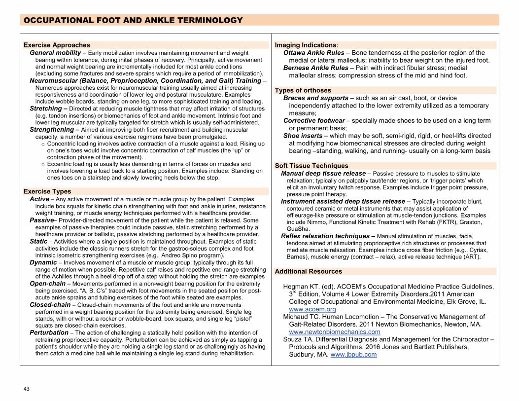
43
OCCUPATIONAL FOOT AND ANKLE TERMINOLOGY
Exercise Approaches General mobility – Early mobilization involves maintaining movement and weight
bearing within tolerance, during initial phases of recovery. Principally, active movement and normal weight bearing are incrementally included for most ankle conditions (excluding some fractures and severe sprains which require a period of immobilization).
Neuromuscular (Balance, Proprioception, Coordination, and Gait) Training – Numerous approaches exist for neuromuscular training usually aimed at increasing responsiveness and coordination of lower leg and postural musculature. Examples include wobble boards, standing on one leg, to more sophisticated training and loading.
Stretching – Directed at reducing muscle tightness that may affect irritation of structures (e.g. tendon insertions) or biomechanics of foot and ankle movement. Intrinsic foot and lower leg muscular are typically targeted for stretch which is usually self-administered.
Strengthening – Aimed at improving both fiber recruitment and building muscular capacity, a number of various exercise regimens have been promulgated. o Concentric loading involves active contraction of a muscle against a load. Rising up
on one’s toes would involve concentric contraction of calf muscles (the “up” orcontraction phase of the movement).
o Eccentric loading is usually less demanding in terms of forces on muscles andinvolves lowering a load back to a starting position. Examples include: Standing onones toes on a stairstep and slowly lowering heels below the step.
Exercise Types Active – Any active movement of a muscle or muscle group by the patient. Examples
include box squats for kinetic chain strengthening with foot and ankle injuries, resistance weight training, or muscle energy techniques performed with a healthcare provider.
Passive– Provider-directed movement of the patient while the patient is relaxed. Some examples of passive therapies could include passive, static stretching performed by a healthcare provider or ballistic, passive stretching performed by a healthcare provider.
Static – Activities where a single position is maintained throughout. Examples of static activities include the classic runners stretch for the gastroc-soleus complex and foot intrinsic isometric strengthening exercises (e.g., Andreo Spino program).
Dynamic – Involves movement of a muscle or muscle group, typically through its full range of motion when possible. Repetitive calf raises and repetitive end-range stretching of the Achilles through a heel drop off of a step without holding the stretch are examples
Open-chain – Movements performed in a non-weight bearing position for the extremity being exercised. “A, B, C’s” traced with foot movements in the seated position for post-acute ankle sprains and tubing exercises of the foot while seated are examples.
Closed-chain – Closed-chain movements of the foot and ankle are movements performed in a weight bearing position for the extremity being exercised. Single leg stands, with or without a rocker or wobble-board, box squats, and single leg “pistol” squats are closed-chain exercises.
Perturbation – The action of challenging a statically held position with the intention of retraining proprioceptive capacity. Perturbation can be achieved as simply as tapping a patient’s shoulder while they are holding a single leg stand or as challengingly as having them catch a medicine ball while maintaining a single leg stand during rehabilitation.
Imaging Indications: Ottawa Ankle Rules – Bone tenderness at the posterior region of the
medial or lateral malleolus; inability to bear weight on the injured foot. Bernese Ankle Rules – Pain with indirect fibular stress; medial
malleolar stress; compression stress of the mid and hind foot.
Types of orthoses Braces and supports – such as an air cast, boot, or device
independently attached to the lower extremity utilized as a temporary measure;
Corrective footwear – specially made shoes to be used on a long term or permanent basis;
Shoe inserts – which may be soft, semi-rigid, rigid, or heel-lifts directed at modifying how biomechanical stresses are directed during weight bearing –standing, walking, and running- usually on a long-term basis
Soft Tissue Techniques Manual deep tissue release – Passive pressure to muscles to stimulate
relaxation; typically on palpably taut/tender regions, or ‘trigger points’ which elicit an involuntary twitch response. Examples include trigger point pressure, pressure point therapy.
Instrument assisted deep tissue release – Typically incorporate blunt, contoured ceramic or metal instruments that may assist application of effleurage-like pressure or stimulation at muscle-tendon junctions. Examples include Nimmo, Functional Kinetic Treatment with Rehab (FKTR), Graston, GuaSha.
Reflex relaxation techniques – Manual stimulation of muscles, facia, tendons aimed at stimulating proprioceptive rich structures or processes that mediate muscle relaxation. Examples include cross fiber friction (e.g., Cyriax, Barnes), muscle energy (contract – relax), active release technique (ART).
Additional Resources
Hegman KT. (ed). ACOEM’s Occupational Medicine Practice Guidelines, 3rd Edition, Volume 4 Lower Extremity Disorders.2011 American College of Occupational and Environmental Medicine, Elk Grove, IL. www.acoem.org
Michaud TC. Human Locomotion – The Conservative Management of Gait-Related Disorders. 2011 Newton Biomechanics, Newton, MA. www.newtonbiomechanics.com
Souza TA. Differential Diagnosis and Management for the Chiropractor – Protocols and Algorithms. 2016 Jones and Bartlett Publishers, Sudbury, MA. www.jbpub.com
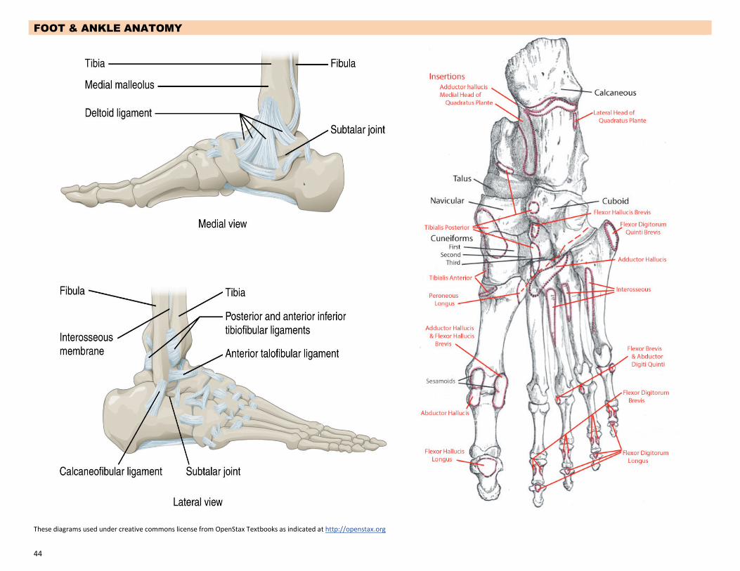
44
FOOT & ANKLE ANATOMY
These diagrams used under creative commons license from OpenStax Textbooks as indicated at http://openstax.org

45
EVIDENCE & METHODOLOGY Intervention/Experimental Studies
Randomized Controlled Trial (RCT) – A study that randomly allocates patients to treatment groups, usually blinding patients, therapists and/or study evaluators. Typically of high quality as randomization assures similarities of subjects within treatment groups.
Observational Studies
Cohort Design – Cohort (retrospective or prospective) – A study that follows patients who self-allocate to treatment groups through the course of their care for a given occurrence of a condition. Larger, well-designed cohort studies may be of good quality, but lack of randomization predisposes to heterogeneity issues within groups, some of which may be able to be adjusted for with statistical methods.
Cross sectional – Involves observing a population to measure disease and exposure status. It is usually thought to be a “snapshot” of the frequency and characteristics of a disease in a population at a specific given time.
Case control – Is a study that compares patients who have an outcome (cases) of interest with patients who do not have the disease or outcome (controls). The study may retrospectively to compare how frequently the exposure was present in a group to determine risk factors.
Case series – Is a study that describes a series of patients with an outcome of interest, may be of variable quality. Better designs use consecutive patients and include robust baseline and follow up outcome measures.
Case reports – Describes an individual case, typically only achieving publication if it represent a unique or unusual clinical experience.
Blinding
Blinding minimizes potential bias. Typically three levels of blinding are sought: patient, treating provider and evaluator. Many conservative interventions do not allow for patient blinding (e.g. someone is likely to know if they received a splint or a pill). At a minimum, single blinding of the evaluator as to what group a subject was in is expected.
Literature Reviews
Quantitative systematic reviews – Studies that review previously published clinical trials that include quantitative comparisons (e.g. meta-analyses). Systematic reviews should have rigorous and comprehensive methodology to identify relevant published research and include appraisal of study quality. Cochrane reviews frequently are of this type.
Qualitative systematic reviews – Similar to quantitative reviews but without systematic quantitative comparison or data pooling.
Narrative literature reviews – Such reviews typically do not include rigorous study selection methodology and may be subject to significant author bias
Literature Retrieval and Review
1. Initial systematic searches of electronic databases (e.g. PubMed). Search terms used typically included MeSH terms for tests and interventions with conditions being addressed. Follow-up searches also included population attributes (e.g., workers compensation, occupational).
2. Abstract screening for relevance. 3. Original paper retrieval with review for relevance, quality, outcome meaningfulness, and effect
magnitude. 4. Additional studies identified through clinical summaries (e.g., reviews, texts), citation tracking,
and feedback from public.
About Evidence for Physical Examination and Conservative Interventions Conservative musculoskeletal care is typically care of first resort based on long standing practices. Typically ‘low tech,’ low cost, with minimal and rare side effects, it is frequently delivered in primary care settings, and by various health providers. The rigor and quality expected of high cost, higher risk, emerging, and tertiary interventions is less common for many routine physical examination procedures and conservative interventions. Much of the evidence summarized here would be considered Class “C” or “III” in ratings systems. Thus, the committee has not presented explicit recommendations, rather, evidence summaries guided by expert consensus to assist in formulating care options. Further, significant emphasis is made regarding tracking and documenting meaningful functional improvement with patients. Study attributes most likely to strengthen or limit confidence are characterized in the evidence descriptions. Assessing Study Methodologic Quality Attributes of study methodology quality vary according to the clinical procedure (eg, diagnostic, therapeutic intervention) looked at, and specific research questions being studied. The American Academy of Neurology’s Clinical Practice Guideline Process Manual offers a comprehensive guide to systematic evidence review, quality attributes and consensus process that generally serves as the approach taken by IICAC. General attributes identified when extracting evidence from studies include identification of population, the intervention and co-interventions and outcomes being addressed in each study. The clinical questions addressed such as diagnostic accuracy, therapeutic effectiveness, or causation are determined. Studies are extracted into evidence tables including quality attributes and/or ratings which are reviewed both by department staff and committee members (usually 2 per study). Specific quality attributes include: Diagnostic Accuracy – design, spectrum of patients, validity and relevance of outcome metric; Therapeutic Interventions – comparison groups (no treatment, placebo, comparative intervention), treatment allocation, blinding/masking (method and degree: single, double, independent), follow-up (period and completion), and analysis (statistical power, intent-to-treat). Specific attention is paid to several factors including reporting of outcomes (primary vs. secondary), relevance of outcome (eg, function vs. pain), and meaningfulness (clinically important change vs minimally detectable change). Synthesizing Evidence Consideration of study quality (class), significance (statistical precision), consistency across studies, magnitude of effect, and relevance to populations and procedures were taken into account in preparing draft summaries. Special attention was given to clarifying conclusions related to the clinical questions of interest. Evidence, particularly with low tech and highly diffused examination and conservative procedures addressed here, is rarely truly “definitive,” even when multiple studies exist. Inconsistent conclusions typically reflect error (systematic, random) and/or bias in studies. Data pooling via meta-analysis is useful to reduce random error when studies are of sufficient power and methodologic strength. Larger meaningful effect size may increases confidence in findings.
Citations

46
1. Souza, T.A., Differential Diagnosis and Management for the Chiropractor: Protocols and Algorithms. 5th ed. 2016: Jones and Bartlett Publishers: Sudbury, MA.
2. Young, C.C., et al., Clinical examination of the foot and ankle. Prim Care, 2005. 32(1): p. 105-32. 3. Michaud, T., The Conservative Management of Gait-Related Disorders. 2011, Newton MA: Newton Biomechanics. 4. ACOEM's Occupational Medicine Practice Guidelines 3rd ed. Vol. 4. 2011, Elk Grove, IL: American College of Occupational and Environmental Medicine. 5. Maffulli, N., The clinical diagnosis of subcutaneous tear of the Achilles tendon. A prospective study in 174 patients. Am J Sports Med, 1998. 26(2): p. 266-
70. 6. Chisholm, M.D., et al., Reliability and validity of a weight-bearing measure of ankle dorsiflexion range of motion. Physiother Can, 2012. 64(4): p. 347-55. 7. O'Shea, S. and K. Grafton, The intra and inter-rater reliability of a modified weight-bearing lunge measure of ankle dorsiflexion. Man Ther, 2013. 18(3): p.
264-8. 8. Coster, M., et al., Validity, reliability, and responsiveness of a self-reported foot and ankle score (SEFAS). Acta Orthop, 2012. 83(2): p. 197-203. 9. Coster, M.C., et al., Comparison of the Self-reported Foot and Ankle Score (SEFAS) and the American Orthopedic Foot and Ankle Society Score (AOFAS).
Foot Ankle Int, 2014. 35(10): p. 1031-6. 10. Eechaute, C., et al., The clinimetric qualities of patient-assessed instruments for measuring chronic ankle instability: a systematic review. BMC
Musculoskelet Disord, 2007. 8: p. 6. 11. Martin, R.L. and J.J. Irrgang, A survey of self-reported outcome instruments for the foot and ankle. J Orthop Sports Phys Ther, 2007. 37(2): p. 72-84. 12. Martin, R.L., et al., Evidence of validity for the Foot and Ankle Ability Measure (FAAM). Foot Ankle Int, 2005. 26(11): p. 968-83. 13. Hale, S.A. and J. Hertel, Reliability and Sensitivity of the Foot and Ankle Disability Index in Subjects With Chronic Ankle Instability. J Athl Train, 2005. 40(1):
p. 35-40. 14. Budiman-Mak, E., K.J. Conrad, and K.E. Roach, The Foot Function Index: a measure of foot pain and disability. J Clin Epidemiol, 1991. 44(6): p. 561-70. 15. Robinson, J.M., et al., The VISA-A questionnaire: a valid and reliable index of the clinical severity of Achilles tendinopathy. Br J Sports Med, 2001. 35(5): p.
335-41. 16. Iversen, J.V., E.M. Bartels, and H. Langberg, The victorian institute of sports assessment - achilles questionnaire (visa-a) - a reliable tool for measuring
achilles tendinopathy. Int J Sports Phys Ther, 2012. 7(1): p. 76-84. 17. Hosman, A.H., et al., A New Zealand national joint registry review of 202 total ankle replacements followed for up to 6 years. Acta Orthop, 2007. 78(5): p.
584-91. 18. Lau, J.T., N.M. Mahomed, and L.C. Schon, Results of an Internet survey determining the most frequently used ankle scores by AOFAS members. Foot Ankle
Int, 2005. 26(6): p. 479-82. 19. Ibrahim, T., et al., Reliability and validity of the subjective component of the American Orthopaedic Foot and Ankle Society clinical rating scales. J Foot
Ankle Surg, 2007. 46(2): p. 65-74. 20. Roos, E.M., S. Brandsson, and J. Karlsson, Validation of the foot and ankle outcome score for ankle ligament reconstruction. Foot Ankle Int, 2001. 22(10):
p. 788-94. 21. Hiller, C.E., et al., The Cumberland ankle instability tool: a report of validity and reliability testing. Arch Phys Med Rehabil, 2006. 87(9): p. 1235-41. 22. Wright, C.J., et al., Recalibration and validation of the Cumberland Ankle Instability Tool cutoff score for individuals with chronic ankle instability. Arch
Phys Med Rehabil, 2014. 95(10): p. 1853-9. 23. Pourkazemi, F., et al., Predictors of chronic ankle instability after an index lateral ankle sprain: a systematic review. J Sci Med Sport, 2014. 17(6): p. 568-
73. 24. Turner, J.A., et al., Back pain in primary care. Patient characteristics, content of initial visit, and short-term outcomes. Spine (Phila Pa 1976), 1998. 23(4):
p. 463-9. 25. Von Korff, M., et al., Comparison of back pain prognostic risk stratification item sets. J Pain, 2014. 15(1): p. 81-9. 26. Croy, T., et al., Anterior talocrural joint laxity: diagnostic accuracy of the anterior drawer test of the ankle. J Orthop Sports Phys Ther, 2013. 43(12): p.
911-9.

47
27. Lee, K.T., et al., New method of diagnosis for chronic ankle instability: comparison of manual anterior drawer test, stress radiography and stress ultrasound. Knee Surg Sports Traumatol Arthrosc, 2014. 22(7): p. 1701-7.
28. Vaseenon, T., Y. Gao, and P. Phisitkul, Comparison of two manual tests for ankle laxity due to rupture of the lateral ankle ligaments. Iowa Orthop J, 2012. 32: p. 9-16.
29. Rosen, A.B., J. Ko, and C.N. Brown, Diagnostic accuracy of instrumented and manual talar tilt tests in chronic ankle instability populations. Scand J Med Sci Sports, 2014.
30. Sman, A.D., et al., Diagnostic accuracy of clinical tests for ankle syndesmosis injury. Br J Sports Med, 2013. 31. Sekir, U., et al., Reliability of a functional test battery evaluating functionality, proprioception, and strength in recreational athletes with functional ankle
instability. Eur J Phys Rehabil Med, 2008. 44(4): p. 407-15. 32. Bussieres, A.E., J.A. Taylor, and C. Peterson, Diagnostic imaging practice guidelines for musculoskeletal complaints in adults--an evidence-based
approach. Part 1. Lower extremity disorders. J Manipulative Physiol Ther, 2007. 30(9): p. 684-717. 33. Bachmann, L.M., et al., Accuracy of Ottawa ankle rules to exclude fractures of the ankle and mid-foot: systematic review. BMJ, 2003. 326(7386): p. 417. 34. Eggli, S., et al., The Bernese ankle rules: a fast, reliable test after low-energy, supination-type malleolar and midfoot trauma. J Trauma, 2005. 59(5): p.
1268-71. 35. Garras, D.N., et al., MRI is unnecessary for diagnosing acute Achilles tendon ruptures: clinical diagnostic criteria. Clin Orthop Relat Res, 2012. 470(8): p.
2268-73. 36. Magnussen, R.A., W.R. Dunn, and A.B. Thomson, Nonoperative treatment of midportion Achilles tendinopathy: a systematic review. Clin J Sport Med,
2009. 19(1): p. 54-64. 37. van Dijk, C.N., et al., Terminology for Achilles tendon related disorders. Knee Surg Sports Traumatol Arthrosc, 2011. 19(5): p. 835-41. 38. Wiegerinck, J.I., et al., Treatment for insertional Achilles tendinopathy: a systematic review. Knee Surg Sports Traumatol Arthrosc, 2013. 21(6): p. 1345-
55. 39. Janig, W. and R. Baron, Complex regional pain syndrome: mystery explained? Lancet Neurol, 2003. 2(11): p. 687-97. 40. Industrial Insurance Medical Advisory Committee (IIMAC). Work-Related Complex Regional Pain Syndrome (CRPS): Diagnosis and Treatment, 2011. State
of Washington Department of Labor & Industries. Olympia WA. 41. Guyton, G.P., et al., Cumulative industrial trauma as an etiology of seven common disorders in the foot and ankle: what is the evidence? Foot Ankle Int,
2000. 21(12): p. 1047-56. 42. Melhorn JM, T.J., Ackerman WE, Hyman MH. , AMA Guides to the Evaluation of Disease and Injury Causation. 2nd ed. 2014: American Medical
Association. 43. Milgrom, C., et al., Cold weather training: a risk factor for Achilles paratendinitis among recruits. Foot Ankle Int, 2003. 24(5): p. 398-401. 44. Thomas, M.J., et al., The population prevalence of foot and ankle pain in middle and old age: a systematic review. Pain, 2011. 152(12): p. 2870-80. 45. Fu, S.C., et al., Deciphering the pathogenesis of tendinopathy: a three-stages process. Sports Med Arthrosc Rehabil Ther Technol, 2010. 2: p. 30. 46. Murtaugh, B. and J.M. Ihm, Eccentric training for the treatment of tendinopathies. Curr Sports Med Rep, 2013. 12(3): p. 175-82. 47. Knobloch, K., Re: Acute Achilles tendon rupture: minimally invasive surgery versus nonoperative treatment with immediate full weightbearing. Am J
Sports Med, 2008. 36(12): p. E3-4; author reply E4. 48. Knobloch, K., H. Thermann, and T. Hufner, [Achilles tendon rupture--early functional and surgical options with special emphasis on rehabilitation issues].
Sportverletz Sportschaden, 2007. 21(1): p. 34-40. 49. Brinks, A., et al., Adverse effects of extra-articular corticosteroid injections: a systematic review. BMC Musculoskelet Disord, 2010. 11: p. 206. 50. Wiegerinck, J.I., A.C. Kok, and C.N. van Dijk, Surgical treatment of chronic retrocalcaneal bursitis. Arthroscopy, 2012. 28(2): p. 283-93. 51. Holm, C., M. Kjaer, and P. Eliasson, Achilles tendon rupture - treatment and complications: A systematic review. Scand J Med Sci Sports, 2015. 25(1): p.
e1-e10. 52. Metz, R., et al., Acute Achilles tendon rupture: minimally invasive surgery versus nonoperative treatment with immediate full weightbearing--a
randomized controlled trial. Am J Sports Med, 2008. 36(9): p. 1688-94.

48
53. Saleh, M., et al., The Sheffield splint for controlled early mobilisation after rupture of the calcaneal tendon. A prospective, randomised comparison with plaster treatment. J Bone Joint Surg Br, 1992. 74(2): p. 206-9.
54. van der Linden-van der Zwaag, H.M., R.G. Nelissen, and J.B. Sintenie, Results of surgical versus non-surgical treatment of Achilles tendon rupture. Int Orthop, 2004. 28(6): p. 370-3.
55. Mattila, V.M., et al., Declining incidence of surgery for Achilles tendon rupture follows publication of major RCTs: evidence-influenced change evident using the Finnish registry study. Br J Sports Med, 2013.
56. Thomas, J.L., et al., The diagnosis and treatment of heel pain: a clinical practice guideline-revision 2010. J Foot Ankle Surg, 2010. 49(3 Suppl): p. S1-19. 57. Goff, J.D. and R. Crawford, Diagnosis and treatment of plantar fasciitis. Am Fam Physician, 2011. 84(6): p. 676-82. 58. Schepsis, A.A., R.E. Leach, and J. Gorzyca, Plantar fasciitis. Etiology, treatment, surgical results, and review of the literature. Clin Orthop Relat Res,
1991(266): p. 185-96. 59. Kavlak, Y. and F. Uygur, Effects of nerve mobilization exercise as an adjunct to the conservative treatment for patients with tarsal tunnel syndrome. J
Manipulative Physiol Ther, 2011. 34(7): p. 441-8. 60. Mackinnon, S.E., et al., Peripheral nerve injection injury with steroid agents. Plast Reconstr Surg, 1982. 69(3): p. 482-90. 61. Gkotsoulias, E.N., D.C. Simonson, and T.S. Roukis, Outcomes and safety of endoscopic tarsal tunnel decompression: a systematic review. Foot Ankle Spec,
2014. 7(1): p. 57-60. 62. Batt, J. and M.M. Neeki, Osteopathic manipulative treatment in tarsal somatic dysfunction: a case study. J Am Osteopath Assoc, 2013. 113(11): p. 857-
61. 63. Menetrey, J. and D. Fritschy, Subtalar subluxation in ballet dancers. Am J Sports Med, 1999. 27(2): p. 143-9. 64. Mooney, M. and L. Maffey-Ward, Cuboid plantar and dorsal subluxations: assessment and treatment. J Orthop Sports Phys Ther, 1994. 20(4): p. 220-6. 65. Patterson, S.M., Cuboid syndrome: a review of the literature. J Sports Sci Med, 2006. 5(4): p. 597-606. 66. Brantingham, J.W., et al., Manipulative therapy for lower extremity conditions: update of a literature review. J Manipulative Physiol Ther, 2012. 35(2): p.
127-66. 67. Dilnot, M. and T.C. Michaud, Plantar plate rupture. Australasian Journal of Podiatric Medicine, 2003. 37(2): p. 43-46. 68. Nwawka, O.K., et al., Sesamoids and accessory ossicles of the foot: anatomical variability and related pathology. Insights Imaging, 2013. 4(5): p. 581-93. 69. Dean, B.J., et al., The jones fracture classification, management, outcome, and complications: a systematic review. Foot Ankle Spec, 2012. 5(4): p. 256-9. 70. Polzer, H., et al., Acute fractures to the proximal fifth metatarsal bone: development of classification and treatment recommendations based on the
current evidence. Injury, 2012. 43(10): p. 1626-32. 71. Kim, J.Y., et al., An anatomical study of Morton's interdigital neuroma: the relationship between the occurring site and the deep transverse metatarsal
ligament (DTML). Foot Ankle Int, 2007. 28(9): p. 1007-10. 72. Shibuya N, H.J., Agarwal MR, Jupiter DC., Efficacy and safety of high-dose vitamin C on complex regional pain syndrome in extremity trauma and surgery--
systematic review and meta-analysis. J Foot Ankle Surg, 2013. 52(1): p. 62-66. 73. Bleakley, C.M., S.M. McDonough, and D.C. MacAuley, Some conservative strategies are effective when added to controlled mobilisation with external
support after acute ankle sprain: a systematic review. Aust J Physiother, 2008. 54(1): p. 7-20. 74. Black JD, B.M., Al-Hadithy N, Hakmi A, Kitson J., Early weight-bearing in operatively fixed ankle fractures: a systematic review. Foot (Edinb), 2013. 23(2-
3): p. 78-85. 75. Dettori, J.R. and C.J. Basmania, Early ankle mobilization, Part II: A one-year follow-up of acute, lateral ankle sprains (a randomized clinical trial). Mil Med,
1994. 159(1): p. 20-4. 76. Dettori, J.R., et al., Early ankle mobilization, Part I: The immediate effect on acute, lateral ankle sprains (a randomized clinical trial). Mil Med, 1994.
159(1): p. 15-20. 77. Eiff, M.P., A.T. Smith, and G.E. Smith, Early mobilization versus immobilization in the treatment of lateral ankle sprains. Am J Sports Med, 1994. 22(1): p.
83-8. 78. Karlsson, J., et al., Early mobilization versus immobilization after ankle ligament stabilization. Scand J Med Sci Sports, 1999. 9(5): p. 299-303.
49
79. van Ochten, J.M., et al., Chronic complaints after ankle sprains: a systematic review on effectiveness of treatments. J Orthop Sports Phys Ther, 2014. 44(11): p. 862-C23.
80. Lehtonen, H., et al., Use of a cast compared with a functional ankle brace after operative treatment of an ankle fracture. A prospective, randomized study. J Bone Joint Surg Am, 2003. 85-A(2): p. 205-11.
81. Kaikkonen, A., P. Kannus, and M. Jarvinen, Surgery versus functional treatment in ankle ligament tears. A prospective study. Clin Orthop Relat Res, 1996(326): p. 194-202.
82. Cruz-Diaz, D., et al., Effects of joint mobilization on chronic ankle instability: a randomized controlled trial. Disabil Rehabil, 2014: p. 1-10. 83. Green, T., et al., A randomized controlled trial of a passive accessory joint mobilization on acute ankle inversion sprains. Phys Ther, 2001. 81(4): p. 984-94. 84. Loudon, J.K., M.P. Reiman, and J. Sylvain, The efficacy of manual joint mobilisation/manipulation in treatment of lateral ankle sprains: a systematic
review. Br J Sports Med, 2014. 48(5): p. 365-70. 85. Vicenzino, B., et al., Initial changes in posterior talar glide and dorsiflexion of the ankle after mobilization with movement in individuals with recurrent
ankle sprain. J Orthop Sports Phys Ther, 2006. 36(7): p. 464-71. 86. Collins, N., P. Teys, and B. Vicenzino, The initial effects of a Mulligan's mobilization with movement technique on dorsiflexion and pain in subacute ankle
sprains. Man Ther, 2004. 9(2): p. 77-82. 87. Lubbe, D., et al., Manipulative therapy and rehabilitation for recurrent ankle sprain with functional instability: a short-term, assessor-blind, parallel-group
randomized trial. J Manipulative Physiol Ther, 2015. 38(1): p. 22-34. 88. Truyols-Domi Nguez, S., et al., Efficacy of thrust and nonthrust manipulation and exercise with or without the addition of myofascial therapy for the
management of acute inversion ankle sprain: a randomized clinical trial. J Orthop Sports Phys Ther, 2013. 43(5): p. 300-9. 89. Eisenhart, A.W., T.J. Gaeta, and D.P. Yens, Osteopathic manipulative treatment in the emergency department for patients with acute ankle injuries. J Am
Osteopath Assoc, 2003. 103(9): p. 417-21. 90. Wassinger, C.A., et al., Acute effects of rearfoot manipulation on dynamic standing balance in healthy individuals. Man Ther, 2014. 19(3): p. 242-5. 91. Grindstaff, T.L., et al., Immediate effects of a tibiofibular joint manipulation on lower extremity H-reflex measurements in individuals with chronic ankle
instability. J Electromyogr Kinesiol, 2011. 21(4): p. 652-8. 92. Beazell, J.R., et al., Effects of a proximal or distal tibiofibular joint manipulation on ankle range of motion and functional outcomes in individuals with
chronic ankle instability. J Orthop Sports Phys Ther, 2012. 42(2): p. 125-34. 93. van der Wees, P.J., et al., Effectiveness of exercise therapy and manual mobilisation in ankle sprain and functional instability: a systematic review. Aust J
Physiother, 2006. 52(1): p. 27-37. 94. Whitman, J.M., et al., Predicting short-term response to thrust and nonthrust manipulation and exercise in patients post inversion ankle sprain. J Orthop
Sports Phys Ther, 2009. 39(3): p. 188-200. 95. Pellow, J.E. and J.W. Brantingham, The efficacy of adjusting the ankle in the treatment of subacute and chronic grade I and grade II ankle inversion
sprains. J Manipulative Physiol Ther, 2001. 24(1): p. 17-24. 96. Andersen, S., G.A. Fryer, and P. McLaughlin, The effect of talo-crural joint manipulation on range of motion at the ankle joint in subjects with a history of
ankle injury. Australas Chiropr Osteopathy, 2003. 11(2): p. 57-62. 97. Cleland, J.A., et al., Manual physical therapy and exercise versus supervised home exercise in the management of patients with inversion ankle sprain: a
multicenter randomized clinical trial. J Orthop Sports Phys Ther, 2013. 43(7): p. 443-55. 98. Marron-Gomez, D., A.L. Rodriguez-Fernandez, and J.A. Martin-Urrialde, The effect of two mobilization techniques on dorsiflexion in people with chronic
ankle instability. Phys Ther Sport, 2015. 16(1): p. 10-5. 99. Bisset, L., et al., A systematic review and meta-analysis of clinical trials on physical interventions for lateral epicondylalgia. Br J Sports Med, 2005. 39(7):
p. 411-22; discussion 411-22. 100. Muth, S., et al., The effects of thoracic spine manipulation in subjects with signs of rotator cuff tendinopathy. J Orthop Sports Phys Ther, 2012. 42(12): p.
1005-16. 101. McClinton, S.M., J.A. Cleland, and T.W. Flynn, Predictors of Response to Physical Therapy Intervention for Plantar Heel Pain. Foot Ankle Int, 2014.

50
102. Cleland, J.A., et al., Manual physical therapy and exercise versus electrophysical agents and exercise in the management of plantar heel pain: a multicenter randomized clinical trial. J Orthop Sports Phys Ther, 2009. 39(8): p. 573-85.
103. Dimou E, B.J., Wood T. , A randomized controlled trial (with blinded observer) of chiropractic manipulation and Achilles stretching vs. orthotics for treatment of plantar fasciitis. . J Am Chiropr Assoc 2004. 41(9): p. 32-42.
104. Davis, P.T., et al., Comparative efficacy of conservative medical and chiropractic treatments for carpal tunnel syndrome: a randomized clinical trail. J Manipulative Physiol Ther, 1998. 21(5): p. 317-26.
105. Burke, J., et al., A pilot study comparing two manual therapy interventions for carpal tunnel syndrome. J Manipulative Physiol Ther, 2007. 30(1): p. 50-61. 106. Petersen, S., J. Brantingham, and H. Kretzmann, The efficacy of chiropractic adjustment in the treatment of primary metatarsalgia. Euro J Chiropr, 2003.
49(3): p. 267-279. 107. Brantingham, J.W. and T.G. Wood, Hallux rigidus. J Chiropr Med, 2002. 1(1): p. 31-7. 108. Manral, D.B., Hallux rigidus: A case report of successful chiropractic management and review of the literature. J Chiropr Med, 2004. 3(1): p. 6-11. 109. Solan, M.C., J.D. Calder, and S.P. Bendall, Manipulation and injection for hallux rigidus. Is it worthwhile? J Bone Joint Surg Br, 2001. 83(5): p. 706-8. 110. Brumm, L.F., et al., Preventive osteopathic manipulative treatment and stress fracture incidence among collegiate cross-country athletes. J Am Osteopath
Assoc, 2013. 113(12): p. 882-90. 111. Andres, B.M. and G.A. Murrell, Treatment of tendinopathy: what works, what does not, and what is on the horizon. Clin Orthop Relat Res, 2008. 466(7):
p. 1539-54. 112. Verhagen, E.A., What does therapeutic ultrasound add to recovery from acute ankle sprain? A review. Clin J Sport Med, 2013. 23(1): p. 84-5. 113. Terada, M., B.G. Pietrosimone, and P.A. Gribble, Therapeutic interventions for increasing ankle dorsiflexion after ankle sprain: a systematic review. J Athl
Train, 2013. 48(5): p. 696-709. 114. Kaminski, T.W., et al., National Athletic Trainers' Association position statement: conservative management and prevention of ankle sprains in athletes. J
Athl Train, 2013. 48(4): p. 528-45. 115. Khan, K.M., et al., Time to abandon the "tendinitis" myth. BMJ, 2002. 324(7338): p. 626-7. 116. Sussmilch-Leitch, S.P., et al., Physical therapies for Achilles tendinopathy: systematic review and meta-analysis. J Foot Ankle Res, 2012. 5(1): p. 15. 117. Chapman-Jones D, H.D., Novel microcurrent treatment is more effective than conventional therapy for chronic Achilles tendinopathy: randomised
comparative trial. Physiotherapy, 2002. 88(8): p. 471-480. 118. Brummitt, J., The role of massage in sports performance and rehabilitation: current evidence and future direction. N Am J Sports Phys Ther, 2008. 3(1): p.
7-21. 119. Renan-Ordine, R., et al., Effectiveness of myofascial trigger point manual therapy combined with a self-stretching protocol for the management of plantar
heel pain: a randomized controlled trial. J Orthop Sports Phys Ther, 2011. 41(2): p. 43-50. 120. Davis, F., Therapeutic Massage Provides Pain Relief to a Client with Morton's Neuroma: A Case Report. Int J Ther Massage Bodywork, 2012. 5(2): p. 12-9. 121. Abdel-Aziem, A.A. and W.S. Mohammad, Plantar-flexor Static Stretch Training Effect on Eccentric and Concentric Peak Torque - A comparative Study of
Trained versus Untrained Subjects. J Hum Kinet, 2012. 34: p. 49-58. 122. Avloniti, A., et al., The Acute Effects of Static Stretching on Speed and Agility Performance Depend on Stretch Duration and Conditioning Level. J Strength
Cond Res, 2014. 123. van Rijn, R.M., et al., Effectiveness of additional supervised exercises compared with conventional treatment alone in patients with acute lateral ankle
sprains: systematic review. BMJ, 2010. 341: p. c5688. 124. van Os, A.G., et al., Comparison of conventional treatment and supervised rehabilitation for treatment of acute lateral ankle sprains: a systematic review
of the literature. J Orthop Sports Phys Ther, 2005. 35(2): p. 95-105. 125. Hubscher, M., et al., Neuromuscular training for sports injury prevention: a systematic review. Med Sci Sports Exerc, 2010. 42(3): p. 413-21. 126. Struijs, P.A. and G.M. Kerkhoffs, Ankle sprain. BMJ Clin Evid, 2010. 2010. 127. Verhagen, E.A. and K. Bay, Optimising ankle sprain prevention: a critical review and practical appraisal of the literature. Br J Sports Med, 2010. 44(15): p.
1082-8.

51
128. Hupperets, M.D., E.A. Verhagen, and W. van Mechelen, Effect of unsupervised home based proprioceptive training on recurrences of ankle sprain: randomised controlled trial. BMJ, 2009. 339: p. b2684.
129. Chaiwanichsiri, D., E. Lorprayoon, and L. Noomanoch, Star excursion balance training: effects on ankle functional stability after ankle sprain. J Med Assoc Thai, 2005. 88 Suppl 4: p. S90-4.
130. Hupperets, M.D., et al., Potential savings of a program to prevent ankle sprain recurrence: economic evaluation of a randomized controlled trial. Am J Sports Med, 2010. 38(11): p. 2194-200.
131. Hupperets, M.D., E.A. Verhagen, and W. van Mechelen, Effect of sensorimotor training on morphological, neurophysiological and functional characteristics of the ankle: a critical review. Sports Med, 2009. 39(7): p. 591-605.
132. McGuine, T.A. and J.S. Keene, The effect of a balance training program on the risk of ankle sprains in high school athletes. Am J Sports Med, 2006. 34(7): p. 1103-11.
133. McHugh, M.P., et al., The effectiveness of a balance training intervention in reducing the incidence of noncontact ankle sprains in high school football players. Am J Sports Med, 2007. 35(8): p. 1289-94.
134. McKeon, P.O. and J. Hertel, Systematic review of postural control and lateral ankle instability, part II: is balance training clinically effective? J Athl Train, 2008. 43(3): p. 305-15.
135. Postle, K., D. Pak, and T.O. Smith, Effectiveness of proprioceptive exercises for ankle ligament injury in adults: a systematic literature and meta-analysis. Man Ther, 2012. 17(4): p. 285-91.
136. Quigley, P.A., et al., Exercise interventions, gait, and balance in older subjects with distal symmetric polyneuropathy: a three-group randomized clinical trial. Am J Phys Med Rehabil, 2014. 93(1): p. 1-12; quiz 13-6.
137. Zech, A., et al., Neuromuscular training for rehabilitation of sports injuries: a systematic review. Med Sci Sports Exerc, 2009. 41(10): p. 1831-41. 138. Wasielewski, N.J. and K.M. Kotsko, Does eccentric exercise reduce pain and improve strength in physically active adults with symptomatic lower extremity
tendinosis? A systematic review. J Athl Train, 2007. 42(3): p. 409-21. 139. Rompe, J.D., J. Furia, and N. Maffulli, Eccentric loading versus eccentric loading plus shock-wave treatment for midportion achilles tendinopathy: a
randomized controlled trial. Am J Sports Med, 2009. 37(3): p. 463-70. 140. Rompe, J.D., et al., Eccentric loading, shock-wave treatment, or a wait-and-see policy for tendinopathy of the main body of tendo Achillis: a randomized
controlled trial. Am J Sports Med, 2007. 35(3): p. 374-83. 141. Silbernagel, K.G., et al., Continued sports activity, using a pain-monitoring model, during rehabilitation in patients with Achilles tendinopathy: a
randomized controlled study. Am J Sports Med, 2007. 35(6): p. 897-906. 142. Mafi, N., R. Lorentzon, and H. Alfredson, Superior short-term results with eccentric calf muscle training compared to concentric training in a randomized
prospective multicenter study on patients with chronic Achilles tendinosis. Knee Surg Sports Traumatol Arthrosc, 2001. 9(1): p. 42-7. 143. Knobloch, K., et al., Gender and eccentric training in Achilles mid-portion tendinopathy. Knee Surg Sports Traumatol Arthrosc, 2010. 18(5): p. 648-55. 144. Bek, N., et al., Home-based general versus center-based selective rehabilitation in patients with posterior tibial tendon dysfunction. Acta Orthop
Traumatol Turc, 2012. 46(4): p. 286-92. 145. Alvarez RG, M.A., Schmitt C, Saltzman CL, Stage I and II posterior tibial tendon dysfunction treated by a structured nonoperative management protocol:
an orthosis and exercise program. . Foot Ankle Int, 2006. 27(1): p. 2-8. 146. Hensley, C.P. and A.J. Kavchak, Novel use of a manual therapy technique and management of a patient with peroneal tendinopathy: a case report. Man
Ther, 2012. 17(1): p. 84-8. 147. Boyce, S.H., M.A. Quigley, and S. Campbell, Management of ankle sprains: a randomised controlled trial of the treatment of inversion injuries using an
elastic support bandage or an Aircast ankle brace. Br J Sports Med, 2005. 39(2): p. 91-6. 148. Cooke, M.W., et al., Treatment of severe ankle sprain: a pragmatic randomised controlled trial comparing the clinical effectiveness and cost-effectiveness
of three types of mechanical ankle support with tubular bandage. The CAST trial. Health Technol Assess, 2009. 13(13): p. iii, ix-x, 1-121. 149. Lamb, S.E., et al., Mechanical supports for acute, severe ankle sprain: a pragmatic, multicentre, randomised controlled trial. Lancet, 2009. 373(9663): p.
575-81.

52
150. Janssen, K.W., W. van Mechelen, and E.A. Verhagen, Bracing superior to neuromuscular training for the prevention of self-reported recurrent ankle sprains: a three-arm randomised controlled trial. Br J Sports Med, 2014. 48(16): p. 1235-9.
151. Gabriner, M.L., et al., The effectiveness of foot orthotics in improving postural control in individuals with chronic ankle instability: a critically appraised topic. J Sport Rehabil, 2015. 24(1): p. 68-71.
152. Mattila, V.M., et al., Can orthotic insoles prevent lower limb overuse injuries? A randomized-controlled trial of 228 subjects. Scand J Med Sci Sports, 2011. 21(6): p. 804-8.
153. Terrier, P., F. Luthi, and O. Deriaz, Do orthopaedic shoes improve local dynamic stability of gait? An observational study in patients with chronic foot and ankle injuries. BMC Musculoskelet Disord, 2013. 14: p. 94.
154. Petersen, W., R. Welp, and D. Rosenbaum, Chronic Achilles tendinopathy: a prospective randomized study comparing the therapeutic effect of eccentric training, the AirHeel brace, and a combination of both. Am J Sports Med, 2007. 35(10): p. 1659-67.
155. Scott, L.A., S.E. Munteanu, and H.B. Menz, Effectiveness of orthotic devices in the treatment of Achilles tendinopathy: a systematic review. Sports Med, 2015. 45(1): p. 95-110.
156. Kulig, K., et al., Nonsurgical management of posterior tibial tendon dysfunction with orthoses and resistive exercise: a randomized controlled trial. Phys Ther, 2009. 89(1): p. 26-37.
157. Alvarez, R.G., et al., Stage I and II posterior tibial tendon dysfunction treated by a structured nonoperative management protocol: an orthosis and exercise program. Foot Ankle Int, 2006. 27(1): p. 2-8.
158. Stuber, K. and K. Kristmanson, Conservative therapy for plantar fasciitis: a narrative review of randomized controlled trials. J Can Chiropr Assoc, 2006. 50(2): p. 118-33.
159. Landorf, K.B., A.M. Keenan, and R.D. Herbert, Effectiveness of foot orthoses to treat plantar fasciitis: a randomized trial. Arch Intern Med, 2006. 166(12): p. 1305-10.
160. Pfeffer, G., et al., Comparison of custom and prefabricated orthoses in the initial treatment of proximal plantar fasciitis. Foot Ankle Int, 1999. 20(4): p. 214-21.
161. Roos, E., M. Engstrom, and B. Soderberg, Foot orthoses for the treatment of plantar fasciitis. Foot Ankle Int, 2006. 27(8): p. 606-11. 162. Donley, B.G., et al., The efficacy of oral nonsteroidal anti-inflammatory medication (NSAID) in the treatment of plantar fasciitis: a randomized,
prospective, placebo-controlled study. Foot Ankle Int, 2007. 28(1): p. 20-3. 163. Derry, S., R.A. Moore, and R. Rabbie, Topical NSAIDs for chronic musculoskeletal pain in adults. Cochrane Database Syst Rev, 2012. 9: p. CD007400. 164. Massey, T., et al., Topical NSAIDs for acute pain in adults. Cochrane Database Syst Rev, 2010(6): p. CD007402. 165. Mason, L., et al., Topical NSAIDs for chronic musculoskeletal pain: systematic review and meta-analysis. BMC Musculoskelet Disord, 2004. 5: p. 28. 166. Hart, L., Corticosteroid and other injections in the management of tendinopathies: a review. . Clin J Sport Med. , 2011. 21(6). 167. Gelberman, R.H., D. Aronson, and M.H. Weisman, Carpal-tunnel syndrome. Results of a prospective trial of steroid injection and splinting. J Bone Joint
Surg Am, 1980. 62(7): p. 1181-4. 168. McMillan, A.M., et al., Ultrasound guided corticosteroid injection for plantar fasciitis: randomised controlled trial. BMJ, 2012. 344: p. e3260. 169. Mulherin, D. and M. Price, Efficacy of tibial nerve block, local steroid injection or both in the treatment of plantar heel pain syndrome. Foot (Edinb), 2009.
19(2): p. 98-100. 170. Franklin, G.M., et al., Early opioid prescription and subsequent disability among workers with back injuries: the Disability Risk Identification Study Cohort.
Spine (Phila Pa 1976), 2008. 33(2): p. 199-204. 171. Interagency Guideline on Opioid Dosing for Chronic Non-cancer Pain (2010). Agency Medical Directors Group. Oympia WA. 172. Chou, R., et al., Clinical guidelines for the use of chronic opioid therapy in chronic noncancer pain. J Pain, 2009. 10(2): p. 113-30. 173. Daniels, S.E. and M. Golf, Clinical efficacy and safety of tapentadol immediate release in the postoperative setting. J Am Podiatr Med Assoc, 2012. 102(2):
p. 139-48. 174. Kim, B.S., et al., Comparison of multi-drug injection versus placebo after hallux valgus surgery. Foot Ankle Int, 2011. 32(9): p. 856-60.

53
175. Yadeau, J.T., et al., Addition of pregabalin to multimodal analgesic therapy following ankle surgery: a randomized double-blind, placebo-controlled trial. Reg Anesth Pain Med, 2012. 37(3): p. 302-7.
176. Guideline for Prescribing Opioids to Treat Pain in Injured Workers (2013). Washington State Department of Labor & Industries. Olympia WA. . 177. Moraes, V.Y., et al., Platelet-rich therapies for musculoskeletal soft tissue injuries. Cochrane Database Syst Rev, 2014. 4: p. CD010071. 178. de Vos, R.J., et al., Platelet-rich plasma injection for chronic Achilles tendinopathy: a randomized controlled trial. JAMA, 2010. 303(2): p. 144-9. 179. Vannini, F., et al., Platelet-rich plasma for foot and ankle pathologies: a systematic review. Foot Ankle Surg, 2014. 20(1): p. 2-9. 180. Chew, K.T., et al., Comparison of autologous conditioned plasma injection, extracorporeal shockwave therapy, and conventional treatment for plantar
fasciitis: a randomized trial. PM R, 2013. 5(12): p. 1035-43. 181. Monto, R.R., Platelet-rich plasma efficacy versus corticosteroid injection treatment for chronic severe plantar fasciitis. Foot Ankle Int, 2014. 35(4): p. 313-
8. 182. Martinelli, N., et al., Platelet-rich plasma injections for chronic plantar fasciitis. Int Orthop, 2013. 37(5): p. 839-42. 183. O'Malley, M.J., J.T. Vosseller, and Y. Gu, Successful use of platelet-rich plasma for chronic plantar fasciitis. HSS J, 2013. 9(2): p. 129-33. 184. Kumar, V., et al., The treatment of intractable plantar fasciitis with platelet-rich plasma injection. Foot (Edinb), 2013. 23(2-3): p. 74-7. 185. Huang Z, C.J., Ma J, Shen B, Pei F, Kraus VB. , Effectiveness of low-level laser therapy in patients with knee osteoarthritis: a systematic review and meta-
analysis. . Osteoarthritis Cartilage, 2015. pii: S1063-4584((15)): p. 01125-5. 186. Desmeules F, B.J., Roy JS, Dionne C, Frémont P, MacDermid JC. , The efficacy of therapeutic ultrasound for rotator cuff tendinopathy: A systematic review
and meta-analysis. Phys Ther Sport, 2014. pii: S1466-853X((14)): p. 00077-7. 187. Stergioulas, A., et al., Effects of low-level laser therapy and eccentric exercises in the treatment of recreational athletes with chronic achilles
tendinopathy. Am J Sports Med, 2008. 36(5): p. 881-7. 188. Kiritsi, O., et al., Ultrasonographic evaluation of plantar fasciitis after low-level laser therapy: results of a double-blind, randomized, placebo-controlled
trial. Lasers Med Sci, 2010. 25(2): p. 275-81. 189. Zhang, S.P., T.P. Yip, and Q.S. Li, Acupuncture treatment for plantar fasciitis: a randomized controlled trial with six months follow-up. Evid Based
Complement Alternat Med, 2011. 2011: p. 154108. 190. Petersen W, R.I., Koppenburg AG, Ellermann A, Liebau C, Brüggemann GP, Best R. . Treatment of acute ankle ligament injuries: a systematic review. Arch
Orthop Trauma Surg., 2013. 133(8): p. 1129-1141. 191. Erken, H.Y., et al., Prospective study of percutaneous radiofrequency nerve ablation for chronic plantar fasciitis. Foot Ankle Int, 2014. 35(2): p. 95-103. 192. Landsman, A.S., et al., A prospective, randomized, double-blinded study with crossover to determine the efficacy of radio-frequency nerve ablation for the
treatment of heel pain. J Am Podiatr Med Assoc, 2013. 103(1): p. 8-15. 193. Notarnicola A, M.B., The biological effects of extracorporeal shock wave therapy (ESTW) on tendon tissue. Muscles Ligaments Tendons J., 2012. 2(1): p.
33-37. 194. Aqil A, S.M., Solan M, Redfern DJ, Gulati V, Cobb JP. , Extracorporeal shock wave therapy is effective in treating chronic plantar fasciitis: a meta-analysis
of RCTs. Clin Orthop Relat Res., 2013. 471(11): p. 3645-3652. 195. Yin MC, Y.J., Yao M, Cui XJ, Xia Y, Shen QX, Tong ZY, Wu XQ, Ma JM, Mo W. , Is extracorporeal shock wave therapy clinical efficacy for relief of chronic,
recalcitrant plantar fasciitis? A systematic review and meta-analysis of randomized placebo or active-treatment controlled trials. Arch Phys Med Rehabil, 2014 95(8): p. 1585-1593.
196. Al-Abbad, H. and J.V. Simon, The effectiveness of extracorporeal shock wave therapy on chronic achilles tendinopathy: a systematic review. Foot Ankle Int, 2013. 34(1): p. 33-41.
197. MacEachen, E., et al., The "toxic dose" of system problems: why some injured workers don't return to work as expected. J Occup Rehabil, 2010. 20(3): p. 349-66.
198. Redfern MS, C.R., The influence of flooring on standing comfort and fatigue. AIHAJ, 2000. 61(5): p. 700-708. 199. Cheng, A.S. and L.K. Hung, Randomized controlled trial of workplace-based rehabilitation for work-related rotator cuff disorder. J Occup Rehabil, 2007.
17(3): p. 487-503.

54


![14438524 Physical Therapy Protocols for Ankle and Foot Conditions[1]](https://static.fdocuments.net/doc/165x107/54691e16b4af9f1c348b4993/14438524-physical-therapy-protocols-for-ankle-and-foot-conditions1.jpg)