Wiskott–Aldrich syndrome protein (WASP) and N … during lymphopoiesis and sought to investigate...
6
Wiskott–Aldrich syndrome protein (WASP) and N-WASP are critical for T cell development Vinicius Cotta-de-Almeida* †‡§ , Lisa Westerberg* †‡ , Michel H. Maillard* †‡ , Dilek Onaldi* †‡ , Heather Wachtel* ¶ , Parool Meelu* †‡ , Ung-il Chung , Ramnik Xavier* †‡¶ , Frederick W. Alt** ††‡‡§§ , and Scott B. Snapper* †‡§§ *Gastrointestinal Unit, † Center for the Study of Inflammatory Bowel Disease, Endocrine Unit, ¶ Department of Molecular Biology, Massachusetts General Hospital, Boston, MA 02114; **Howard Hughes Medical Institute, Children’s Hospital, Boston, MA 02115; †† Center for Blood Research, Boston, MA 02115; Departments of ‡‡ Genetics and ‡ Medicine, Harvard Medical School, Boston, MA 02115; and § Oswaldo Cruz Institute, FIOCRUZ, Rio de Janeiro, RJ, 21045-900, Brazil Contributed by Frederick W. Alt, July 27, 2007 (sent for review January 9, 2007) Although T cell dysfunction and lymphopenia are key features of immunodeficient patients with the Wiskott–Aldrich syndrome and Wiskott–Aldrich syndrome protein (WASP)-deficient mice, T cell development appears relatively normal. We hypothesized that N-WASP, a ubiquitously expressed homologue of WASP, may serve a redundant function with WASP. To examine the unique and redundant activities of WASP and N-WASP, we generated ES cells devoid of WASP and N-WASP [double knockout (DKO)] and used the RAG-2-deficient blastocyst complementation system to gener- ate DKO lymphocytes. Moreover, we mated WASP KO mice with mice containing a conditionally targeted N-WASP allele and used the Cre-loxP system to generate mice lacking WASP and N-WASP in T cells [conditional DKO (cDKO)]. In both systems, N-WASP- deficient cells were indistinguishable from WT cells. In contrast, T cell development in DKO and cDKO mice was markedly altered, as shown by thymic hypocellularity and reduced numbers of periph- eral T cells. We found that the combined activity of WASP and N-WASP was important for CD4 CD8 double-negative (DN)-to- CD4 CD8 double-positive (DP) cell transition, and this may be partly explained by reduced cycling DN3 cells. In addition, de- creased migratory responses of CD4 CD8 and CD4 CD8 single- positive (SP) cells and increased percentage of CD69 low CD24 low and CD62L low SP cells in cDKO cells imply retention of SP cells in the thymus. In summary, this study suggests that, although WASP serves a unique role for peripheral T cell function, T cell develop- ment depends on the combined activity of WASP and N-WASP. cytoskeleton thymus migration colitis knockout mice L ymphoid progenitors enter the thymus to initiate a complex differentiation process resulting in maturation of T cells (1, 2). The well characterized maturation steps of T cells in the thymus are also governed by trafficking events and positional cues driven by guidance molecules, including chemokines, ad- hesion molecules, and extracellular matrix components (3–6). Rearrangement of the actin cytoskeleton is regulated during both T cell receptor signaling and T cell migration (7–9), but much less is known about its requirement during T cell devel- opment. The small Rho-family GTPases and their guanine nucleotide exchange factors (GEFs), proteins known to regulate the actin cytoskeleton, are clearly required during thymocyte development (10 –12). Although these molecules are also known to regulate cell adhesion and migration, their involvement in thymocyte trafficking has not been thoroughly assessed. The small GTPase effector molecule Wiskott–Aldrich syndrome protein (WASP) has also been demonstrated to be a critical regulator of antigen receptor signaling, actin cytoskeletal rear- rangements, and lymphocyte migration (13–22). Although WASP deficiency has been correlated with lower numbers of naı ¨ve T cells (23), WASP does not seem to play a critical role in T cell development. Moreover, despite a large body of evidence showing a role for WASP in T cell signaling, its role in thymocyte migration has not been studied. WASP belongs to the WASP family of proteins, including WASP, N-WASP, and WAVE/SCAR molecules 1–3 (24). In particular, N-WASP, which shares 50% homology with WASP, may serve specific and redundant function with WASP in hematopoietic cells. In fact, we have shown that expression of N-WASP in WASP-deficient T cells partly restores CD3- mediated proliferation, implying that WASP and N-WASP might share functions in T cells (25). Here we sought to explore the importance of cytoskeletal regulation for thymocyte development by examining the unique and redundant roles of WASP and N-WASP. Using two com- plementary approaches, we analyzed T cells devoid of WASP and N-WASP and demonstrated that thymopoiesis cannot pro- ceed in the absence of WASP and N-WASP. Results Deletion of WASP and N-WASP in Lymphocytes Using the RAG-2- Deficient Complementation System Leads to a Block in T Cell Devel- opment. We have demonstrated that lymphoid development is normal in WASP knockout (WKO) mice (20). We hypothesized that N-WASP might have some overlapping functions with WASP during lymphopoiesis and sought to investigate the unique and redundant activities for WASP and N-WASP in T cell development and function. To circumvent the embryonic lethality of N-WASP germ-line inactivation in mice (26), we used the RAG2-deficient blastocyst complementation system (27). Blastocysts from RAG-2-deficient mice implanted into foster mothers generate animals that fail to rearrange antigen receptor genes and consequently lack mature B and T cells. Injection of gene-targeted ES cells into RAG-2-deficient blastocysts leads to the generation of somatic chimeras in which all mature B and T cells derive from the injected ES cells. N-WASP-KO (NWKO) and WASP/N-WASP double-KO (DKO) ES cells were isolated from blastocysts resulting from heterozygous matings (WASP / ; N-WASP neo/ )[supporting information (SI) Fig. 6] and injected into RAG2-deficient blastocysts. Lymphoid organs were analyzed from chimeric mice and compared with control WKO and WT mice. T cell development in WKO and NWKO chimeric mice was Author contributions: L.W., M.H.M., and D.O. contributed equally to this work; S.B.S. designed research; V.C.-d.-A., L.W., M.H.M., D.O., H.W., P.M., U.-i.C., R.X., and S.B.S. performed research; F.W.A. contributed new reagents/analytic tools; V.C.-d.-A., L.W., M.H.M., D.O., and H.W. analyzed data; and V.C.-d.-A., L.W., and S.B.S. wrote the paper. The authors declare no conflict of interest. Abbreviations: WASP, Wiskott–Aldrich syndrome protein; KO, knockout; WKO, WASP KO; NWKO, N-WASP-KO; DKO, WASP/N-WASP double KO; cDKO, WASP/N-WASP conditional DKO; GEF, guanine nucleotide exchange factor; DN, CD4 CD8 double-negative; DP, CD4 CD8 double-positive; SP, CD4 CD8 and CD4 CD8 single-positive. §§ To whom correspondence may be addressed. E-mail: [email protected] or [email protected]. This article contains supporting information online at www.pnas.org/cgi/content/full/ 0706881104/DC1. © 2007 by The National Academy of Sciences of the USA 15424 –15429 PNAS September 25, 2007 vol. 104 no. 39 www.pnas.orgcgidoi10.1073pnas.0706881104
Transcript of Wiskott–Aldrich syndrome protein (WASP) and N … during lymphopoiesis and sought to investigate...

Wiskott–Aldrich syndrome protein (WASP) andN-WASP are critical for T cell developmentVinicius Cotta-de-Almeida*†‡§, Lisa Westerberg*†‡, Michel H. Maillard*†‡, Dilek Onaldi*†‡, Heather Wachtel*¶,Parool Meelu*†‡, Ung-il Chung�, Ramnik Xavier*†‡¶, Frederick W. Alt**††‡‡§§, and Scott B. Snapper*†‡§§
*Gastrointestinal Unit, †Center for the Study of Inflammatory Bowel Disease, �Endocrine Unit, ¶Department of Molecular Biology, Massachusetts GeneralHospital, Boston, MA 02114; **Howard Hughes Medical Institute, Children’s Hospital, Boston, MA 02115; ††Center for Blood Research, Boston, MA 02115;Departments of ‡‡Genetics and ‡Medicine, Harvard Medical School, Boston, MA 02115; and §Oswaldo Cruz Institute, FIOCRUZ, Rio de Janeiro, RJ,21045-900, Brazil
Contributed by Frederick W. Alt, July 27, 2007 (sent for review January 9, 2007)
Although T cell dysfunction and lymphopenia are key features ofimmunodeficient patients with the Wiskott–Aldrich syndrome andWiskott–Aldrich syndrome protein (WASP)-deficient mice, T celldevelopment appears relatively normal. We hypothesized thatN-WASP, a ubiquitously expressed homologue of WASP, may servea redundant function with WASP. To examine the unique andredundant activities of WASP and N-WASP, we generated ES cellsdevoid of WASP and N-WASP [double knockout (DKO)] and usedthe RAG-2-deficient blastocyst complementation system to gener-ate DKO lymphocytes. Moreover, we mated WASP KO mice withmice containing a conditionally targeted N-WASP allele and usedthe Cre-loxP system to generate mice lacking WASP and N-WASP inT cells [conditional DKO (cDKO)]. In both systems, N-WASP-deficient cells were indistinguishable from WT cells. In contrast, Tcell development in DKO and cDKO mice was markedly altered, asshown by thymic hypocellularity and reduced numbers of periph-eral T cells. We found that the combined activity of WASP andN-WASP was important for CD4�CD8� double-negative (DN)-to-CD4�CD8� double-positive (DP) cell transition, and this may bepartly explained by reduced cycling DN3 cells. In addition, de-creased migratory responses of CD4�CD8� and CD4�CD8� single-positive (SP) cells and increased percentage of CD69lowCD24low andCD62Llow SP cells in cDKO cells imply retention of SP cells in thethymus. In summary, this study suggests that, although WASPserves a unique role for peripheral T cell function, T cell develop-ment depends on the combined activity of WASP and N-WASP.
cytoskeleton � thymus � migration � colitis � knockout mice
Lymphoid progenitors enter the thymus to initiate a complexdifferentiation process resulting in maturation of T cells (1,
2). The well characterized maturation steps of �� T cells in thethymus are also governed by trafficking events and positionalcues driven by guidance molecules, including chemokines, ad-hesion molecules, and extracellular matrix components (3–6).Rearrangement of the actin cytoskeleton is regulated duringboth T cell receptor signaling and T cell migration (7–9), butmuch less is known about its requirement during T cell devel-opment. The small Rho-family GTPases and their guaninenucleotide exchange factors (GEFs), proteins known to regulatethe actin cytoskeleton, are clearly required during thymocytedevelopment (10–12). Although these molecules are also knownto regulate cell adhesion and migration, their involvement inthymocyte trafficking has not been thoroughly assessed. Thesmall GTPase effector molecule Wiskott–Aldrich syndromeprotein (WASP) has also been demonstrated to be a criticalregulator of antigen receptor signaling, actin cytoskeletal rear-rangements, and lymphocyte migration (13–22). AlthoughWASP deficiency has been correlated with lower numbers ofnaı̈ve T cells (23), WASP does not seem to play a critical role inT cell development. Moreover, despite a large body of evidenceshowing a role for WASP in T cell signaling, its role in thymocytemigration has not been studied.
WASP belongs to the WASP family of proteins, includingWASP, N-WASP, and WAVE/SCAR molecules 1–3 (24). Inparticular, N-WASP, which shares 50% homology with WASP,may serve specific and redundant function with WASP inhematopoietic cells. In fact, we have shown that expression ofN-WASP in WASP-deficient T cells partly restores CD3-mediated proliferation, implying that WASP and N-WASPmight share functions in T cells (25).
Here we sought to explore the importance of cytoskeletalregulation for thymocyte development by examining the uniqueand redundant roles of WASP and N-WASP. Using two com-plementary approaches, we analyzed T cells devoid of WASPand N-WASP and demonstrated that thymopoiesis cannot pro-ceed in the absence of WASP and N-WASP.
ResultsDeletion of WASP and N-WASP in Lymphocytes Using the RAG-2-Deficient Complementation System Leads to a Block in T Cell Devel-opment. We have demonstrated that lymphoid development isnormal in WASP knockout (WKO) mice (20). We hypothesizedthat N-WASP might have some overlapping functions withWASP during lymphopoiesis and sought to investigate theunique and redundant activities for WASP and N-WASP in Tcell development and function. To circumvent the embryoniclethality of N-WASP germ-line inactivation in mice (26), we usedthe RAG2-deficient blastocyst complementation system (27).Blastocysts from RAG-2-deficient mice implanted into fostermothers generate animals that fail to rearrange antigen receptorgenes and consequently lack mature B and T cells. Injection ofgene-targeted ES cells into RAG-2-deficient blastocysts leads tothe generation of somatic chimeras in which all mature B and Tcells derive from the injected ES cells. N-WASP-KO (NWKO)and WASP/N-WASP double-KO (DKO) ES cells were isolatedfrom blastocysts resulting from heterozygous matings(WASP�/�; N-WASPneo/�) [supporting information (SI) Fig. 6]and injected into RAG2-deficient blastocysts. Lymphoid organswere analyzed from chimeric mice and compared with controlWKO and WT mice.
T cell development in WKO and NWKO chimeric mice was
Author contributions: L.W., M.H.M., and D.O. contributed equally to this work; S.B.S.designed research; V.C.-d.-A., L.W., M.H.M., D.O., H.W., P.M., U.-i.C., R.X., and S.B.S.performed research; F.W.A. contributed new reagents/analytic tools; V.C.-d.-A., L.W.,M.H.M., D.O., and H.W. analyzed data; and V.C.-d.-A., L.W., and S.B.S. wrote the paper.
The authors declare no conflict of interest.
Abbreviations: WASP, Wiskott–Aldrich syndrome protein; KO, knockout; WKO, WASP KO;NWKO, N-WASP-KO; DKO, WASP/N-WASP double KO; cDKO, WASP/N-WASP conditionalDKO; GEF, guanine nucleotide exchange factor; DN, CD4�CD8� double-negative; DP,CD4�CD8� double-positive; SP, CD4�CD8� and CD4�CD8� single-positive.
§§To whom correspondence may be addressed. E-mail: [email protected] [email protected].
This article contains supporting information online at www.pnas.org/cgi/content/full/0706881104/DC1.
© 2007 by The National Academy of Sciences of the USA
15424–15429 � PNAS � September 25, 2007 � vol. 104 � no. 39 www.pnas.org�cgi�doi�10.1073�pnas.0706881104

indistinguishable from WT mice (Fig. 1A), as were the T cellnumbers in the periphery (Fig. 1B). In contrast, analyses of DKOchimeric mice revealed a profound defect in T cell developmentwith a marked reduction in thymic cellularity (Fig. 1 A). Flowcytometric analyses showed an increased percentage ofCD4�CD8� double-negative (DN) cells with an increase in DN3(CD44�CD25�) cells and a decrease in DN4 (CD44�CD25�)cells (Fig. 1 A). The decrease in DN4 thymocytes was associatedwith a corresponding decrease in the percentages of CD4�CD8�
double-positive (DP) and CD4�CD8� and CD4�CD8� single-positive (SP) thymocytes (Fig. 1 A). A relative increase in CD8SP cells was observed. These are not immature SP, because theyexpress high levels of TCR (data not shown). This alteredthymocyte development was associated with a decreased numberof mature T cells in the periphery of DKO mice (Fig. 1B).Normal contribution to the B-cell compartment (Fig. 1B) arguesagainst chimerism artifacts being responsible for the T celldevelopmental abnormalities, because the B cell compartment isgenerally more difficult to populate using this approach (28).
We next investigated TCR-mediated proliferative responses ofNWKO and DKO peripheral T lymphocytes. Proliferation ofNWKO peripheral T cells was similar to WT cells (Fig. 1C). Aspreviously shown, WKO T cells showed a markedly reducedproliferative response upon TCR activation that could be par-tially rescued by addition of exogenous IL-2 (20, 22). Similar to
WKO T cells, DKO T cells exhibited very low proliferativeresponses (Fig. 1C). Notably, DKO cells also had reducedresponses to receptor-independent mitogenic stimuli (i.e., phor-bol 12-myristate 13-acetate and ionomycin; Fig. 1C). In conclu-sion, deletion of WASP and N-WASP leads to thymic hypocel-lularity with a profound block at the DN stage, associated withreduced number of T lymphocytes in the periphery.
Conditional Deletion of WASP and N-WASP in Early ThymocytesResults in Aberrant Thymic Development. To gain further insightinto the role of WASP and N-WASP in intrathymic T celldifferentiation, we specifically inactivated the N-WASP gene inthymocytes of WKO mice using the Cre-loxP system (29). Usingstandard homologous recombination approaches, exon 2 ofN-WASP was flanked by loxP sites (SI Fig. 7). We determinedthis exon was essential for N-WASP function (26). We bredN-WASP conditionally targeted WKO mice with transgenicmice expressing the Cre recombinase under the T lymphocyte-specific lck promoter (lck-Cre) to generate WASP/N-WASPconditional DKO mice (termed cDKO hereafter). The use of thelck-Cre transgenic mouse results in homogeneous deletion ofloxP flanked genes at the DN3 stage (30). Expression of Crerecombinase in cDKO thymocytes was confirmed by intracellu-lar staining for Cre protein (SI Fig. 8C). PCR and Westernblotting analyses determined nearly complete excision of N-WASP exon 2 and loss of N-WASP protein expression, respec-tively, in thymocytes expressing Cre (SI Fig. 8 A, B, and D).
Analogous to studies on chimeric mice described above, DN,DP, and SP thymocyte lineages were present in WT, WKO,
Fig. 2. T cell development is impaired in cDKO T lymphocytes. (A) Thymocytenumbers are markedly reduced in cDKO mice. Numbers represent total thy-mocytes � SD (n � 4–8 mice per group). *, P � 0.01. (B) Representativeexample of flow cytometric analyses demonstrating a modest decrease in thepercentage of DP thymocytes and increase in the percentage of DN3 subset incDKO mice. Overall thymocyte numbers associated with each histogram arenoted under each genotype.
Fig. 1. Deletion of WASP and N-WASP in lymphocytes leads to a block in Tcell development. (A) Thymocyte numbers are reduced in DKO mice; numbersrepresent total thymocytes � SD (n � 4–8 mice per group). Flow cytometricanalysis reveals a marked increase in the percentage of total DN thymocytesand of DN3 subset in DKO mice. (B) T lymphocyte numbers are reduced in DKOlymph nodes. Absolute numbers represent total Thy1.2� staining T cells � SD(n � 4–8 mice per group). *, P � 0.01. (C) Reduction in proliferative responsesof WKO and DKO T cells. Values are representative of four independentexperiments. cpm indicates the cpm ([3H]thymidine) � SD of triplicate wells.
*, P � 0.01.
Cotta-de-Almeida et al. PNAS � September 25, 2007 � vol. 104 � no. 39 � 15425
IMM
UN
OLO
GY
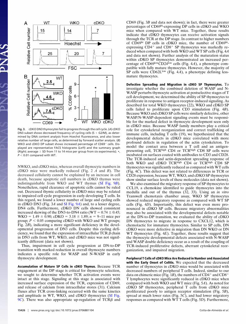
NWKO, and cDKO mice, whereas overall thymocyte numbers incDKO mice were markedly reduced (Fig. 2 A and B). Thedecreased cellularity cannot be explained by an increase in celldeath, because apoptotic cell numbers in cDKO thymus wereindistinguishable from WKO and WT thymus (SI Fig. 9B).Nonetheless, rapid clearance of apoptotic cells cannot be ruledout. Decreased thymic cellularity in cDKO mice may be relatedto impaired cell cycle progression in early developing T cells. Inthis regard, we found a lower number of large and cycling cellsin cDKO DN3 (Fig. 3A and SI Fig. 9A) and, to a lesser degree,DN4 cells. Furthermore, cDKO DN cells showed consistentincreased skewing of the DN3-to-DN4 ratio (WT � 0.74 � 0.45;WKO � 1.49 � 0.90; cDKO � 3.18 � 1.89; n � 9–11 mice pergroup; P � 0.05 comparing cDKO with WKO and WT groups;Fig. 2B), indicating a more significant deficiency in the devel-opmental progression of DN3 cells. Despite this cycling defi-ciency, we found that the expression of intracellular TCR � chainin DN3 cells from WT, WKO, and cDKO mice was not signif-icantly different (data not shown).
Thus, impairment in cell cycle progression at DN-to-DPtransition with marked reduction in overall thymocyte numbersindicates a specific role for WASP and N-WASP in earlythymocyte development.
Accumulation of Mature SP Cells in cDKO Thymus. Because TCRengagement at the DP stage is critical for thymocyte selection,we sought to determine whether TCR activation events wereintact at this stage. Signaling at this stage is associated withincreased surface expression of the TCR, expression of CD69,and release of calcium from intracellular stores (31). Calciumfluxes after TCR cross-linking occurred with the same kineticsand amplitude in WT, WKO, and cDKO thymocytes (SI Fig.9C). There was also appropriate up-regulation of TCR� and
CD69 (Fig. 3B and data not shown); in fact, there were greaterpercentages of CD69hi-expressing DP cells in cDKO and WKOmice when compared with WT mice. Together, these resultsindicate that cDKO thymocytes can receive activation signalsthrough the TCR at the DP stage. In contrast to higher numbersof CD69hi DP cells in cDKO mice, the number of CD69hi-expressing CD4� and CD8� SP thymocytes was markedly re-duced when compared with both WKO and WT SP cells (Fig. 4Aand data not shown). Further analysis of the maturation statuswithin cDKO SP thymocytes demonstrated an increased per-centage of CD69lowCD24low cells (Fig. 4A), a phenotype com-patible with fully mature thymocytes. However, the majority ofSP cells were CD62Llow (Fig. 4A), a phenotype defining less-mature thymocytes.
Defective Spreading and Migration in cDKO SP Thymocytes. Toinvestigate whether the combined deletion of WASP and N-WASP perturbs thymocyte activation at postselective stages of Tcell development, we determined the ability of SP thymocytes toproliferate in response to antigen receptor-induced signaling. Asdescribed for total WKO thymocytes (22), WKO and cDKO SPcells failed to proliferate upon CD3 stimulation (Fig. 4B).Because WKO and cDKO SP cells were similarly defective, otherWASP/N-WASP-dependent signaling events must be responsi-ble for the marked defect in thymocyte development seen onlyin cDKO mice. Because WASP family members play a criticalrole for cytoskeletal reorganization and correct trafficking ofimmune cells, including T cells (19), we hypothesized that thedevelopmental defects in cDKO mice correlated with moreprofound defects in regulation of the actin cytoskeleton. Tomodel the contact area between a T cell and an antigen-presenting cell, TCRhigh CD4 or TCRhigh CD8 SP cells wereincubated on surfaces coated with antibodies to CD3 and CD28.The TCR-induced and actin-dependent spreading response ofboth WKO and cDKO TCRhigh CD4 or TCRhigh CD8 SPthymocytes was significantly reduced as compared with WT cells(Fig. 4C). This defect was not related to differences in TCR orCD28 expression, because WT, WKO, and cDKO SP thymocytesshow similar surface levels of these molecules (data not shown).
We also examined the migratory response of SP thymocytes toCCL19, a chemokine identified to guide thymocytes into themedulla and out of the thymus (32, 33). Using an in vitroTranswell chemotaxis chamber assay, WKO SP thymocytesshowed reduced migratory response as compared with WT SPcells (Fig. 4D). Importantly, this defect was even more pro-nounced in cDKO SP cells. Because migratory abnormalitiesmay also be associated with the developmental defects notableat the DN-to-DP transition, we evaluated the ability of cDKODN thymocytes to migrate in response to CXCL12, which ischemotactic for immature thymocytes. Similar to SP cells, DNcDKO were more defective in migration than DN WKO or DNWT thymocytes (Fig. 4E). Together, these results suggest thatthe thymocyte developmental defects associated with N-WASPand WASP double deficiency occur as a result of the coupling ofTCR-induced proliferative defects, aberrant cytoskeletal reor-ganization, and impaired migration.
Peripheral T Cells of cDKO Mice Are Reduced in Number and Associatedwith the Early Onset of Colitis. We expected that the decreasednumber of thymocytes in cDKO mice would be associated withdecreased numbers of peripheral T cells. Indeed, similar to ourdata on chimeric mice (Fig. 1B), the numbers of CD4� and CD8�
T lymphocytes were significantly reduced in cDKO mice whencompared with both WKO and WT mice (Fig. 5A). As noted forcDKO SP thymocytes, peripheral T cells from cDKO miceproliferated poorly to antigen receptor stimulation (Fig. 5B),spread at much lower rates (Fig. 5C), and had lower migratoryresponses as compared with WT T cells (Fig. 5D). Furthermore,
Fig. 3. cDKO DN3 thymocytes fail to progress through the cell cycle. (A) cDKODN3 subset shows decreased frequency of cycling cells (S � G2/M), as deter-mined by DNA content analysis from Hoechst fluorescence, and also lowerrelative number of large cells, as determined by forward scatter analysis. (B)WKO and cDKO DP subset shows increased percentage of CD69� cells. Dis-played are representative FACS histograms (Left) and the summary graph(Right; average � SD from 11 to 14 mice per group from six experiments). *,P � 0.01 compared with WT.
15426 � www.pnas.org�cgi�doi�10.1073�pnas.0706881104 Cotta-de-Almeida et al.

CD4� T cells from cDKO mice were predominantlyCD44�CD62L�, a phenotype of effector/memory cells (SI Fig.10A). Recently, we identified a reduction in the number andfunction of naturally occurring regulatory T cells (CD4�CD25�
FoxP3�) in WKO mice (34). In cDKO mice, we observed asimilar defect when compared with WT mice [percent CD25�
cells in CD4� SP thymocytes: cDKO, 2.25 � 1.21 (n � 6); WKO,2.59 � 0.38 (n � 4); WT, 5.81 � 0.53 (n � 4)]. Because the totalnumbers of CD4� SP thymocytes are markedly reduced incDKO, the absolute numbers of nTregs in cDKO mice weredramatically reduced in comparison with WT and WKO (datanot shown). Interestingly, these aberrant numbers of effector andregulatory T cells were associated with more severe earlier onsetof colitis in cDKO mice when compared with WKO mice, withthe majority of animals developing clinical (SI Fig. 10B) andhistopathologic (Fig. 5E) signs of colitis by 2 months of age.
DiscussionIn this study, we sought to determine the unique and redundantfunctions of WASP and N-WASP in T cell development. BecauseWASP plays a critical role in peripheral T cell activation andcytoskeletal regulation, it seemed somewhat surprising to us that
Fig. 4. Reduced CD69 up-regulation, proliferation, spreading, and migra-tion in cDKO SP thymocytes. (A) Representative FACS profiles show decreasedCD69 and CD62L expression and increased percentage of CD69lowCD24low
subset within cDKO CD4� SP thymocytes. (B) Proliferative response to anti-CD3plus IL-2 stimulation is similarly reduced in WKO and cDKO SP thymocytes. Barsare representative mean values of cpm ([3H]thymidine) � SD of triplicate wellsfrom one of three independent experiments. *, P � 0.01 compared with WT.(C) Antigen-receptor-induced spreading is markedly decreased in both WKOand cDKO SP thymocytes. (Left) Phalloidin staining [�40 (Top) and �100(Bottom)] depicts the formation of large sheet-like lamellipodia of spread WTSP thymocytes (white arrowheads), which are absent in the representativefields from WKO and cDKO cells. (Right) Graphs show the average of relativenumbers of spread TCRhigh CD4 and TCRhigh CD8 SP thymocytes (�SD) repre-sentative of two experiments. *, P � 0.01 compared with WT. (D) CCL19-drivenmigratory activity of cDKO SP thymocytes is reduced. The percentage ofmigrating cells is shown as mean values (�SD) from at least two experiments(n � 3–4 mice per group). *, P � 0.05 when compared with WT. (E) CXCL12-driven transmigration of cDKO DN cells is reduced. The percentage of migrat-ing cells is shown as mean values (�SD) from two experiments (n � 3–4 miceper group). *, P � 0.05 compared with WT.
Fig. 5. Reduced number and activation of T cells in the periphery and earlyonset of colitis in cDKO mice. (A) CD4� and CD8� T cell numbers in spleens ofcDKO mice are significantly reduced. Bars represent average of absolutelymphocyte numbers (�SD) (n � 4–6 mice per group). *, P � 0.01 comparedwith WT and WKO. (B) T cells purified from lymph nodes of WKO and cDKOmice show marked decrease in proliferative response to anti-CD3 plus IL-2stimulation. Bars indicate mean values of cpm ([3H]thymidine) � SD of tripli-cate wells from one of four experiments. *, P � 0.01 compared with WT. (C)Antigen-receptor-induced spreading is markedly decreased in WKO and cDKOperipheral T cells. Bar graphs show the average of relative numbers of spreadT cells from triplicate samples (�SD) representative of three experiments. *,P � 0.01 compared with WT. (D) CCL19-induced chemotaxis of peripheral Tcells is significantly diminished in both WKO and cDKO mice. Data are shownas average � SD of triplicates from one of two experiments. (E) Histologicalanalyses of distal colons show signs of severe inflammation with crypt elon-gation and mucosal thickening in samples from cDKO mice. Age-matched (2months) WT and WKO mice show no sign of colitis in this representativeexperiment.
Cotta-de-Almeida et al. PNAS � September 25, 2007 � vol. 104 � no. 39 � 15427
IMM
UN
OLO
GY

T cell development is relatively normal in WAS patients and inWASP-deficient mice. We hypothesized that N-WASP mightplay redundant roles with WASP in some of the signaling eventsrequired for T cell development.
We have deleted WASP and N-WASP in T cells by using twodifferent gene-targeting approaches, RAG-2-deficient blasto-cyst complementation assay and conditional deletion by Crerecombinase. Deletion of WASP and N-WASP severely com-promised T cell development with reduced numbers of thymo-cytes and peripheral T cells. In contrast, T cell numbers weresimilar to normal in WKO and NWKO mice, suggesting thatWASP and N-WASP have overlapping functions during T celldevelopment. We found slight differences in results using the twoapproaches for deletion. The RAG-2 chimeric animals showeda block in progression from the DN3-to-DN4 stage. Our datawith the conditional deletion approach also showed a relativeblock in the DN3-to-DN4 transition, but the defect was lesspronounced. Although we failed to detect N-WASP by Westernblotting of total thymocytes in cDKO mice, some residualN-WASP expression might be present at the DN3 and DN4stage, because the lck promoter seems to drive homogeneousand higher expression of Cre recombinase at the DN3 stage (30).Thus, whereas deletion of N-WASP occurs within the thymus incDKO mice, WASP and N-WASP are deleted in lymphoidprogenitors before their arrival in the thymus in the RAG-2chimeric mice. Notably, WASP-deficient bone marrow-derivedhematopoietic stem cells were shown to have defective chemo-taxis and homing properties (35).
Our study suggests a complex regulation of T cell maturationby WASP and N-WASP. Despite marked hypocellularity and apartial block in progression at the DN stage in cDKO mice, theexisting DP cells responded to TCR activation, as determined byexpression of CD69, TCR�, and calcium fluxes. In markedcontrast, cDKO SP cells exhibited pronounced deficiency inTCR-triggered proliferation, spreading, and chemokine-drivenmigration. These results support a fundamental role for WASPand N-WASP for correct overall T cell development, whereastheir activity may be overlapping with other proteins for some ofthe receptor-mediated signaling events at different stages duringdevelopment. In this context, many functional activities are quitedistinct during the stages of DP and SP cells. For example, PKC-�is crucial for mature T cell activation but dispensable forTCR-dependent thymocyte development (36). Moreover, DPthymocytes seem to form an immunological synapse patterndistinct from that of mature T cells (37). Single-positive cellsrespond to TCR triggering by spreading, but freshly isolated DPcells fail to polarize actin and spread (38). Likewise, the migra-tory properties of cortical thymocytes change dramatically uponTCR engagement, changing from random and slow migrationpatterns to rapid and inwardly directed to the medulla (39).
Although WKO thymocytes have defective TCR-inducedproliferative response and spreading, thymocytes undergo fairlynormal development and maturation (20, 22). Thymocytes fromcDKO mice also have abolished proliferation and spreading, butin addition have more pronounced defects in migratory re-sponses and development. Our data are consistent with recentdata demonstrating that the thymocyte expression of a verprolinhomology, cofilin homology, and acidic (VCA) domain-deletedWASP resulted in a severe block in T cell development withdefective DN3 progression and impaired positive selection (40).However, in addition to reduced cell cycle progression at DN-to-DP transition in cDKO thymus, we also observed an accu-mulation of CD69lowCD24low and CD62Llow SP cells that mightindicate retention of mature cells with reduced migratory func-tion (41). One potential unifying hypothesis is that deletion ofboth WASP and N-WASP results in defective intrathymic mi-gration, preventing cells from reaching specific locations to
receive appropriate stimulating signals and to egress from thethymus (42).
The importance of cytoskeletal polarization and remodelingfor lymphocyte activation has been highlighted (7, 43). Mole-cules that regulate the actin cytoskeleton upstream of WASPfamily members have been implicated as critical for thymocytedevelopment. Mice lacking Vav-1, a guanine exchanging factorfor Rho GTPases, share many features of WASP/N-WASPdouble-deleted mice by having thymic hypocellularity associatedwith a block at the DN3 stage of development (44). Likewise,cells that escape the DN3 block show inefficient selection,resulting in reduced numbers of peripheral T cells. Similar to ourfindings on cDKO SP cells, T cells from Vav-1-deficient micehave defective receptor-induced actin polymerization (45–47).Interestingly, Vav-1 deficiency can be largely corrected byexpression of activated Rac-1 (48), and Rac activation of actinrearrangements is mediated by WAVE proteins, moleculesclosely related to WASP and N-WASP (49). Together with ourresults, these studies provide compelling evidence for the im-portance of regulated actin remodeling for correct thymopoiesis.
Materials and MethodsSee also SI Text.
Animal Handling. Mice were housed at Children’s Hospital andMassachusetts General Hospital under specific pathogen-freeconditions. Animal experiments were carried out after approvaland in accordance with guidelines from the Subcommittee onResearch Animal Care of Massachusetts General Hospital.
Proliferation Analysis. Purified T cells (1 � 105 per well) wereadded to 96-well tissue culture plates and cultured in RPMImedium 1640, supplemented with 2 mM L-glutamine/1 mMsodium pyruvate/100 units/ml penicillin/streptomycin/50 �M2-mercapthoethanol/10% FCS. Murine IL-2 (5 ng/ml; BD Bio-sciences, San Jose, CA) was added to the wells where anti-CD3�stimulating antibody (10 �g/ml; clone 145-2C11; BD Bio-sciences) was previously allowed to bind (1 h at 37°C). To assessreceptor-independent activation, cells were stimulated withphorbol-myristate-acetate and ionomycin (Sigma–Aldrich, St.Louis, MO) at 10 ng/ml and 0.5 �M, respectively. T cells werecultured for 56–60 h and pulsed with 1 �Ci [3H]thymidine (1Ci � 37 GBq) for an additional 12–16 h, harvested, andscintillation counted.
Migration Assay. The migration of thymocytes to CCL19 andCXCL12 (R&D Systems, Minneapolis, MN) was determined ina Transwell chemotaxis chamber (5 �m; Corning, Lowell, MA).Total thymocytes (5 � 104) were diluted in RPMI medium 1640supplemented with 0.5% BSA and added to the upper chamber;25 nM CCL19 or 20 nM CXCL12 was added to the lowerchamber. Cells that migrated after 1.5 h at 37°C were collectedand counted by flow cytometry with unlabeled caliBRITE beads(BD Biosciences) as reference counting. Cells were labeled withanti-CD4 and -CD8 to determine the percentage of DP and SPcells.
Thymocyte-Spreading Assay. To determine the spreading responseof TCRhigh CD4 and TCRhigh CD8 SP cells (isolated by FACS)stimulating antibodies to CD3 and CD28 (clones 145-2C11 and37.51, respectively, both at 10 �g/ml; BD Biosciences) wereallowed to bind for 10 min at 37°C onto glass slides (precoatedwith poly-L-lysine). Cells were fixed with 3.8% paraformalde-hyde for 10 min at room temperature (RT) and permeabilizedwith PBS/0.1% Triton X-100 for 5 min at RT. Actin was stainedwith AlexaFluor 488-conjugated phalloidin (Molecular Probes/Invitrogen, Carlsbad, CA) for 30 min at RT. Spread cells weredefined as having a flattened appearance and formation of
15428 � www.pnas.org�cgi�doi�10.1073�pnas.0706881104 Cotta-de-Almeida et al.
actin-rich broad lamellipodia as compared with nonspread roundcells. The percentage of spread cells of the total number of cellswas calculated by counting 300–400 cells per condition intriplicate or quadruplicate at �100 magnification.
Statistical Analyses. Statistical analyses were performed by usingraw data or arcsin-transformed percentage data with the Graph-Pad Prism program (ver. 4.00, GraphPad, San Diego, CA).Comparison among all groups was done by one-way ANOVA orthe Kruskal–Wallis test, and differences among group pairs weredetermined by unpaired Student’s t test or the Mann–Whitneytest. Differences were considered significant when P � 0.05.
We thank Drs. Raif Geha, Daniel K. Podolsky, Vijay Yajnik, EileenRemold-O’Donnell, and Cathy Nagler and the members of the labora-tories of S.B.S., V. Yajnik (Massachusetts General Hospital, Boston,MA) and R. Geha (Children’s Hospital, Boston, MA) for critical readingand helpful discussions. We also thank Yuko Fujiwara (Children’sHospital) for blastocyst injections, David Dombkowski for cell sortingand cell cycle analysis, and Florentina Anastasoaie and Long Hang forassistance. This work was supported by the National Institutes of Health[Grants HL59561 (to F.W.A. and S.B.S.) and AI50950 (to S.B.S.)], theGerman Research Council (D.O.), and the Wenner–Gren Foundation(L.W.); by postdoctoral support from the SICPA Foundation andLausanne University Hospital (M.H.M.); and by CAPES/Brazil(V.C.-d-A.).
1. von Boehmer H, Aifantis I, Gounari F, Azogui O, Haughn L, Apostolou I,Jaeckel E, Grassi F, Klein L (2003) Immunol Rev 191:62–78.
2. Germain RN (2002) Nat Rev Immunol 2:309–322.3. Ladi E, Yin X, Chtanova T, Robey EA (2006) Nat Immunol 7:338–343.4. Petrie HT (2003) Nat Rev Immunol 3:859–866.5. Savino W, Mendes-da-Cruz DA, Silva JS, Dardenne M, Cotta-de-Almeida V
(2002) Trends Immunol 23:305–313.6. Norment AM, Bevan MJ (2000) Semin Immunol 12:445–455.7. Acuto O, Cantrell D (2000) Annu Rev Immunol 18:165–184.8. Badour K, Zhang J, Siminovitch KA (2003) Immunol Rev 192:98–112.9. Samstag Y, Eibert SM, Klemke M, Wabnitz GH (2003) J Leukocyte Biol
73:30–48.10. Cantrell DA (2003) Immunol Rev 192:122–130.11. Fujikawa K, Miletic AV, Alt FW, Faccio R, Brown T, Hoog J, Fredericks J,
Nishi S, Mildiner S, Moores SL, et al. (2003) J Exp Med 198:1595–1608.12. Henning S, Cleverley S (1999) Immunol Res 20:29–42.13. Badour K, Zhang J, Shi F, McGavin MK, Rampersad V, Hardy LA, Field D,
Siminovitch KA (2003) Immunity 18:141–154.14. Barda-Saad M, Braiman A, Titerence R, Bunnell SC, Barr VA, Samelson LE
(2005) Nat Immunol 6:80–89.15. Cannon JL, Burkhardt JK (2004) J Immunol 173:1658–1662.16. Dupre L, Aiuti A, Trifari S, Martino S, Saracco P, Bordignon C, Roncarolo MG
(2002) Immunity 17:157–166.17. Gallego MD, de la Fuente MA, Anton IM, Snapper S, Fuhlbrigge R, Geha RS
(2006) Int Immunol 18:221–232.18. Sasahara Y, Rachid R, Byrne MJ, de la Fuente MA, Abraham RT, Ramesh N,
Geha RS (2002) Mol Cell 10:1269–1281.19. Snapper SB, Meelu P, Nguyen D, Stockton BM, Bozza P, Alt FW, Rosen FS,
von Andrian UH, Klein C (2005) J Leukocyte Biol 77:993–998.20. Snapper SB, Rosen FS, Mizoguchi E, Cohen P, Khan W, Liu CH, Hagemann
TL, Kwan SP, Ferrini R, Davidson L, et al. (1998) Immunity 9:81–91.21. Westerberg L, Larsson M, Hardy SJ, Fernandez C, Thrasher AJ, Severinson E
(2005) Blood 105:1144–1152.22. Zhang J, Shehabeldin A, da Cruz LA, Butler J, Somani AK, McGavin M,
Kozieradzki I, dos Santos AO, Nagy A, Grinstein S, et al. (1999) J Exp Med190:1329–1342.
23. Park JY, Kob M, Prodeus AP, Rosen FS, Shcherbina A, Remold-O’Donnell E(2004) Clin Exp Immunol 136:104–110.
24. Takenawa T, Miki H (2001) J Cell Sci 114:1801–1809.25. Klein C, Nguyen D, Liu CH, Mizoguchi A, Bhan AK, Miki H, Takenawa T,
Rosen FS, Alt FW, Mulligan RC, Snapper SB (2003) Blood 101:2159–2166.26. Snapper SB, Takeshima F, Anton I, Liu CH, Thomas SM, Nguyen D, Dudley
D, Fraser H, Purich D, Lopez-Ilasaca M, et al. (2001) Nat Cell Biol 3:897–904.
27. Chen J, Lansford R, Stewart V, Young F, Alt FW (1993) Proc Natl Acad SciUSA 90:4528–4532.
28. Chen J, Alt FW (1994) in Transgenesis and Targeted Mutagenesis, ed BluthmannHO, P (Academic, San Diego), pp 35–49.
29. Kuhn R, Schwenk F, Aguet M, Rajewsky K (1995) Science 269:1427–1429.30. Lee PP, Fitzpatrick DR, Beard C, Jessup HK, Lehar S, Makar KW, Perez-
Melgosa M, Sweetser MT, Schlissel MS, Nguyen S, et al. (2001) Immunity15:763–774.
31. Mariathasan S, Bachmann MF, Bouchard D, Ohteki T, Ohashi PS (1998)J Immunol 161:6030–6037.
32. Kwan J, Killeen N (2004) J Immunol 172:3999–4007.33. Ueno T, Saito F, Gray DH, Kuse S, Hieshima K, Nakano H, Kakiuchi T, Lipp
M, Boyd RL, Takahama Y (2004) J Exp Med 200:493–505.34. Maillard MH, Cotta-de-Almeida V, Takeshima F, Nguyen DD, Michetti P,
Nagler C, Bhan AK, Snapper SB (2007) J Exp Med 204:381–391.35. Lacout C, Haddad E, Sabri S, Svinarchouk F, Garcon L, Capron C, Foudi A,
Mzali R, Snapper SB, Louache F, et al. (2003) Blood 102:1282–1289.36. Sun Z, Arendt CW, Ellmeier W, Schaeffer EM, Sunshine MJ, Gandhi L, Annes
J, Petrzilka D, Kupfer A, Schwartzberg PL, Littman DR (2000) Nature404:402–407.
37. Hailman E, Burack WR, Shaw AS, Dustin ML, Allen PM (2002) Immunity16:839–848.
38. Hailman E, Allen PM (2005) J Immunol 175:4847–4857.39. Witt CM, Raychaudhuri S, Schaefer B, Chakraborty AK, Robey EA (2005)
PLoS Biol 3:e160.40. Zhang J, Shi F, Badour K, Deng Y, McGavin MK, Siminovitch KA (2002) Proc
Natl Acad Sci USA 99:2240–2245.41. Carlson CM, Endrizzi BT, Wu J, Ding X, Weinreich MA, Walsh ER, Wani MA,
Lingrel JB, Hogquist KA, Jameson SC (2006) Nature 442:299–302.42. Kurobe H, Liu C, Ueno T, Saito F, Ohigashi I, Seach N, Arakaki R, Hayashi
Y, Kitagawa T, Lipp M, et al. (2006) Immunity 24:165–177.43. Dustin ML, Cooper JA (2000) Nat Immunol 1:23–29.44. Turner M, Mee PJ, Walters AE, Quinn ME, Mellor AL, Zamoyska R,
Tybulewicz VL (1997) Immunity 7:451–460.45. Fischer K-D, Kong Y-Y, Nishina H, Tedfor D, Manengere LEM, Kozieradzki
I, Sasaki T, Starr M, Chan G, Gardener S, et al. (1998) Curr Biol 8:554–562.46. Holsinger LJ, Graef IA, Swat W, Chi T, Bautista DM, Davidson L, Lewis RS,
Alt FW, Crabtree GR (1998) Curr Biol 8:563–572.47. Wulfing C, Bauch A, Crabtree GR, Davis MM (2000) Proc Natl Acad Sci USA
97:10150–10155.48. Gomez M, Tybulewicz V, Cantrell DA (2000) Nat Immunol 1:348–352.49. Miki H, Suetsugu S, Takenawa T (1998) EMBO J 17:6932–6941.
Cotta-de-Almeida et al. PNAS � September 25, 2007 � vol. 104 � no. 39 � 15429
IMM
UN
OLO
GY