Why Learn About Neonatal EEG? - University of South …. Robert Clancy & Eilon Shany: Jan. 18, 2008...
13
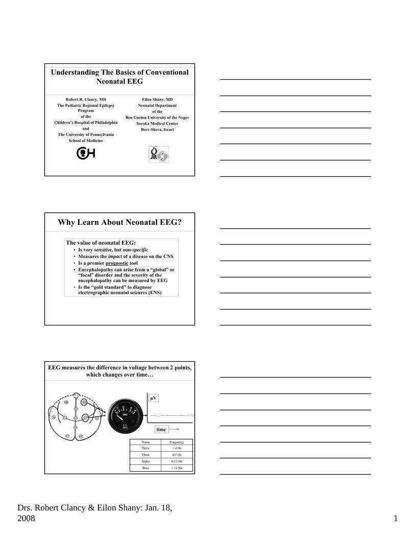
Drs. Robert Clancy & Eilon Shany: Jan. 18, 2008 1 Understanding The Basics of Conventional Neonatal EEG Robert R. Clancy, MD The Pediatric Regional Epilepsy Program of the Children’s Hospital of Philadelphia and The University of Pennsylvania School of Medicine Eilon Shany, MD Neonatal Department of the Ben Gurion University of the Negev Soroka Medical Center Beer-Sheva, Israel Why Learn About Neonatal EEG? The value of neonatal EEG: • Is very sensitive, but non-specific • Measures the impact of a disease on the CNS • Is a premier prognostic tool • Encephalopathy can arise from a “global” or “focal” disorder and the severity of the encephalopathy can be measured by EEG • Is the “gold standard” to diagnose electrographic neonatal seizures (ENS) EEG measures the difference in voltage between 2 points, which changes over time… Name Frequency Delta < 4 Hz Theta 4-7 Hz Alpha 8-12 Hz Beta > 12 Hz time μV
Transcript of Why Learn About Neonatal EEG? - University of South …. Robert Clancy & Eilon Shany: Jan. 18, 2008...
Drs. Robert Clancy & Eilon Shany: Jan. 18, 2008 1
Understanding The Basics of Conventional Neonatal EEG
Robert R. Clancy, MDThe Pediatric Regional Epilepsy
Programof the
Children’s Hospital of Philadelphiaand
The University of PennsylvaniaSchool of Medicine
Eilon Shany, MDNeonatal Department
of theBen Gurion University of the Negev
Soroka Medical CenterBeer-Sheva, Israel
Why Learn About Neonatal EEG?
The value of neonatal EEG:• Is very sensitive, but non-specific• Measures the impact of a disease on the CNS• Is a premier prognostic tool• Encephalopathy can arise from a “global” or
“focal” disorder and the severity of the encephalopathy can be measured by EEG
• Is the “gold standard” to diagnose electrographic neonatal seizures (ENS)
EEG measures the difference in voltage between 2 points, which changes over time…
Name Frequency
Delta < 4 Hz
Theta 4-7 Hz
Alpha 8-12 Hz
Beta > 12 Hz
time
µV
Drs. Robert Clancy & Eilon Shany: Jan. 18, 2008 2
EEG Voltage Fields: Phase Reversals
(Olejniczak, Journal of Clinical Neurophysiology, 2006)
μV
Time
A→B
B→C
A
B
C
Montage: An Array or Collection of Electrode Couples
10-20 system:
Full array
10-20 system:
modified for neonates
aEEG
(P3 →P4)
Conventional Versus Limited Channel EEG monitoring
Drs. Robert Clancy & Eilon Shany: Jan. 18, 2008 3
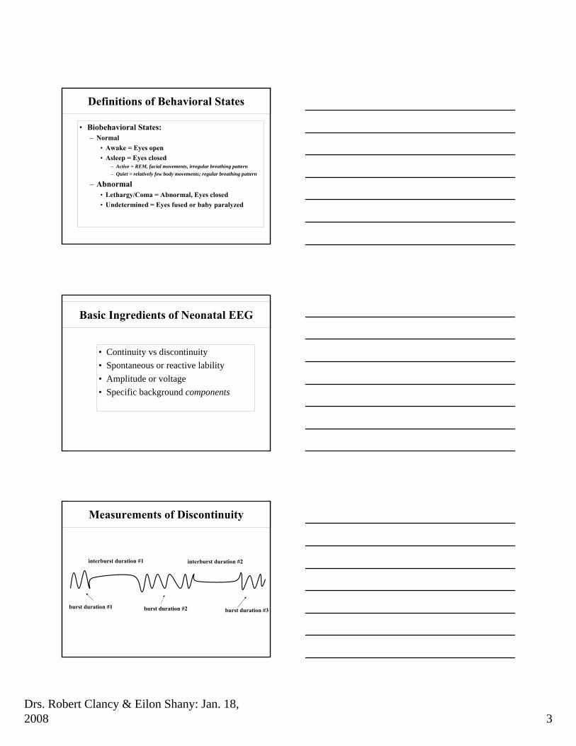
Definitions of Behavioral States
• Biobehavioral States:– Normal
• Awake = Eyes open• Asleep = Eyes closed
– Active = REM, facial movements, irregular breathing pattern – Quiet = relatively few body movements; regular breathing pattern
– Abnormal• Lethargy/Coma = Abnormal, Eyes closed• Undetermined = Eyes fused or baby paralyzed
Basic Ingredients of Neonatal EEG
• Continuity vs discontinuity• Spontaneous or reactive lability• Amplitude or voltage• Specific background components
Measurements of Discontinuity
burst duration #1
interburst duration #2interburst duration #1
burst duration #3burst duration #2
Drs. Robert Clancy & Eilon Shany: Jan. 18, 2008 4
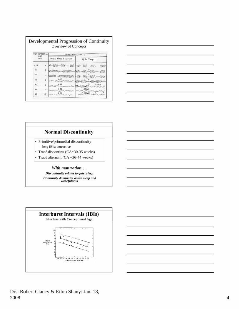
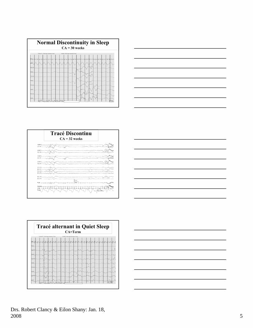
Developmental Progression of ContinuityOverview of Concepts
Quiet SleepActive Sleep & Awake
Normal Discontinuity
• Primitive/primordial discontinuity– long IBIs; unreactive
• Tracé discontinu (CA~30-35 weeks)• Tracé alternant (CA ~36-44 weeks)
With maturation….Discontinuity relates to quiet sleep
Continuity dominates active sleep and wakefulness
Interburst Intervals (IBIs)Shortens with Conceptional Age
Drs. Robert Clancy & Eilon Shany: Jan. 18, 2008 5
Normal Discontinuity in SleepCA = 30 weeks
Tracé DiscontinuCA = 32 weeks
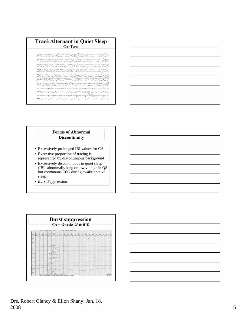
Tracé alternant in Quiet SleepCA=Term
Drs. Robert Clancy & Eilon Shany: Jan. 18, 2008 6
Tracé Alternant in Quiet SleepCA=Term
Forms of Abnormal Discontinuity
• Excessively prolonged IBI values for CA• Excessive proportion of tracing is
represented by discontinuous background• Excessively discontinuous in quiet sleep
(IBIs abnormally long or low voltage in QS but continuous EEG during awake / active sleep)
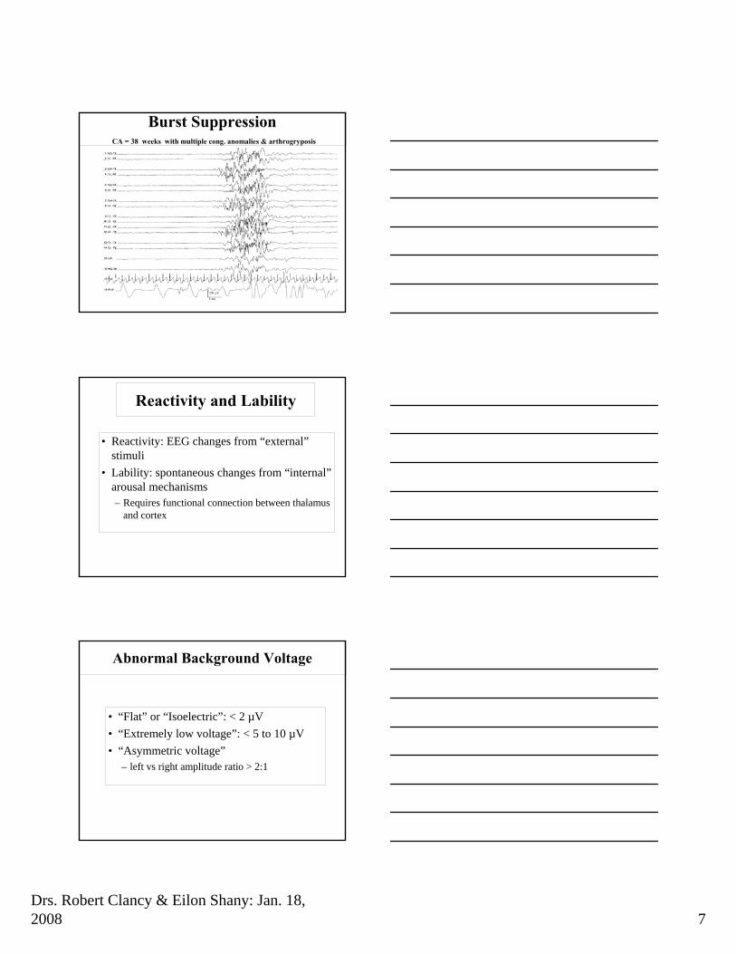
• Burst Suppression
Burst suppressionCA = 42weeks 2º to HIE
Drs. Robert Clancy & Eilon Shany: Jan. 18, 2008 7
Burst SuppressionCA = 38 weeks with multiple cong. anomalies & arthrogryposis
Reactivity and Lability
• Reactivity: EEG changes from “external”stimuli
• Lability: spontaneous changes from “internal”arousal mechanisms– Requires functional connection between thalamus
and cortex
Abnormal Background Voltage
• “Flat” or “Isoelectric”: < 2 µV• “Extremely low voltage”: < 5 to 10 µV• “Asymmetric voltage”
– left vs right amplitude ratio > 2:1
Drs. Robert Clancy & Eilon Shany: Jan. 18, 2008 8
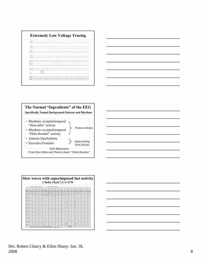
Extremely Low Voltage Tracing
The Normal “Ingredients” of the EEGSpecifically Named Background Patterns and Rhythms
• Rhythmic occipital/temporal “theta delta” activity
• Rhythmic occipital/temporal “Delta Brushes” activity
• Anterior Dysrhythmia• Encoches Frontales Approaching
Term Infants
Preterm Infants
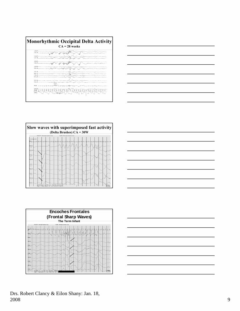
With MaturationFrom Slow Delta and Theta to faster “Delta Brushes”
Slow waves with superimposed fast activity(“Delta Theta”) CA=27W
Drs. Robert Clancy & Eilon Shany: Jan. 18, 2008 9
Monorhythmic Occipital Delta ActivityCA = 28 weeks
Slow waves with superimposed fast activity(Delta Brushes) CA = 30W
Encoches Frontales(Frontal Sharp Waves)
The Term Infant
Drs. Robert Clancy & Eilon Shany: Jan. 18, 2008 10
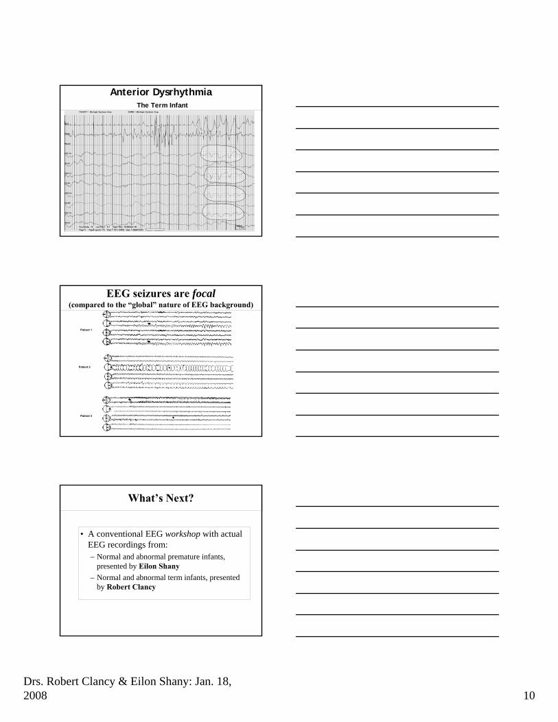
Anterior DysrhythmiaThe Term Infant
Patient 1
Patient 2
Patient 3
EEG seizures are focal(compared to the “global” nature of EEG background)
What’s Next?
• A conventional EEG workshop with actual EEG recordings from:– Normal and abnormal premature infants,
presented by Eilon Shany– Normal and abnormal term infants, presented
by Robert Clancy
Drs. Robert Clancy & Eilon Shany: Jan. 18, 2008 11
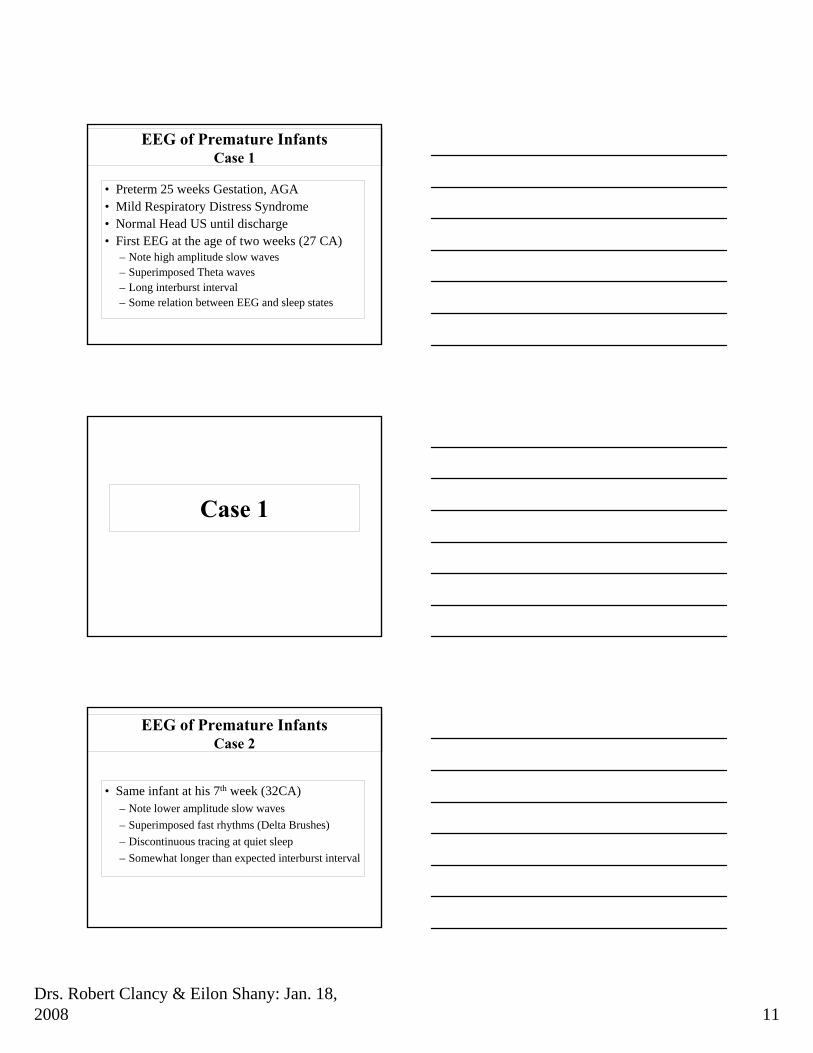
EEG of Premature InfantsCase 1
• Preterm 25 weeks Gestation, AGA• Mild Respiratory Distress Syndrome• Normal Head US until discharge• First EEG at the age of two weeks (27 CA)
– Note high amplitude slow waves– Superimposed Theta waves– Long interburst interval– Some relation between EEG and sleep states
Case 1
EEG of Premature InfantsCase 2
• Same infant at his 7th week (32CA)– Note lower amplitude slow waves– Superimposed fast rhythms (Delta Brushes)– Discontinuous tracing at quiet sleep– Somewhat longer than expected interburst interval
Drs. Robert Clancy & Eilon Shany: Jan. 18, 2008 12
Case 2
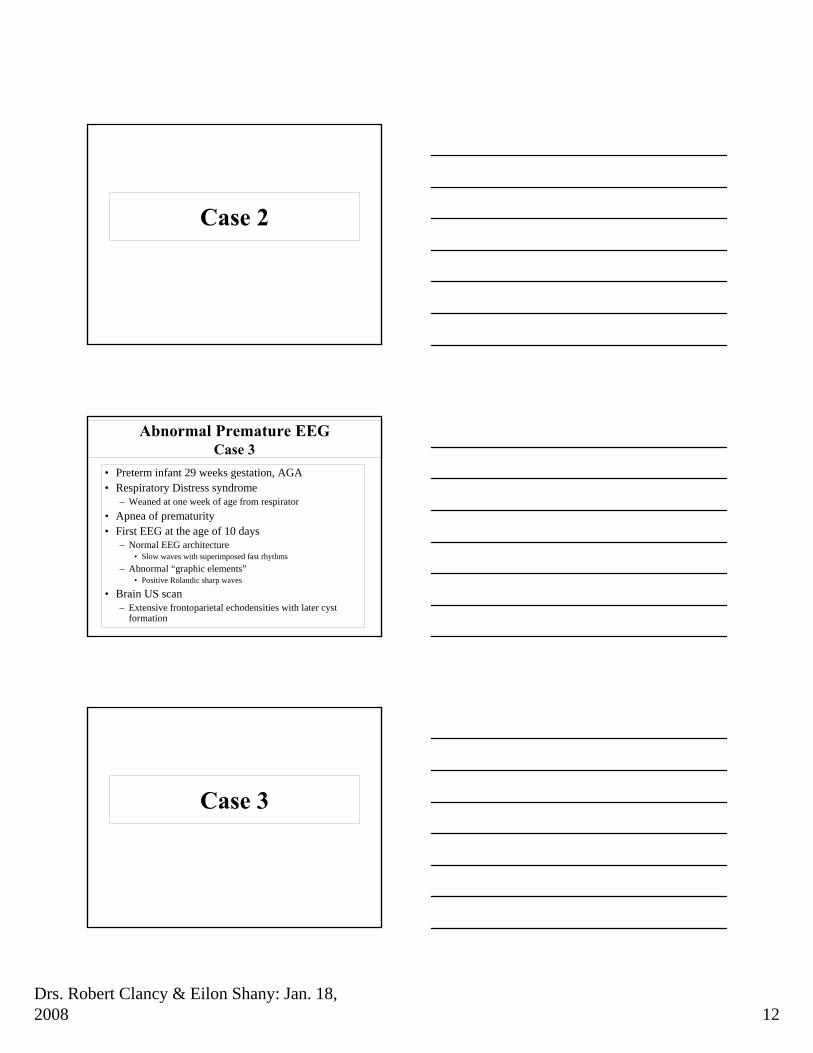
Abnormal Premature EEGCase 3
• Preterm infant 29 weeks gestation, AGA• Respiratory Distress syndrome
– Weaned at one week of age from respirator• Apnea of prematurity• First EEG at the age of 10 days
– Normal EEG architecture• Slow waves with superimposed fast rhythms
– Abnormal “graphic elements”• Positive Rolandic sharp waves
• Brain US scan– Extensive frontoparietal echodensities with later cyst
formation
Case 3
Drs. Robert Clancy & Eilon Shany: Jan. 18, 2008 13
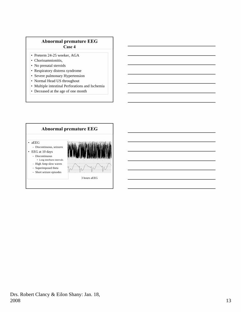
Abnormal premature EEGCase 4
• Preterm 24-25 weeker, AGA• Chorioamnionitis, • No prenatal steroids• Respiratory distress syndrome• Severe pulmonary Hypertension• Normal Head US throughout• Multiple intestinal Perforations and Ischemia• Deceased at the age of one month
Abnormal premature EEG
• aEEG– Discontinuous, seizures
• EEG at 10 days– Discontinuous
• Long interburst intervals
– High Amp slow waves– Superimposed theta– Short seizure episodes
3 hours aEEG