White matter structural connectivity and episodic memory ...
13
Contents lists available at ScienceDirect Developmental Cognitive Neuroscience journal homepage: www.elsevier.com/locate/dcn White matter structural connectivity and episodic memory in early childhood Chi T. Ngo a, ⁎ , Kylie H. Alm a , Athanasia Metoki a , William Hampton a , Tracy Riggins b , Nora S. Newcombe a , Ingrid R. Olson a a Temple University, United States b University of Maryland, College Park, United States ARTICLE INFO Keywords: White matter Memory development Episodic memory Diffusion weighted imaging ABSTRACT Episodic memory undergoes dramatic improvement in early childhood; the reason for this is poorly understood. In adults, episodic memory relies on a distributed neural network. Key brain regions that supporting these processes include the hippocampus, portions of the parietal cortex, and portions of prefrontal cortex, each of which shows different developmental profiles. Here we asked whether developmental differences in the axonal pathways connecting these regions may account for the robust gains in episodic memory in young children. Using diffusion weighted imaging, we examined whether white matter connectivity between brain regions implicated in episodic memory differed with age, and were associated with memory performance differences in 4- and 6-year-old children. Results revealed that white matter connecting the hippocampus to the inferior parietal lobule significantly predicted children’s performance on episodic memory tasks. In contrast, variation in the white matter connecting the hippocampus to the medial prefrontal cortex did not relate to memory per- formance. These findings suggest that structural connectivity between the hippocampus and lateral parietal regions is relevant to the development of episodic memory. 1. Introduction Remembering a past event and the specific spatiotemporal context in which the event occurred is a hallmark of episodic memory. Early child- hood marks an important developmental period for episodic memory, as substantial growth in this ability is observed. Many studies have shown robust age differences between 4- and 6-year-old children, with 4-year- olds performing worse than 6-year-olds on tasks that require relational memory, i.e., memory linking multiple items (Drummey and Newcombe, 2002; Lloyd et al., 2009; Newcombe et al., 2014; Ngo et al., 2017; Sluzenski et al., 2006), or memory for contextual details (Bauer et al., 2012; Riggins, 2014; Riggins et al., 2015; Riggins and Rollins, 2015). The enhancement in episodic memory during childhood is thought to rely, at least in part, on complex and dynamic developmental changes in the brain, in an interplay with social and other cognitive factors (Riggins, 2012). Understanding the neural bases of episodic memory development requires investigation of the relation among key regions of episodic memory, including the hippocampus, the parietal cortex, and the pre- frontal cortex. The goal of our study was to better understand this inter- action by examining the structural connectivity among these brain areas via white matter pathways. In the last two decades, there have been substantial efforts in characterizing the developmental profiles of white matter pathways in the brain. Convergent findings from cross-sectional (e.g., Bonekamp et al., 2007; Lebel et al., 2008; Loenneker et al., 2011; Moon et al., 2011; Qiu et al., 2010; Rollins et al., 2010; Sadeghi et al., 2014) and longitudinal studies (e.g., Krogsrud et al., 2016; Lebel and Beaulieu, 2011; Simmonds et al., 2014) show a protracted timeline of white matter development from early childhood until adulthood, with dif- ferential maturational rate across white matter tracts (reviewed in Lebel et al., 2017). It is believed that the information transmission properties of any given white matter tract can be predicted by the function of the gray matter regions that it connects (Maunsell and van Essen, 1983; Passingham et al., 2002). Thus, it is likely that specific white matter pathways connecting brain regions implicated in episodic memory should play a role in age-related improvements in memory performance in children. The focus of this paper is to examine such relations. An essential role of the hippocampus is to construct relational memories by binding together multiple elements of an event to form a cohesive episode (Backus et al., 2016; Cohen and Eichenbaum, 1993; Horner and Doeller, 2017). Developmental changes in hippocampal structure and function relate to improvement in episodic memory in https://doi.org/10.1016/j.dcn.2017.11.001 Received 21 August 2017; Received in revised form 7 November 2017; Accepted 7 November 2017 ⁎ Corresponding author at: Department of Psychology, Temple University, 1701 North 13th Street, Philadelphia, PA 19122, United States. E-mail address: [email protected] (C.T. Ngo). Developmental Cognitive Neuroscience 28 (2017) 41–53 Available online 16 November 2017 1878-9293/ © 2017 The Authors. Published by Elsevier Ltd. This is an open access article under the CC BY-NC-ND license (http://creativecommons.org/licenses/BY-NC-ND/4.0/). T
Transcript of White matter structural connectivity and episodic memory ...

Contents lists available at ScienceDirect
Developmental Cognitive Neuroscience
journal homepage: www.elsevier.com/locate/dcn
White matter structural connectivity and episodic memory in earlychildhood
Chi T. Ngoa,⁎, Kylie H. Alma, Athanasia Metokia, William Hamptona, Tracy Rigginsb,Nora S. Newcombea, Ingrid R. Olsona
a Temple University, United StatesbUniversity of Maryland, College Park, United States
A R T I C L E I N F O
Keywords:White matterMemory developmentEpisodic memoryDiffusion weighted imaging
A B S T R A C T
Episodic memory undergoes dramatic improvement in early childhood; the reason for this is poorly understood.In adults, episodic memory relies on a distributed neural network. Key brain regions that supporting theseprocesses include the hippocampus, portions of the parietal cortex, and portions of prefrontal cortex, each ofwhich shows different developmental profiles. Here we asked whether developmental differences in the axonalpathways connecting these regions may account for the robust gains in episodic memory in young children.Using diffusion weighted imaging, we examined whether white matter connectivity between brain regionsimplicated in episodic memory differed with age, and were associated with memory performance differences in4- and 6-year-old children. Results revealed that white matter connecting the hippocampus to the inferiorparietal lobule significantly predicted children’s performance on episodic memory tasks. In contrast, variation inthe white matter connecting the hippocampus to the medial prefrontal cortex did not relate to memory per-formance. These findings suggest that structural connectivity between the hippocampus and lateral parietalregions is relevant to the development of episodic memory.
1. Introduction
Remembering a past event and the specific spatiotemporal context inwhich the event occurred is a hallmark of episodic memory. Early child-hood marks an important developmental period for episodic memory, assubstantial growth in this ability is observed. Many studies have shownrobust age differences between 4- and 6-year-old children, with 4-year-olds performing worse than 6-year-olds on tasks that require relationalmemory, i.e., memory linking multiple items (Drummey and Newcombe,2002; Lloyd et al., 2009; Newcombe et al., 2014; Ngo et al., 2017;Sluzenski et al., 2006), or memory for contextual details (Bauer et al.,2012; Riggins, 2014; Riggins et al., 2015; Riggins and Rollins, 2015). Theenhancement in episodic memory during childhood is thought to rely, atleast in part, on complex and dynamic developmental changes in thebrain, in an interplay with social and other cognitive factors (Riggins,2012). Understanding the neural bases of episodic memory developmentrequires investigation of the relation among key regions of episodicmemory, including the hippocampus, the parietal cortex, and the pre-frontal cortex. The goal of our study was to better understand this inter-action by examining the structural connectivity among these brain areasvia white matter pathways.
In the last two decades, there have been substantial efforts incharacterizing the developmental profiles of white matter pathways inthe brain. Convergent findings from cross-sectional (e.g., Bonekampet al., 2007; Lebel et al., 2008; Loenneker et al., 2011; Moon et al.,2011; Qiu et al., 2010; Rollins et al., 2010; Sadeghi et al., 2014) andlongitudinal studies (e.g., Krogsrud et al., 2016; Lebel and Beaulieu,2011; Simmonds et al., 2014) show a protracted timeline of whitematter development from early childhood until adulthood, with dif-ferential maturational rate across white matter tracts (reviewed in Lebelet al., 2017). It is believed that the information transmission propertiesof any given white matter tract can be predicted by the function of thegray matter regions that it connects (Maunsell and van Essen, 1983;Passingham et al., 2002). Thus, it is likely that specific white matterpathways connecting brain regions implicated in episodic memoryshould play a role in age-related improvements in memory performancein children. The focus of this paper is to examine such relations.
An essential role of the hippocampus is to construct relationalmemories by binding together multiple elements of an event to form acohesive episode (Backus et al., 2016; Cohen and Eichenbaum, 1993;Horner and Doeller, 2017). Developmental changes in hippocampalstructure and function relate to improvement in episodic memory in
https://doi.org/10.1016/j.dcn.2017.11.001Received 21 August 2017; Received in revised form 7 November 2017; Accepted 7 November 2017
⁎ Corresponding author at: Department of Psychology, Temple University, 1701 North 13th Street, Philadelphia, PA 19122, United States.E-mail address: [email protected] (C.T. Ngo).
Developmental Cognitive Neuroscience 28 (2017) 41–53
Available online 16 November 20171878-9293/ © 2017 The Authors. Published by Elsevier Ltd. This is an open access article under the CC BY-NC-ND license (http://creativecommons.org/licenses/BY-NC-ND/4.0/).
T

school-aged children (e.g., DeMaster et al., 2013; DeMaster and Ghetti,2013; Ofen et al., 2007; reviewed in Ghetti and Bunge, 2012). Graymatter volume of the hippocampal head predicts children’s ability torecall contexts in which events occur, but this relation only exists in 6-year-olds, not in 4-year-old children (Riggins et al., 2015). A recentstudy using resting state functional connectivity in 4- and 6-year-oldsshowed that the hippocampal-cortical network supporting episodicmemory varies with age, such that with age, the hippocampus becomesmore functionally integrated with cortical regions associated with theadult-like memory network (Riggins et al., 2016). Thus, age-relateddifferences in the hippocampus and its functional connectivity withcortical regions contribute to the rapid memory improvements ex-hibited in young children. However, the role of structural connectivityhas not been investigated.
1.1. Memory-related cortical regions
The inferior parietal lobe (IPL) has been strongly linked to episodicmemory in adults, yet its precise role remains controversial. A largenumber of fMRI studies have reported activations in the IPL duringepisodic memory retrieval. For instance, it is more active during re-trieval of studied, versus unstudied items, and during source, as com-pared to item memory judgments (reviewed in Cabeza et al., 2008).Despite the consistency of neuroimaging findings, evidence from pa-tients with lesions to the IPL suggests that its role in episodic memory isquite nuanced. Patients with bilateral IPL lesions are not amnesic; ra-ther, they exhibit normal performance on many episodic memory tasks(Berryhill et al., 2009; Haramati et al., 2008; Simons et al., 2010).However, these same patients show diminished detail, and vividness ofrecollection when recalling autobiographical memories based on a cue(Berryhill et al., 2007). They also consistently show decreases in sub-jective aspects of recollection (Drowos et al., 2010; Hower et al., 2014;Simons et al., 2010). Most recently, it was reported that unilateral IPLlesions can cause deficits in cued recall (Ben-Zvi et al., 2015).
The medial prefrontal cortex (mPFC) is also believed to play animportant role in episodic memory. In rodents, an axonal pathwayconnecting the mPFC to the hippocampus is critical for several forms ofmemory including the classic Morris water maze (Goto and Grace,2008; Wang and Cai, 2008). This evidence has led to the proposal thatthe mPFC takes inputs from the hippocampus about the past andcombines this with information about the current context to predictadaptive responses (reviewed in Euston et al., 2012). Less is knownabout the functional significance of hippocampal-mPFC structuralconnectivity in humans, although it is known that such connectivityexists. Theories about the frontal lobe in episodic memory have focusedon its role in retrieval strategy and control. For instance, functionalconnectivity between the hippocampus and PFC has been related tomnemonic control in adults (Benoit and Anderson, 2012). It has beenproposed that age-related improvements in episodic memory depend onthe development of strategic processes mediated by portions of theprefrontal cortex (DeMaster and Ghetti, 2013; Shing et al., 2008).However, little is known about whether structural connectivity betweenthe hippocampus and mPFC relates to the improvements of episodicmemory in early childhood.
Taken together, the interactions between the hippocampus and theIPL, as well as between the hippocampus and the mPFC, are likely toplay a key role in the development of episodic memory in young chil-dren. To better understand the interplay among these regions, it isimportant to examine the underlying structural connectivity amongthese regions, given that developmental changes in white matter con-nectivity are crucial aspects of cognitive development (reviewed inGhetti and Bunge, 2012). To our knowledge, no study has linked age-related changes in white matter connectivity and memory performanceduring early childhood, an imperative developmental period for epi-sodic memory development.
1.2. Current study
The goal of the current study was to examine the relation betweenwhite matter connectivity of the hippocampus and specific cortical re-gions hypothesized to be related to episodic memory enhancementduring early childhood. Specifically, we focused on the children agesfour and six, which marks a critical transition from fragile to robustepisodic memory (Lloyd et al., 2009; Riggins, 2014; Sluzenski et al.,2006). The currently study had two aims: (1) to test age-related dif-ferences in the macrostructure and microstructure of white matterconnectivity among brain regions implicated in episodic memory infour- and six-year-olds; and (2) to relate variations in hippocampal-cortical white matter connectivity to episodic memory performance.
We administered the Children’s Memory Scale (CMS; Cohen, 1997),as well as an Episodic Memory task developed to test young children(Riggins et al., 2015; Riggins and Rollins, 2015). The CMS is a stan-dardized and well-known measure of episodic memory (e.g., Willfordet al., 2004; Jack et al., 2009), which provides a “gross” measure ofepisodic memory. The Episodic memory task is a lab-based task de-signed to specifically probe context details surrounding an event, tap-ping memory for what happened and where it happened. We collecteddiffusion-weighted imaging data in the same group of children andemployed probabilistic tractography to examine macro- and micro-structural properties of white matter connecting key brain regionsshown to support episodic memory. These regions included the hip-pocampus, the inferior parietal lobule, and the medial prefrontal cortex.In addition, we delineated a control tract (hippocampus – primary vi-sual cortex), which should not be associated with memory functions.
Furthermore, we conducted an exploratory analysis to examinewhether memory performance related to two major limbic pathways:the fornix and the uncinate fasciculus, both of which have been im-plicated in memory functions (fornix: reviewed in Douet and Chang,2014; uncinate fasciculus: reviewed in Olson et al., 2015). The fornix isthe largest efferent pathway from the hippocampus and projects fromthe posterior hippocampus to the septal area, mammillary bodies, andportions of the hypothalamus, and has long been linked to episodicmemory (e.g., Metzler-Baddeley et al., 2011; Mielke et al., 2012; Oishiet al., 2011; Sexton et al., 2010; Tsivilis et al., 2008; Zhuang et al.,2013, reviewed in Douet and Chang, 2014). The uncinate fasciculusconnects the anterior temporal lobe, as well as perirhinal and en-trorhinal cortex and possibly portions of the anterior hippocampus tolateral and orbitofrontal prefrontal cortex. It has also been linked tomemory functions in older children (ages 7–11: Wendelken et al., 2015)and adults (Alm et al., 2016; reviewed in Von Der Heide et al., 2013 andOlson et al., 2015). Given these findings, we tested whether variationsin the macrostructure or microstructure of the fornix and uncinate re-lates to episodic memory performance using probabilistic tractography.
To preview, we found that, although no age differences emergedacross the white matter connectivity measures, the microstructure ofthe white matter connecting the hippocampus to the inferior parietallobule predicted children’s episodic memory performance. All othertracts examined did not relate to memory performance.
2. Methods
2.1. Participants
The sample in this report included 29 4-year-old (19 females;Mmonth = 53.14 ± 3.73; range = 48.00–59.00) and 23 6-year-oldchildren (14 females; Mmonth = 77.35 ± 3.19; range = 73.00–83.00).Of these, DTI data from 5 children were excluded due to incompletescans (n= 4) and excessive head motion (n= 1). The final sampleincluded 47 (24 4-year-old and 23 6-year-old children). The racialbreak down was as follow: 53.84% Caucasian, 9.62% African American,3.85% Native American or Native Alaskan, and 32.69% undisclosed/unreported or wished to not disclose. The majority of the children’s
C.T. Ngo et al. Developmental Cognitive Neuroscience 28 (2017) 41–53
42

families had high SES: 73.08% of the families earned more than$75,000/year. This study was a part of a larger study, such that addi-tional children (n= 23) were tested but were not included in the pre-sent report due to not completing the memory assessments of interest,DTI assessment or both (see Riggins et al., 2015 for report on the samesample).
2.2. Memory tasks
2.2.1. Children’s Memory Scale (Cohen, 1997)At encoding, children were told that the experimenter would read
them some short stories, and that they should listen carefully and try toremember as much as they could so that they could retell the story at alater time. The experimenter read the stories aloud. Each story included7 short sentences.
2.2.1.1. Immediate recall. Immediately after each story was read,children were asked to retell the story without leaving out details.After the recall of the second story, children were told to rememberboth stories because they would be asked to retell the stories at a latertime point. The proportion of correctly recalled details (out of 57 pre-determined content details) was calculated for each of the two stories,which were then averaged for each child.
2.2.1.2. CMS delayed recall. After approximately an hour, childrenwere asked to recall each story, and were prompted with the generaltopic of the story (e.g., “Remember the story I read to you about thecat? I want you to tell me the story one more time.”).
2.2.1.3. CMS delayed recognition. After the delayed recall, childrenwere given a yes/no recognition test consisting of 15 items for eachstory. The questions asked details about the story. Mean proportion ofcorrect trials was calculated for each child.
2.2.2. Episodic Memory Task (Riggins et al., 2015)At encoding, children were shown 36 object toys in two different
rooms (18 toys/room). The rooms were made to be engaging to chil-dren and perceptually distinct from one another. Children were in-structed to interact with each item by carrying out one of the threeactions (put it on your head, beat on it like a drum, or hug it). Theexperimenter first carried out the action and asked the child to imitatethe action. The order of rooms visited was randomized across children.After a one-hour delay, children were tested on 54 toy items, presentedsequentially. Among 54 items, 36 were old and 18 were novel toy items,presented in a randomized order across children. Children were askedto identify each item as either old or new. For the items identified asold, children were then asked to recall which action was associated withthose items, and the location in which they were encountered. Theproportion of correctly recalled contextual details was averaged andused as an index of context memory for each child. Five training trialswere administered to ensure all children understood the task at en-coding and retrieval.
2.2.3. Kaufman brief intelligence test (KBIT)The verbal and nonverbal subtests of the KBIT were administered to
each child as an assessment of general intelligence. This measure wasused as a control variable in our statistical analyses. In the KBIT Verbaltest, the experimenter showed the child a page consisting of 6 colorimages. The experimenter read aloud names (e.g., “socks”) to the childand asked the child to point to the object images corresponding to eachname. The total number of correct trials was used to compute a KBITverbal score. In the KBIT Nonverbal test (Riddles), the experimentershowed the child a page consisting of 6 color images and read aloud averbal description of an item/concept “what hops, eats carrots, and haslong ears?” The child was asked to point to the image that correspondedto the verbal description. The first 8 items were with images, the later
items required verbal responses. Administration was discontinued after4 consecutive incorrect responses.
3. DWI acquisition and analyses
3.1. Image acquisition
Images were collected on a 12-channel head coil on a Siemens 3Tscanner (MAGNETOM TrioTim) at the University of Maryland. Imageacquisition included one T1-weighted 3D MPRAGE sequence (176contiguous sagittal slices, voxel size = 1.0 × 1.0 × 1.0 mm, TR/TE/inversion time = 1900 ms/2.52 ms/900 ms; flip angle = 90, pixel ma-trix = 256 × 256). Diffusion-weighted images included three non-dif-fusion-weighted volumes (b = 0) and 30 non-collinear gradient direc-tions (b = 1000 s/mm2 with 3 sequence repetitions) at 128 × 128resolution and voxel size of 1.8 × 1.8 × 4 mm3.
3.2. Diffusion weighted imaging preprocessing
Diffusion-weighted images were preprocessed and analyzed usingFSL (Smith et al., 2004). Preprocessing included correction for headmovements and eddy current distortions. Similar to previous research(e.g., Westlye et al., 2010), we averaged the three acquisitions andremoved non-brain tissue. Non-brain tissue was removed using FSL’sautomated brain extraction tool (BET).
3.3. Selection of regions of interest
The neural regions of interest (ROIs) in this study included: hippo-campus (HC), inferior parietal lobule (IPL), medial prefrontal cortex(mPFC), and primary visual cortex (V1). V1 was included as a controlbrain region. Bilateral hippocampal ROIs and mPFC ROIs were obtainedfrom the Harvard-Oxford Atlas. Because the IPL and V1 masks were notavailable in the Harvard-Oxford Atlas, bilateral IPL ROIs and V1 ROIswere obtained from Juelich Histological Atlas. Other ROIs used tocreate exclusion masks included the left and right hemisphere, brainstem, cerebellum, and four lobes from the MNI Structural Atlas, again,given that these ROIs were not available from either the Harvard-Oxford or the Juelich Histology Atlases. All aforementioned atlaseswere available through FSL tools. It is worth noting that these atlasesare based on adult brain templates. However, they have been usedsuccessfully in children in previous studies (e.g., Chaddock-Heymanet al., 2013; Krogsrud et al., 2016; Wendelken et al., 2015).
3.4. Probabilistic tractography
Probabilistic tractography models the anisotropic movement ofwater molecules in restricted compartments, such as axons, to infer thepresence of white matter fibers. Virtual reconstruction of white matterpathways and their associated diffusion properties are derived fromdiffusion data.
Tractography analyses were performed in participants’ native ana-tomical space and the results were output in Montreal NeurologicalInstitute (MNI) standard space according to transformation parameters.First, the FA image was registered to each subject’s T1-weighted imageusing six degrees of freedom and a mutual information cost function.Next, the T1-weighted image was registered to the 2 × 2× 2 mm3 MNItemplate using a nonlinear warping algorithm. These transformationparameters were then used as a conversion matrix to transform fromdiffusion space to MNI space.
We used BEDPOSTX to build the probability distributions of diffu-sion parameters at each voxel in the brain to model crossing fiberswithin each voxel. Then, we employed “seeded” probabilistic tracto-graphy to delineate tracts connecting the hippocampus to cortical ROIsusing the FMRIB Diffusion Toolbox (FDT, http://fsl.fmrib.ox.ac.uk/fsl/fslwiki/FDT) with a partial volume model (Behrens et al., 2003),
C.T. Ngo et al. Developmental Cognitive Neuroscience 28 (2017) 41–53
43

allowing for up to two fiber directions in each voxel. The model esti-mates the probability distribution of the diffusion parameters to de-termine the most likely location of a pathway that connects the assignedseed and target ROIs (Behrens et al., 2007). The connectivity distribu-tion between seed and target ROIs were generated using 5000streamline samples that travelled along the probability function at eachvoxel (curvature threshold = 0.2, step length = 0.5, maximumsteps = 2000) (Behrens et al., 2007). The hippocampus was assigned asthe seed ROI – the departure location of subsequent tractography, withthe cortical ROIs (IPL and mPFC) assigned as targets for each individualtractography. To assess specificity of findings, a control tract betweenthe hippocampus and V1, a region that should not be involved in epi-sodic memory, was examined and correlated with performance.
All tractographies were performed separately for the left and righthemispheres. Exclusion masks were used for each tractography suchthat lobes that did not include either the seed or target ROI were ex-cluded. For example, for the tractography between left hippocampusand mPFC, exclusion masks were placed on the following regions of
non-interest: brain stem, cerebellum, occipital lobe, and parietal lobe.In addition, the brain hemisphere contralateral to the tractography wasalso excluded to ensure modeled tracts were fully lateralized (seeFig. 1A and B).
3.5. Exploratory analysis: tractography of major limbic tracts
We used Wake Forest Atlas to generate 10 mm spheres surroundingthe x, y, z coordinates that mark the starting and end points of both thefornix and uncinate fasciculus based on the FSL atlas. For the fornix,seed, waypoint, and target ROIs encompassed the left and right anteriorpillars, body, and left and right fimbria of the fornix, respectively. Thecoordinates were determined based on the white matter fornix ROIfrom the Juelich Histological Atlas (see Table 1). For the uncinate, weperformed separate tractography for the left and right hemispheres.Seeds, waypoints, and targets were determined based on the whitematter left and right uncinate fasciculus ROI from the John HopkinsUniversity White Matter Atlas (see Table 1). To ensure that probabilistic
Fig. 1. (A) Examples of reconstructed streamlines of hippocampus – IPL connectivity and hippocampus – V1 connectivity in the left and right hemispheres. Scatterplots of (red) or controltract (purple) and memory performances. (B) An example of reconstructed streamlines of the hippocampus – mPFC connectivity (left), and reconstructed streamlines of the fornix anduncinate fasciculus (right).
C.T. Ngo et al. Developmental Cognitive Neuroscience 28 (2017) 41–53
44

tractography delineates specific limbic tracts of interest, we createdcustomized exclusion masks in FSL to ensure that tractography was onlyperformed for voxels that belong to a given tract. We visually inspectedthe reconstructed streamlines for each participant to ensure tracto-graphy was successful and acceptable for further analyses (see Fig. 1B).
3.6. DWI analysis
Following tractography, we extracted white matter fractional ani-sotropy (FA), a measure of microstructure and white matter volume, ameasure of macrostructure, for each delineated tract. We calculated FAto assess white matter microstructural properties of specific tracts andthe whole brain. FA quantifies the dispersion of water moleculesin a given voxel, such that voxels within which water moleculesdiffuse in a similar direction yield higher FA values. FA values rangesfrom 0 (isotropic diffusion) to 1 (highly anisotropic diffusion).Estimates of FA were calculated using the following equation:
− + − + −
+ +
12
(λ λ ) (λ λ ) (λ λ )λ λ λ
1 2 2 2 3 2 3 1 2
12 22 32, where λ1, λ2, and λ3 represent each of the
three eigenvalues. FA depends on several factors including axonalpacking, membrane thickness, myelination, and crossing fibers(Beaulieu, 2002; Jones et al., 2013). We used the average FA withinspecific delineated tracts to index white matter microstructure. It isworth noting that there are other diffusivity parameters such as meandiffusivity, radial diffusivity, and axial diffusivity that can be measured.To minimize multiple comparisons, we only calculated FA, given that itis the most commonly used diffusivity index in the DWI literature.
To examine whether memory performance specifically correlateswith white matter connectivity among regions of interest as opposed toglobal changes in white matter in the brain, we calculated whole-brainFA for each participant as a control variable. Structural segmentationfrom T1-weighted images was performed using FAST (Zhang et al.,2001) to create separate partial volume maps for gray matter, whitematter, and cerebrospinal fluid. Partial volume maps for white matterwere used as a mask to extract mean FA isolated to white matter tissues
in the whole brain.In addition to microstructure, we measured white matter macro-
structure of each delineated tract by calculating the number of voxels ofthe reconstructed streamlines. In addition, we used partial volumemaps for white matter as a mask to extract the mean number of voxelsof white matter tissue in the whole brain for each participant.
3.7. Statistical analyses
Statistical analyses were performed using SPSS (Version 24.0).Hierarchical linear regression analyses were conducted to examine thespecificity of the relation between white matter and memory perfor-mance. Separate regression models were conducted for white mattermacrostructure (volume) and microstructure (FA) for each tract of in-terest to avoid multicollinearity. In Step 1, we included five variables:age, whole-brain FA/volume, KBIT score, and FA of the bilateral controltract (hippocampus – V1). In Step 2, we entered two additional pre-dictors: the left and right FA values of a given tract of interest.Regression models were constructed to predict mean performances on 3dependent variables from the Children’s Memory Scale test: ImmediateRecall, Delayed Recall, and Delayed Recognition, and 1 dependentvariable from the Episodic Memory test: context memory. To control formultiple comparisons, we applied Bonferroni correction in Step 1(corrected α= 0.01, with 5 predictors), and Step 2 (correctedα= 0.007, with 7 predictors) for all regression analyses.
4. Results
4.1. Behavioral tasks
(1) Children’s Memory Scale (CMS)
On the Immediate Recall portion of the CMS task, six-year-oldsperformed better than four-year-olds, (M= 0.62, SE = 0.03 vs.M= 0.40, SE = 0.03, t(49) = −4.58, p < 0.001, d = 1.30). A similarresult was found on the Delayed Recall portion of the CMS task with six-year-olds out-performing four-year-olds (M = 0.58, SE = 0.03 vs.M= 0.37, SE = 0.04; t(46) = −3.91, p < 0.001, d = 1.30). Last, onthe Delayed Recognition task, six-year-olds again performed better thanfour-year-olds (M= 0.82, SE = 0.02 vs. M= 0.73, SE = 0.02; t(48)= −2.72, p < 0.01, d = 0.79) (see Fig. 2 Left). Male and femalechildren performed similarly on all CMS tests (all p’s > 0.47).
(2) Episodic Memory Task
As previously reported in Riggins et al. (2015), four- and six-year-
Table 1X, Y, Z coordinates of seed, waypoint, and target ROIs generated.
Tracts Seeds Waypoints Targets
Left Right Left Right Left Right
Fornix −35; 10,−36
35, 10,−36
0, −15, 24(midline)
−6, 9,−16
6, 9,−16
Uncinate −16, 54,−8
12, 54,−12
−20, 26, −2 20, 26,−2
−38, 10,−26
34, 0,−16
Fig. 2. Mean proportion correct in the immediate recall, de-layed recall, and delayed recognition tests of the Children’sMemory Scale (Left), and mean proportion correct in the itemrecognition and context memory recall tests of the EpisodicMemory task (Right) in 4- and 6-year-olds.
C.T. Ngo et al. Developmental Cognitive Neuroscience 28 (2017) 41–53
45

olds performed similarly (and near ceiling) on the item memory task, t(50) =−1.68, p= 0.10. Given the restricted range of performance, wedid not examine the relation between item memory and white matterindices in the subsequent analyses. Six-year-olds recalled numericallymore context details than four-year-olds; however this difference failedto meet the conventional threshold for statistical significance(M = 0.50, SE = 0.03 vs. M= 0.42, SE = 0.02; t(50) = −1.86,p = 0.07, d = 0.51 (see Fig. 2 Right). Male and female children per-formed similarly on both tasks (all p’s > 0.71). Collinearity statisticsshowed that the assumption of collinearity was not violated (all VIFvalues ranged from 1.07 to 2.45).
(3) KBIT
Four- and six-year-olds did not differ on KBIT standardized scores(M = 116.90, SE = 1.80 vs. M= 115.74, SE= 2.62; t(48) = 0.38,p = 0.052, d = 0.52). However, six-year-olds scored higher on theKBIT nonverbal portions than four-year-olds (M= 111.78, SE = 3.88vs. M= 107.07, SE = 2.37; t(48) = −1.08, p= 0.02, d = 1.47). Nosex differences were found, all p’s > 0.10.
4.2. Diffusion-weighted imaging
Mean FA and volume of each tract as a function of age group arepresented in Fig. 3. Mean FA and volume did not differ between four- andsix-year-olds in any of the white matter tracts examined (all p’s > 1.00).Moreover there were no statistically significant age differences in either
whole-brain FA or whole-brain white matter (all p’s > 0.60). Last, no sexdifferences were found in any DWI measure (all p’s > 0.06).
Hemispheric differences in macro- and microstructure for each tractwere tested for four- and six-year-olds separately. For macrostructure,hippocampus – V1 volume was significantly greater for the right than theleft hemisphere in 4-year-olds (M=58573.67, SE=4912.83 vs.M=123170.33, SE=7981.29; t(23) =−8.88, p < 0.001), and in 6-year-olds, (M=636300.61, SE=6188.62 vs. M=115979.48,SE=10130.40; t(22) =−6.85, p < 0.001). No hemispheric differenceswere found for the other two tracts, all p’s > 0.14. For microstructure, infour-year-olds, hippocampus – V1 FA was significantly higher in left thanin right hemisphere (M=0.24, SE=0.004 vs. M=0.23, SE=0.002; t(23) = 2.25, p=0.03). No differences were found for the other twotracts. In six-year-olds, hippocampus – IPL FA was significantly greater inthe right than in the left hemisphere (M=0.25, SE=0.003 vs.M=0.26, SE=0.002; t(23) =−2.62, p=0.02), with no hemisphericdifferences found for the other two tracts (see Fig. 3).
4.3. White matter microstructure – behavioral performance relations
Pearson correlations of behavioral performances and the whitematter microstructure are presented Table 2. Our goal was to examinethe unique variances of memory performance accounted for by eachtract of interest, thus we focused on the results of the regression ana-lyses of hippocampus – IPL, and hippocampus – mPFC connectivitypredicting memory performance, presented in Tables 3 and 4, respec-tively. We conducted hierarchical regression models for each tract of
Fig. 3. Mean FA (top) and volume (bottom) of tracts of interest: bi-lateral hippocampus – inferior parietal lobule, bilateral hippocampus– medial prefrontal cortex, and the control tract: bilateral hippo-campus – primary visual cortex in each age group. Mean FA and vo-lume did not differ between four- and six-year-olds in any of the whitematter tracts examined.
C.T. Ngo et al. Developmental Cognitive Neuroscience 28 (2017) 41–53
46
interest’s microstructure and macrostructure separately to predict eachmemory performance index.
4.4. Hippocampus – inferior parietal lobule (see Table 3)
4.4.1. CMS Immediate RecallAt Step 1, all control variables were entered including age, whole-
brain FA, KBIT Verbal standardized score, and bilateral hippocampus –V1 FA. The model was significant, F(5, 37) = 6.29, R2 = 0.46,p = 0.001. Age, β = 0.49, t(44) = 4.00, p < 0.001, and KBIT verbal,β = 0.39, t(44) = 3.17, p = 0.02, were significant predictors. In Step 2,introducing bilateral hippocampus – IPL FA explained an additional19% of the variance, and this change in R2 was significant, ΔF(2, 35)= 9.36, p= 0.001. Both left, β= −0.61, t(44) = −4.05, p = 0.002,and right, β= 0.44, t(44) = 3.19, p= 0.02, hippocampus – IPL FAsignificantly predicted CMS immediate recall. The left and right hip-pocampus – IPL FA accounted for 32% and 23% of variance, respec-tively. Importantly, using Steiger’s Z tests, we found that the effect wassignificantly greater in the left hippocampus – IPL (R2 = 0.32) than inits respective control tract, the left hippocampus – V1 (R2 = 0.003),z = 2.60, p = 0.009 (see Fig. 1A). However, the effect in the righthippocampus – IPL (R2 = 0.23) was not significantly greater than thatin the right hippocampus – V1 (R2 = 0.08), Z= 0.96, p= 0.34. Thesefindings suggest that connectivity between the left hippocampus andleft IPL microstructure significantly predicts performance on the CMSimmediate recall above and beyond age, global FA, and verbal in-telligence. There was no multicollinearity violation (all VIFs < 2.33).
4.4.2. CMS delayed recallWhen all 5 control variables were included in Step 1, the model
significantly predicted performance on the CMS delayed recall, F(5, 34)= 4.44, R2 = 0.40, p = 0.003. Age, β = 0.44, t(40) = 3.26, p = 0.01,and KBIT verbal, β = 0.41, t(40) = 3.03, p = 0.02, significantly pre-dicted CMS delayed recall performance. Adding bilateral hippocampus– IPL FA in Step 2 explained an additional 13% of the variance, ΔF(2,32) = 4.20, p = 0.02. Neither the left, t(40) =−2.72, p = 0.07, orright, t(40) = 2.16, p= 0.28 tract, when taken alone significantlypredicted CMS delayed recall performance. Again, no violation ofmulticollinearity was detected (all VIFs < 2.29).
4.4.3. CMS delayed recognitionThe regression model in Step 1 did not predict CMS delayed re-
cognition accuracy, F(5, 36) = 1.82, R2 = 0.20, p= 0.14. None of thecontrol variables significantly predicted CMS Delayed Recognition, all
p’s > 0.34. However, introducing bilateral hippocampus – IPL FA ex-plained an additional 27% of the variance, ΔF(2, 34) = 8.61,p = 0.001. Left hippocampus – IPL FA was the only significant pre-dictor of CMS delayed recognition, β = −0.77, t(42) = −4.12,p = 0.002, accounting for 33% of the variances. Importantly, the effectwas significantly greater in the left hippocampus – IPL (R2 = 0.33) thanin its respective control tract, the left hippocampus – V1 (R2 = 0.00),Z= 2.80, p = 0.005 (see Fig. 1A). These results suggest that the lefthippocampus – IPL white matter microstructure significantly predictsperformance on the CMS delayed recognition above and beyond age,global FA, and verbal intelligence. In addition, our data did not violatethe assumption of collinearity (all VIFs < 2.34).
4.4.4. Context memoryEntering five control variables (age, whole-brain FA, and KBIT non-
verbal, bilateral hippocampus – V1 FA) in Step 1 did not significantlypredict context memory accuracy, F(5, 38) = 1.45, R2 =0.16, p=0.23.None of the control variables significantly predicted context memory, allp’s > 0.41. In Step 2, entering bilateral hippocampus – IPL FA explainedan additional 23% o the variance, ΔF(2, 36) = 6.62, p=0.004. Righthippocampus – IPL FA was the only significant predictor, β=0.60, t(44)= 3.19, p=0.02, accounting for 22% of the variance. Importantly, theeffect was significantly greater in the right hippocampus – IPL (R2 =0.22)than in its respective control tract, the right hippocampus – V1(R2 =0.00), Z=2.18, p=0.03 (see Fig. 1A). White matter connectingthe right hippocampus and right IPL microstructure significantly predictscontext memory accuracy and beyond age, global FA, and nonverbal in-telligence (see Fig. 1A). Furthermore, no violation of the assumption ofcollinearity was found (all VIFs < 2.45).
4.4.5. KBITTo examine whether bilateral hippocampal – IPL FA predict behavioral
variable of non-interest, i.e., verbal and nonverbal intelligence, we con-ducted hierarchical regression models predicting KBIT Verbal andNonverbal standardized scores separately. For KBIT Verbal, entering vari-ables including age, whole-brain FA, bilateral hippocampus – V1 FA in Step1 did not predict children’s scores on the KBIT Verbal test, F(4, 40) = 0.14,R2 =0.01, p=0.97. Adding bilateral hippocampus – IPL FA in Step 2 onlyexplained an additional 6% of the variance, and this change in R2 was notsignificant, F(2, 38) = 1.15, p=0.82. The results on the KBIT Nonverbalwere similar. The control variables entered in Step 1 significantly predictedKBIT nonverbal, F(4, 40)= 0.70, R2 =0.07, p=0.60. Only an additional7% of the variance was accounted for by adding bilateral hippocampus –IPL FA to the model at Step 2, F(2, 38) = 1.54, p=0.45. These results
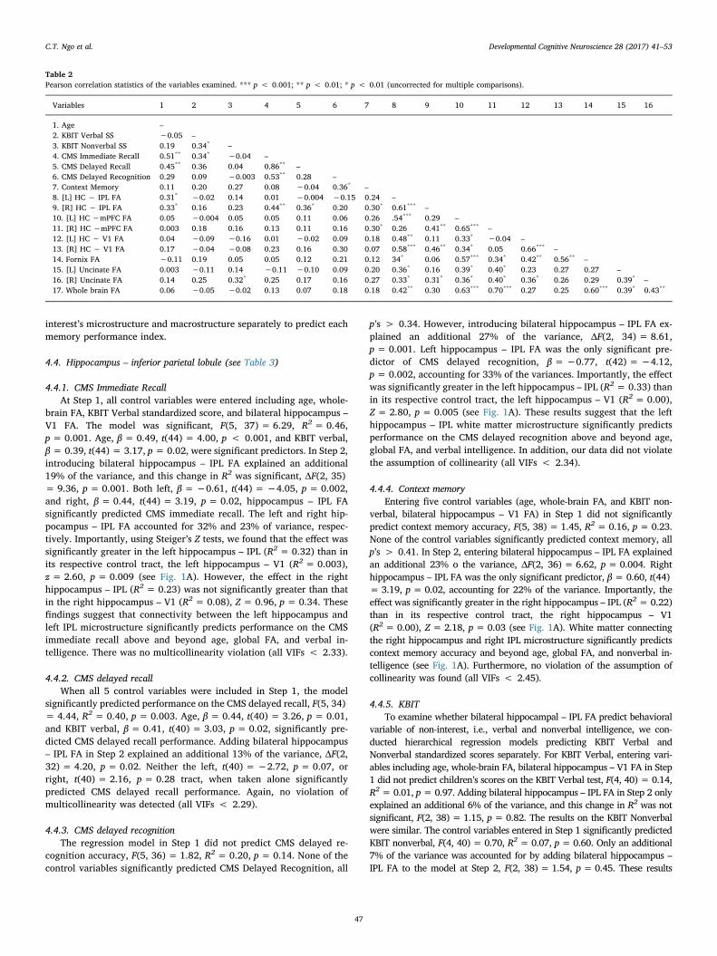
Table 2Pearson correlation statistics of the variables examined. *** p < 0.001; ** p < 0.01; * p < 0.01 (uncorrected for multiple comparisons).
Variables 1 2 3 4 5 6 7 8 9 10 11 12 13 14 15 16
1. Age –2. KBIT Verbal SS −0.05 –3. KBIT Nonverbal SS 0.19 0.34* –4. CMS Immediate Recall 0.51** 0.34* −0.04 –5. CMS Delayed Recall 0.45** 0.36 0.04 0.86** –6. CMS Delayed Recognition 0.29 0.09 −0.003 0.53** 0.28 –7. Context Memory 0.11 0.20 0.27 0.08 −0.04 0.36* –8. [L] HC − IPL FA 0.31* −0.02 0.14 0.01 −0.004 −0.15 0.24 –9. [R] HC − IPL FA 0.33* 0.16 0.23 0.44** 0.36* 0.20 0.30* 0.61*** –10. [L] HC −mPFC FA 0.05 −0.004 0.05 0.05 0.11 0.06 0.26 .54*** 0.29 –11. [R] HC −mPFC FA 0.003 0.18 0.16 0.13 0.11 0.16 0.30* 0.26 0.41** 0.65*** –12. [L] HC − V1 FA 0.04 −0.09 −0.16 0.01 −0.02 0.09 0.18 0.48** 0.11 0.33* −0.04 –13. [R] HC − V1 FA 0.17 −0.04 −0.08 0.23 0.16 0.30 0.07 0.58*** 0.46** 0.34* 0.05 0.66*** –14. Fornix FA −0.11 0.19 0.05 0.05 0.12 0.21 0.12 34* 0.06 0.57*** 0.34* 0.42** 0.56** –15. [L] Uncinate FA 0.003 −0.11 0.14 −0.11 −0.10 0.09 0.20 0.36* 0.16 0.39* 0.40* 0.23 0.27 0.27 –16. [R] Uncinate FA 0.14 0.25 0.32* 0.25 0.17 0.16 0.27 0.33* 0.31* 0.36* 0.40* 0.36* 0.26 0.29 0.39* –17. Whole brain FA 0.06 −0.05 −0.02 0.13 0.07 0.18 0.18 0.42** 0.30 0.63*** 0.70*** 0.27 0.25 0.60*** 0.39* 0.43**
C.T. Ngo et al. Developmental Cognitive Neuroscience 28 (2017) 41–53
47

Table 3Summary of hierarchical linear regression models of white matter connectivity between hippocampus and inferior parietal lobe microstructure (FA) predicting memory performances.
Dependent variable Predictors β t-value F ΔF R2 ΔR2
Children’s Memory ScaleImmediate RecallStep 1 6.29*** 0.46
Age 0.49 4.00**
Whole-brain FA 0.10 0.78KBIT Verbal SS 0.39 3.17*
[L] HC − V1 FA −0.20 −1.22[R] HC − V1 FA 0.03 1.84
Step 2 9.36** 0.19Age 0.52 4.83***
Whole-brain FA 0.16 1.41KBIT Verbal SS 0.33 3.18*
[L] HC − V1 FA 0.05 0.34[R] HC − V1 FA 0.27 1.77[L] HC − IPL FA −0.61 −4.05**
[R] HC − IPL FA 0.44 3.19*
Delayed RecallStep 1 4.44** 0.40
Age 0.44 3.26*
Whole-brain FA 0.08 0.59KBIT Verbal SS 0.41 3.03*
[L] HC − V1 FA −0.18 −1.00[R] HC − V1 FA 0.26 1.42
Step 2 4.20* 0.13Age 0.47 3.63**
Whole-brain FA 0.13 0.93KBIT Verbal SS 0.37 2.94*
[L] HC − V1 FA 0.03 0.16[R] HC − V1 FA 0.22 1.26[L] HC − IPL FA −0.50 −2.62[R] HC − IPL FA 0.36 12.16
Delayed RecognitionStep 1 1.82 0.20
Age 0.25 1.64Whole-brain FA 0.14 0.87KBIT Verbal SS 0.12 0.80[L] HC − V1 FA −0.21 −1.02[R] HC − V1 FA 0.38 1.88
Step 2 8.61** 0.27Age 0.35 2.63Whole-brain FA 0.28 2.03KBIT Verbal SS 0.10 0.76[L] HC − V1 FA 0.03 0.13[R] HC − V1 FA 0.50 2.63[L] HC − IPL FA −0.77 −4.12**
[R] HC − IPL FA 0.24 1.39
Episodic MemoryContext MemoryStep 1 1.45 0.16
Age 0.18 1.79Whole-brain FA 0.18 1.23KBIT Nonverbal SS −0.14 −1.52[L] HC − V1 FA −0.31 −1.62[R] HC − V1 FA 0.33 1.66
Step 2 6.62 0.23Age 0.06 0.40Whole-brain FA 0.04 0.29KBIT Nonverbal SS −0.27 −1.90[L] HC − V1 FA −0.14 −0.70[R] HC − V1 FA −0.03 −0.16[L] HC − IPL FA 0.03 0.16[R] HC − IPL FA 0.60 3.19*
FA: fractional anisotropy, β: standardized regression coefficient, HC – mPFC: white matter connecting hippocampus and medial prefrontal cortex, HC – V1: white matter connectinghippocampus and primary visual cortex.ΔF: the change in F values between models 1 and 2. ΔR2: the change in R2 between models 1 and 2.*** p < 0.001; ** p < 0.01; * p < 0.01 Bonferroni corrected (corrected α= 0.01 for Model 1 and corrected α= 0.007 for Model 2).
C.T. Ngo et al. Developmental Cognitive Neuroscience 28 (2017) 41–53
48

suggest that the white matter tract connecting the hippocampus and IPLrelates to performance on various memory tasks, but not to performance ontasks with a low declarative memory demand. Again, multicollinearity wasnot an issue, all VIFs < 2.40.
4.5. Hippocampus – medial prefrontal cortex (see Table 4)
Unlike the HC – IPL microstructure, neither the left nor right hip-pocampus – mPFC FA predicted performances on any of the CMS tests
Table 4Summary of hierarchical linear regression models of white matter connectivity between hippocampus and medial prefrontal cortex microstructure (FA) predicting memory performances.
Dependent variable Predictors β t-value F ΔF R2 ΔR2
Children’s Memory ScaleImmediate RecallStep 1 5.83*** 0.43
Age 0.48 3.88**
Whole-brain FA 0.12 0.93KBIT Verbal SS 0.36 2.92*
[L] HC − V1 FA −0.13 −0.76[R] HC − V1 FA 0.31 1.78
Step 2 0.64** 0.02Age 0.49 3.94**
Whole-brain FA 0.04 0.19KBIT Verbal SS 0.33 2.60[L] HC − V1 FA −0.07 −0.41[R] HC − V1 FA 0.34 1.94[L] HC − mPFC FA −0.18 −0.93[R] HC − mPFC FA 0.23 1.03
Delayed RecallStep 1 4.00** 0.36
Age 0.42 3.13*
Whole-brain FA 0.10 0.72KBIT Verbal SS 0.38 2.78[L] HC − V1 FA −0.10 −0.55[R] HC − V1 FA 0.26 1.37
Step 2 0.27 0.01Age 0.43 3.10*
Whole-brain FA −0.03 −0.13KBIT Verbal SS 0.36 2.51[L] HC − V1 FA −0.08 −0.37[R] HC − V1 FA 0.25 1.30[L] HC − mPFC FA 0.02 0.09[R] HC − mPFC FA 0.15 0.57
Delayed RecognitionStep 1 1.95 0.20
Age 0.26 1.77Whole-brain FA 0.14 0.89KBIT Verbal SS 0.12 0.81[L] HC − V1 FA −0.23 −1.08[R] HC − V1 FA 0.39 1.92
Step 2 1.00 0.04Age 0.28 1.89Whole-brain FA 0.08 0.32KBIT Verbal SS 0.09 0.58[L] HC − V1 FA −0.14 −0.65[R] HC − V1 FA 0.45 2.14[L] HC − mPFC FA −0.31 −1.33[R] HC − mPFC FA 0.29 1.09
Episodic MemoryContext MemoryStep 1 1.45 0.15
Age 0.22 1.48Whole-brain FA 0.17 1.12KBIT Nonverbal SS −0.12 −0.77[L] HC − V1 FA −0.25 −1.21[R] HC − V1 FA 0.30 1.46
Step 2 0.52 0.02Age 0.24 1.55Whole-brain FA 0.10 0.44KBIT Nonverbal SS −0.14 −0.88[L] HC − V1 FA −0.18 −0.82[R] HC − V1 FA 0.31 1.51[L] HC − mPFC FA −0.19 −0.84[R] HC − mPFC FA 0.23 0.90
FA: fractional anisotropy, β: standardized regression coefficient, HC – mPFC: white matter connecting hippocampus and medial prefrontal cortex, HC – V1: white matter connectinghippocampus and primary visual cortex.ΔF: the change in F values between models 1 and 2. ΔR2: the change in R2 between models 1 and 2.*** p < 0.001; ** p < 0.01; * p < 0.01 Bonferroni corrected (corrected α = 0.01 for Step 1 and corrected α = 0.007 for Step 2).
C.T. Ngo et al. Developmental Cognitive Neuroscience 28 (2017) 41–53
49

or Episodic Memory tasks, all p’s > 0.99.
4.6. White matter macrostructure– behavioral performance relations
The same statistical approach was conducted for the white mattermacrostructure of each tract predicting each memory performance. InStep 1, the control variables included age, whole-brain white mattervolume, KBIT standardized score, and bilateral control tract (hippo-campus – V1) volume; and bilateral tract of interest volumes were en-tered in Step 2. Neither the left nor right hippocampus – IPL volumepredicted any of the CMS tests, all p’s > 0.56, or context memory onthe Episodic Memory task, all p’s > 0.99. Similarly, the hippocampus –mPFC volume did not predict performance on any of the CMS or contextmemory on the Episodic Memory task, all p’s > 0.99.
4.7. Exploratory analyses: limbic white matter pathways
It is possible that although the hippocampus – mPFC connectivitydid not correlate with memory performance, an analysis with highergranularity might unveil a relation between medial temporal-frontalconnectivity and the memory of young children. Visual inspection ofthe hippocampus mPFC tractography revealed that this white matterpathway includes portions of the fornix and the uncinate fasciculus (seeFig. 1B).
Neither the Fornix FA nor volume differed between 4 and 6-year-olds (all p’s > 0.16). To examine the whether variations in the fornixmicrostructure and macrostructure relate to memory, we conductedhierarchical regression analyses with five control variables (same asdescribed above) entered in Step 1, and fornix FA/volume entered inStep 2. Separate regression models were conducted for microstructureand macrostructure. Our results showed that entering fornix FA to themodel did not explain a significant additional amount of variance inany of the memory tests (ΔR2 = 0.00–0.04, all p’s > 0.13).
Similar results were found for the uncinate fasciculus. No age dif-ferences were found for either the left or right uncinate fasciculus FA orvolume (all p’s = 0.40). In the hierarchical regression models, addingbilateral uncinate FA or volume did not explain a significant additionalamount of variances on any of the memory tests (ΔR2 = 0.00–0.02, allp’s > 0.66).
In sum, we found no age effect in the fornix and uncinate micro- andmacrostructure between 4- and 6-year-old children. Furthermore, var-iations in the micro- and macrostructure of the fornix and uncinatefasciculus did not relate to any of the memory measures. These findingscorroborate our findings on the hippocampus – mPFC connectivity,suggesting that neither of the specific major fronto-temporal whitematter tracts – fornix and uncinate – related to memory performance inyoung children (for results summary, see Table 5).
5. General discussion
Early childhood, particularly between the ages of four and six, ismarked by important and robust growth in episodic memory (e.g.,Newcombe et al., 2014; Ngo et al., 2017; Lloyd et al., 2009; Riggins,2014). Gains in episodic memory performance may partly reflect ma-turation of brain networks essential for episodic memory (reviewed inOlson and Newcombe, 2014). These networks mature along severaldimensions: in terms of gray matter volume (Riggins et al., 2015),functional connectivity within the memory networks (Riggins et al.,2016), and in terms of age-related differences in white matter macroand microstructure. Importantly, different brain regions mature at dif-ferent rates. Thus the dynamic interplay of maturational processes bothwithin and outside of the hippocampus will affect episodic memoryfunctioning (reviewed in Olson and Newcombe, 2014).
Specifically, we examined age-related differences in white mattertracts that support episodic memory systems. We assessed episodicmemory using the Children’s Memory Scale standardized test – a verbalrecall memory paradigm, and an episodic memory test that tapsmemory for single objects, as well as memory for context – a hallmarkof episodic memory. We delineated two main white matter tracts ofinterests: one connecting the hippocampus to the inferior parietal lo-bule, and one connecting the hippocampus to the medial prefrontalcortex. We found that the microstructural properties of the white matterpathway connecting the hippocampus to the inferior parietal lobulesignificantly correlated with performance on several memory tasks.
Specifically, the left hippocampus – IPL microstructure predictedchildren’s performance on the CMS immediate recall and delayed re-cognition, whereas the right hippocampus – IPL microstructure pre-dicted children’s ability to recall contextual details in the EpisodicMemory task. The hemispheric effect aligns with the nature of thesetasks: CMS is a verbal memory task in which children recalled stories,whereas the context memory contains less verbal demand (recalling thelocation and action associated with an object). White matter micro-structure of this pathway explained significant amounts of variance(10%–26%), above and beyond age, whole-brain FA, verbal/nonverbalintelligence, and a bilateral control white matter tracts that should notbe implicated in episodic memory.
5.1. Age-related differences in white matter
Although previous studies have found a general increase in whitematter volume, as well as FA, throughout development (e.g., Lebel andBeaulieu, 2011; Westlye et al., 2010; reviewed in Lebel et al., 2017), wefound that white matter macrostructure and microstructure did notdiffer between 4- and 6-year-olds. However, our sample had a severelyrestricted age range. We suspect that including a wider age range mayyield an age effect – a potential future direction of this work. In addi-tion, higher resolution imaging, such as HARDI imaging, may revealmore subtle age-related white matter changes.
5.2. The role of the inferior parietal lobe in episodic memory
It is believed that the information transmission properties of anygiven white matter tract can be predicted by the function of the graymatter regions that it connects (Maunsell and van Essen, 1983;Passingham et al., 2002). For this reason, we chose the hippocampus asour seed region, as its role in episodic memory is well established fromdecades of research across a range of species. One question that must beasked is whether our findings of hippocampal-IPL structural con-nectivity have any support from the neuroanatomy literature, especiallythose studies using techniques that are more precise than diffusionimaging. Studies in monkeys using injected radiotracers have identifiedseveral monosynaptic axonal pathways between the IPL and medialtemporal lobe. The cingulum bundle, which begins in the medial tem-poral lobe and circles through the cingulate cortex, connects lateral and
Table 5Results summary of white matter microstructure of the tracts examined and their rela-tions with each memory task.
Memory Tasks HC–IPL HC–mPFC Fornix UF
Left Right Left Right Left Right
Children’s Memory ScaleImmediate Recall ✓* ✓ ✗ ✗ ✗ ✗ ✗
Delayed Recall ✗ ✗ ✗ ✗ ✗ ✗ ✗
Delayed Recognition ✓* ✗ ✗ ✗ ✗ ✗ ✗
Episodic MemoryContext Memory ✗ ✓* ✗ ✗ ✗ ✗ ✗
✓: Significant predictor.*: Significantly greater than that of the respective control tract (left/right hippocampus –V1 FA).
C.T. Ngo et al. Developmental Cognitive Neuroscience 28 (2017) 41–53
50

medial regions of the posterior IPL to the parahippocampal gyrus(Seltzer and Pandya, 1984) and connections exist between Area 7 in theIPL and entorhinal cortex (Insausti and Amaral, 2008; Wellman andRockland, 1997). Most interestingly, there are connections betweenhippocampal area CA1, in the anterior hippocampus, and Area 7a and7b of the IPL (Clower et al., 2001; Rockland and Van Hoesen, 1999).Connections have also been identified between the presubiculum andArea 7a of the IPL (Cavada and Goldman-Rakic, 1989; Ding et al.,2000). Thus it is likely that the tractography results from the currentwork closely reflect the ground-truth evidence from gross anatomicaldissection.
A second question that must be asked is what is the nature andmanner of IPL involvement in episodic memory? As noted in the in-troduction, fMRI studies in adults have consistently linked the inferiorparietal cortex to memory retrieval accompanied by recollection (re-viewed in Wagner et al., 2005). For example, studies have reported thatIPL activity is greater for items recognized with recollection judgmentsthan those with familiarity judgments (Cansino et al., 2002; Hensonet al., 1999; Dobbins et al., 2003; Hutchinson et al., 2014; Wheeler andBuckner, 2004; reviewed in Cabeza et al., 2012), and when retrieval issupported by recollection as opposed to familiarity (Dobbins et al.,2003; Dobbins et al., 2002; Dobbins and Wagner, 2005; reviewed inWagner et al., 2005). Corroborating this view, patients with bilateralparietal lobe lesions report few details when recalling autobiographicalmemories compared to healthy controls (Berryhill et al., 2007), andhave reduced certainty in their memories, as indexed by reduced sub-jective confidence (Hower et al., 2014; Simons et al., 2010).
Several theories have been proposed to explain this relationship,ranging from theories relating the IPL to a mnemonic accumulator,tracking memory signal strength to help make old/new decisions, anddecisions related to subjective aspects of memory (Ally et al., 2008;Hower et al., 2014; Simons et al., 2010), to theories linking this regionto “internal attention”, essential for retrieval (reviewed in Cabeza et al.,2008). Our findings cannot adjudicate between these different views.
However, our findings do add to the growing literature linking theIPL to episodic memory function. Our findings are consistent with theliterature implicating the IPL in recollection, but cannot speak tofindings on subjective aspects of memory since young children lack themeta-cognitive abilities required to report on such things. Importantly,our findings highlight the role of structural connectivity between thehippocampus and IPL in episodic memory development, using recalland recognition accuracy as measures of interest.
5.3. Frontal connectivity
In contrast to the hippocampus – IPL connectivity, structural con-nectivity between the hippocampus and mPFC did not relate to any ofthe memory measures. The same results were found when we deli-neated specific medial temporal-frontal pathways, the fornix and un-cinate fasciculus. At first glance, these findings may appear to contra-dict findings showing involvement of the prefrontal cortex in episodicmemory (reviewed in Raj and Bell, 2010) and neuropsychologicalfindings showing that damage to the PFC impedes source memory re-trieval (e.g., Ciaramelli and Spanoil, 2009; Duarte et al., 2005), in-creases false recognition (Curran et al., 1997), and increases suscept-ibility to interference (Shimamura et al., 1995).
Several speculations can be made about our null results in regards tohippocampal-mPFC connectivity. First, one possibility is that the agegroups examined in this study are quite young and the mPFC is stillundergoing regional maturation (Gogtay et al., 2004). If true, regionalchanges in the mPFC should relate to memory development (Ofen et al.,2007). Second, it is possible that the white matter connectivity betweenthe hippocampus and the mPFC is underdeveloped and has a long toway to reach adult-like form. White matter volume gradually increasesaround the age of 2 until adulthood (Groeschel et al., 2010), withfrontal-temporal connections showing the most prolonged development
(Lebel and Beaulieu, 2011). If true, we should expect that age-relateddifferences in this white matter would closely track differences inmemory performance. These hypotheses merit further investigations,possibly at a later stage in development.
5.4. Laterality and directionality
The direction of the left hippocampus – IPL FA and CMS memoryperformance and the right hippocampus – IPL FA and context memoryare in the opposite directions. Lower left hippocampus – IPL FA wasassociated with higher performances on the CMS tests, whereas higherright hippocampus – IPL FA was associated with higher context memoryrecall. Interestingly, diffusion-weighted imaging studies in adults havefound similar patterns in young adults (e.g., Alm et al., 2016; Nugielet al., 2016; Metoki et al., 2017). It is a possibility that the relation ofthe hippocampus – IPL connectivity and memory differs depending onthe nature of the stimuli in the memory tasks. The fact that CMS is averbal task, whereas context memory is primarily visual, may influencethe directionality of effects. Furthermore, the extent of myelination mayhave different effects on signal transduction depending on differentbrain areas. Although these patterns of findings have been reported inother studies, the issue of directionality and white matter indices meritsfurther investigation.
5.5. Specificity and generality of findings
The level of neural specificity of any brain-behavior effect is criticalfor its interpretation. We used several levels of control to assess speci-ficity. First, we used white matter connecting the hippocampus to V1 asa control fiber pathway. As expected, we found no reliable relationbetween this tract and memory. Second, we carefully controlled forseveral variables, including whole brain white matter and IQ, reasoningthat these variables could potentially explain differences in behavior,microstructure or both. Indeed, IQ was related to memory performance;however, it did not correlate with microstructural differences in ourpathways of interest. Therefore, there seems to be some specificity toour findings.
It is also important to consider how the findings of any given studygeneralize to other tasks and populations. In our study, episodicmemory was indexed by immediate and delayed verbal recall tasks(CMS) and a delayed context recall memory task (Episodic Memorytasks). Our delay intervals were half an hour and one hour. Futurestudies should examine whether these results would generalize tomemory tasks with longer delay such as 24 h or a one-week delay. Inaddition, episodic memory tasks vary in the complexity of relationalstructure. In this study, the episodic memory task required children tobind a toy to specific action and a specific context. Other episodicmemory tasks used in previous studies employed a non-overlapping AB-CD associations (e.g., Lloyd et al., 2009; Sluzenski et al., 2006), ordifferential extents of overlapping elements (e.g., AB-AC associations:Ngo et al., 2017; Darby and Sloutsky, 2015; Yim et al., 2013; AB-ABr:Yim et al., 2013). Relational memory for different extents of over-lapping features may result in differential demands on retrieval stra-tegies – potentially recruited from frontal regions – to minimize po-tential inference. Thus, our findings may not generalize to othervariants of episodic memory tasks. Lastly, given that both white matterconnectivity and episodic memory change drastically in early devel-opment, different patterns in the relation between white matter andmemory performance may be observed with different age ranges, par-ticularly in later development.
5.6. Limitations
One limitation of the study is its cross-sectional design, which al-lowed us a glimpse into brain development. However, brain maturationaccompanying memory development is a complex process,
C.T. Ngo et al. Developmental Cognitive Neuroscience 28 (2017) 41–53
51

encompassing the dynamic interplay among many key players. Alongitudinal design would be ideal. In addition, due to our sample size,we limited our investigation of hippocampal connectivity to a subset ofcortical target regions. With larger sample sizes, future studies shouldexplore other white matter tracts that may also be important for thedevelopment of episodic memory.
Another limitation of the current work lies in the technical chal-lenge of diffusion weighted imaging as indirect measurements of whitematter tissue based on estimates of water diffusivity. Diffusionweighted imaging techniques only provide computational models ofWM tissue with many assumptions about the underlying processes andstructures. Thus, the success of diffusion-weighted imaging in deli-neating white matter pathways in the brain is highly dependent on dataquality, the chosen diffusion model, and the analysis pipeline (Joneset al., 2013). In this study, we acquired diffusion tensor imaging withthree repetitions, hence increasing the signal-to-noise ratio in our data.In addition, we employed the dual-fiber model, which accounts forcrossing fibers in the brain, as opposed to the a single tensor model witha deterministic tracing algorithm, which only calculates a single prin-ciple diffusion direction in each voxel. Nevertheless, one must be waryof technical limitations and pitfalls in interpretation of white matterconnectivity using diffusion weighted imaging (Jbabdi et al., 2015;Jones et al., 2013), as there is unlikely to be a one-to-one correspon-dence between diffusion parameter and the underlying tissue structure(Assaf et al., 2017).
6. Conclusions
Typical episodic memory functioning relies on the operations of afar-flung yet exquisitely orchestrated network of brain regions. The“wires” connecting the nodes of this network are axons, bundled intotracts that can be measured with diffusion imaging. However, whitematter matures slowly and variably, making its measurement criticalfor understanding the emergence of cognition over development. Ourstudy was among the first to identify and characterize the relation be-tween white matter connectivity and episodic memory in young chil-dren, ages four and six. Our results show that hippocampal-inferiorparietal lobe white matter is a key variable in predicting episodicmemory performance in this age range.
Acknowledgement
We would like to thank the members of the NeurocognitiveDevelopment Lab and Developmental Social Cognitive NeuroscienceLab at the University of Maryland, especially, Elizabeth Redcay, SarahBlankenship and Katherine Rice for their contribution to this work. Weadditionally thank the families who participated in this study and theMaryland Neuroimaging Center for their support in data acquisition.Portions of these data were presented at the Society for Research inChild Development (Austin, TX) 2017). This work was supported by aNational Institutes of Health (RO1 MH091113 to I.R.O andF31HD090872 to C.T.N), a National Science Foundation (SBE-1041707to N.S.N), and by a Maryland Neuroimaging Center seed grant, theBehavioral and Social Sciences Dean’s Research Initiative, theUniversity of Maryland Department of Psychology and an NationalInstitutes of Health (R01HD079518A to T.R.). The content is solely theresponsibility of the authors and does not necessarily represent theofficial views of the National Institute of Mental Health or the NationalInstitutes of Health. The authors declare no competing or conflictingfinancial interests.
References
Ally, B.A., Simons, J.S., McKeever, J.D., Peers, P.V., Budson, A.E., 2008. Parietal con-tributions to recollection: electrophysiological evidence from aging and patients withparietal lesions. Neuropsychologia 46 (7), 1800–1812.
Alm, K.H., Rolheiser, T., Olson, I.R., 2016. Inter-individual variation in fronto-temporalconnectivity predicts the ability to learn different types of associations. Neuroimage132, 213–224.
Assaf, Y., Johansen-Berg, Schotten, M.T., 2017. The role of diffusion MRI in neuroscience.NMR Biomed. http://dx.doi.org/10.1002/nbm.3762.
Backus, A.R., Bosch, S.E., Ekman, M., Grabovetsky, A.V., Doeller, C.F., 2016. Mnemonicconvergence in the human hippocampus. Nat. Commun. http://dx.doi.org/10.1038/ncomms11991.
Bauer, P.J., Doydum, A.O., Pathman, T., Larkina, M., Guler, O.E., Burch, M., 2012. It’s allabout location, location, location: children’s memory for the where of personallyexperienced events. J. Exp. Child Psychol. 133 (4), 510–522.
Beaulieu, C., 2002. The basis of anisotropic water diffusion in the nervous system – atechnical review. NMR Biomed. 15, 435–455.
Behrens, T.E., Johansen-Berg, H., Woolrich, M.W., Smith, S.M., Wheeler-Kingshott, C.A.,Boulby, P.A., Barker, G.J., Sillery, E.L., Sheehan, K., Ciccarelli, O., Thompson, A.J.,Brady, J.M., Matthews, P.M., 2003. Non-invasive mapping of connections betweenhuman thalamus and cortex using diffusion imaging. Nat. Neurosci. 6, 750–757.
Behrens, T.E., Berg, H.J., Jbabdi, S., Rushworth, M.F., Woolrich, M.W., 2007.Probabilistic diffusion tractography with multiple fibre orientations: what can wegain? Neuroimage 34 (1), 144–155.
Ben-Zvi, S., Soroker, N., Levy, D.A., 2015. Parietal lesion effects on cued recall followingpair associate learning. Neuropsychologia 73, 176–194.
Benoit, R.G., Anderson, M.C., 2012. Opposing mechanisms supporting the voluntaryforgetting of unwanted memories. Neuron 76 (2), 450–460.
Berryhill, M.E., Phuong, L., Picasso, L., Cabeza, R., Olson, I.R., 2007. Parietal lobe andepisodic memory: bilateral damage causes impaired free recall of autobiographicalmemory. J. Neurosci. 27, 14415–14423.
Berryhill, M.E., Drowos, D.B., Olson, I.R., 2009. Bilateral parietal cortex damage does notimpair associative memory for paired stimuli. Cognit. Neuropsychol. 26 (7),606–619.
Bonekamp, D., Nagae, L.M., Degaonkar, M., Matson, M., Abdalla, W.M., Peter, B., et al.,2007. Diffusion tensor imaging in children and adolescents: reproducibility, hemi-spheric, and age-related differences. Neuroimage 34 (2), 733–742.
Cabeza, R., Ciaramelli, E., Olson, I.R., Moscovitch, M., 2008. The parietal cortex andepisodic memory: an attentional account. Nat. Rev. Neurosci. 9 (8), 613–625.
Cabeza, R., Ciaramelli, E., Moscovitch, M., 2012. Cognitive contributions of the ventralparietal cortex: an integrative theoretical account. Trends Cognit. Sci. 16 (6),338–352.
Cansino, S., Maquet, P., Dolan, R., Rugg, M., 2002. Brain activity underlying encodingand retrieval of source memory. Cereb. Cortex 12 (10), 1048–1056.
Cavada, C., Goldman-Rakic, P.S., 1989. Posterior parietal cortex in rhesus monkey: i.Parcellation of areas based on distinctive limbic and sensory corticocortical con-nections. J. Comp. Neurol. 287 (4), 393–421.
Chaddock-Heyman, L., Erickson, K., Voss, M.W., Powers, J.P., Knecht, A., Hillman, C.H.,Kramer, A.F., 2013. White matter microstructure is associated with cognitive controlin children. Biol. Psychol. 94 (1), 109–115.
Ciaramelli, E., Spanoil, J., 2009. Ventromedial prefrontal damage and memory for con-text: perceptual versus semantic features. Neuropsychology 23 (5), 649–657.
Clower, D.M., West, R.A., Lynch, J.C., Strick, P.L., 2001. The inferior parietal lobule is thetarget output from the superior colliculus, hippocampus, and cerebellum. J. Neurosci.21 (16), 6283–6291.
Cohen, N.J., Eichenbaum, H., 1993. Memory, Amnesia, and the Hippocampal System.MIT Press, Cambridge, MA.
Cohen, M.J., 1997. Children’s Memory Scale Manual The Psychological Corporation.Harcourt Brace and Company, San Antonio, TX.
Curran, T., Schacter, D.L., Norman, K.A., Galluccio, L., 1997. False recognition after aright frontal lobe infarction: memory for general and specific information.Neuropsychologia 35 (7), 1035–1049.
Darby, K.P., Sloutsky, V.M., 2015. The cost of learning: interference effects in memorydevelopment. J. Exp. Psychol. Gen. 144, 410–431.
DeMaster, D.M., Ghetti, S., 2013. Developmental differences in hippocampal and corticalcontributions to episodic retrieval. Cortex 49 (6), 1482–1493.
DeMaster, D.M., Pathman, T., Lee, J., Ghetti, S., 2013. Structural development of thehippocampus and episodic memory: developmental differences along the anterior-posterior axis. Cereb. Cortex 24 (11), 3036–3045.
Ding, S.L., Van Hoessen, G., Rockland, K.S., 2000. Inferior parietal lobule projections tothe presubiculum and neighboring ventromedial temporal cortical areas. J. Comp.Neurol. 425 (4), 510–530.
Dobbins, I.G., Wagner, A.D., 2005. Domain-general and domain-sensitive prefrontalmechanisms for recollecting events and detecting novelty. Cereb. Cortex 15 (11),1768–1778.
Dobbins, I.G., Foley, H., Schacter, D., Wagner, A.D., 2002. Executive control duringepisodic retrieval: multiple prefrontal processes subserve source memory. Neuron 35(5), 989–996.
Dobbins, I.G., Rice, H.J., Wagner, A.D., Schacter, D.L., 2003. Memory orientation andsuccess: separable cognitive components underlying episodic recognition.Neuropsychologia 41, 318–333.
Douet, V., Chang, L., 2014. Fornix as an imaging marker for episodic memory deficits inhealthy changing and in various neurological disorders. Front. Aging Neurosci. 6,343.
Drowos, D.B., Berryhill, M., André, J.M., Olson, I.R., 2010. True memory, false memory,and subjective recollection deficits after focal parietal lobe lesions. Neuropsychology24 (4), 465–475.
Drummey, A.B., Newcombe, N.S., 2002. Developmental changes in source memory. Dev.Sci. 5 (4), 502–513.
Duarte, A., Ranganath, C., Knight, R.T., 2005. Effects of unilateral prefrontal lesions onfamiliarity, recollection, and source memory. J. Neurosci. 25 (36), 8333–8337.
Euston, D.R., Gruber, A.J., McNaughton, B.L., 2012. The role of medial prefrontal cortexin memory and decision making. Neuron 76 (6), 1057–1070.
Ghetti, S., Bunge, S.A., 2012. Neural changes underlying the development of episodic
C.T. Ngo et al. Developmental Cognitive Neuroscience 28 (2017) 41–53
52

memory during middle childhood. Dev. Cognit. Neurosci. 2 (4), 381–395.Gogtay, N., Giedd, J.N., Lusk, L., Hayashi, K.M., Greenstein, D., Vaituzis, A.C., III, Clasen,
L.S., Toga, A.W., Rapoport, J.L., Thompson, P.M., 2004. Dynamic mapping of humancortical development during childhood through early adulthood. Proc. Natl. Acad.Sci. 101 (21), 8174–8179.
Goto, Y., Grace, A.A., 2008. Dopamine modulation of the hippocampal-prefrontal corticalinteraction drives memory-guided behavior. Cereb. Cortex 18, 1407–1414.
Groeschel, S., Vollmer, B., King, M.D., Connelly, A., 2010. Developmental changes incerebral grey and white matter volume from infancy to adulthood. Int. J. Dev.Neurosci. 6, 481–489.
Haramati, S., Soroker, S., Dudai, Y., Levy, D.A., 2008. The posterior parietal cortex inrecognition memory: a neuropsychological study. Neuropsychologia 46 (7),1756–1766.
Henson, R.N., Rugg, M.D., Shallice, T., Joshephs, O., Dolan, R.J., 1999. Recollection andfamiliarity in recognition memory: an event-related functional magnetic resonanceimaging study. J. Neurosci. 19, 3962–3972.
Hower, K.H., Wixted, J., Berryhill, M.E., Olson, I.R., 2014. Impaired perception of mne-monic oldness, but not mnemonic newness: after parietal lobe damage.Neuropsychologia 56, 409–417.
Hutchinson, B., Uncapher, M., Weiner Bressler, D., Silver, M., Preston, A., Wagner, A.,2014. Functional heterogeneity in posterior parietal cortex across attention andepisodic memory retrieval. Cereb. Cortex 24 (1), 49–66.
Insausti, R., Amaral, D.G., 2008. Entorhinal cortex of the monkey: IV. Topographical andlaminar organization of cortical afferents. J. Comp. Neurol. 509, 608–641.
Jack, F., MacDonald, S., Reese, E., Hayne, H., 2009. Maternal reminiscing style duringearly childhood predicts the age of adolescents’ earliest memories. Child Dev. 80 (2),496–505.
Jbabdi, S., Sotiropoulos, S.N., Haber, S.N., Van Essen, D.C., Behrens, T.E., 2015.Measuring macroscopic brain connections in vivo. Nat. Neurosci. 18 (11),1546–1555. http://dx.doi.org/10.1038/nn.4134.
Jones, D.K., Knösche, T.R., Turner, R., 2013. White matter integrity, fiber count, andother fallacies: the do’s and don’ts of diffusion MRI. Neuroimage 73, 239–254.
Krogsrud, S.K., Fjell, A., Tamnes, C.K., Grydeland, J., Mork, L., Due-Tonnessen, P., et al.,2016. Changes in white matter microstructure in the developing brain – a long-itudinal diffusion tensor imaging study of children from 4 to 11 years of age.Neuroimage 124, 473–486.
Lebel, C., Beaulieu, C., 2011. Longitudinal development of human brain wiring continuesfrom childhood into adulthood. J. Neurosci. 31 (30), 10937–10947.
Lebel, C., Walker, L., Leemans, A., Phillips, L., Beaulieu, C., 2008. Microstructural ma-turation of the human brain from childhood to adulthood. Neuroimage 40 (3),1044–1055.
Lebel, C., Treit, S., Beaulieu, C., 2017. A review of diffusion MRI of typical white matterdevelopment from early childhood to young adulthood. NMR Biomed. http://dx.doi.org/10.1002/nbm.3778.
Lloyd, M.E., Doydum, A.O., Newcombe, N.S., 2009. Memory binding in early childhood:evidence for a retrieval deficit. Child Dev. 80 (5), 1321–1328. http://dx.doi.org/10.1111/j.1467-8624.2009.01353.x.
Loenneker, T., Klaver, P., Bucher, K., Lichtensteiger, J., Imfeld, A., Martin, E., 2011.Microstructural development: organizational differences of the fiber architecturebetween children and adults in dorsal and ventral visual streams. Hum. Brain Mapp.32 (6), 935–946.
Maunsell, J.H., van Essen, D.C., 1983. The connections of the middle temporal visual area(MT) and their relationship to a cortical hierarchy in the macaque monkey. J.Neurosci. 3 (12), 2563–2586.
Metoki, A., Alm, K., Wang, Y., Ngo, C., Olson, I.R., (2017). Never forget a name: whitematter connectivity predicts person memory.
Metzler-Baddeley, C., Jones, D.K., Belaroussi, B., Aggleton, J., O’Sullivan, M.J., 2011.Frontotemporal connections in episodic memory and aging: a diffusion MRI tracto-graphy study. J. Neurosci. 31 (37), 13236–13245.
Mielke, M.M., Okonkwo, O.C., Oishi, K., Mori, S., Tighe, S., Miller, M.I., Ceritoglu, C.,Brown, T., Albert, M., Lyketsos, C.G., 2012. Fornix integrity and hippocampal volumepredict memory decline and progression to Alzheimer’s disease. Alzheimer’sDementia 8 (2), 105–113.
Moon, W.J., Provenzale, J.M., Sarikaya, B., Ihn, Y.K., Morlese, J., Chen, S., et al., 2011.Diffusion tensor imaging assessment of white matter maturation in childhood andadolescence. Am. J. Roentgenol. 193 (3), 704–712.
Newcombe, N.S., Balcomb, F., Ferrara, K., Hansen, M., Koski, J., 2014. Two rooms, tworepresentations. Episodic-like memory in toddlers and preschoolers. Dev. Sci. 17 (5),743–756.
Ngo, C.T., Newcombe, N.S., Olson, I.R., 2017. The ontogeny of relational memory andpattern separation. Dev. Sci. http://dx.doi.org/10.1111/desc.12556.
Nugiel, T., Alm, K.H., Olson, I.R., 2016. Individual differences in white matter micro-structure predict semantic control. Cognit. Affect. Behav. Neurosci. 16 (6),1003–1016.
Ofen, N., Kao, Y., Sokol-Hessner, P., Kim, H., Whitfield-Gabrieli, S., Gabrieli, J.D.E., 2007.Development of the declarative memory system in the human brain. Nat. Neurosci.10, 1198–1205.
Oishi, K., Mielke, M.M., Albert, M., Lyketsos, C.G., Mori, S., 2011. The fornix sign: apotential sign for Alzheimer’s disease based on diffusion tensor imaging. Neuroimage22 (4), 365–374.
Olson, I.R., Newcombe, N.S., 2014. Binding together the elements of episodes: relationalmemory and the developmental trajectory of the hippocampus. In: Patricia J. Bauerand Robyn Fivush. The Wiley Handbook and the Development of Children’s MemoryVolume I/II.
Olson, I.R., Von Der Heide, R.J., Ahm, K., Vyas, G., 2015. Development of the uncinate
fasciculus: implications for theory and developmental disorder. Dev. Cognit.Neurosci. 14, 50–61.
Passingham, R.E., Stephan, K.E., Kötter, R., 2002. The anatomical basis of functionallocalizer in the cortex. Nat. Rev. Neurosci. 3, 606–616.
Qiu, M.G., Li, Q.Y., Liu, G.J., Xie, B., Wang, J.A., 2010. Voxel-based analysis of whitematter during adolescence and young adulthood. Brain Dev. 32 (7), 531–537.
Raj, V., Bell, M.A., 2010. Cognitive processes supporting episodic memory formation inchildhood. The role of source memory, binding, and executive function. Dev. Rev. 30,384–402.
Riggins, T., Rollins, L., 2015. Developmental differences in recollection and familiarityduring early childhood: insights from event-related potentials. Child Dev. 86 (3),889–902.
Riggins, T., Blankenship, S., Mulligan, E., Rice, K., Redcay, E., 2015. Developmentaldifferences in relations between episodic memory and hippocampal subregion vo-lume during early childhood. Child Dev. 86 (6), 1710–1718.
Riggins, T., Geng, F., Blankenship, S., Redcay, E., 2016. Hippocampal functional con-nectivity and episodic memory in early childhood. Dev. Cognit. Neurosci. 19, 58–69.
Riggins, T., 2012. Building blocks of recollection. In: Ghetti, Bauer, P.J. (Eds.), Originsand Development of Recollection: Perspectives from Psychology and Neuroscience.Oxford University Press, New York, NY, pp. 42–72.
Riggins, T., 2014. Longitudinal investigation of source memory reveals qualitative dif-ferences between item memory and binding. Dev. Psychol. 50 (2), 449–459.
Rockland, K.S., Van Hoesen, G.W., 1999. Some temporal and parietal cortical connectionsconverge in CA1 of the primate hippocampus. Cereb. Cortex 9, 232–237.
Rollins, N.K., Glasier, P., Seo, Y., Morriss, M.C., Chua, J., Wang, Z.Y., 2010. Age-relatedvariations in white matter anisotropy in school-age children. Pediatr. Radiol. 40 (12),1918–1930.
Sadeghi, N., Nayak, A., Walker, L., Irganoglu, Okan, Albert, M., Pierpaoli, P.S., BrainDevelopment Cooperative Group, C., 2014. Analysis of the contribution of experi-mental bias, experimental noise, and inter-subject biological variability on the as-sessment of developmental trajectories in diffusion MRI studies of the brain.Neuroimage 109, 480–492.
Seltzer, B., Pandya, D.N., 1984. Further observations on parieto-temporal connections inthe rhesus monkey. Exp. Brain Res. 55 (2), 301–312.
Sexton, C.E., Mackay, C.E., Lonie, J.A., Bastin, M.E., Terriere, E., O’Carroll, R.E., Ebmeier,K.P., 2010. MRI correlates of episodic memory in Alzheimer’s disease, mild cognitiveimpairment, and healthy aging. Psychiatry Res.: Neuroimaging 184 (1), 57–62.
Shimamura, A., Jurica, P., Mangels, J.A., Gershberg, F.B., Knight, R., 1995. Susceptibilityto memory interference effects following frontal lobe damage: findings from tests ofpaired-associate learning. J. Cognit. Neurosci. 7 (2), 144–152.
Shing, Y.L., Werkle-Bergner, M., Li, S., Lindenberger, U., 2008. Associative and strategiccomponents of episodic memory: a life-span dissociation. J. Exp. Psychol. Gen. 137(3), 495–513.
Simmonds, D.J., Hallquist, M.N., Asato, M., Luna, B., 2014. Developmental stages and sexdifferences of white matter and behavioral development through adolescence: alongitudinal diffusion tensor imaging (DTI) study. Neuroimage 92, 356–368.
Simons, J.S., Peers, P.V., Mazuz, Y.S., Berryhill, M.E., Olson, I.R., 2010. Dissociationbetween memory accuracy and memory confidence following bilateral parietal le-sions. Cereb. Cortex 20 (2), 479–485.
Sluzenski, J., Newcombe, N.S., Kovacs, S.L., 2006. Binding, relational memory: and recallof naturalistic events: a developmental perspective. J. Exp. Psychol. 32, 89–100.
Smith, S.M., Jenkinson, M., Woolrich, M.W., Beckmann, C.F., Behrens, T.E., Johansen-Berg, H., Matthews, P.M., 2004. Advances in functional and structural MR imageanalysis and implementation as FSL. NeuroImage 23 (S1), S208–219.
Tsivilis, D., Vann, S.D., Denby, C., Roberts, N., Mayes, A.R., Montaldi, D., Aggleton, J.P.,2008. A disproportionate role of the fornix and mammillary bodies in recall versusrecognition memory. Nat. Neurosci. 11 (7), 834–842.
Von Der Heide, R.J., Skipper, L.M., Klobusicky, E., Olson, I.R., 2013. Dissecting the un-cinate fasciculus: disorder controversies and a hypothesis. Brain 136, 1692–1707.
Wagner, A.D., Shannon, B.J., Kahn, I., Buckner, R.L., 2005. Parietal lobe contributions toepisodic memory retrieval. Trends Cognit. Neurosci. 9 (9), 445–453.
Wellman, B.J., Rockland, K.S., 1997. Divergent cortical connections to entorhinal cortexfrom area TF in the macaque. J. Comp. Neurol. 389 (3), 361–376.
Wendelken, C., Lee, J.K., Pospisil, J., Sastre, M., Ross, J.M., Bunge, S.A., Ghetti, S., 2015.White matter tracts connected to the medial temporal lobe support the developmentof mnemonic control. Cereb. Cortex 18 (9), 2116–2208.
Westlye, L.T., Walhovd, K.B., Dale, A.M., Bjornerud, A., Due-Tonnessen, P., Engvig, A.,Grydeland, H., Tamnes, C.L., Ostby, Y., Fjell, A.M., 2010. Life-span changes of thehuman brain white matter: diffusion tensor imaging (DTI) and volumetry. Cereb.Cortex 20 (9), 2055–2068.
Wheeler, M.E., Buckner, R.L., 2004. Functional anatomic correlates of remembering andknowing. Neuroimage 21, 1337–1349.
Willford, J.A., Richardson, G.A., Leech, S.L., Day, N.L., 2004. Verbal and visuospatiallearning and memory function in children with moderate prenatal alcohol exposure.Alcohol Clin. Exp. Res. 28 (3), 497–507.
Yim, H., Dennis, S.J., Sloutsky, V.M., 2013. The development of episodic memory: items,contexts and relations. Psychol. Sci. 24, 2153–2172.
Zhang, Y., Brady, M., Smith, S., 2001. Segmentation of brain MR images through a hiddenMarkov random field model and the expectation-maximization algorithm. IEEETrans. Med. Imaging 20, 45–57.
Zhuang, L., Sachdev, P.S., Trollor, J.N., Reppermund, S., Kochan, N.A., Brodaty, H., et al.,2013. Microstructural white matter changes, not hippocampal atrophy, detect earlyamnestic mild cognitive impairment. PLoS One 8 (3), e58887. http://dx.doi.org/10.1371/journal.pone.0058887.
C.T. Ngo et al. Developmental Cognitive Neuroscience 28 (2017) 41–53
53