WEDNESDAY SLIDE CONFERENCE 2015-2016 - … Joint Pathology Center Veterinary Pathology Services...
21
1 Joint Pathology Center Veterinary Pathology Services WEDNESDAY SLIDE CONFERENCE 2015-2016 C o n f e r e n c e 17 10 February 2016 Casey J. LeBlanc, DVM, PhD Diplomate, ACVP Clinical Pathologist & CEO KDL VetPath Knoxville, TN & Springfield, VA CASE I: TAMU-1-2013 (JPC 4033379). Signalment: 18-day-old thoroughbred colt (Equus caballus) History: A healthy newborn foal nursed and the next day, became lethargic and had hemoglobinuria. With progressive stupor, icterus, hyperbilirubinemia and anemia, the foal was presented to the clinic at 2 days of age. Gross Pathology: Yellow mucous membranes and tissues (icterus); ~200ml abdominal transudate with fibrin strands; myocardial mottling grey and red (hemorrhage and necrosis) a small Thebsian vein in the noncoronary aortic sinus; urachus patent. Laboratory Results: PCV=12 (32-53) no Nrbc’s Platelets 470,000/ul (100-350,000) TP 4.7g/dl (5.3-7.3) Total bilirubin 14.5 mg/dl (0-1.9) GGT 124U/L (0-53) Alkaline phosphatase 553U/L (128-512) ALT 850U/L (134-643) Saline agglutination test positive Mare serum Anti-Qab positive Histopathologic Description: The section of liver has widespread atrophy of hepatocytes with hypertrophied and Mucous membranes, foal. The mucous membranes of this foal are extremely yellow, indicating icterus. (Photo courtesy of: Dept Vet Pathobiology, College Vet Med Texas A&M University, College Station, TX 77843-4467 http://vetmed.tamu.edu/vtpb)
Transcript of WEDNESDAY SLIDE CONFERENCE 2015-2016 - … Joint Pathology Center Veterinary Pathology Services...

1
Joint Pathology Center
Veterinary Pathology Services
WEDNESDAY SLIDE CONFERENCE 2015-2016
C o n f e r e n c e 17 10 February 2016
Casey J. LeBlanc, DVM, PhD
Diplomate, ACVP
Clinical Pathologist & CEO
KDL VetPath
Knoxville, TN & Springfield, VA
CASE I: TAMU-1-2013 (JPC 4033379).
Signalment: 18-day-old thoroughbred colt
(Equus caballus)
History: A healthy newborn foal nursed and
the next day, became lethargic and had
hemoglobinuria. With progressive stupor,
icterus, hyperbilirubinemia and anemia, the
foal was presented to the clinic at 2 days of
age.
Gross Pathology: Yellow mucous
membranes and tissues (icterus); ~200ml
abdominal transudate with fibrin strands;
myocardial mottling grey and red
(hemorrhage and necrosis) a small Thebsian
vein in the noncoronary aortic sinus; urachus
patent.
Laboratory Results: PCV=12 (32-53) no Nrbc’s
Platelets 470,000/ul (100-350,000)
TP 4.7g/dl (5.3-7.3)
Total bilirubin 14.5 mg/dl (0-1.9)
GGT 124U/L (0-53)
Alkaline phosphatase 553U/L (128-512)
ALT 850U/L (134-643)
Saline agglutination test positive
Mare serum Anti-Qab positive
Histopathologic Description: The section
of liver has widespread atrophy of
hepatocytes with hypertrophied and
Mucous membranes, foal. The mucous membranes of
this foal are extremely yellow, indicating icterus. (Photo
courtesy of: Dept Vet Pathobiology, College Vet Med
Texas A&M University, College Station, TX 77843-4467
http://vetmed.tamu.edu/vtpb)

2
occasionally binucleate nuclei. Some
hepatocyte syncytia contain 6-8 nuclei.
Often, the hepatocytes exhibit feathery
degeneration, but centrilobular hepatocytes
are degenerating or have individual cell
necrosis. The spaces of Disse are expanded
(edema), and sinus leukocytosis is common
with leucocytes often concentrated in areas
of hepatocyte loss. Scattered lipofuscin-
laden macrophages are often near the pale
staining triads, and bile casts and mild bile
duct proliferation are noted. Throughout the
section are syncytia similar to Langhan’s
type multinucleate giant cells with nuclei
surrounding a tan, granular cytoplasm
(lipofuscin). Erythrophagocytosis by
Kupffer cells is noted, and iron-staining
Kupffer cells are in sinusoids. Hepatic cords
are disassociated (shock).
Contributor’s Morphologic Diagnosis:
Liver: Subacute hepatopathy with centrilo-
bular hepatocellular degeneration, necrosis
and collapse; edema, lipofuscinosis, bile
casts and mild bile duct proliferation;
hepatocyte syncytia; erythrophagocytosis
and hemosiderosis; multifocal neutrophilic
hepatitis.
Contributor’s Comment: This is a classic
case of equine neonatal isoerythrolysis
/isoimmune hemolytic anemia (NI). The
animal was treated with dexamethasone,
banamine, and antibiotics; clinical and post-
mortem blood cultures were sterile. No
organisms were noted with Gram or GMS
stains. While there is inflammation is in the
lesion, it was felt to reflect secondary,
cholestasis-induced hepatitis.
Liver, foal. There is extensive pigment deposition throughout the section, to include brown granules in hepatocytes
(lipofuscin- yellow arrows), brown spicular material (acid hematin – green arrows) and distended bile canaliculi (cholestasis
– black arrows). Syncytial hepatocytes, often with peripheralized nuclei are scattered throughout the section. (HE, 40X)

3
When reviewing the case with the resident,
the pathologist commented that the giant
cells were classically seen with NI;
however, the resident noted that this lesion
is not mentioned in textbooks as a lesion
associated with NI. Young horse
hepatocytes often form syncytia, and giant
cell hepatopathies/ hepatidides are
described; especially associated with lepto-
spirosis,13,20
but “idiopathic” hepatocyte
syncytia have been described in 5-7 month-
old, equine abortuses3 and have been seen
by this contributor in leptospira PCR-
negative cases. In human infants with
jaundice, it was described as “giant cell
hepatitis”17
or “syncytial giant cell
hepatitis.15
Initially, some considered it an
expression of viral hepatitis, particularly
serum hepatitis, in infancy. Some feel the
lesion reflects a specific insult such as blood
group incompatibility between mother and
infant. It has been considered a nonspecific
response of liver regeneration to any
injury.10,19
Still others consider it a
congenital defect in the formation of bile
canaliculi or another genetically transmitted
abnormality.5,14
Popper and Schaffner14
concluded hepatocyte giant cell formation
was a feature of cholestasis in infancy, and
that it may be associated with some inflam-
matory changes. Syncytial cell hepatitis has
been reported in humans and is associated
with bacterial sepsis (especially
toxoplasmosis, syphilis listeriosis,
tuberculosis), viral diseases (cyto-
megalovirus, herpes simplex, varicella,
echovirus, parvovirus B19, enterovirus,
rubella, human herpesvirus-6, human
immunodeficiency virus, hepatitis types A,
B and C, Marburg virus and paramyxo-
virus), liver transplantation and death from
severe liver failure.8,16
However, in the
neonatal period it is considered a non-
specific reaction of immature hepatocytes to
various forms of “aggression,”10
and the
syncytia stain with nuclear proliferation and
growth factors.7 Just as in autoimmune
hemolytic anemia in early childhood,
syncytial cell hepatopathy has been reported
in cases of equine NI.2
It was noted in a case
of NI presented in the WSC of 5-14-86.
How does one explain the lack of the lesion
description? Foal livers in fatal cases of NI
are “busy,” and the syncytial cells, while
typical, may be down-played in face of the
catastrophic clinical and autopsy findings.
Because our cases are often referral cases
and are subacute to chronic, it may also be
that syncytia are just more frequent in long-
standing, fatal cases. Syncytia are
characteristic. Foals seem prone to respond
this way. Although neonatal infections may
complicate clinical NI, infectious agents are
not always documentable. We should
remember that only recently a viral agent of
equine Theiler’s disease has been
identified,4 which may play a role (though I
doubt it).
Kernicterus is always a concern in cases of
prolonged unconjugated hyperbilirubinemia
in neonates. This foal had no macroscopic
lesions of kernicterus, the cortex did not
fluoresce and no neuronal lesions were
noted in the cerebral cortex, Purkinje cells or
hippocampus, where it has been described in
foals with NI.12
It’s of interest that syncytia
were not reported in any of their autopsy
cases, even their chronic cases. There is a
case report of NI in a three-day-old foal with
kernicterus and neuronal necrosis
represented macroscopically by yellow-
discolored nuclei.6
For completeness, it is interesting that this
colt had a persistent neutrophilia and
developed a thrombocytosis. Alloimmune,
neonatal neutropenia has been described in a
foal with Ka antibodies in a recent report of
NI.21
The most complete study of anti-
erythrocyte antibodies in mare serum and
colostrum was done in thoroughbred and

4
standardbred mares and Qa and Aa seemed
to be the most common alloantibodies found
at large;1 however, these alloantibodies may
not be the most common seen in clinical
cases of NI.2, 9
Alloantibodies to Qa antigens
were found in the mare of our case. The colt
of our study did not show a regenerative
erythroid response. Over the 2-week
hospitalization, no nucleated erythrocytes
were noted and the bone marrow had
erythrocytic hypoplasia. We wondered if
this may have reflected a specific
destruction of precursors of the erythroid
series.
JPC Diagnosis: Liver: Hepatocellular
degeneration and atrophy, diffuse, severe
with Kupffer siderosis, cholestasis, and
hepatocellular syncytial cell formation.
Conference Comment: Neonatal iso-
erythrolysis is a type II hypersensitivity
reaction that results from antibodies directed
against neonatal red blood cells, most
frequently IgG or IgM. This leads to
activation of complement component C1q
and eventual formation of the membrane
attack complex which lyses the red blood
cell. Red blood cell lysis can also result
from opsonization of red blood cells by
complement component C3b, or by
antibody, followed by phagocytosis.18
Lipofuscin is an intracellular pigment
thought to accumulate as a result of aging
and cellular “wear and tear,” including
processing of senescent cellular organelles
and other material. It accumulates within
lysosomes most significantly in post mitotic
or slowly dividing cells as an end product of
autophagy and is often seen as a perinuclear
golden brown pigment. It looks very similar
histologically to ceroid, which is more
commonly associated with pathologic
conditions such as severe malnutrition and
vitamin E deficiency or can be seen in
certain inherited conditions such as neuronal
ceroid lipofuscinosis (see WSC 2015-16,
conference 10, case 1). Ceroid is known to
accumulate in both Kupffer cells and
hepatocytes as well as other tissues and can
be intracellular or extracellular. Lipofuscin
is thought to accumulate slowly over the life
of the animal, whereas ceroid usually
accumulates rapidly depending on the
associated condition. Histochemical tech-
niques which can aid in identifying both
ceroid and lipofuscin include Sudan black,
oil-red-O, PAS and Ziehl-Neelsen stains.
Although differences in content of the two
pigments have been demonstrated by special
histochemical techniques, it is not possible
to differentiate the two pigments in standard
H&E stained sections.11
The primary histologic features in this case
include hepatocyte degeneration and
atrophy, with formation of syncytial cells.
Extensive pigmentation of hepatocytes and
Kupffer cells was present throughout the
section prompting extensive discussion. .
The brown granular to globular intracellular
pigment within hepatocytes was interpreted
as lipofuscin and within Kupffer cells as
hemosiderin. An iron stain confirmed the
presence of moderate amounts of hemo-
Liver, foal: There are moderate amounts of iron within
Kupffer cells. (Perl’s, 40X)
5
siderin within Kupffer cells and occasionally
within hepatocytes. Viewing under fluor-
escence confirmed the presence of small
amounts of autoflourescent pigment within
hepatocytes consistent with lipofuscin.
Abundant birefringent, yellow-brown spi-
culated material was ultimately identified as
acid hematin. Numerous dilated bile canal-
iculi were also noted, indicating cholestatic
disease. A Hall’s stain for bile confirmed
this finding.
We thank the contributor for providing
clinical pathology data with the submission,
which greatly adds to the teaching value of
the case. One of the best indicators of
regenerative anemia is reticulocyte count,
but horses do not release reticulocytes. The
absence of nucleated red blood cells may
suggest a nonregenerative anemia; however,
the best way to determine regenerative status
would be a bone marrow sample. In this
case, a bone marrow aspirate revealed
erythrocytic hypoplasia, as indicated above
in the contributor’s comment. Conference
participants briefly discussed bilirubin
metabolism and the elevated total bilirubin
in this case was interpreted as both pre-
hepatic and hepatic, which fits with both the
pathogenesis and histologic findings.
Contributing Institution:
Dept Vet Pathobiology, College Vet Med
Texas A&M University
http://vetmed.tamu.edu/vtpb
References:
1. Bailey E. Prevalence of anti-red blood
cell antibodies in the serum and colostrum
of mares and its relationship to neonatal
isoerythrolysis. Am J Vet Res. 1982;
43:1917-21.
2. Boyle A, Magdesian KC, Ruby RE.
Neonatal isoerythrolysis in horse foals and a
mule foal: 18 cases (1988-2003). J Am Vet
Med Assoc. 2005; 227: 1276-1283.
3. Car BD, Anderson WI. Giant cell
hepatopathy in three aborted midterm equine
fetuses. Vet Pathol. 1988; 25:389-91.
4. Chandriani S, et al. (2013) Identification
of a previously undescribed divergent virus
from the Flaviviridae family in an outbreak
of equine serum hepatitis. ProcNatl Acad
Sci U S A. 2013 Apr 9; 110(15):E1407-15.
doi: 10.1073/pnas.1219217110.
5. Clayton PT, Disorders of bile acid
synthesis. J Inherit Metab Dis. 2011;
34:593-604.
6. David JB, Byers TD, Braniecki A,
Chaffin MK, Storts RW. Kernicterus in a
foal with neonatal isoerythrolysis. Comp
Cont Educ Prac Veterinarian. 1988; 20:517-
20.
7. Fang JWS, González-Peralta RP, Chong
SKF, Lau GM. Hepatic expression of cell
proliferation markers and growth factors in
giant cell hepatitis: Implications for the
pathogenetic mechanisms involved. JPGN.
2010; 52:65-72.
8. Hicks J, Barrish J, Zhu SH. Neonatal
syncytial giant cell hepatitis with
paramyxoviral-like inclusions.
Ultrastructural Pathol. 2001; 25:65-71.
9. MacLeay JM. Neonatal isoerythrolysis
involving Qc and Db antigens in a foal. J
Am Vet Med Assoc. 2001; 219:79-81.
10. Maggiore G, Sciveres M, Fabre M, Gori
L, et al. Giant cell hepatitis with auto-
immune hemolytic anemia in early
childhood: Long-term outcome in 16
children. J Pediat. 2012; 159:127-132.
11. Myers RK, McGavin MD, Zachary JF.
Cellular adaptions, injury and death:
Morphologic, biochemical and genetic

6
bases. In: McGavin MD, Zachary JF, eds.
Pathologic Basis of Veterinary Disease. 5th
ed. St. Louis, MO: Mosby Elsevier;
2012:43-44.
12. Polkes AC, Giguere S, Lester GD, Bain
FT. Factors associated with outcome in foals
with neonatal isoerythrolysis (72 Cases,
1988 –2003). J Vet Intern Med. 2008;
22:1216-1222.
13. Poonacha KB, Smith BJ, Donahue JM,
Tramontin RR. Leptospiral abortion in
horses in central Kentucky. Proc 36th
Ann
Convention Am Asso Eq Practitioners. 1991;
36:397-402.
14. Popper H, Schaffner F. Pathophysiology
of cholestasis. Hum Pathol. 1970; 1:1-24.
15. Portenza L, Luppi M, Barozzi P, Rossi
G. HHV-6 in syncytial giant cell hepatitis.
NE J Med. 2008; 359:593-602.
16. Raj S, Stephen T, Debski RF. Giant cell
hepatitis with autoimmune hemolytic
anemia: A case report and review of
pediatric literature. Clin Pediatr. 2011;
50:357-9.
17. Shaffner F, Popper H. Morphologic
studies in neonatal cholestasis with
emphasis on giant cells. Ann NY Acad Sci.
1963; 111:358-374.
18. Snyder PW. Diseases of immunity. In:
McGavin MD, Zachary JF, eds. Pathologic
Basis of Veterinary Disease. 5th ed. St.
Louis, MO: Mosby Elsevier; 2012:263-264.
19. Torbenson M, Hart J, Westerhoff M,
Azzam RK. Neonatal giant cell hepatitis:
Histological and etiological findings. Am J
Surg Pathol. 2010; 34:1498-1503.
20. Wilke IW, Prescott JF, Hazlett MJ,
Maxie MG, van Dreumel AA. Giant cell
hepatitis in four aborted foals: A possible
leptospiral infection. Can Vet J. 1988;
29:1003-4.
21. Wong DM, Alcott CJ, Clark SK, Jones
DE. Alloimmune neonatal neutropenia and
neonatal isoerythrolysis in a Thoroughbred
colt. J Vet Diag Invest. 2013; 24: 219-226.
CASE II: A15-9631 (JPC 4065767).
Signalment: 1-year-old, gelding, Arabian
horse (Equus ferus caballus)
History: Forty days after being gelded,
vaccinated (annual vaccines plus tetanus
antitoxin), and de-wormed (ivermectin), this
horse was acutely anorectic and ataxic in the
morning with progression to recumbency by
afternoon. On physical examination at the
Veterinary Teaching Hospital later that day,
the horse was recumbent in the trailer,
depressed but responsive, and hypothermic
(92.2 F). Other abnormal physical
examination findings included hyperpnea
(28 breaths per minute), intermittent vertical
nystagmus, bilateral inconsistent menace
response, mydriasis and delayed pupillary
light reflex, prolonged capillary and jugular
refill times, icterus, and dehydration.
Gross Pathology: Icterus, hemoabdomen,
perirenal hemorrhage, subendocardial
hemorrhage
Liver—pale yellow-brown, flaccid, 2.3 Kg
(0.99% body weight)
Laboratory Results:
Test Result Reference
Range
Packed cell
volume (PCV)
58% 35-50%
Segmented
neutrophils
0.4 x
103/μL
6.0-12.0 x
103/μL

7
Total Protein 7.6 g/dL 5.7-8.1
g/dL
Lactate 7.2
mmol/L
<2 mmol/L
Fibrinogen 112
mg/dL
115-289
mg/dL
Glucose <20
mg/dL
73-124
mg/dL
Blood urea
nitrogen (BUN)
3 mg/dL 8-27 mg/dL
Creatinine 2.0
mg/dL
0.6-1.8
mg/dL
TCO2 19
mmol/L
23-31
mmol/L
Anion gap 26.4
mmol/L
12-20
mmol/L
Aspartate
aminotransferase
(AST)
3,563
IU/L
206-810
IU/L
Alkaline
phosphatase
933 IU/L 109-331
IU/L
γ-Glutamyl
transferase (GGT)
158 IU/L 12-46 IU/L
Creatine kinase
(CK)
9811
IU/L
88-453
IU/L
Bilirubin 21.1
mg/dL
0.10-2.60
mg/dL
Unconjugated
15.3
mg/dL
Conjugated
5.8
mg/dL
Blood ammonia 254.4
μmol/L
25.0-75.0
μmol/L
Aerobic and anaerobic bacterial culture: no
significant growth.
PCR for Theiler’s disease-associated
flavivirus (TDAV), Cornell University
Animal Health Diagnostic Center:
nNegative.
Histopathologic Description: Swelling,
lysis, and drop-out of hepatocytes affected
all hepatic lobules and were centrilobular to
massive. Hepatocytes in lobular centers
were generally not recognizable. In the
lobular periphery, where viable parenchyma
remained, hepatic plates were disrupted and
hepatocytes had vacuolated cytoplasm.
Brown crystals were in the cytoplasm of
periportal hepatocytes. Brown granular
pigment was in Kupffer cells. Inspissated
bile was observed in a few canaliculi. There
was patchy increase in fibrous tissue and
mononuclear leukocytes (mainly lymph-
ocytes) in portal tracts, but neither
inflammation nor fibrosis was severe. Bile
duct proliferation was not appreciated.
Changes in the cerebrum (slide not
submitted to WSC) included increased space
around vessels and cortical neurons with
Alzheimer type II astrocytes (hypertrophied
and hypochromatic nuclei). These changes
were considered consistent with hepatic
encephalopathy. In addition, many deep
cortical neurons had ischemic change with a
shrunken angular profile, intense cyto-
plasmic eosinophilia, and pyknosis.
Contributor’s Morphologic Diagnosis:
Submassive hepatic necrosis

8
Contributor’s Comment: The laboratory
results—especially hypofibrinogenemia,
decreased BUN, hypoglycemia, elevated
hepatic enzyme activity, hyperbilirubinemia,
and hyperammonemia—and the history and
clinical signs all pointed to liver disease
with hepatic encephalopathy as a likely
explanation for the neurologic signs. The
initial differential diagnosis included
Theiler’s disease (equine serum hepatitis),
pyrrolizidine alkaloid or other hepa-
totoxicosis, ascending cholangiohepatitis,
chronic hepatitis, and hepatic steatosis.
Postmortem gross and histologic lesions
were typical of Theiler’s disease, hence the
PCR testing for Theiler’s disease-associated
virus (TDAV). This horse was negative by
PCR for TDAV, but the serum was positive
for a previously unreported non-flavivirus
that is under investigation (personal comm-
unication, Drs. Bud Tennant, Tom Divers
and colleagues at Cornell University and
Columbia University).
Equine serum hepatitis or Theiler’s disease
was first described in 1919, when Arnold
Theiler reported acute hepatic atrophy and
hepatitis in South African horses vaccinated
against African horse sickness with an
equine antiserum-containing live virus
vaccine.6 Today, Theiler’s disease is usually
recognized in horses 4-10 weeks after
injection with a product that contains equine
serum or plasma, though some affected
horses have no history of injection with
equine blood-containing biologic products.
Although the incubation period is long (42-
90 days), clinical disease (subclinical cases
are also recognized) is fulminating with
acute hepatic failure and death within 24
hours in up to 90% of clinically ill horses.
At autopsy, the carcass is icteric. The liver
is generally close to normal size, but flaccid
(“dish-rag” liver) due to massive necrosis
with loss of so many hepatocytes. The
histologic lesion is centrilobular to massive
necrosis with mild, mainly lymphocytic,
inflammation of portal tracts. Surviving
periportal hepatocytes are swollen with
vacuolated cytoplasm.
Theiler’s disease has long been suspected to
be a viral hepatitis, but the first candidate
virus, Theiler’s disease-associated virus
(TDAV, a pegivirus in the Flaviviridae
family), was only identified a few years ago
in horses treated with equine antiserum to
botulinum toxin.1 Another pegivirus called
equine pegivirus has been identified in
horses, but has not been documented to
cause hepatitis.4 The recently discovered
non-primate hepacivirus (NPHV, equine
hepacivirus) is another member of the
Flaviviridae family that is hepatotropic and
can result in transient or chronic infection
with elevated hepatic enzymes and hepatitis
in horses.3-5
Many horses (30-40%) have
serum antibodies to NPHV; fewer horses
have detectable viral RNA in the peripheral
blood. The horse has been proposed as an
animal model of viral hepatitis because
NPHV is so closely related to the human
hepatitis C virus.
Liver, horse. The retiform pattern noted at subgross
results from diffuse centrilobular and midzonal
hepatocellular necrosis (pale areas), contrasted with the
remaining viable periportal hepatocytes, which stain more
intensely. (HE, 4X)

9
JPC Diagnosis: Liver: Necrosis, centrilo-
bular to midzonal, diffuse with hepa-
tocellular lipidosis.
Conference Comment: Although the
majority of cases of equine serum hepatitis
are associated with prior administration of
equine biologics, cases have also been
documented in horses with no history of
prior injection. While Theiler’s disease-
associated virus has been proposed as an
etiology for this condition,1 additional
research is necessary to prove definitive
causation and fulfill Koch’s postulates.2 It is
also unclear what percentage of horses
develop subclinical hepatic disease after
exposure to equine origin biologics as the
disease is rarely diagnosed prior to
development of hepatic failure; however,
some animals have been known to survive
after mild disease. Histologic findings often
suggest a subacute process, which contrasts
with the relatively acute clinical course of
disease. Lesions range from the presence of
abundant degenerate and/or necrotic
hepatocytes to complete loss of parenchymal
cells. Many hepatocytes are often
completely lost, leaving behind variable
numbers of degenerate and lipid-filled
hepatocytes, dilated congested sinusoids,
and collapsed stromal remnants. Hem-
orrhage and acute necrosis are not typical
findings. Other common features, albeit
varying in severity, include low numbers of
inflammatory cells, mild fibrosis in portal
areas and an increase in bile duct profiles.2
In this case, conference participants
described diffuse loss of hepatic cord
architecture with hepatocellular deg-
eneration, necrosis and loss. There is
stromal collapse and mild portal bridging
fibrosis. The brown pigment present within
Kupffer cells and hepatocytes was
interpreted as hemosiderin. The section is
Liver, horse. Higher magnification demonstrating the differential staining between hepatocytes in centrilobular and
midzonal areas and those in the periportal areas. (HE 80X)

10
moderately autolytic and there is abundant
acid hematin present.
We thank the contributor for providing
clinical pathology data with the submission,
which greatly enhances the teaching and
learning value of the case. Severe
neutropenia is most commonly associated
with endotoxemia, but the preciseetiology is
difficult to ascertain in this case. Elevated
packed cell volume (PCV) is likely
secondary to dehydration and the normal
total protein is indicative of hypo-
proteinemia in a dehydrated animal. Low
fibrinogen, blood urea nitrogen, elevated
ammonia and low glucose are indicators of
liver failure. The profoundly low glucose is
not compatible with life and may indicate
some degree of artifact. The decreased
TCO2 and elevated anion gap are indicative
of a metabolic acidosis and the unmeasured
anions in this case include lactate and renal
acids. Elevated AST is secondary to both
myocyte and hepatocyte damage and
elevated CK provides additional evidence of
muscle damage. Elevated GGT and ALP
are both indicators of cholestasis and the
most common cause of elevated total
bilirubin horses is anorexia. The hyper-
bilirubinemia in this case is higher than can
be attributed to anorexia alone and,
therefore; the remarkable hepatic changes
likely contribute to the elevation. Elevated
bilirubin can also be seen in cases of
endotoxemia due to impaired excretion of
conjugated bilirubin into the biliary tract.
Contributing Institution:
Purdue University
Animal Disease Diagnostic Laboratory:
http://www.addl.purdue.edu/
Department of Comparative Pathobiology:
http://www.vet.purdue.edu/cpb/
References:
1. Chandriani S, Skewes-Cox P, Zhong W,
Ganem DE, et al. Identification of a
previously undescribed divergent virus from
the Flaviviridae family in an outbreak of
equine serum hepatitis. 2013. Proc Natl
Acad Sci USA. 2013; 110(15):E1407-E1415.
2. Cullen JM, Stalker MJ. Liver and Biliary
System. In: Maxie MG, ed. Jubb, Kennedy,
and Palmer's Pathology of Domestic
Animals. 6th ed. Vol 2. St. Louis, MO:
Elsevier; 2015:312-313.
3. Pfaender S, Cavalleri JMV, Walter S,
Doerrbecker J, et al. Clinical course of
infection and viral tissue tropism of hepatitis
C virus-like nonprimate hepaciviruses in
horses. Hepatology. 2015; 61(5):447-459.
4. Ramsay JD, Evanoff R, Wilkinson TE,
Divers TJ, et al. Experimental transmission
of equine hepacivirus in horses as a model
for hepatitis C virus. Hepatology. 2015;
61(5):1533-1546.
5. Scheel TKH, Kapoor A, Nishiuchi E,
Brock KV, et al. Characterization of
nonprimate hepacivirus and construction of
a functional molecular clone. Proc Natl
Acad Sci USA. 2015; 112:2192-2197.
6. Stalker MJ, Hayes MA. Liver and
biliary system. In: Maxie MG, ed. Jubb,
Kennedy, and Palmer’s Pathology of
Domestic Animals. 5th
ed. Vol. 2. Elsevier,
St. Louis, MO; 2007:297-388.
CASE III: 705-14 (JPC 4066679).
Signalment: 10-year-old, neutered female,
dog, Pekingese (Canis familiaris)
History: The dog was presented to the
veterinary hospital with a history of apathy
and severe anemia. Due to a previous

11
diagnosis of hemolytic anemia, the dog
received prednisolone (10 mg BID) and
doxycycline (5 mg/kg IV bid), for two
weeks, and a blood transfusion was
performed 40 days prior to admission. One
week after the admission, a blood sample
was collected for total blood cell count and
serological tests for detecting anti-
Leishmania antibodies. At physical
examination, the cornea of the left eye was
diffusely opaque, and there was moderate
exophthalmos. Ophthalmic examination
revealed retinal detachment. The clinical
condition of the dog was followed-up for
two months, and total blood cell count was
performed weekly. During this period, the
dog received three blood transfusions. The
animal died two and half months after the
admission, despite medical treatment.
Gross Pathology: The dog was in good
body condition. The cornea of the left eye
was diffusely opaque and whitish. There
was moderate exophthalmos of the left eye.
The upper and lower incisor teeth were
absent. The cervical, popliteal and
mesenteric lymph nodes were enlarged. On
the cut surface, these lymph nodes were
diffusely light brown and soft (interpreted as
hyperplasia and hemosiderosis). The spleen
was markedly enlarged. The splenic capsule
was thickened by fibrosis and with several
whitish plaques (1.0 to 3.0 mm in thickness)
of chronic active inflammation (perisplenitis
with multifocal hyperemia). On the cut
surface, the splenic parenchyma protruded
and it was firm (carnous) and reddish, with
multiple small white foci. The liver was
diffusely pale red with multifocal white foci
(1.0 mm in diameter) in the parenchyma.
The lungs were moderately congested and
edematous. Mild myxomatous valvular
degeneration was observed in the mitral
valve. There were several petechiae in the
epicardium of left ventricle and in the
peritoneum. Dark red areas (approximately
2.0 cm in diameter), with sharp edge were
observed in the parenchyma of the right and
left kidney (acute infarction).
Laboratory Results: The initial serological
tests resulted in positive indirect immuno-
fluorescence reaction (RIFI) (1:40 dilution),
and negative by ELISA, whereas the second
serological tests were positive by both
techniques (i.e. RIFI and ELISA).
The dog was observed for two months and
total blood cell count analyses were
performed weekly. Overall, the dog had
marked anemia, leukopenia, and thromb-
ocytopenia. Clinical pathology results are
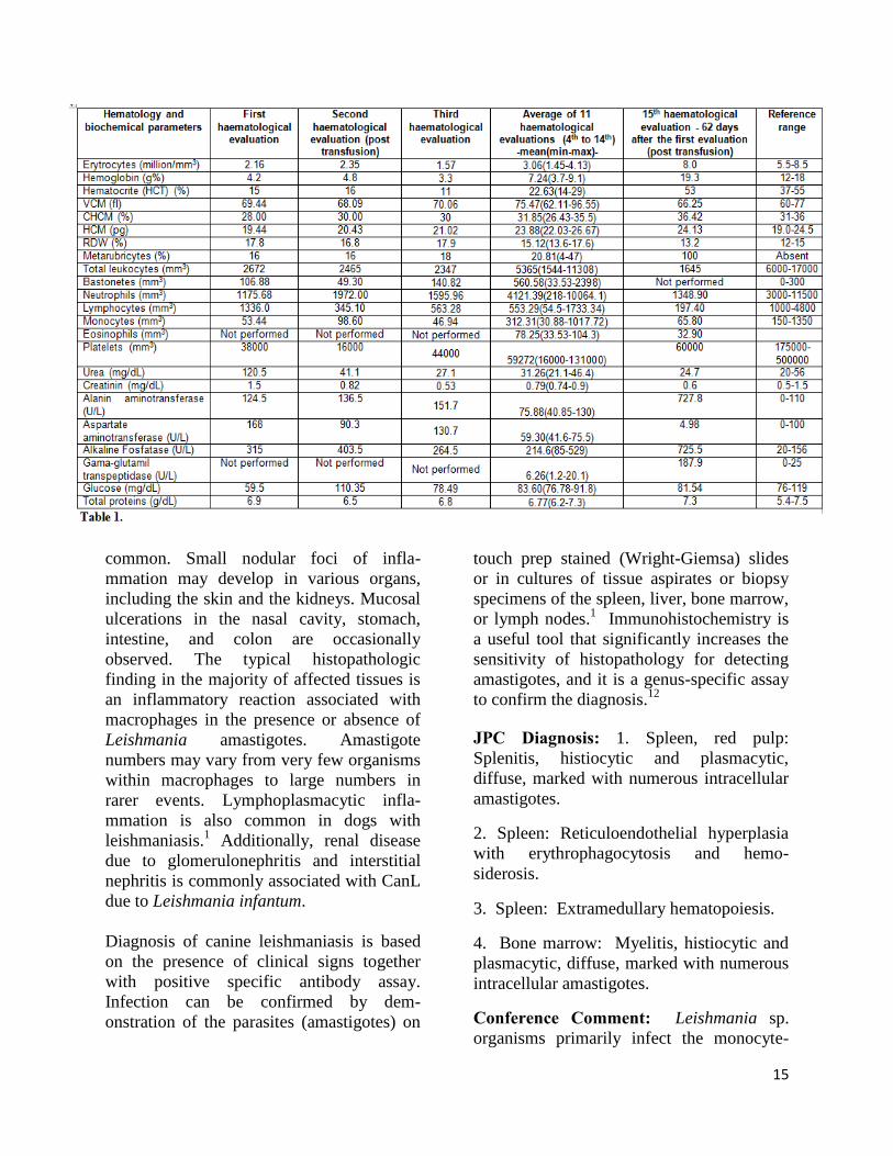
presented in Table 1.
After the last transfusion, i.e. 62 days after
the first hematological evaluation, the RBC,
HCT, and hemoglobin values were within
the reference range.
Concomitantly with the fourth hem-
atological evaluation, a bone marrow
aspiration was performed. It was noticed the
paucity of hematopoietic precursors, mainly
from the erythroid lineage. Serological tests
at this time resulted in positive ELISA and
negative RIFI.
All hematological analyses revealed reduced
numbers of platelets and lymphocytes. The
numbers of monocytes remained below the
Spleen, dog. The spleen parenchyma is expanded by
large numbers of macrophages, effacing the normal red
and white pulp architecture. (HE, 5X).

12
reference range in most of the hematological
exams.
Histopathologic Description: Spleen: The
normal splenic architecture is markedly
distorted due to complete replacement of the
red and white pulp by numerous
macrophages, moderate numbers of plasma
cells, fewer lymphocytes, and occasional
neutrophils, that expand the cords, fill the
sinuses and invade the splenic trabeculae.
Macrophages are enlarged and contain
myriad of 2.0-4.0 m, ovoid, intra-
cytoplasmic amastigotes (Fig. 3) with a 1.0
m nucleus, surrounded by a 1.0-2.0 m
clear zone and a kinetoplast perpendicular to
the nucleus (compatible with amastigotes of
Leishmania spp.). Multifocal areas of the
capsule are expanded due to deposition of
variable amounts of fibrous connective
tissue and infiltration of scattered lymph-
ocytes, plasma cells, and macrophages.
Occasional areas of
neovascularization are
within the capsule.
There are scattered
areas of karyorrhectic
and cellular debris
(lytic necrosis)
containing occasional
viable and degenerate
neutrophils admixed
with variable amounts
of an eosinophilic
beaded to fibrillar
material (fibrin). There
is multifocal histiocytic
erythrophagocytosis,
rare megakaryocytes,
and numerous
macrophages
containing intra-
cytoplasmic granular
and brown pigment
(hemosiderosis).
Bone marrow (proximal femur): The
marrow parenchyma is completely replaced
(myelopthisis) by numerous macrophages,
moderate plasma cells and fewer
lymphocytes. Macrophages contain myriad
of basophilic structures similar to those
described in the spleen (Fig. 4).
Multifocally, are areas of karyorrhectic and
cellular debris (lytic necrosis) containing
occasional viable and degenerate neutrophils
admixed with variable amounts of an
eosinophilic beaded to fibrillar material
(fibrin). There is multifocal hemosiderosis.
Tissues not submitted:
Lymph nodes present changes similar to
those described in the spleen. Additionally,
moderate lymphoid hyperplasia and
increased plasma cell differentiation are
observed. Macrophages containing myriad
of amastigotes of Leishmania spp. are seen
within lymphatic sinus, medullary cords,
Spleen, red pulp. The red pulp is replaced by large numbers of amastigote-laden
macrophages admixed with numerous plasma cells and fewer lymphocytes. Moderate
numbers of macrophages (arrows) contain phagocytized erythrocytes.

13
cortical and paracortical regions. In the liver
there are multiple areas with loss of
hepatocytes and infiltration of macrophages,
plasma cells and lymphocytes. Some
macrophages are loaded with amastigotes of
Leishmania spp. In both organs there is
moderate multifocal hemosiderosis.
The kidneys presented with moderate
membranous glomerulonephropathy. The
Lumina of several tubules are ectatic, and
contain aggregates of eosinophilic material
(protein casts).
Amastigotes of Leishmania spp. were
detected by immunohistochemistry
(Streptavidin-biotin-peroxidase) in the
spleen (Fig. 5) and in the bone marrow (Fig.
6 and 7).
Contributor’s Morphologic Diagnosis:
Spleen: Splenitis and perisplenitis,
histiocytic and plasmacytic, diffuse, severe,
with myriad of intrahistiocytic amastigotes,
etiology consistent with Leishmania spp.
Bone marrow: Myelitis, histiocytic and
lymphoplasmacytic, diffuse, severe, with
myriad of intrahistiocytic amastigotes,
etiology consistent with Leishmania spp.
Contributor’s Comment: Canine
leishmaniasis (CanL) is a major global
zoonosis, potentially fatal to humans and
dogs. Dogs are the main reservoir of the
infection to humans.3 The disease is
endemic in more than 70 countries in the
world and occurs predominantly in the
southern Europe, Africa, South and Central
America.11
While Leishmania infantum
(synonym L. chagasi) occurs in Latin
America, as well as in the Mediterranean
climate regions of the Old World,
Leishmania donovani infections are
restricted to the (sub-) tropics of Asia and
Africa. In these former regions, transmission
is mostly anthroponotic.9 In Latin America,
approximately 90% of the cases of human
visceral leishmaniosis are reported in Brazil,
and occur in almost all of the states of the
country. The higher incidence of the disease
in humans takes place in the northeast
region of the country (65% of the cases),
followed by the southeast (14%) north
(14%) and centerwest (14%).3,6
Currently,
the disease is gradually spreading to the
south and west regions of the country.3,10
Studies comparing the incidence or
prevalence of the disease in dogs in different
regions of the country are scarce. In Belo
Horizonte, capital of the state of Minas
Gerais, CanL is considered endemic with
increasing seroprevalence in the canine
population. From 1994 to 2000, this
prevalence was estimated in 3.6% of all
dogs of the county. In 2007, this index
increased to 9.3%.3,8
The life cycle of Leishmania spp. involves
two hosts – a phlebotomine sand fly vector
(genus Lutzomyia in the New World) and a
mammal (including rodents, canids, or
humans). Leishmania spp. occurs as
flagellated, extracellular promastigotes in
the gut of sand fly vectors. Infection occurs
when a feeding sand fly deposits metacyclic
promastigotes into the dermis of the host. In
mammalian hosts, Leishmania spp. occur as
amastigotes (2.0 to 3.0 m in diameter)
within mononuclear phagocytes in the skin,
bone marrow, and visceral organs.11
Although vector-borne is the most important
route of transmission, CanL can also be
transmitted in the absence of the invertebrate
vector.13
CanL is manifested by a broad spectrum of
clinical signs and degrees of severity. In the
typical CanL case, history and physical
examination include skin lesions, local or
generalized lymphadenomegaly, emaciation,

14
cachexia, splenomegaly, anorexia or
increased appetite, lethargy, temporal
muscle atrophy, exercise intolerance,
polyuria/polydipsia, ocular lesions,
epistaxis, onychogryphosis, lameness,
vomiting, and diarrhea.4
Serum
biochemistry findings in dogs with clinical
leishmaniosis include, most commonly,
serum hypoproteinemia with hyper-
globulinemia and hypoalbuminemia, re-
sulting in a decreased albumin/globulin
ratio. Mild increases of liver enzyme
activities are frequent; however, grossly
elevated liver enzyme activities, severe
azotemia, or both, are found in only a
minority of dogs with leishmaniosis.
Proteinuria and some renal abnormalities
develop in most dogs with this disease.1
Hematological parameters and the serum
biochemical profile in L. infantum-infected
dogs have limited diagnostic value.
However, they can be useful biomarkers for
evaluating the
clinical progress of
infected animals
and may also
contribute to the
understanding of
CanL
pathogenesis. One
the most
remarkable
characteristics of
CanL-associated
hematological
disorders is
anemia,
characterized as
non-regenerative
normocytic or
normochromic.
The leucocyte
alteration in the
blood of
symptomatic dogs
includes leucopenia characterized by mono-
cytopenia, lymphopenia, and eosinopenia.1
The pathogenesis of hematological changes
in both red and white blood cells is often
related to bone marrow disorders associated
with diminished erythropoiesis due to
intense bone marrow parasitism. In addition,
anemia can be related to reduced plasma
iron due to abnormal iron retention by
macrophages and increased levels of
hepcidin, typical of anemia of chronic
diseases. Anemia could also be related to
increased hemolysis in enlarged spleen and
liver associated with inflammatory response
to L. infantum and to decreased production
of erythropoietin by damaged kidneys.7
Severely affected dogs are usually cachectic
and suffer from muscle atrophy. The skin
and hemolymphatic organs are primarily
affected. Generalized lymphadenomegaly
and splenomegaly are usually present.
Hepatomegaly may be present, but is less
Bone marrow. Macrophages, which largely replace immature erythrpoietic elements, contain
numerous 2-3um amastigotes with a central nucleus and a rod-shaped kinetoplast. (HE, 400X)
15
common. Small nodular foci of infla-
mmation may develop in various organs,
including the skin and the kidneys. Mucosal
ulcerations in the nasal cavity, stomach,
intestine, and colon are occasionally
observed. The typical histopathologic
finding in the majority of affected tissues is
an inflammatory reaction associated with
macrophages in the presence or absence of
Leishmania amastigotes. Amastigote
numbers may vary from very few organisms
within macrophages to large numbers in
rarer events. Lymphoplasmacytic infla-
mmation is also common in dogs with
leishmaniasis.1 Additionally, renal disease
due to glomerulonephritis and interstitial
nephritis is commonly associated with CanL
due to Leishmania infantum.
Diagnosis of canine leishmaniasis is based
on the presence of clinical signs together
with positive specific antibody assay.
Infection can be confirmed by dem-
onstration of the parasites (amastigotes) on
touch prep stained (Wright-Giemsa) slides
or in cultures of tissue aspirates or biopsy
specimens of the spleen, liver, bone marrow,
or lymph nodes.1 Immunohistochemistry is
a useful tool that significantly increases the
sensitivity of histopathology for detecting
amastigotes, and it is a genus-specific assay
to confirm the diagnosis.12
JPC Diagnosis: 1. Spleen, red pulp:
Splenitis, histiocytic and plasmacytic,
diffuse, marked with numerous intracellular
amastigotes.
2. Spleen: Reticuloendothelial hyperplasia
with erythrophagocytosis and hemo-
siderosis.
3. Spleen: Extramedullary hematopoiesis.
4. Bone marrow: Myelitis, histiocytic and
plasmacytic, diffuse, marked with numerous
intracellular amastigotes.
Conference Comment: Leishmania sp.
organisms primarily infect the monocyte-

16
macrophage system and the visceral
manifestation bears many similarities to
histoplasmosis. Leishmania amastigotes are
able to survive and reproduce in macrophage
phagolysosomes due in part to an increase in
pH, and macrophage destruction occurs
secondary to proliferation of the protozoa.14
The immune response includes both T
helper 1 (Th1) and T helper 2 (Th2)
mediated mechanisms and tissue damage
occurs via a variety of methods including
granulomatous inflammation, immune
complex deposition and the formation of
autoantibodies. Inefficient killing of the
organism by macrophages leads to the
various immune responses, eventually
resulting in damage to a variety of tissues
and an array of clinical manifestations
discussed above.2
Resistance to infection is
based on a strong Th1 response, whereas a
strong Th2 response can be deleterious5 due
to immune complex deposition, particularly
in renal glomeruli. The level of parasite
burden also appears to play a role in the
immune response effectiveness against the
invading organisms. In dogs with a high
parasite load CD8+ T Cells are less effective
at lysing infected macrophages. Genetic
susceptibility also plays a role in outcome of
infection with certain breeds such as the
German Shepherd, cocker spaniel, and boxer
being more susceptible.4 The most common
clinical manifestations of disease include
skin, renal and/or ocular disease as well as
epistaxis, and the disease is broadly divided
into cutaneous, mucocutaneous and visceral
forms. Enlargement of the spleen is very
common and nearly always occurs in cases
of visceral leishmaniasis.14
While leish-
maniasis is more common in Mediterranean
countries and South America, endemic foci
are also present in North America including
in Texas, Oklahoma, Ohio and Michigan.5
The conference histologic description was
aligned very closely with the contributor’s
description above. Conference participants
were struck by the marked degree of
effacement of both the splenic red pulp and
bone marrow.
We thank the contributor for providing
clinical pathology data with the submission,
which greatly enhances the teaching value of
the case. Participants discussed the third
hematological evaluation and whether the
anemia is regenerative or nonregenerative,
but without a reticulocyte count it is difficult
to determine with certainty. The presence of
nucleated red blood cells (metarubricytosis)
can be indicative of regeneration with
concurrent reticulocytosis, but nucleated red
blood cells can also be present in other
conditions such as bone marrow damage
from inflammation or necrosis, lead
poisoning and due to splenectomy among
other things. A normal MCV would support
a nonregenerative anemia; additionally, the
pancytopenia and histologic changes in the
bone marrow also provide support for a
nonregenerative anemia. Elevated ALT and
AST are indicative of mild hepatocellular
damage, which correlates with the
histopathologic description mentioned above
by the contributor in the tissues not
submitted. The total protein is normal but a
decrease in albumin and increase in globulin
is likely present in this case, based on the
degree of infection and ongoing infla-
mmation, which is a common pattern in
dogs afflicted with leishmaniasis, as
mentioned above by the contributor.
Contributing Institution:
Veterinary School. Universidade Federal de
Minas Gerais
www.vet.ufmg.br

17
References:
1. Baneth G, Solano-Gallego L.
Leishmaniasis. In: Greene CE, ed. Infectious
diseases of the dog and cat. 4th ed.
Philadelphia, US: Elsevier Saunders;
2012:735-748.
2. Cooper BJ, Valentine BA. Muscle and
Tendon. In: Maxie MG, ed. Jubb, Kennedy,
and Palmer's Pathology of Domestic
Animals. 6th ed. Vol 1. St. Louis, MO:
Elsevier; 2015:240.
3. Diniz SA, Silva FL, Carvalho Neta AV,
et al. Animal reservoirs for visceral
leishmaniasis in densely populated urban
areas. J Infect Dev Ctries. 2008; 2:24-33.
4. Koutinas AF, Koutinas CK. Pathologic
mechanism underlying the clinical findings
in canine Leishmaniosis due to Leishmania
infantum/chagasi. Vet Pathol.
2014;51(2)527-538.
5. Mauldin EA, Peters-Kennedy J.
Integumentary system. In: Maxie MG, ed.
Jubb, Kennedy, and Palmer's Pathology of
Domestic Animals. 6th ed. Vol 1. St. Louis,
MO: Elsevier; 2015:663.
6. Ministério da Saúde. Manual de
vigilância e controle da leishmaniose
visceral. Brasília, BR: Editora MS; 2006.
7. Nicolato RC, Abreu RT, Roatt BM, et al.
Clinical forms of canine visceral
Leishamniasis in naturally Leishmania
infantum-infected dogs and related
myelogram and hemogram changes. Plos
One. 2013;8(12):1-9.
8. Papa DN. Perfil epidemiologico da
Leishmaniose Visceral em cães
diagnosticasdos no Laboratório da Escola de
Veterinária da Universidade Federal de
Minas Gerais Belo Horizonte 2004 a 2008.
Master’s degree dissertation, Escola de
Veterinária, Universidade Federal de Minas
Gerais, 2010.
9. Ready PD. Epidemiology of visceral
leishmaniasis. Clin Epidemiol. 2014; 6:147-
154.
10. Romero GAS, Boelaert M. Control of
visceral Leishmaniasis in Latin America – A
systematic review. Plos Negl Trop Dis.
2010;4(1):1-17.
11. Solano-Gallego L, Miró G, Koutinas A,
et al. LeishVet guidelines for practical
management of canine leishmaniosis.
Parasite Vect. 2011;4:86.
12. Tafuri WL, Santos RL, Arantes MRE, et
al. An alternative immunohistochemical
method for detecting Leishmania
amastigotes in paraffin-embedded canine
tissues. J Immunol Methods. 2004; 292:17-
23.
13. Turchetti AP, Souza TD, Paixão TA,
Santos RL. Sexual and vertical transmission
of visceral leishmaniasis. J Infect Dev
Ctries. 2014; 8:403-407.
14. Valli VEO, Kiupel M, Bienzle D.
Hematopoietic System. In: Maxie MG, ed.
Jubb, Kennedy, and Palmer's Pathology of
Domestic Animals. 6th ed. Vol 3. St. Louis,
MO: Elsevier; 2015:1

18
CASE IV: 21966 (JPC 4066536).
Signalment: 10-year-old spayed female
Miniature Dachshund dog, (Canis
familiaris)
History: The dog was presented with a
gingival mass on right maxilla, which had
been enlarging for 2 to 3 weeks. Both right
and left mandibular lymph nodes were
enlarged by palpation. Thoracic CT scan
revealed no metastasis in the lung.
Gross Pathology: The mass measured 3.7
x 2.4 x 1.1 cm and was partly red and black.
Laboratory Results: None
Cytologic description: Fine-needle asp-
iration of the mass was performed. The
smear was highly cellular and consisted of
monomorphic cells, most of which are in
clusters. The cells had a round nucleus in
pale abundant cytoplasm. The cytoplasm
was mild to moderately vacuolated and
small black pigment noted in a few cells.
Cells showed mild to moderate anisocytosis
and anisokaryosis, and mitotic figures were
rarely seen.
From the left mandibular lymph node,
similar morphologic cells were mainly
acquired, and several small lymphocytes,
plasma cells, and eosinophils were noted.
On the smear of right mandibular lymph
node, lymphoid cells, predominantly small
mature lymphocytes with some lymph-
oblasts and plasma cells were seen.
Scattered melanophages were also observed.
Contributor’s Interpretation:
1. Gingival mass: Malignant melanoma with
metastasis to the left mandibular lymph node
was suspected.
2. Right mandibular lymph node: Reactive
lymph node.
Histologic result: The mass was partly
resected for histopathology. On histologic
examination, the neoplastic cells invaded
almost all of the mass and were arranged in
nests supported by a fine fibrovascular
stroma. The neoplastic cells had clear
abundant cytoplasm and round to oval
nucleus. The cytoplasm was vacuolated and
small amount of brown pigment were
observed in several cells. A few mitotic
figures are noted. Both left and right
mandibular lymph nodes were removed and
submitted for the histopathology. Neoplastic
cells infiltrated both lymph nodes, and
especially in the left lymph node, they
spread across a wide area.
Contributor’s Comment: Tumors of
melanocytic origin are relatively common in
the dog. The malignancy of melanoma
greatly depends on anatomic location, and
oral melanoma is highly aggressive.
Cytologic morphology of melanoma is
various, showing the feature of epithelial
cells, mesenchymal cells or discrete round
cells. Balloon cell melanoma, which is a rare
variant of melanocytic tumor, has been
Fine needle aspirate, oral mass, dog. A densely cellular,
fine needle aspirate was submitted digitally for
examination. (Wright’s-Giemsa, 4X).

19
reported in human, dog and cat.1, 3,8,9,10,11
Microscopically, the neoplastic cells are
amelanotic and cytoplasm is vacuolated.
Sebaceous carcinoma, liposarcoma, other
clear cell neoplasm and granulomatous
inflammation are listed as differentials. In
this case, sebaceous carcinoma and lipo-
sarcoma were unlikely due to the location.
The malignant neoplasia was more sus-
picious than inflammatory disease because
of the morphologic atypia. Due to the small
amount of granules that can be melanin
pigments, melanoma was the primary rule-
out; immunohistochemistry such as Melan-
A is helpful for further diagnosis.
JPC Diagnosis: Fine needle aspirate, oral
mass: Malignant neoplasm; differentials
include amelanotic melanoma (balloon cell),
rhabdomyoma, granular cell tumor, and
oncocytoma.
Conference Comment: The neoplastic cells
are round to spindled and arranged
individually and in small aggregates; the
cytoplasm is amphophilic and indistinctly
vacuolated and a low to moderate subset of
cells contain a pale, granular eosinophilic
material in the cytoplasm, the origin of
which is unclear. The cells have a single,
central, round to oval nucleus with finely
stippled chromatin, and 1-2 variably distinct
nucleoli. Anisocytosis is mild and
anisokaryosis is moderate, and the N:C is
low; rare mitoses are observed. Other
salient features include nuclear molding, a
central fold in the nuclear membrane of few
cells, and rare multinucleate cells. Features
of malignancy are not striking in this
sample, but the anisokaryosis, nuclear
molding and presence of mitoses warrants a
malignant interpretation. Many conference
participants commented melanomas are us-
ually more aggressive looking, particularly
in the oral cavity. A search for melanin was
largely unrewarding, particularly with the
digital slide only being scanned to 20X, but
small amounts are seen in images of the
histologic section provided by the con-
tributor. Aside from the differential dia-
gnoses listed above, conference participants
remarked the cells resemble hepatocytes in
animals with steroid hepatopathy, sug-
gesting glycogen or another similar substrate
may be present within the cytoplasm.
Differential diagnoses discussed include
balloon cell melanoma, rhabdomyoma,
granular cell tumor and oncocytoma. A
definitive diagnosis is not possible without
the use of immunohistochemistry, which
was not performed by the contributor.
Balloon cell melanoma is an uncommon
variant of melanoma most commonly
described in the skin in both domestic
animals and humans, and in cats is often
localized to the head. The cells are
described as epithelioid, large and round,
with abundant finely vacuolated cytoplasm
that generally lacks pigment.6,11
Glycogen
(PAS positive) and lipid rich variants have
been described, primarily in humans.
Although the balloon cells may contain
substances other than glycogen and other
similar appearing non-melanocytic tumors
may contain glycogen,11
warranting a
cautious interpretation of cytoplasmic
features. The background of glycogen rich
neoplasms is occasionally described as
“tigroid,” referring to a vague tiger striping
pattern imparted by background con-
stituents.4 Ultrastructural findings in balloon
cell melanomas indicate the cytoplasmic
vacuoles represent enlarged melanosomes.
In addition to Melan A, balloon cell
melanomas may stain for S-100 and neuron-
specific enolase.1,11

20
Rhabdomyomas, oncocytomas and granular
cell tumors have a similar cytologic app-
earance with eosinophilic granular material
in the cytoplasm, comparable to what is seen
in a subset of cells in this case. The similar
cytoplasmic appearance in oncocytomas and
rhabdomyomas is related to the presence of
large numbers of mitochondria, and in
granular cell tumors is postulated to be due
to metabolic abnormalities and accumulation
of lysosomes.5 Rhabomyomas are benign
tumors of striated muscle and may occur in
the larynx and tongue of domestic animals.
Cells may have abundant cytoplasmic
glycogen, suggested by PAS positivity.7
Oncocytomas are epithelial or neur-
oendocrine origin neoplasms which arise
from oncocytes or oxyphil cells, and are
described in the larynx of young dogs. The
exact origin of granular cell tumors is
unclear but they primarily occur in the oral
cavity and have been conjectured to be of
Schwann cell origin. Rhabdomyomas and
oncocytomas have benign behavior, and the
behavior of granular cell tumors is not well
defined. Both rhadomyoma and oncocytoma
have large pale cells with granular or foamy
cytoplasm. The nucleus of oncocytoma is
central, while the nucleus of rhadomyoma
may be central or peripheral and both have
finely clumped chromatin and a single
indistinct nucleolus. Multinucleate cells
may be seen in rhabdomyoma. The in-
distinct nucleolus is a feature which may
help distinguish them from balloon cell
melanoma. Granular cell tumors are variably
sized but typically have abundant granular
eosinophilic cytoplasm and an eccentric
nucleus. Pleomorphism may be moderate in
each of the three tumors. Additional
diagnostics which may help differentiate the
tumors include desmin positivity in
Fine needle aspirate, oral mass, dog. Neoplastic cells are polygonal with distinct cell borders, abundant vacuolated
amphophilic cytoplasm with adherent pink fibrillar matrix. Nuclei exhibit ropy chromatin with mild anisokaryosis, and
binucleated cells are frequent. (HE, 120X)

21
rhabdomyoma, cytokeratin positivity in
oncocytoma and PAS positivity (diastase
resistance) in granular cell tumor.2
Contributing Institution:
Laboratory of Comparative Pathology and
Department of Diagnostic Pathology,
Graduate School of Veterinary Medicine,
Hokkaido University, Japan
http://www.vetmed.hokudai.ac.jp/
References:
1. Blanchard TW, Bryant NJ, Mense MG.
Balloon cell melanoma in three dogs: a
histopathological, immunohistochemical and
ultrastructural study. J Comp Pathol. 2001;
125:254-261.
2. Burkhard MJ. Respiratory Tract. In:
Raskin RE, Meyer DJ, eds. Canine and
Feline Cytology. 3rd ed. St. Louis, MO:
Elsevier; 2016:155-156.
3. Cangul IT, van Garderen E, van der
Linde-Sipman JS, van den Ingh TS,
Schalken JA. Canine balloon and signet-ring
cell melanomas: a histological and imm-
unohistochemical characterization. J Comp
Pathol. 2001; 125:166-173.
4. DeMay RM. The Art and Science of
Cytopathology. Vol 2. 2nd ed. Hong Kong:
American Society for Clinical Pathology;
2012:592.
5. Dunbar MD, Ginn P, Winter M, Miller
KB, Craft W. Laryngeal rhabdomyoma in a
dog. Vet Clin Pathol. 2012; 41(4):590-593.
6. Goldschmidt MH, Dunstan RW, Stannard
AA, Tscharner CV, et al. Histological
classification of epithelial and melanocytic
tumors of the skin of domestic animals. Vol
III. 2nd series. Washington D.C.: Armed
Forces Institute of Pathology. 1998:38-39.
7. Hendrick MJ, Mahaffey EA, Moore FM,
Vos JH, Walder EJ. Histological class-
ification of mesenchymal tumors of the skin
and soft tissues of domestic animals. 2nd
series. Washington D.C.: Armed Forces
Institute of Pathology. 1998:21.
8. Kao GF, Helwig EB, Graham JH. Balloon
cell malignant melanoma of the skin. A
clinicopathologic study of 34 cases with
histochemical, immunohistochemical, and
ultrastructural observations. Cancer. 1992;
69:2942-2952.
9. Mowat A, Reid R, Mackie R. Balloon cell
metastatic melanoma: an important diff-
erential in the diagnosis of clear cell tumors.
Histopathology. 1994; 24:469-472.
10. van der Linde-Sipman JS, de Wit MM,
van Garderen E, Molenbeek RF, et al.
Cutaneous malignant melanomas in 57 cats:
identification of (amelanotic) signet-ring and
balloon cell types and verification of their
origin by immunohistochemistry, electron
microscopy, and in situ hybridization. Vet
Pathol. 1997; 34:31-38.
11. Wilkerson MJ, Dolce K, DeBey BM,
Heeb H, et al. Metastatic Balloon Cell
Melanoma in a Dog. Vet Clin Pathol. 2003;
32(1):31-6.