web.ics.purdue.eduweb.ics.purdue.edu/~jfleary/nanomedicine_course_2011/Lecture_6... · that leads...
14
Polyethyleneimine Coating Enhances the Cellular Uptake of Mesoporous Silica Nanoparticles and Allows Safe Delivery of siRNA and DNA Constructs Tian Xia, †, Michael Kovochich, †, Monty Liong, ‡, Huan Meng, † Sanaz Kabehie, ‡ Saji George, † Jeffrey I. Zink, ‡, and Andre E. Nel †,§,, * † Division of NanoMedicine, Department of Medicine, ‡ Department of Chemistry & Biochemistry, § The Southern California Particle Center, and California NanoSystems Institute, University of California, Los Angeles, California 90095. These authors contributed equally to this work. O n the basis of properties such as large surface area and or- dered porous channels that can be used to encapsulate molecules, meso- porous silica nanoparticles (MSNP) have emerged as an efficient drug delivery platform. 15 In addition to the well- developed surface chemistry, silica ma- terials are known to be safe, biodegrad- able, and potentially biocompatible. 6,7 We and others have demonstrated that this drug transport system is suitable for the delivery of anticancer drugs, includ- ing camptothecin, paclitaxel, and doxorubicin. 1,2,8 The chemical stability of the particles contributes to their thera- peutic utility by allowing the attachment of functional groups for imaging and tar- geting applications along with the place- ment of a series of nanovalves for on- demand drug release. 2,9,10 In addition to being used for the deliv- ery of chemical therapeutic agents, MSNP have the potential as hybrid organicinorganic materials that can act as carriers for nucleic acids and therefore are potentially useful for the delivery of small interfering RNAs (siRNAs) and other forms of gene therapy. 1113 As opposed to other gene delivery systems based on inorganic materials, the porous structure of MSNP allows both the binding of nu- cleotides on the surface as well as the en- capsulation of small molecules within the particles. It is even possible to com- bine these modules to achieve dual deliv- ery of drugs and nucleic acids. 13 To maxi- mize the delivery of negatively charged nucleic acids to cells, the silica surface has to be converted into positive charge in order to bind DNA and siRNA. Some of the methods for introducing cationic charge on inorganic materials, which in- clude silica, iron oxide, and gold, typically involve surface grafting with amine groups and coating with cationic poly- mers (e.g., polyethyleneimine, polyami- doamine, and polylysine) through either covalent or electrostatic association. 12,1420 The gene delivery ca- pabilities of these surface-modified ma- terials have been demonstrated and may prove useful as alternatives to traditional viral vectors. *Address correspondence to [email protected]. Received for review July 1, 2009 and accepted August 26, 2009. Published online September 9, 2009. 10.1021/nn900918w CCC: $40.75 © 2009 American Chemical Society ABSTRACT Surface-functionalized mesoporous silica nanoparticles (MSNP) can be used as an efficient and safe carrier for bioactive molecules. In order to make the MSNP a more efficient delivery system, we modified the surface of the particles by a functional group that enhances cellular uptake and allows nucleic acid delivery in addition to traditional drug delivery. Noncovalent attachment of polyethyleneimine (PEI) polymers to the surface not only increases MSNP cellular uptake but also generates a cationic surface to which DNA and siRNA constructs could be attached. While efficient for intracellular delivery of these nucleic acids, the 25 kD PEI polymer unfortunately changes the safety profile of the MSNP that is otherwise very safe. By experimenting with several different polymer molecular weights, it was possible to retain high cellular uptake and transfection efficiency while reducing or even eliminating cationic MSNP cytotoxicity. The particles coated with the 10 kD PEI polymer were particularly efficient for transducing HEPA-1 cells with a siRNA construct that was capable of knocking down GFP expression. Similarly, transfection of a GFP plasmid induced effective expression of the fluorescent protein in >70% cells in the population. These outcomes were quantitatively assessed by confocal microscopy and flow cytometry. We also demonstrated that the enhanced cellular uptake of the nontoxic cationic MSNP enhances the delivery of the hydrophobic anticancer drug, paclitaxel, to pancreatic cancer cells. In summary, we demonstrate that, by a careful selection of PEI size, it is possible to construct cationic MSNP that are capable of nucleotide and enhanced drug delivery with minimal or no cytotoxicity. This novel use of a cationic MSNP extends its therapeutic use potential. KEYWORDS: mesoporous silica nanoparticles (MSNP) · polyethyleneimine (PEI) · transfection · plasmid DNA · siRNA · drug delivery ARTICLE www.acsnano.org VOL. 3 ▪ NO. 10 ▪ 3273–3286 ▪ 2009 3273
-
Upload
nguyenkhuong -
Category
Documents
-
view
213 -
download
0
Transcript of web.ics.purdue.eduweb.ics.purdue.edu/~jfleary/nanomedicine_course_2011/Lecture_6... · that leads...

Polyethyleneimine Coating Enhances theCellular Uptake of Mesoporous SilicaNanoparticles and Allows Safe Deliveryof siRNA and DNA ConstructsTian Xia,†,� Michael Kovochich,†,� Monty Liong,‡,� Huan Meng,† Sanaz Kabehie,‡ Saji George,†
Jeffrey I. Zink,‡,� and Andre E. Nel†,§,�,*†Division of NanoMedicine, Department of Medicine, ‡Department of Chemistry & Biochemistry, §The Southern California Particle Center, and �California NanoSystemsInstitute, University of California, Los Angeles, California 90095. �These authors contributed equally to this work.
On the basis of properties suchas large surface area and or-dered porous channels that can
be used to encapsulate molecules, meso-
porous silica nanoparticles (MSNP) have
emerged as an efficient drug delivery
platform.1�5 In addition to the well-
developed surface chemistry, silica ma-
terials are known to be safe, biodegrad-
able, and potentially biocompatible.6,7
We and others have demonstrated that
this drug transport system is suitable for
the delivery of anticancer drugs, includ-
ing camptothecin, paclitaxel, and
doxorubicin.1,2,8 The chemical stability of
the particles contributes to their thera-
peutic utility by allowing the attachment
of functional groups for imaging and tar-
geting applications along with the place-
ment of a series of nanovalves for on-
demand drug release.2,9,10
In addition to being used for the deliv-
ery of chemical therapeutic agents, MSNP
have the potential as hybrid
organic�inorganic materials that can act
as carriers for nucleic acids and therefore
are potentially useful for the delivery of
small interfering RNAs (siRNAs) and other
forms of gene therapy.11�13 As opposed
to other gene delivery systems based on
inorganic materials, the porous structure
of MSNP allows both the binding of nu-
cleotides on the surface as well as the en-
capsulation of small molecules within
the particles. It is even possible to com-
bine these modules to achieve dual deliv-
ery of drugs and nucleic acids.13 To maxi-
mize the delivery of negatively charged
nucleic acids to cells, the silica surface has
to be converted into positive charge inorder to bind DNA and siRNA. Some ofthe methods for introducing cationiccharge on inorganic materials, which in-clude silica, iron oxide, and gold, typicallyinvolve surface grafting with aminegroups and coating with cationic poly-mers (e.g., polyethyleneimine, polyami-doamine, and polylysine) through eithercovalent or electrostaticassociation.12,14�20 The gene delivery ca-pabilities of these surface-modified ma-terials have been demonstrated and mayprove useful as alternatives to traditionalviral vectors.
*Address correspondence [email protected].
Received for review July 1, 2009and accepted August 26, 2009.
Published online September 9, 2009.10.1021/nn900918w CCC: $40.75
© 2009 American Chemical Society
ABSTRACT Surface-functionalized mesoporous silica nanoparticles (MSNP) can be used as an efficient and
safe carrier for bioactive molecules. In order to make the MSNP a more efficient delivery system, we modified the
surface of the particles by a functional group that enhances cellular uptake and allows nucleic acid delivery in
addition to traditional drug delivery. Noncovalent attachment of polyethyleneimine (PEI) polymers to the surface
not only increases MSNP cellular uptake but also generates a cationic surface to which DNA and siRNA constructs
could be attached. While efficient for intracellular delivery of these nucleic acids, the 25 kD PEI polymer
unfortunately changes the safety profile of the MSNP that is otherwise very safe. By experimenting with several
different polymer molecular weights, it was possible to retain high cellular uptake and transfection efficiency
while reducing or even eliminating cationic MSNP cytotoxicity. The particles coated with the 10 kD PEI polymer
were particularly efficient for transducing HEPA-1 cells with a siRNA construct that was capable of knocking down
GFP expression. Similarly, transfection of a GFP plasmid induced effective expression of the fluorescent protein in
>70% cells in the population. These outcomes were quantitatively assessed by confocal microscopy and flow
cytometry. We also demonstrated that the enhanced cellular uptake of the nontoxic cationic MSNP enhances the
delivery of the hydrophobic anticancer drug, paclitaxel, to pancreatic cancer cells. In summary, we demonstrate
that, by a careful selection of PEI size, it is possible to construct cationic MSNP that are capable of nucleotide and
enhanced drug delivery with minimal or no cytotoxicity. This novel use of a cationic MSNP extends its therapeutic
use potential.
KEYWORDS: mesoporous silica nanoparticles (MSNP) · polyethyleneimine(PEI) · transfection · plasmid DNA · siRNA · drug delivery
ARTIC
LE
www.acsnano.org VOL. 3 ▪ NO. 10 ▪ 3273–3286 ▪ 2009 3273

In this article, we studied the effects of polyethylene-
imine (PEI) coating on the MSNP in terms of cellular up-
take, cytotoxicity, and efficiency of nucleic acid deliv-
ery. PEIs are synthetic cationic polymers that
compact DNA and siRNA into complexes that are ef-
fectively taken up in cells to make nucleic acid deliv-
ery and gene therapy possible.21�23 Although the
polymer itself is used as a delivery vehicle, PEI can
also be attached to nanoparticle surfaces through
covalent and electrostatic interactions to achieve the
same goal.11,16,17,19,24 Complexing the polymer with
the nanoparticles has the potential advantage of fa-
cilitating DNA and siRNA delivery by a multifunc-
tional platform that also allows imaging, targeting,
and concurrent drug delivery. PEI was chosen as the
polymer coating to enhance the particle uptake
into cells and facilitate endosomal escape for the nu-
cleotide delivery.25 It is documented that, while low
MW PEI is not cytotoxic, these polymers are ineffec-
tive at transfecting nucleotides in contrast to the
high MW PEI. In this regard, it has been demon-
strated that the size (MW), compactness, and chemi-
cal modification of PEI affect the efficacy and toxic-
ity of this polymer.26,27 Therefore, we experimented
with several PEI polymer sizes ranging from MW of
0.6 to 25 kD in order to balance the efficiency of nu-
cleic acid delivery and cellular toxicity. PEI cytotoxic-
ity occurs via a proton sponge effect, which in-
volves proton sequestration by the polymer surface
Figure 1. TEM of the MSNP and cell viability detection by the MTS assay. (A) TEM image shows the particle size and the or-dered pore structure. (B) After addition of appropriately dispersed MSNP exhibiting a range of surface modifications to pan-creatic cancer cell lines at doses ranging from 12.5 to 50 �g/mL for 16 h, cells were incubated with the MTS reagent for 30min and the absorbance was measured at 490 nm. All the MTS values were normalized according to the value of the con-trol (no particle exposure); this was regarded as 100% cell viability. The IC50 values of MSNP-PEI-25 kD in PANC-1 and BxPC3cells were 37 and 46 �g/mL, respectively. The results were reproduced three times.
TABLE 1. Size Distribution of MSNP in Aqueous Solutionsa
Size (nm) � potential (mV)
H2ODMEM 10%
serum BEGM
BEGM 2mg/mL
BSA
H2O/DMEM �
serum/BEGM �
BSA
MSNP-OH 1966 408 1096 416 �10.5/�6.8/�7.2MSNP-phosphate 1975 306 867 439 �8.9/�6.5/�5.8MSNP-PEG 2675 405 1215 542 �10.4/�5.9/�4.5MSNP-PEI 0.6 kD 1689 415 1243 474 �29.5/�7.8/�6.5MSNP-PEI 1.2 kD 1684 452 1298 502 �38.7/�6.5/�7.1MSNP-PEI 1.8 kD 1053 510 1087 550 �36.9/�5.4/�3.2MSNP-PEI 10 kD 614 702 917 684 �34.1/�7.5/�6.9MSNP-PEI 25 kD 1473 1043 1544 825 �30.8/�5.9/�4.0
aParticle size and � potential in solution were measured by a ZetaSizer Nano (Mal-vern). DMEM � complete Dulbecco’s Modified Eagle Media, which contains 10% fe-tal calf serum (FCS). BEGM � bronchial epithelial growth medium, which includesgrowth factors, cytokines, and supplements (no serum).
ART
ICLE
VOL. 3 ▪ NO. 10 ▪ XIA ET AL. www.acsnano.org3274

that leads to heightened pro-ton pump activity inside thecell, osmotic swelling of theendocytic compartment, endo-somal rupture, and ultimatelycell death by a mitochondrial-mediated mechanism.28,29 Wedemonstrate that the reduc-tion of the polymer size is ca-pable of scaling back the cyto-toxic effect and that particlescoated with polymers of 10 kDor less still maintain the featureof facilitated cellular uptake ofcationic nanoparticles, mostlikely due to high avidity mem-brane binding and efficientmembrane wrapping that al-lows these particles to entercellular endocytic compart-ments. We show that the cellu-lar uptake of PEI-coated MSNP,irrespective of polymer size, isconsiderably enhanced com-pared to unmodified MSNP (si-lanol surface) or particlescoated with phosphonate orpoly(ethylene gylcol) groups.Finally, we demonstrate thatPEI-coated particles bind plas-mid DNA and siRNA with highaffinity, enabling us to achieveefficient cellular delivery ofthese nucleic acids with non-toxic MSNP coated with 10 kDPEI.
RESULTSPhysicochemical Characterization
of the NP. MSNP were synthesized according to a modi-fied procedure previously described.12,30 The primaryparticle size is in the 100�130 nm range with a uni-form pore size of �2.5 nm, as shown by TEM (Figure1A). To conduct biological experimentation, particlesize and � potential were measured in water as well astissue culture media.31 For the purposes of this study,we used DMEM supplemented with 10% fetal calf se-rum (FCS) or BEGM as is or supplemented with 2 mg/mLBSA (Table 1). We observed that the addition of pro-tein leads to improved particle dispersion by counter-ing the colloidal forces that promote particle aggrega-tion in salt containing media (Supporting InformationFigure 1). While all of the non-PEI-coated particles ex-hibited a negative � potential, PEI-coated particlesshowed a positive charge (Table 1). However, with theaddition of FCS or BSA, all PEI-coated particles assumednegative � potential.
Differences in the Cytotoxic Potential of Anionic versus Cationic
MSNP. To screen for particle hazard, we used the MTS as-
say, which reflects dehydrogenase activity in healthy
cells.29 While most of the MSNP did not interfere in MTS
activity in the PANC-1 and BxPC3 pancreatic cancer
cell lines, particles coated with the 25 kD PEI polymer
showed decreased cellular viability (Figure 1B). The par-
ticles coated with the 25 kD polymer also induced tox-
icity in macrophage (RAW 264.7) and bronchial epithe-
lial (BEAS-2B) cell line staining (Supporting Information
Figure 2). The toxicity was confirmed by propidium io-
dide (PI) staining, which showed that the rate of cell
death was progressive over 15 h period (Supporting In-
formation Figure 2). We also confirmed that, similar to
our previous results with cationic polystyrene nanopar-
ticles,29 the toxicity of cationic MSNP involves an effect
on mitochondria as determined by JC-1 fluorescence
(Supporting Information Figure 2). JC-1 measures mito-
Figure 2. Cellular uptake of FITC-labeled MSNP in PANC-1 cells. MSNP were labeled with FITC as de-scribed in Materials and Methods. (A) Representative histogram showing the shift in fluorescence in-tensity in PANC-1 cells treated with 25 �g/mL FITC-MSNP that contain different surface modifications(left panel). The fold increase in MFI after 3 h was calculated and used to generate the graph. RITC-labeled MSNP-Phos served as a control particle to show that coating with PEI leads to enhanced up-take in the same particle type in the same cell (right panel). (B) Confocal microscopy was used to studythe cellular uptake of FITC-MSNP in PANC-1 cells. Cells were exposed to 25 �g/mL FITC-labeled par-ticles for 3 h. After cell membrane staining with 5 �g/mL red-fluorescent wheat germ agglutinin (WGA),cells were visualized using a confocal 1P/FCS inverted microscope. Data are representative of threeseparate experiments; *p � 0.01 compared with control.
ARTIC
LE
www.acsnano.org VOL. 3 ▪ NO. 10 ▪ 3273–3286 ▪ 2009 3275

chondrial membrane potential (MMP). In contrast,
phosphonate-coated nanoparticles (MSNP-Phos) were
nontoxic and did not perturb the mitochondrial func-
tion (Supporting Information Figure 2).
To confirm that PEI toxicity is due to cationic charge,
succinic anhydride was used to convert the primary NH2
to COOH groups.29 Supporting Information Figure 3A
demonstrates that this conversion reduced the toxicity
of MSNP-PEI-25 kD in a dose-dependent fashion. Deple-
tion of the primary NH2 groups was confirmed by fluo-
rescamine, which yields green fluorescence when it re-
acts with primary amines (Supporting Information
Figure 2B). Thus, succinic anhydride reduced the mean
fluorescence intensity (MFI) in a dose-dependent fash-
ion. The stability of PEI attached to the MSNP surface
was confirmed by coating FITC-labeled MSNP with
rhodamine-B-labeled PEI. Confocal microscopy con-
firmed that both labels colocalize as shown by the com-
posite yellow fluorescent spots in the cell (Supporting
Information Figure 4).
Differences in the Cellular Uptake of Anionic versus Cationic
MSNP. We have previously shown that the cellular tox-
icity of cationic polystyrene nanoparticles involves
a high rate of cellular uptakedue to tight surface membranebinding, which facilitates par-ticle wrapping and cellularuptake.28,29 Cellular uptake ofFITC-labeled MSNP was assessedby flow cytometry and confocalmicroscopy.29 FITC-labeledMSNP were incubated withPANC-1, BxPC3, RAW 264.7, andBEAS-2B cells. This resulted in aclear shift in the MFI of the dif-ferent cellular populations fol-lowing treatment with particlescoated with 1.2 and 25 kD PEIpolymers as compared to thephosphonate-coated MSNP (Fig-ures 2 and 3; Supporting Infor-mation Figure 5). When ex-pressed as fold increase in theMFI compared to untreatedcells, the relative abundance ofMSNP-PEI-25 kD and MSNP-PEI-1.2 kD uptake was 2 orders ofmagnitude higher than thephosphonate or PEG-coatedparticles (Figures 2 and 3; Sup-porting Information Figure 5). Incontrast, there was no differ-ence in the uptake of RITC-labeled MSNP in the same cellstreated with the FITC-labeledparticles. This confirms that thecationic charge is responsible
for high cellular uptake. The flow data were corrobo-rated by confocal studies that showed a significantincrease in the number of PEI-coated particles in allcell types (Figures 2 and 3 and Supporting Informa-tion Figure 5). Please notice that a large fraction ofthe MSNP-PEI-25 kD particles localized at the cellmembrane as demonstrated by the composite yel-low fluorescence in pancreatic cancer cells stainedwith the red-fluorescent wheat germ agglutinin (Fig-ures 2 and 3). It is possible that this membrane bind-ing might contribute to the toxicity of theseparticles.
Reducing PEI Polymer Length Maintains Effective Nucleic AcidDelivery but Eliminates Cellular Toxicity. While PEI is quite ef-fective for complexing and delivering nucleic acids,polymer-based delivery of DNA and siRNA oftenleads to cytotoxicity due to damage to the surfacemembrane, lysosomes, and mitochondria.21,26,29
However, on the basis of the observation that deco-ration of MSNP with a 1.2 kD polymer resulted in anontoxic particle, we explored the effect of a rangeof PEI polymers in terms of achieving cellular deliv-ery versus reduction of toxicity. MSNP were coated
Figure 3. Cellular uptake of FITC-labeled MSNP in BxPC3 cells. BxPC3 cells were exposed to FITC-labeled MSNP, and flow cytometry and confocal microscopy were conducted as in Figure 2. PanelsA and B represent similar observations.
ART
ICLE
VOL. 3 ▪ NO. 10 ▪ XIA ET AL. www.acsnano.org3276

with 0.6, 1.2, 1.8, 10, and 25 kD polymers and their
cytotoxic potential assessed in various cell types.
This included the use of HEPA-1 cells for which there
is a commercial variant expressing green fluores-
cent protein (GFP) for the purposes of assessing
siRNA knockdown. No toxicity was seen with par-
ticles coated with 0.6, 1.2, and 1.8 kD PEI polymers
(Figure 4 and Supporting Information Figure 6).
While MSNP-PEI 10 kD exerted toxic effects at the
highest dose (50 �g/mL) tested, MSNP-PEI 25 kD was
responsible for the decline in MTS activity at doses
�12.5 �g/mL (Figure 4 and Supporting Information
Figure 6). This demonstrates that it is possible to ad-
just MSNP toxicity according to the polymer length
used and is the first demonstration that the choice of
the PEI polymer length can be used to modify the
toxicity of MSNP while still maintaining a useful
function.
To assess the efficacy of different PEI-coated MSNP
in terms of siRNA and plasmid DNA delivery, we first de-
termined the nucleic acid binding capacity of the par-
ticles using a gel retardation assay. The data show high
siRNA and plasmid DNA binding capacity that was
mostly independent of the PEI MW (Figure 5). All avail-
able DNA and siRNA molecules were bound to the cat-
ionic particle surface at particle-to-nucleic acid ratios of
10�100 (Figure 5B). Noteworthy, the high binding effi-
cacy of MSNP was accompanied by protection of DNA
to DNase I degradation (Figure 5C). By contrast,
phosphonate-coated particles did not show effective
DNA binding.
To test whether siRNA delivery to its GFP-
expressing HEPA-1 cells could provide knockdown
of the expression of this fluorescent protein, 100 ng
of GFP-siRNA was complexed with MSNP-PEI and in-
cubated with the cells for 48 h. Comparison of the
MFI of transfected versus control cells showed that
particles coated with 10 and 25 kD polymers were
capable of knocking down GFP expression by 55 and
60%, respectively (Figure 6A). This was comparable
to the transfection efficiency of commercially avail-
able transfection agent, Lipofectamine 2000. By con-
trast, scrambled siRNA delivery did not have a GFP
knockdown effect (Figure 6A). Moreover, siRNA de-
livery by 0.6, 1.2, and 1.8 kD PEI-coated particles did
not exert an effect on GFP expression. GFP knock-
Figure 4. Cell viability detection by the MTS assay. After incubation with particles coated with polymers of MW 0.6�25 kDat doses of 6�100 �g/mL for 16 h, PANC-1, BxPC3, and HEPA-1 cells were incubated with the MTS reagent for 30 min andthe absorbance was measured at 490 nm. All the MTS values were normalized as described in Figure 1. The experiment wasreproduced three times.
ARTIC
LE
www.acsnano.org VOL. 3 ▪ NO. 10 ▪ 3273–3286 ▪ 2009 3277

down was confirmed by confocal microscopy (Fig-
ure 6B) and immunoblotting for GFP protein expres-
sion (Supporting Information Figure 7). Please notice
that, although the toxicity of the 25 kD polymer
may contribute to decreased GFP expression, the
10 kD polymers are not toxic at this dose (Figure 4).
Scrambled siRNA delivery had no effect on GFP ex-
pression (Figure 6B). Confocal studies using Texas
Red-labeled siRNA were performed to track the intra-
cellular fate of the particles (Figure 6B). This con-
firmed that the particles coated with longer poly-
mers were taken up in larger numbers than the
shorter range polymers in HEPA-1 cells (Figure 6B
and Supporting Information Figure 7B).
In the next set of experiments, we assessed the effi-
cacy of DNA delivery by transfecting a GFP plasmid into
HEPA-1 cells. While plasmid delivery by the 10 and 25
kD PEI-coated particles resulted in abundant cellular
fluorescence, only faint fluorescence was observed
when the carrier particle was coated with a shorter
length polymer (Figure 7). The transfection efficiency
with the longer length polymers compares favorably
to the results with Lipofectamine 2000. Moreover, while
this commercial agent only transduced a fraction of
the cells in the population, MSNP-PEI 10 kD transfected
�70% cells in the population (Figure 7B).This was confirmed by the magnitude of theMFI increase with the particles versus Lipo-fectamine 2000 (Figure 7A). Although also ef-ficient for DNA delivery, MSNP-PEI 25 kD didresult in toxicity as explained previously. Thismay not constitute a problem when stabletransfections are being performed becauseone selects for viable and proliferating cellscontaining the expressed gene.
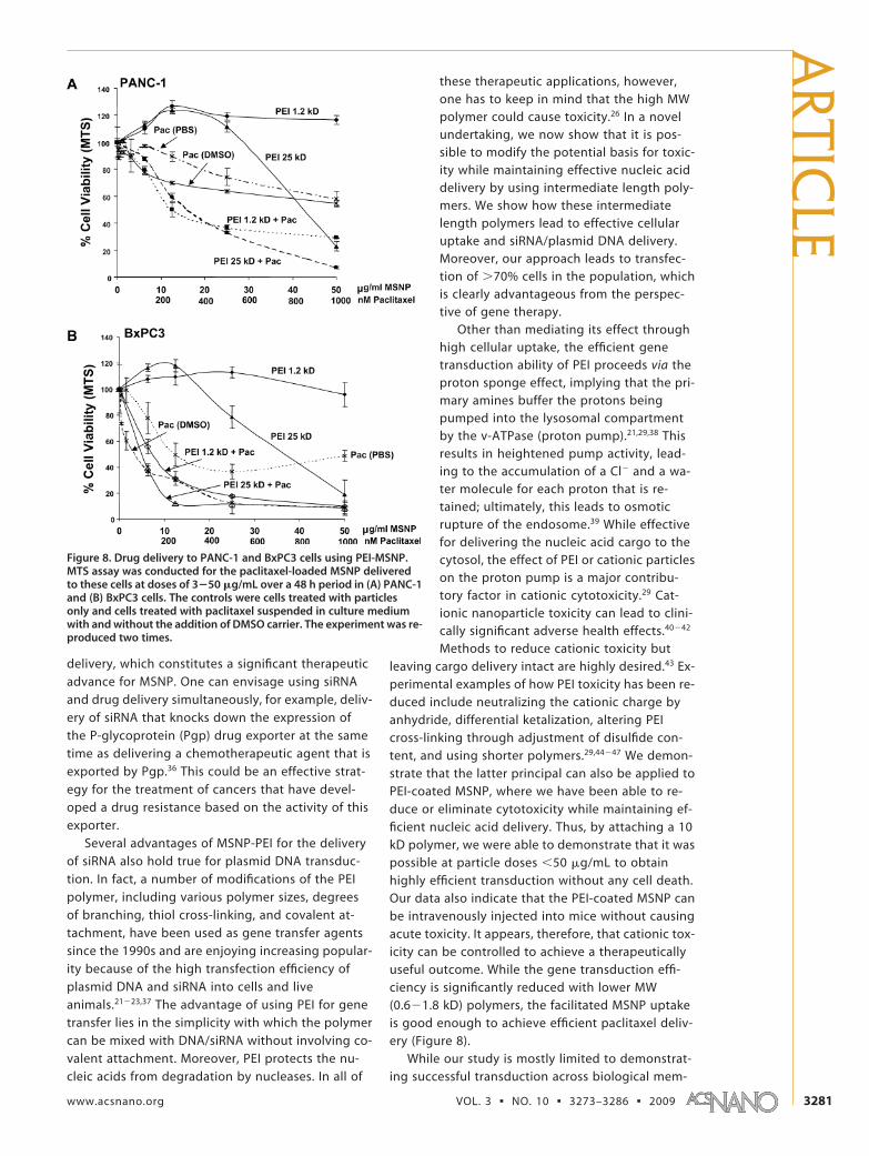
Cationic MSNP is Effective for Delivering Paclitaxelto Pancreatic Cancer Cells. We have recently dem-onstrated that MSNP are capable of deliver-ing water-insoluble drugs to cancer cells.2 Inlight of the high cellular uptake of PEI-coatedparticles, it was logical to ask whether, inthe traditional use of the MSNP, the polymerattachment still allows effective delivery ofthe hydrophobic cancer drug, paclitaxel, toPANC-1 and BxPC3 cells. Paclitaxel wasloaded into MSNP-PEI 1.2 kD and 25 kD inDMSO, followed by washing in aqueousbuffer to entrap the hydrophobic drug inthe particle pores. Release of the drug fromthe pores using methanol confirmed thatequal amounts of paclitaxel were loaded intoMSNP irrespective of the polymer MW (Sup-porting Information Figure 8). Subsequentassessment of the impact of paclitaxel on theviability of PANC-1 and BxPC3 cells showedthe efficacy of PEI-coated MSNP in drug de-livery (Figure 8). Thus, paclitaxel delivery by
MSNP-PEI 1.2 kD induced a rate of cytotoxicity that issuperior to the drug delivery by an aqueous suspensionof paclitaxel and as efficient as the drug suspended inDMSO (Figure 8). While MSNP-PEI 25 kD showed intrin-sic particle-related toxicity at doses �25 �g/mL, the ef-fect was comparable to MSNP-PEI 1.2 kD (Figure 8).These data demonstrate that the enhanced cellular up-take of nontoxic cationic MSNP is capable of enhanc-ing the delivery of hydrophobic cancer drugs. This isalso a novel demonstration for the delivery function ofMSNP.
PEI-Coated MSNP Are Devoid of Toxicity In Vivo. In order toachieve therapeutic delivery of nucleic acids anddrugs, it is required that cationic particles are well-tolerated and without any evidence of toxicity invivo. Hence, it was necessary for us to demonstratethat PEI-coated MSNP can be safely administered toanimals, and we chose intravenous injection of 25 kDPEI-coated particles to perform this analysis. MSNP-phosphonate and saline-only injections were used ascontrols. Particles were injected via the tail vein atdoses of 40 mg/kg once a week for 2 weeks. In or-der to keep the particles appropriately dispersed, itwas necessary to stabilize them against aggregatingeffects of the saline carrier. Saline alone yielded par-
Figure 5. Gel retardation and DNase I protection assays. Agarose gel electrophoresis ofPEI-MSNP/plasmid DNA (pEGFP) (A) and PEI-MSNP/siRNA (B) complexes at various nano-particle to nucleic acid (N/P) ratios. Anionic phosphonate-coated MSNP was used as acontrol. M � MW marker. (C) DNase I protection assay. M: DNA marker. �: naked plas-mid DNA (pEGFP), as negative control. Lane 1, pDNA/PEI 1.2 kD complex. Lane 2, pDNA/PEI 25 kD complex. Lane 3, naked pDNA treated with DNase I, positive control. Lane 4,pDNA/PEI 1.2 kD complex treated with DNase I before pDNA was released by 1% SDS.Lane 5, pDNA/PEI 25 kD complex treated with DNase I before pDNA was released by 1%SDS. Lane 6, pDNA/PEI 1.2 kD complex treated with 1% SDS. Lane 7, pDNA/PEI 25 kDcomplex treated with 1% SDS.
ART
ICLE
VOL. 3 ▪ NO. 10 ▪ XIA ET AL. www.acsnano.org3278

ticles sizes of 984 and 842 nm, respectively, for
MSNP-phosphonate and MSNP-PEI 25 kD. Particle
dispersion was improved by adding 2% mouse se-
rum, which reduced the particle sizes to 249 and 278
nm, respectively, in saline. Animals were sacrificed
2 weeks after the first injection, and blood and ma-
jor organs such as liver, kidney, spleen, lung, heart,
and brain were removed for study. Biochemical
analysis of the serum did not show any significant
changes in liver function, kidney function, choles-
terol and triglycerides, glucose, CO2 content, and
electrolytes of PEI versus phosphonate particles or
the saline control (Table 2). Moreover, histological
examination of the major organs did not show any
Figure 6. GFP knockdown by siRNA in stable transfected GFP-HEPA cells. HEPA-1 cells with stable GFP expression were usedfor siRNA knockdown assays. MSNP coated with different size PEI polymers were used to transfect GFP-specific or scrambledsiRNA and the results compared with Lipofectamine 2000 as transfection agent. (A) GFP knockdown was assessed by flow cy-tometry in which GFP MFI was normalized to the value of control untransduced cells (100%). (B) Confocal pictures were takenshowing GFP knockdown in GFP-HEPA cells. TEX 615-labeled siRNA was used to show the cellular localization of the nu-cleic acid bound particles (red dots). X, scrambled siRNA. The experiment was reproduced three times.
ARTIC
LE
www.acsnano.org VOL. 3 ▪ NO. 10 ▪ 3273–3286 ▪ 2009 3279

gross pathology; representative liver, spleen, and
kidney sections are shown in Supporting Informa-
tion Figure 9. In summary, these data confirm the
safety of phosphonate MSNP and demonstrate that
particles coated with 25 kD PEI, which is responsible
for considerable in vitro cytotoxicity, is also well-
tolerated in vivo. This demonstrates that PEI-coated
in MSNP could in principle be safely implemented for
in vivo drug and siRNA delivery. However, for this to
become a therapeutically useful option, additional
experimentation is required to show that the circu-
latory half-life and pharmacokinetics of MSNP deliv-
ery system that can achieve effective drug and nu-
cleic acid delivery.
DISCUSSIONIn this paper, we demonstrate that surface coating
with PEI yields cationic MSNP with therapeutically use-
ful nucleic acid delivery properties that include high
binding avidity of DNA and siRNA as well as a high rate
of cellular uptake. We show that siRNA complexed to
the MSNP-PEI surface is quite effective to achieve GFP
knockdown in transduced HEPA-1 cells, while plasmid
DNA delivery is comparable to a commercially available
transfection agent. We further demonstrate that the fa-
cilitated cellular uptake of cationic particles enhances
the ability of MSNP to deliver the hydrophobic chemo-therapeutic agent, paclitaxel, to pancreatic cancer cells.A potential downside of cationic functionalization fordrug or nucleic delivery is the possibility of cytotoxic-ity, best demonstrated by the use of MSNP-PEI 25 kD.However, we demonstrate that this toxicity could be re-duced or eliminated by attaching shorter length poly-mers that retain nucleic acid and drug delivery capabili-ties. Moreover, injection of MSNP coated with PEI 25kD did not lead to obvious toxicity in vivo. Thus, wedemonstrate that the therapeutic use of the MSNP plat-form can be extended to delivery of DNA and siRNAconstructs. From the perspective of MSNP as a thera-peutic platform, these are novel and encouragingfindings.
RNA interference describes natural processesthat lead to gene silencing by siRNA.32 siRNA hasbeen widely used as an experimental tool that is nowalso becoming the focus of the pharmaceutical in-dustry.33 Currently, there are a number of clinical tri-als underway that include the use of siRNAs to treatvarious disease processes.34,35 As for most moleculartherapies, in vivo delivery is a major hurdle in suc-cessful implementation and has sparked a numberof strategies to increase siRNA circulatory half-life,facilitate transduction across biological membranes,and achieve cell-specific delivery.34,35 We proposethat packaging of siRNA on the surface of cationicMSNP is capable of meeting each of these objectives.First, the MSNP surface can be functionalized to en-hance siRNA binding through the attachment of PEIpolymers, which in their own right have been usedas effective siRNA compacting and transducingagents. The tight complexing between PEI and nu-cleic acids on the particle surface protects these mol-ecules from enzymatic degradation as we demon-strated for DNA. Furthermore, the positive charge ofPEI-coated nanoparticles leads to strong electro-static interaction with the negatively charged cellsurface membrane, leading to facilitated particlewrapping and cellular uptake. This is in agreementwith the recent demonstration that PEI-coated nano-particles are taken up into cells at high efficiency.10,25
However, the latter study did not look at nucleicacid delivery but did show that the attachment of aligand such as folic acid further enhances uptake incancer cells.2,10 While PEI can be used in various waysto make siRNA delivery complexes, coating it ontothe surface of MSNP is particularly advantageous be-cause this therapeutic platform acts as a carrier withlarge surface area, is inexpensive and simple to syn-thesize, can be decorated with functional groupsand fluorescent tags, and can be used for magneticresonance imaging through the inclusion of super-paramagnetic iron oxide nanocrystals.2 Not onlyhave we shown PEI coating can enhance siRNA de-livery, but the particles also are capable of paclitaxel
Figure 7. GFP plasmid DNA transfection into HEPA-1 cells. HEPA-1cells were used for GFP plasmid DNA transfection. MSNP coated withdifferent size PEI polymers were used to transfect GFP plasmid DNA,and the results were compared with Lipofectamine 2000 as transfec-tion agent. (A) Representative histogram showing the shift in greenfluorescence intensity in HEPA-1 cells after transfection with Lipo-fectamine 2000 or MSNP-PEI 10 kD. (B) Confocal pictures showing GFPexpression in transfected HEPA-1 cells. This demonstrates differencesin the transfection efficiency as judged by fluorescent intensity andproportion of cells in the population showing GFP expression. The ex-periment was reproduced three times.
ART
ICLE
VOL. 3 ▪ NO. 10 ▪ XIA ET AL. www.acsnano.org3280
delivery, which constitutes a significant therapeuticadvance for MSNP. One can envisage using siRNAand drug delivery simultaneously, for example, deliv-ery of siRNA that knocks down the expression ofthe P-glycoprotein (Pgp) drug exporter at the sametime as delivering a chemotherapeutic agent that isexported by Pgp.36 This could be an effective strat-egy for the treatment of cancers that have devel-oped a drug resistance based on the activity of thisexporter.
Several advantages of MSNP-PEI for the deliveryof siRNA also hold true for plasmid DNA transduc-tion. In fact, a number of modifications of the PEIpolymer, including various polymer sizes, degreesof branching, thiol cross-linking, and covalent at-tachment, have been used as gene transfer agentssince the 1990s and are enjoying increasing popular-ity because of the high transfection efficiency ofplasmid DNA and siRNA into cells and liveanimals.21�23,37 The advantage of using PEI for genetransfer lies in the simplicity with which the polymercan be mixed with DNA/siRNA without involving co-valent attachment. Moreover, PEI protects the nu-cleic acids from degradation by nucleases. In all of
these therapeutic applications, however,one has to keep in mind that the high MWpolymer could cause toxicity.26 In a novelundertaking, we now show that it is pos-sible to modify the potential basis for toxic-ity while maintaining effective nucleic aciddelivery by using intermediate length poly-mers. We show how these intermediatelength polymers lead to effective cellularuptake and siRNA/plasmid DNA delivery.Moreover, our approach leads to transfec-tion of �70% cells in the population, whichis clearly advantageous from the perspec-tive of gene therapy.
Other than mediating its effect throughhigh cellular uptake, the efficient genetransduction ability of PEI proceeds via theproton sponge effect, implying that the pri-mary amines buffer the protons beingpumped into the lysosomal compartmentby the v-ATPase (proton pump).21,29,38 Thisresults in heightened pump activity, lead-ing to the accumulation of a Cl� and a wa-ter molecule for each proton that is re-tained; ultimately, this leads to osmoticrupture of the endosome.39 While effectivefor delivering the nucleic acid cargo to thecytosol, the effect of PEI or cationic particleson the proton pump is a major contribu-tory factor in cationic cytotoxicity.29 Cat-ionic nanoparticle toxicity can lead to clini-cally significant adverse health effects.40�42
Methods to reduce cationic toxicity butleaving cargo delivery intact are highly desired.43 Ex-perimental examples of how PEI toxicity has been re-duced include neutralizing the cationic charge byanhydride, differential ketalization, altering PEIcross-linking through adjustment of disulfide con-tent, and using shorter polymers.29,44�47 We demon-strate that the latter principal can also be applied toPEI-coated MSNP, where we have been able to re-duce or eliminate cytotoxicity while maintaining ef-ficient nucleic acid delivery. Thus, by attaching a 10kD polymer, we were able to demonstrate that it waspossible at particle doses �50 �g/mL to obtainhighly efficient transduction without any cell death.Our data also indicate that the PEI-coated MSNP canbe intravenously injected into mice without causingacute toxicity. It appears, therefore, that cationic tox-icity can be controlled to achieve a therapeuticallyuseful outcome. While the gene transduction effi-ciency is significantly reduced with lower MW(0.6�1.8 kD) polymers, the facilitated MSNP uptakeis good enough to achieve efficient paclitaxel deliv-ery (Figure 8).
While our study is mostly limited to demonstrat-ing successful transduction across biological mem-
Figure 8. Drug delivery to PANC-1 and BxPC3 cells using PEI-MSNP.MTS assay was conducted for the paclitaxel-loaded MSNP deliveredto these cells at doses of 3�50 �g/mL over a 48 h period in (A) PANC-1and (B) BxPC3 cells. The controls were cells treated with particlesonly and cells treated with paclitaxel suspended in culture mediumwith and without the addition of DMSO carrier. The experiment was re-produced two times.
ARTIC
LE
www.acsnano.org VOL. 3 ▪ NO. 10 ▪ 3273–3286 ▪ 2009 3281

branes, it would be clearly necessary to conductstudies to demonstrate the therapeutic efficiency ofthe MSNP platform in vivo. To date, only limited invivo data have been published regarding the toxic-ity, biodistribution, and biopersistence ofMSNP.1,48�50 Coating of MSNP by MRI agents hasbeen used to perform in vivo imaging; this demon-strated that the particles could still be detected inthe circulation 30 min after administration, but then
accumulate in the RES system such as liver andspleen, with less signal coming from the kidney,lung, and heart.1,50 Another interesting finding isthat MSNP accumulate preferentially at tumor sitesthrough an enhanced permeability and retention(EPR) effect.1 To achieve the full potential of MSNP,detailed in vivo testing should be done to assess bio-distribution, pharmacokinetics, and targeting. It isencouraging that our in vivo safety study did notshow any signs of toxicity after intravenous injec-tion, including for particles coated by the 25 kD poly-mer. The lack of in vivo toxicity in spite of the invitro cytotoxic effects can best be explained by thedilution of the particles in the circulatory system andthe strong defense capabilities of the body againstforeign substances. The extent to which these pro-tective mechanisms may interfere with the effective-ness of drug delivery still needs to be determinedbut could be dealt with in a rational fashion by dy-namic design features such as demonstrated in thisstudy.
CONCLUSIONPolyethyleneimine-coated mesoporous silica nano-
particle is a versatile delivery system that can facilitatecellular uptake to increase drug delivery payload andalso be utilized to improve nucleic acids delivery fortherapeutic and experimental use. While the potentialcytotoxicity of PEI attachment could interfere with theefficacy of siRNA delivery, it is possible by selecting op-timal polymer lengths to maintain high transfection ef-ficiency while simultaneously reducing or eliminatingtoxic effects. While inefficient for gene delivery, coat-ing with low molecular weight PEI polymers is nonethe-less efficient to increase delivery of antitumor drug pa-clitaxel into cancer cells. These hybridorganic�inorganic porous nanoparticles can poten-tially be useful for the simultaneous delivery of nucle-otides and small molecules into cells.
MATERIALS AND METHODSReagents. Tetraethylorthosilicate (98%), cetyltrimethylammo-
nium bromide (CTAB, 95%), fluorescein isothiocyanate (FITC,90%), polyethyleneimine (MW 1.2 and 25 kD), poly(ethylene gly-col) methyl ether (MW 5 kD), N,N=-disuccinimidyl carbonate (DSC,95%), 4-(dimethylamino)pyridine (DMAP, 99%), aminopropyltri-ethoxysilane (APTS, 99%), 3-(trihydroxysilyl)propyl methylphos-phonate (42%), succinic anhydride (99%), fluorescamine, pacli-taxel, propidium iodide (PI), -actin antibody, and bovine serumalbumin (BSA) were from Sigma (St. Louis, MO). Polyethylene-imine (MW 0.6, 1.8, and 10 kD) were from Alfa Aesar (Ward Hill,MA). The MTS assay kit was from Promega (Madison, WI). Dulbec-co’s Modified Eagle’s medium (DMEM), penicillin/streptomycin,and L-glutamine were purchased from Invitrogen (Carlsbad, CA).Fetal calf serum (FCS) was from Atlanta Biologicals, Inc.(Lawrenceville, GA). siRNA for GFP knockdown was purchasedfrom IDT Technologies (Coralville, IA). For all experiments andanalyses, water was deionized and filtered with a 0.45 �m poresize polycarbonate syringe filter (Millipore, Billerica, MA). All
chemicals were reagent grade and used without further purifica-tion or modification.
Synthesis and Surface Modification of Mesoporous Silica NanoparticlesMSNP. The basic synthesis of MSNP was conducted by mixingthe silicate source tetraethylorthosilicate (TEOS) with the tem-plating surfactants cetyltrimethylammonium bromide (CTAB) inbasic aqueous solution (pH 11). In a round-bottom flask, 100 mgof CTAB was dissolved in a solution of 48 mL of distilled waterand 0.35 mL of sodium hydroxide (2 M). The solution was heatedto 80 °C and stirred vigorously. After the temperature had stabi-lized, 0.5 mL of TEOS was added slowly into the heated CTAB so-lution. After 15 min, 0.23 mmol of the organosilane solutionwas added into the mixture. 3-Trihydroxysilylpropyl methylphos-phonate was used for phosphonate surface modification, andaminopropyltriethoxy silane (APTS) was used for amine surfacemodification. After 2 h, the solution was cooled to room temper-ature, and the materials were washed with methanol using cen-trifugation. In order to incorporate fluorescent dye molecules inthe silicate framework, fluorescein-modified silane was first syn-thesized and then mixed with TEOS. To synthesize fluorescein-
TABLE 2. Serum Biochemistry (avg � SD, n � 6)a
control (saline) MSNP-Phos MSNP-PEI 25 kD
CHOL (mg/dL) 162.5 8.5 153.8 15.4 144.4 6.8CK (U/L) 3000.3 2285.3 2690.5 1129.3 3280.9 1512.7ALT (U/L) 59.5 6.4 55.7 8.4 46.1 22.6AST (U/L) 184.2 70.2 182.2 21.1 217.2 65.6ALP (U/L) 105.2 8.6 102.1 17.5 88.6 48.9TBILI (mg/dL) 0.7 0.1 0.9 0.3 0.6 0.1TPROT (mg/dL) 6.2 0.2 6.0 0.4 5.9 0.3GLU (mg/dL) 140.8 16.5 141.2 29.2 140.8 34.6PHOS (mg/dL) 8.4 0.7 7.4 1.0 8.3 0.6CA (mg/dL) 8.9 1.7 9.2 0.4 9.4 0.2CO2_LC (mEq/L) 10.1 2.2 13.3 3.8 11.3 1.1BUN (mg/dL) 20.8 2.1 22.3 9.3 22.6 2.6CREAT (mg/dL) 0.3 0.0 0.3 0.1 0.3 0.0D-BILI (mg/dL) 0.9 0.2 1.0 0.5 0.7 0.2ALB (g/dL) 3.3 0.1 3.2 0.2 3.2 0.2AGR (N/A) 1.2 0.1 1.2 0.1 1.1 0.1B-CREA (mg/dL) 75.3 16.2 79.2 30.0 82.0 12.4AMYL (U/L) 1252.2 215.1 1266.0 189.5 1139.4 122.0LDH (U/L) 681.5 152.8 638.8 133.3 611.0 141.6MG (mg/dL) 2.0 0.5 2.2 0.2 2.3 0.2TRIG (mg/dL) 201.2 43.8 178.2 65.3 156.0 28.5
aBiochemical parameters includes cholesterol (CHOL), creatine kinase (CK), alanineaminotransferase (ALT), aspartate aminotransferase (AST), alkaline phosphatase(ALP), total bilirubin (TBILI), total protein (TPROT), glucose (GLU), inorganic phos-phorus (PHOS), calcium (CA), carbon dioxide (CO2_LC), blood urea nitrogen (BUN),creatinine (CREAT), direct bilirubin (DBILI), albumin (ALB), albumin-globulin ratio(AGR), blood creatinine (B-CREA), amylase (AMYL), lactate dehydrogenase (LDH),magnesium (Mg), triglycerides (TRIG). There is no statistical significance betweeneach group as analyzed by one-way ANOVA.
ART
ICLE
VOL. 3 ▪ NO. 10 ▪ XIA ET AL. www.acsnano.org3282

modified silane, 2.4 �L of APTS was mixed with 1 mg of fluores-cein isothiocyanate (FITC) in 0.6 mL of absolute ethanol andstirred for 2 h under inert atmosphere. In another formulation,rhodamine-B isothiocyanate (RITC) was used instead of FITC tosynthesize rhodamine-B-modified APTS. The dye-modified silanewas then mixed with TEOS before adding the mixture into theheated CTAB solution. The surfactants were removed from thepores by refluxing the particles in a mixture of 20 mL of metha-nol and 1 mL of hydrochloric acid (12.1 M) for 24 h. The materi-als were then centrifuged and washed with methanol.
For the poly(ethylene glycol) modification, 1 g of poly(ethyl-ene glycol) methyl ether (MW 5 kD, mPEG) was dried undervacuum for 30 min and dissolved in 5 mL of dioxane (with slightheating). mPEG has only one reactive end that can be attachedto the particle surface and limits the coupling process only tothat end, whereas normal PEG has two reactive ends and maycause particle cross-linking; 307.4 mg of disuccinimidyl carbon-ate (DSC) was dissolved in 2 mL of anhydrous DMF (with slightheating) and mixed with the mPEG solution; 146.6 mg of4-(dimethylamino)pyridine was dissolved in 2 mL of acetoneand added slowly into the mPEG solution The mixture was stirredfor 6 h under an inert atmosphere. The polymer was precipi-tated by the addition of 30 mL of diethyl ether to the solutionand separated by centrifugation. After washing the polymertwice with diethyl ether, the activated mPEG was dried undervacuum. Sixty milligrams of amine-modified MSNP was washedand resuspended in 2 mL of anhydrous DMF; 300 mg of the ac-tivated mPEG was dissolved in 9 mL of DMF and mixed with theparticles. The mixture was stirred for 12 h and washed thor-oughly with DMF and PBS.
To perform polyethyleneimine (PEI) modification, 5 mg ofphosphonate-modified MSNP was dispersed in a solution of 2.5mg of PEI (MW 25 kD) and 1 mL of absolute ethanol. The processto coat the particles with other PEI polymers (MW 0.6, 1.2, 1.8,10 kD) was carried out similarly. After the mixture was sonicatedand stirred for 30 min, the particles coated with PEI were washedwith ethanol and PBS. Thermogravimetric analysis showed thatthe amount of PEI on the particles was approximately 5 wt %. Tosuccinylate the PEI 25 kD-coated particles, 1 mg particles was re-suspended in 0.25 mL of anhydrous DMF and mixed with differ-ent amounts of succinic anhydride (0.15, 0.075, and 0.015 mg).The mixture was sonicated and stirred overnight. The succiny-lated particles were washed with DMF and resuspended in PBS.To fluorescently label PEI (MW 25 kD), 60 mg of PEI 25 kD was dis-solved in 10 mL of carbonate buffer (pH 9) and mixed with 1mg of rhodamine-B isothiocyanate dissolved in 1 mL of DMSO.The mixture was stirred for 24 h at 4 °C and dialyzed against dis-tilled water. The rhodamine-B-labeled PEI 25 kD was attachedto the particles by using similar procedure for the unlabeledPEI.
Physicochemical Characterization. All MSNP were characterizedfor size, size distribution, shape, and charge (Table 1). The shapeand structure were characterized using a transmission electronmicroscope (JEOL JEM 2010, JEOL USA, Inc., Peabody, MA). Micro-films for TEM imaging were made by placing a drop of the re-spective particle suspension onto a 200-mesh copper TEM grid(Electron Microscopy Sciences, Washington, PA) and then dryingat room temperature overnight. A minimum of five images ofeach sample was collected to obtain representative views. Par-ticle size and � potential in solution were measured by ZetaSizerNano (Malvern Instruments Ltd., Worcestershire, UK). This instru-ment measures the light scattering (DLS) from a suspension atan angle of 173°. Size measurements were performed on dilutesuspensions in water or complete cell culture media at pH 7.4.The ZetaSizer Nano was also used to measure the electro-phoretic mobility of the MSNP suspended in solution. Electro-phoretic mobility is used as an approximation of particle surfacecharge and can be used to calculate � potential. TheHelmholtz�Smoluchowski equation was used to recalculateelectrophoretic mobility into � potential.
Drug Loading of Paclitaxel. The modified materials were loadedwith paclitaxel by incubating 10 mg of the nanoparticles in a so-lution of 1 mg of paclitaxel and 0.25 mL of DMSO for 6 h. Afterthe drug-laden nanoparticles were removed from the suspen-sion by centrifugation and the supernatant was removed com-
pletely, the materials were dried under vacuum. The drug-ladennanoparticles were washed and sonicated with PBS. In order todetermine the amount of paclitaxel that partitioned to theMSNP, the aqueous particle suspension was incubated for 6 hat 4 °C before centrifugation. Methanol was used to release pa-clitaxel from MSNP to determine the loading capacity. The drug-laden MSNP pellet was resuspended and sonicated in methanolon three occasions, and the supernatants were combined tomeasure the release of the drug by UV absorption at 230 nm;50 �g/mL particles contained about 1 �M of paclitaxel.
MSNP Dispersion and Use To Perform Tissue Culture. All cell cultureswere maintained in 25 cm2 cell culture flasks in which the cellswere passaged at 70�80% confluency every 2�4 days. RAW264.7, BxPC3, PANC-1, and HEPA-1 cell lines were cultured in Dul-becco’s Modified Eagle Medium (DMEM) (Carlsbad, CA) contain-ing 10% fetal calf serum (FCS), 100 U/mL penicillin, 100 �g/mLstreptomycin, and 2 mM L-glutamine (complete medium).BEAS-2B cells were cultured in BEGM (Charles City, IA) in type Irat tail collagen-coated flasks or plates. Cells were cultured at 37°C in a humidified 5% CO2 atmosphere. To disperse MSNP, thestock solution (in water) was sonicated (Tekmar Sonic Disruptorprobe) for 15 s prior to aliquoting. In order to coat the surface ofMSNP with bovine serum albumin (BSA), the aliquoted NP sus-pension (�10 �L) was mixed with an equal volume of 4% BSA.Tissue culture media (1 mL) was added to the BSA-coated MSNPsuspension. Cell culture media deprived of serum (e.g., BEGM)was modified by addition of BSA at a concentration of 2 mg/mL.The cell culture media containing MSNP at the desired concen-tration was sonicated for 15 s and characterized as describedbefore.
Assays for Cellular Viability and Mitochondrial Function. Cellular viabil-ity was determined by the MTS assay, which looks at the re-duction of (3-(4,5-dimethylthiazol-2-yl)-5-(3-carboxymethoxy-phenyl)-2-(4-sulfophenyl)-2H-tetrazolium (MTS) to formazan inviable cells. Briefly, 2 � 104 cells were plated onto 96-multi-wellplates (Costar; Corning, NY). After incubation with the indicateddose of MSNP for various lengths of time at 37 °C, formazan ab-sorbance was measured at 490 nm. The mean absorbance ofnonexposed cells served as the reference for calculating 100%cellular viability.
Cell death and mitochondrial function were detected usingpropidium iodide (PI) uptake and JC-1 fluorescence. Fluorescentprobes were diluted in DMEM before the addition to cells for 30min at 37 °C in the dark: (i) 5 �g/mL propidium iodide (PI) in200 �L of DMEM (assessment of cell death); (ii) 5 �M JC-1 (assess-ment of � m). Flow cytometry was performed using a LSR (Bec-ton Dickinson, Mountain View, CA). PI was analyzed in FL-2, andJC-1 was analyzed in both FL-1 and FL-2. Forward and side scat-ter were used to gate out cellular fragments.
Assessment of Cellular MSNP Uptake by Flow Cytometry and ConfocalMicroscopy. For the performance of flow cytometry, aliquots of 5� 104 cells (RAW 264.7, BEAS-2B, PANC-1, and BxPC3) were cul-tured in 48-well plates in 0.4 mL of medium. RITC-labeled MSNPwith a phosphonate surface coating were added to the abovecultures at a dose of 25 �g/mL for 30 min, followed by incuba-tion with the FITC-labeled MSNP-PEI series at final concentrationsof 25 �g/mL for 3 h. All cell types were trypsinized and washedwith trypan blue to quench the fluorescence of cell-surface-attached MSNP. Cells were analyzed in a LSR flow cytometer us-ing mean FL-2 and FL-1 to assess RITC and FITC fluorescence, re-spectively. Data are reported as fold increase above control(cells without MSNP).
Cellular uptake of MSNP was performed by adding 25 �g/mLof the various MSNP to 8-well chamber slides (Nunc) in which 5� 104 cells were cultured in each well containing 0.4 mL culturemedium. Cell membranes were stained with 5 �g/mL wheatgerm agglutinin (WGA) Alexa Fluor 594 conjugate in PBS for 30min. Slides were mounted with DAPI (Molecular Probes, Eugene,OR) and visualized under a confocal microscope (Leica Confocal1P/FCS) in the UCLA/CNSI Advanced Light Microscopy/Spectros-copy Shared Facility. High-magnification images were obtainedwith a 63� objective. Optical sections were averaged 2�4 timesto reduce noise. Images were processed using Leica ConfocalSoftware.
ARTIC
LE
www.acsnano.org VOL. 3 ▪ NO. 10 ▪ 3273–3286 ▪ 2009 3283

Preparation of PEI-MSNP�pDNA/siRNA Polyplexes and Agarose GelRetardation. Agarose gel retardation assay was used to deter-mine the DNA/siRNA binding ability of PEI-coated MSNP; 0.1 �gof plasmid DNA (pEGFP) or siRNA in aqueous solution was usedto mix with PEI-coated nanoparticles to obtain particle to pDNAratios (N/P) ratios of 5�600. The mixture was incubated at roomtemperature for 30 min for complex formation. Ten microlitersof the polyplex solution mixed with 2 �L of 6� loading bufferwas electrophoresed on 1% agarose gel containing 0.5 �g/mLethidium bromide (EB) with Tris-boric acid (TBE) running buffer(pH 8) at 100 V for 30 min. DNA/RNA bands were visualized by aUV (254 nm) illuminator and photographed with a Bio-Rad imag-ing system (Hercules, CA). The binding capacity was expressedby the N/P ratio that shows total retardation of DNA or siRNA mi-gration (as reflected by the disappearance of DNA/RNA bandson the gel).
DNase I Protection Assay: Particle/pDNA complexes were pre-pared at a MSNP/pDNA ratio of 100 with 100 ng of pDNA in 10�L total volume. The complex solutions were incubated with 1�L of DNase I (2.7 U/�L) in 50 mM Tris-Cl, 10 mM MgCl2, pH 7.4.at 37 °C for 30 min. The DNase I was inactivated by adding 1 �Lof 100 mM ethylenediaminetetraacetic acid (EDTA). The pDNAwas then released from the complex by adding 1% sodiumdodecyl sulfate (SDS) and analyzed by 1% agarose gelelectrophoresis.
Plasmid DNA and siRNA Transfection with the Use of MSNP-PEI. Plas-mid DNA (pDNA) containing a GFP insert was used to transfectHEPA-1 cells cultured either in Nunc chamber slides for perfor-mance of confocal microscopy or in 48-well plates for assess-ment by flow cytometry. Cells were plated at a density of 2 �104 cells per well in 0.4 mL of medium. pDNA/MSNP complexeswere prepared by mixing 100 ng/mL of DNA with 25 �g/mL ofMSNP for 30 min prior to cellular incubation for 24 h. Cells werefixed for confocal microscopy as described above. Harvestedcells were used to conduct flow cytometry on FL-1 channel.
To perform siRNA experiments, HEPA-1 cells were prior trans-fected with a GFP plasmid and then sorted in the FL-1 channelto select stable GFP expressing cells. The sorted cells were platedat a density of 5 � 104 cells per well containing 0.4 mL culturemedium in chamber slides for performance of confocal micros-copy and in 48-well plates for performance of flow cytometry.MSNP/siRNA complexes were prepared by incubating 500 ng/mLof siRNA with 25 �g/mL of MSNP-PEI for 30 min in serum-freeDMEM. Cells were then exposed to the complexes for 3 h. DMEM� 10% FCS was then added to bring the final volume to 400 �Lfor 48 h. Cells were fixed and prepared for confocal microscopyas described above. For flow cytometry, cells were harvested andanalyzed for fold decrease in GFP expression (FL-1 channel).
Paclitaxel Delivery by MSNP-PEI. PANC-1 and BxPC3 cells wereplated at 2 � 104 cells per well in a 96-well plate. MSNP par-ticles, loaded with paclitaxel, were incubated with the cells atdoses of 10�50 �g/mL for 48�72 h. Free paclitaxel correspond-ing to the amount loaded into MSNP particles was suspendedor dissolved in PBS and DMSO to serve as controls. MTS assayswere performed after 48 h to determine the cell viability aftertreatment.
In Vivo MSNP Toxicity Testing. Animal experiment protocols werereviewed and approved by the Chancellor’s Animal ResearchCommittee (ARC) at UCLA. Animal experiments were performedin accordance with UCLA guidelines for care and treatment oflaboratory animals and the NIH Guide for the Care and Use ofLaboratory Animals in Research (DHEW78-23). The mice wererandomly divided into three groups: MSNP-phosphonate, MSNP-PEI 25 kD, and the saline control group. We used six mice pergroup since this number has enough statistical power to dis-cern differences in the toxic responses. We used 40 mg/kg par-ticles for intravenous injection through tail vein once a week for2 weeks. Animal weight was monitored after particle injections.Animals were sacrificed later to obtain blood and organs.
Biochemical Serum Assays. The serum was obtained by centrifug-ing the whole blood at 3000 rpm for 15 min. The biochemical pa-rameters were assayed by UCLA Division of Laboratory AnimalMedicine (DLAM) diagnostic laboratory services.
Histology of Major Organs. A small piece of liver, kidney, spleen,lung, heart, and brain was fixed by 10% formalin and then em-
bedded into paraffin, sectioned for 5 �m thick, and mountedon the glass microscope slides by UCLA Division of LaboratoryAnimal Medicine (DLAM) diagnostic laboratory services. The sec-tions were stained with hematoxylin-eosin and examined bylight microscopy. The slides were read by an experienced veteri-nary pathologist.
Statistical Analysis. All data are expressed as the mean stan-dard deviation. An unpaired Student’s t test was used to assessthe difference between two groups. One-way ANOVA was per-formed when more than two groups were compared with asingle control. Differences between individual groups withinthe set were assessed by a multiple comparison test (Tukey)when the F statistic was �0.05. A p � 0.05 was consideredsignificant.
Acknowledgment. This work is supported by the NationalScience Foundation and the Environmental ProtectionAgency under Cooperative Agreement Number EF 0830117.Any opinions, findings, conclusions, or recommendations ex-pressed herein are those of the author(s) and do not neces-sarily reflect the views of the National Science Foundation orthe Environmental Protection Agency. This work has notbeen subjected to an EPA peer and policy review. Key sup-port was provided by the UC Lead Campus for Nanotoxicol-ogy Training and Research, funded by UC TSR&TP, US PublicHealth Service Grants (U19 AI070453, R01 ES016746, and RO1ES015498) and the US EPA STAR award (RD-83241301) tothe Southern California Particle Center. Key support was alsoprovided by the grant USDOD HDTRA 1-08-1-0041. Fluores-cent microscopy was performed at the CNSI Advanced LightMicroscopy/Spectroscopy Shared Facility at UCLA.
Note added after ASAP publication: In the version publishedASAP September 9, 2009, Figure 5 was missing the � labels,and Figure 8 was missing � prefixes in the axis labels. The cor-rected version was published ASAP September 18, 2009.
Supporting Information Available: Particle size distribution,cell viability, surface modification and fluorescent labeling of PEI-MSNP, flow cytometry, and confocal microscopy for cellular up-take. This material is available free of charge via the Internet athttp://pubs.acs.org.
REFERENCES AND NOTES1. Kim, J.; Kim, H. S.; Lee, N.; Kim, T.; Kim, H.; Yu, T.; Song, I. C.;
Moon, W. K.; Hyeon, T. Multifunctional UniformNanoparticles Composed of a Magnetite Nanocrystal Coreand a Mesoporous Silica Shell for Magnetic Resonanceand Fluorescence Imaging and for Drug Delivery. Angew.Chem., Int. Ed. 2008, 47, 8438–8441.
2. Liong, M.; Lu, J.; Kovochich, M.; Xia, T.; Ruehm, S. G.; Nel,A. E.; Tamanoi, F.; Zink, J. I. Multifunctional InorganicNanoparticles for Imaging, Targeting, and Drug Delivery.ACS Nano 2008, 2, 889–896.
3. Lu, J.; Liong, M.; Zink, J. I.; Tamanoi, F. Mesoporous SilicaNanoparticles as a Delivery System for HydrophobicAnticancer Drugs. Small 2007, 3, 1341–1346.
4. Slowing, I. I.; Vivero-Escoto, J. L.; Wu, C.-W.; Lin, V. S.-Y.Mesoporous Silica Nanoparticles as Controlled ReleaseDrug Delivery and Gene Transfection Carriers. Adv. DrugDelivery Rev. 2008, 60, 1278–1288.
5. Vallet-Regı, M.; Balas, F.; Arcos, D. Mesoporous Materialsfor Drug Delivery. Angew. Chem., Int. Ed. 2007, 46,7548–7558.
6. Borm, P.; Klaessig, F. C.; Landry, T. D.; Moudgil, B.; Pauluhn,J.; Thomas, K.; Trottier, R.; Wood, S. Research Strategies forSafety Evaluation of Nanomaterials, Part V: Role ofDissolution in Biological Fate and Effects of NanoscaleParticles. Toxicol. Sci. 2006, 90, 23–32.
7. Finnie, K.; Waller, D.; Perret, F.; Krause-Heuer, A.; Lin, H.;Hanna, J.; Barbe, C. Biodegradability of Sol�Gel SilicaMicroparticles for Drug Delivery. J. Sol�Gel Sci. Technol.2009, 49, 12–18.
ART
ICLE
VOL. 3 ▪ NO. 10 ▪ XIA ET AL. www.acsnano.org3284

8. Vivero-Escoto, J. L.; Slowing, I. I.; Wu, C.-W.; Lin, V. S.-Y.Photoinduced Intracellular Controlled Release DrugDelivery in Human Cells by Gold-Capped MesoporousSilica Nanosphere. J. Am. Chem. Soc. 2009, 131, 3462–3463.
9. Nguyen, T. D.; Leung, K. C. F.; Liong, M.; Pentecost, C. D.;Stoddart, J. F.; Zink, J. I. Construction of a pH-DrivenSupramolecular Nanovalve. Org. Lett. 2006, 8, 3363–3366.
10. Rosenholm, J. M.; Meinander, A.; Peuhu, E.; Niemi, R.;Eriksson, J. E.; Sahlgren, C.; Linde, M. Targeting of PorousHybrid Silica Nanoparticles to Cancer Cells. ACS Nano2009, 3, 197–206.
11. Park, I. Y.; Kim, I. Y.; Yoo, M. K.; Choi, Y. J.; Cho, M.-H.; Cho,C. S. Mannosylated Polyethylenimine CoupledMesoporous Silica Nanoparticles for Receptor-MediatedGene Delivery. Int. J. Pharm. 2008, 359, 280–287.
12. Radu, D. R.; Lai, C. Y.; Jeftinija, K.; Rowe, E. W.; Jeftinija, S.;Lin, V. S. Y. A Polyamidoamine Dendrimer-CappedMesoporous Silica Nanosphere-Based Gene TransfectionReagent. J. Am. Chem. Soc. 2004, 126, 13216–13217.
13. Torney, F.; Trewyn, B. G.; Lin, V. S.-Y.; Wang, K. MesoporousSilica Nanoparticles Deliver DNA and Chemicals intoPlants. Nat. Nanotechnol. 2007, 2, 295–300.
14. Bharali, D. J.; Klejbor, I.; Stachowiak, E. K.; Dutta, P.; Roy, I.;Kaur, N.; Bergey, E. J.; Prasad, P. N.; Stachowiak, M. K.Organically Modified Silica Nanoparticles: A NonviralVector for In Vivo Gene Delivery and Expression in theBrain. Proc. Natl. Acad. Sci. U.S.A. 2005, 102, 11539–11544.
15. Bonoiu, A. C.; Mahajan, S. D.; Ding, H.; Roy, I.; Yong, K.-T.;Kumar, R.; Hu, R.; Bergey, E. J.; Schwartz, S. A.; Prasad, P. N.Nanotechnology Approach for Drug Addiction Therapy:Gene Silencing Using Delivery of Gold Nanorod�siRNANanoplex in Dopaminergic Neurons. Proc. Natl. Acad. Sci.U.S.A. 2009, 106, 5546–5550.
16. Elbakry, A.; Zaky, A.; Liebl, R.; Rachel, R.; Goepferich, A.;Breunig, M. Layer-by-Layer Assembled Gold Nanoparticlesfor siRNA Delivery. Nano Lett. 2009, 9, 2059–2064.
17. Fuller, J. E.; Zugates, G. T.; Ferreira, L. S.; Ow, H. S.; Nguyen,N. N.; Wiesner, U. B.; Langer, R. S. Intracellular Delivery ofCore�Shell Fluorescent Silica Nanoparticles. Biomaterials2008, 29, 1526–1532.
18. Kneuer, C.; Sameti, M.; Bakowsky, U.; Schiestel, T.; Schirra,H.; Schmidt, H.; Lehr, C.-M. A Nonviral DNA DeliverySystem Based on Surface Modified Silica-NanoparticlesCan Efficiently Transfect Cells In Vitro. Bioconjugate Chem.2000, 11, 926–932.
19. McBain, S. C.; Yiu, H. H. P.; Haj, A. E.; Dobson, J.Polyethyleneimine Functionalized Iron OxideNanoparticles as Agents for DNA Delivery andTransfection. J. Mater. Chem. 2007, 17, 2561–2565.
20. Zhu, S.-G.; Xiang, J.-J.; Li, X.-L.; Shen, S.-R.; Lu, H.-b.; Zhou,J.; Xiong, W.; Zhang, B.-C.; Nie, X.-M.; Zhou, M.; Tang, K.Gui-Yuan, Poly(L-lysine)-Modified Silica Nanoparticles forthe Delivery of Antisense Oligonucleotides. Biotechnol.Appl. Biochem. 2004, 39, 179–187.
21. Boussif, O.; Lezoualc’h, F.; Zanta, M. A.; Mergny, M. D.;Scherman, D.; Demeneix, B.; Behr, J. P. A Versatile Vectorfor Gene and Oligonucleotide Transfer into Cells in Cultureand In Vivo: Polyethylenimine. Proc. Natl. Acad. Sci. U.S.A.1995, 92, 7297–7301.
22. Godbey, W. T.; Wu, K. K.; Mikos, A. G. Tracking theIntracellular Path of Poly(ethylenimine)/DNA Complexesfor Gene Delivery. Proc. Natl. Acad. Sci. U.S.A. 1999, 96,5177–5181.
23. Urban-Klein, B.; Werth, S.; Abuharbeid, S.; Czubayko, F.;Aigner, A. RNAi-Mediated Gene-Targeting throughSystemic Application of Polyethylenimine (PEI)-ComplexedsiRNA In Vivo. Gene Ther. 2005, 12, 461–466.
24. Liong, M.; France, B.; Bradley, K. A.; Zink, J. I. AntimicrobialActivity of Silver Nanocrystals Encapsulated inMesoporous Silica Nanoparticles. Adv. Mater. 2009, 21,1684–1689.
25. Duan, H.; Nie, S. Cell-Penetrating Quantum Dots Based onMultivalent and Endosome-Disrupting Surface Coatings.J. Am. Chem. Soc. 2006, 129, 3333–3338.
26. Florea, B. I.; Meaney, C.; Junginger, H. E.; Borchard, G.Transfection Efficiency and Toxicity of Polyethylenimine inDifferentiated Calu-3 and Nondifferentiated COS-1 CellCultures. AAPS PharmSci. 2002, 4, E12.
27. Neu, M.; Fischer, D.; Kissel, T. Recent Advances in RationalGene Transfer Vector Design Based on Poly(ethyleneimine) and Its Derivatives. J. Gene. Med. 2005, 7, 992–1009.
28. Xia, T.; Kovochich, M.; Brant, J.; Hotze, M.; Sempf, J.;Oberley, T.; Sioutas, C.; Yeh, J. I.; Wiesner, M. R.; Nel, A. E.Comparison of the Abilities of Ambient and ManufacturedNanoparticles to Induce Cellular Toxicity According to AnOxidative Stress Paradigm. Nano Lett. 2006, 6, 1794–1807.
29. Xia, T.; Kovochich, M.; Liong, M.; Zink, J. I.; Nel, A. E.Cationic Polystyrene Nanosphere Toxicity Depends onCell-Specific Endocytic and Mitochondrial Injury Pathways.ACS Nano 2008, 2, 85–96.
30. Cai, Q.; Luo, Z.-S.; Pang, W.-Q.; Fan, Y.-W.; Chen, X.-H.; Cui,F.-Z. Dilute Solution Routes to Various ControllableMorphologies of MCM-41 Silica with a Basic Medium.Chem. Mater. 2001, 13, 258–263.
31. Xia, T.; Kovochich, M.; Liong, M.; Madler, L.; Gilbert, B.; Shi,H.; Yeh, J. I.; Zink, J. I.; Nel, A. E. Comparison of theMechanism of Toxicity of Zinc Oxide and Cerium OxideNanoparticles Based on Dissolution and Oxidative StressProperties. ACS Nano 2008, 2, 2121–2134.
32. Moazed, D. Small RNAs in Transcriptional Gene Silencingand Genome Defence. Nature 2009, 457, 413–420.
33. Blow, N. Small RNAs: Delivering the Future. Nature 2007,450, 1117–1120.
34. Davis, M. E. The First Targeted Delivery of siRNA inHumans via a Self-Assembling, Cyclodextrin Polymer-Based Nanoparticle: From Concept to Clinic. Mol. Pharm.2009, 6, 659–668.
35. Judge, A. D.; Bola, G.; Lee, A. C. H.; MacLachlan, I. Design ofNoninflammatory Synthetic siRNA Mediating Potent GeneSilencing In Vivo. Mol. Ther. 2006, 13, 494–505.
36. Ludwig, J. A.; Szakacs, G.; Martin, S. E.; Chu, B. F.; Cardarelli,C.; Sauna, Z. E.; Caplen, N. J.; Fales, H. M.; Ambudkar, S. V.;Weinstein, J. N.; Gottesman, M. M. Selective Toxicity ofNSC73306 in MDR1-Positive Cells as a New Strategy toCircumvent Multidrug Resistance in Cancer. Cancer Res.2006, 66, 4808–4815.
37. Kircheis, R.; Schuller, S.; Brunner, S.; Ogris, M.; Heider, K. H.;Zauner, W.; Wagner, E. Polycation-Based DNA Complexesfor Tumor-Targeted Gene Delivery In Vivo. J. Gene. Med.1999, 1, 111–120.
38. Yezhelyev, M. V.; Qi, L.; O’Regan, R. M.; Nie, S.; Gao, X.Proton-Sponge Coated Quantum Dots for siRNA Deliveryand Intracellular Imaging. J. Am. Chem. Soc. 2008, 130,9006–9012.
39. Sonawane, N. D.; Thiagarajah, J. R.; Verkman, A. S. ChlorideConcentration in Endosomes Measured Using a RatioableFluorescent Cl� Indicator: Evidence for ChlorideAccumulation During Acidification. J. Biol. Chem. 2002,277, 5506–5513.
40. Clottens, F. L.; Verbeken, E. K.; Demedts, M.; Nemery, B.Pulmonary Toxicity of Components of Textile Paint Linkedto the Ardystil Syndrome: Intratracheal Administration inHamsters. Occup. Environ. Med. 1997, 54, 376–387.
41. Hoet, P. H.; Gilissen, L.; Nemery, B. Polyanions ProtectAgainst the In Vitro Pulmonary Toxicity of PolycationicPaint Components Associated with the Ardystil Syndrome.Toxicol. Appl. Pharmacol. 2001, 175, 184–190.
42. Hoet, P. H.; Gilissen, L. P.; Leyva, M.; Nemery, B. In vitroCytotoxicity of Textile Paint Components Linked to the“Ardystil Syndrome”. Toxicol. Sci. 1999, 52, 209–216.
43. Nel, A. E.; Madler, L.; Velegol, D.; Xia, T.; Hoek, E. M.;Somasundaran, P.; Klaessig, F.; Castranova, V.; Thompson,M. Understanding Biophysicochemical Interactions at theNano-Bio Interface. Nat. Mater. 2009, 8, 543–557.
44. Shim, M. S.; Kwon, Y. J. Controlled Cytoplasmic andNuclear Localization of Plasmid DNA and siRNA byDifferentially Tailored Polyethylenimine. J. ControlledRelease 2009, 133, 206–213.
ARTIC
LE
www.acsnano.org VOL. 3 ▪ NO. 10 ▪ 3273–3286 ▪ 2009 3285

45. Veiseh, O.; Kievit, F. M.; Gunn, J. W.; Ratner, B. D.; Zhang, M.A Ligand-Mediated Nanovector for Targeted GeneDelivery and Transfection in Cancer Cells. Biomaterials2009, 30, 649–657.
46. Hobel, S.; Prinz, R.; Malek, A.; Urban-Klein, B.; Sitterberg, J.;Bakowsky, U.; Czubayko, F.; Aigner, A. PolyethyleniminePEI F25-LMW Allows the Long-Term Storage of FrozenComplexes as Fully Active Reagents in siRNA-MediatedGene Targeting and DNA Delivery. Eur. J. Pharm. Biopharm.2008, 70, 29–41.
47. Peng, Q.; Hu, C.; Cheng, J.; Zhong, Z.; Zhuo, R. Influence ofDisulfide Density and Molecular Weight on DisulfideCross-Linked Polyethylenimine as Gene Vectors.Bioconjugate Chem. 2009, 20, 340–346.
48. Barb, C.; Bartlett, J.; Kong, L.; Finnie, K.; Lin, H.; Larkin, M.;Calleja, S.; Bush, A.; Calleja, G. Silica Particles: A NovelDrug-Delivery System. Adv. Mater. 2004, 16, 1959–1966.
49. Taylor, K. M. L.; Kim, J. S.; Rieter, W. J.; An, H.; Lin, W.; Lin, W.Mesoporous Silica Nanospheres as Highly Efficient MRIContrast Agents. J. Am. Chem. Soc. 2008, 130, 2154–2155.
50. Wu, S.-H.; Lin, Y.-S.; Hung, Y.; Chou, Y.-H.; Hsu, Y.-H.; Chang,C.; Mou, C.-Y. Multifunctional Mesoporous SilicaNanoparticles for Intracellular Labeling and AnimalMagnetic Resonance Imaging Studies. ChemBioChem2008, 9, 53–57.
ART
ICLE
VOL. 3 ▪ NO. 10 ▪ XIA ET AL. www.acsnano.org3286