· Web viewBacillus phage genomes. The Bacillus genus consists of the ATC family (B. anthracis, B....
17
IDENTIFICATION OF HIGHLY CONSERVED BACILLUS ORFS OF UNKNOWN FUNCTION Bacteriophages (Phages) are the most numerous [1] and highly diverse biological entities in the biosphere. The genetic diversity of these biological entities gives rise to numerous novel genes. A recent comparative genomic study done in the Pope lab analyzed 657 genomes [2] . Of these genomes, 69,633 ORFs were identified and grouped into 5,205 phams, of which 1,613 (31%) were orphams. This suggests bacteriophages are not only highly diverse, but also contain an exuberant amount of unexplored genetic information. While much of their genetic information is unknown, many phages such as Lambda [3] and T7 [4] have been studied extensively. These studies can be used as models for further exploration of other phage genomes. In this present study, we propose to investigate the function of highly conserved proteins in Bacillus phages by overexpression in Bacillus bacteria. The data generated by this study will establish a foundation for future functional analysis. Discovering the function of these highly conserved unknown proteins paves the way for a better understanding of viral – host interactions. We will use an overexpression study by Jeroen Wagemans [5] and colleagues completed in 2014 as a model for our investigation of protein function. This study identified 26 proteins of unknown function found to be translated during the early bacterial infection process of Pseudomonas phages. Each of these proteins were cloned into the entry vector pUC18-mini-Tn7T-Lac, which is E. coli and P. aeruginosa compatible. Then transformed into the P.
Transcript of · Web viewBacillus phage genomes. The Bacillus genus consists of the ATC family (B. anthracis, B....

IDENTIFICATION OF HIGHLY CONSERVED BACILLUS ORFS OF UNKNOWN FUNCTION
Bacteriophages (Phages) are the most numerous [1] and highly diverse biological entities
in the biosphere. The genetic diversity of these biological entities gives rise to numerous novel
genes. A recent comparative genomic study done in the Pope lab analyzed 657 genomes [2]. Of
these genomes, 69,633 ORFs were identified and grouped into 5,205 phams, of which 1,613
(31%) were orphams. This suggests bacteriophages are not only highly diverse, but also contain
an exuberant amount of unexplored genetic information. While much of their genetic
information is unknown, many phages such as Lambda [3] and T7 [4] have been studied
extensively. These studies can be used as models for further exploration of other phage genomes.
In this present study, we propose to investigate the function of highly conserved proteins in
Bacillus phages by overexpression in Bacillus bacteria. The data generated by this study will
establish a foundation for future functional analysis. Discovering the function of these highly
conserved unknown proteins paves the way for a better understanding of viral – host interactions.
We will use an overexpression study by Jeroen Wagemans [5] and colleagues completed
in 2014 as a model for our investigation of protein function. This study identified 26 proteins of
unknown function found to be translated during the early bacterial infection process of
Pseudomonas phages. Each of these proteins were cloned into the entry vector pUC18-mini-
Tn7T-Lac, which is E. coli and P. aeruginosa compatible. Then transformed into the P.
aeruginosa PAO1 bacteria with plasmid pTNS2 in order to facilitate the integration of the ORF
into its hosts genome. The result was a single ORF in each host genome to be overexpressed.
Cells were grown in various serial dilutions on media with and without IPTG present as a
transcription inducing agent. Phenotypes at various stages of cell growth were observed. Of the
26 proteins overexpressed, 6 of them (protein 7, 8, 14, 15, 18, and 30) were found to have a
phenotypic impact on host bacterial growth. These 6 proteins were then selected for yeast two-
hybrid assays for more detailed analysis of protein function. This experiment was repeated in
both E. coli MG1655 and P. aeruginosa PA14 to verify the accuracy of results in P. aeruginosa
PAO1 since the experimental phage does not infect the other host bacteria on its own.
The VCU SEA PHAGES program has over the years built a diverse library of sequenced
and annotated Bacillus phage genomes. The Bacillus genus consists of the ATC family (B.
anthracis, B. thuringiensis, B. cereus), which are all closely related by sequence. These bacteria
are rod shaped sporulating gram-positive bacteria [6]. At VCU the B. thuringiensis phages are

studied since its host is not a human pathogen but is still closely related to B. anthracis and B.
cereus, which are human pathogens. Phages that infect these bacteria have the potential to be
used therapeutically to treat their infectious host in humans. Due to the growing problem of
bacterial resistance to antibiotics many scientists are looking elsewhere for alternatives, one such
alternative being phage therapy. However, more must be known about these phages in order to
be used to combat bacterial infections in humans safely. Gaining a better understanding of phage
genomes, protein function and their protein – protein interactions with host bacteria could have a
major impact on the food industry, human health and our quality of life. We propose to
investigate the function of unknown proteins by establishing overexpression assays with an entry
and gateway expression vector system to screen for phenotypes so that we can identify proteins
for further functional analysis.
Preliminary Data,
Detailed analysis of 83 Bacillus phage genomes using various bioinformatics tools
allowed us to identify a set of highly conserved genes of unknown function. These biological
entities can be categorized into clusters based on
sequence similarity and shared gene content.
Analysis of genome sequence similarity, generated
by dot plot (Fig.1), categorized each of the 83
genomes into 13 clusters. Dot plot analysis is used to
organize sequences with 50% or more similarity into
clusters. Dot plot works by comparing each genome
to itself and all other genomes. Lines with darker
shading indicate a higher level of similarity. The
thick dark line diagonally across the plot represents
genomes compared to themselves. Cluster A
containing 11 genomes, shows well defined
darkened lines indicating a high level of conservation for sequence similarity, with the exception
of one genome on the outer edge. Cluster E, also containing 11 genomes, has slightly darkened
lines suggesting a lower level of sequence similarity compared to cluster A.
Figure 1. Dot plot image showing the genome sequence similarity of 70 myoviridae Bacillus phages. Group by sequence similarity reveals 7 distinct clusters.

SplitsTree imaging was done to verify results
observed from Dot Plot analysis (Fig.2). SplitsTree
software organizes genomes into clusters, much like
Dot Plot, however, this program compares genomes by
shared protein content instead of sequence similarity.
Interestingly enough, both Dot Plot and SplitsTree
yielded similar results. Lattice structures within the
tree represent areas of protein diversity, while areas
with thicker and longer lines indicate conserved
proteins between genomes. Cluster A exhibits a long,
darkened line with little lattice networking suggesting a group of conserved proteins shared
among genomes. Cluster E, however, shows a long line with lattice networking throughout,
indicating protein diversity within the cluster. Although the tree organizes majority of the
genomes into distinct clusters with proteins uniquely shared within each cluster, the center of the
tree shows an area where a large number of genomes share proteins in common across multiple
clusters. Given the diverse nature of phages, it can be speculated that proteins present in
numerous genomes and clusters are essential to their viral life cycle, thus making highly
conserved proteins of unknown function valuable candidates to further research.
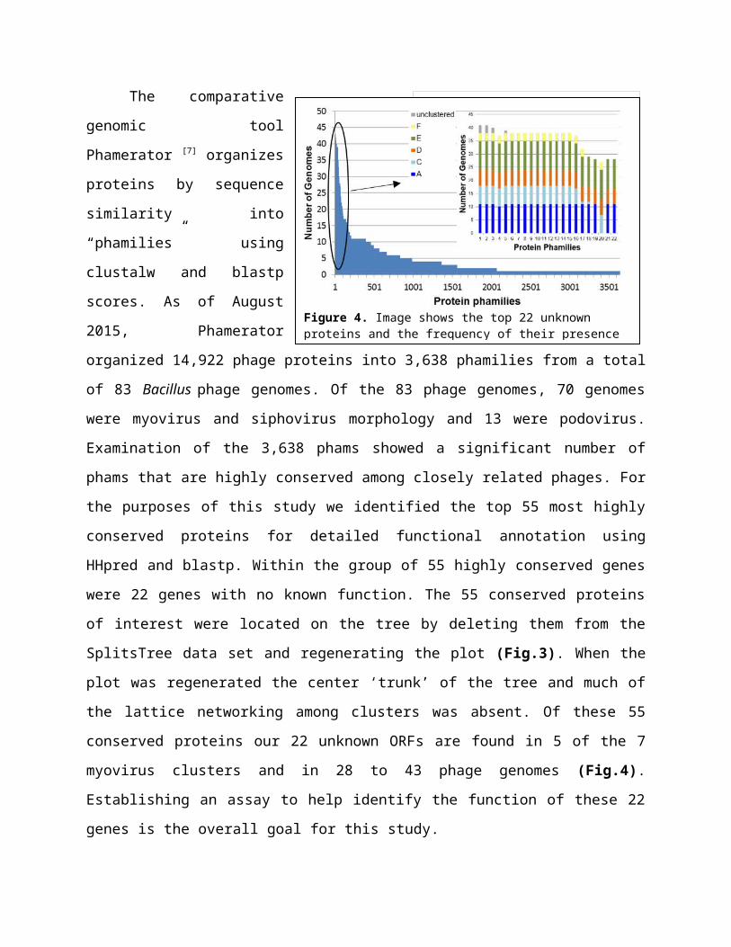
The comparative genomic tool Phamerator [7]
organizes proteins by sequence similarity into
“phamilies” using clustalw and blastp scores. As of
August 2015, Phamerator organized 14,922 phage
proteins into 3,638 phamilies from a total of 83
Bacillus phage genomes. Of the 83 phage genomes, 70
genomes were myovirus and siphovirus morphology
and 13 were podovirus. Examination of the 3,638
phams showed a significant number of phams that are
highly conserved among closely related phages. For
the purposes of this study we identified the top 55 most highly conserved proteins for detailed
functional annotation using HHpred and blastp. Within the group of 55 highly conserved genes
were 22 genes with no known function. The 55 conserved proteins of interest were located on the
Figure 3. SplitsTree image of 83 bacillus phage genomes with the top 55 most highly conserved proteins absent.
Figure 2. SplitsTree image of 83 bacillus phage genomes grouped into clusters based on protein content similarity.

tree by deleting them from the
SplitsTree data set and
regenerating the plot (Fig.3).
When the plot was regenerated
the center ‘trunk’ of the tree and
much of the lattice networking
among clusters was absent. Of
these 55 conserved proteins our
22 unknown ORFs are found in 5
of the 7 myovirus clusters and in
28 to 43 phage genomes (Fig.4).
Establishing an assay to help identify the function of these 22 genes is the overall goal for this
study.
The Bacillus phage Phrodo is a myoviridae with a double stranded DNA genome and a
contractile tail (Fig.5) that was isolated by the SEA PHAGES program at VCU in 2014 using the
host bacteria Bacillus thuringiensis. Phrodo is our experimental phage of choice because its
DNA and genetic information is readily available and a vast majority of our genes of interest are
found within Phrodo’s genome. These genes of interest are also found in majority of the 13
clusters, including cluster E where Phrodo is located. While cluster E is fairly diverse in
comparison to the highly conserved cluster A, it is interesting that majority of the top 55 highly
conserved proteins (including unknowns) are still found in Phrodo’s genome and numerous other
genomes found in cluster E. The diversity of this clusters makes it an
interesting choice to focus on given its representation of the natural
diversity of phage genomes. This observation could further support
the speculation that proteins present in numerous genomes and
clusters are essential to their viral life cycle.
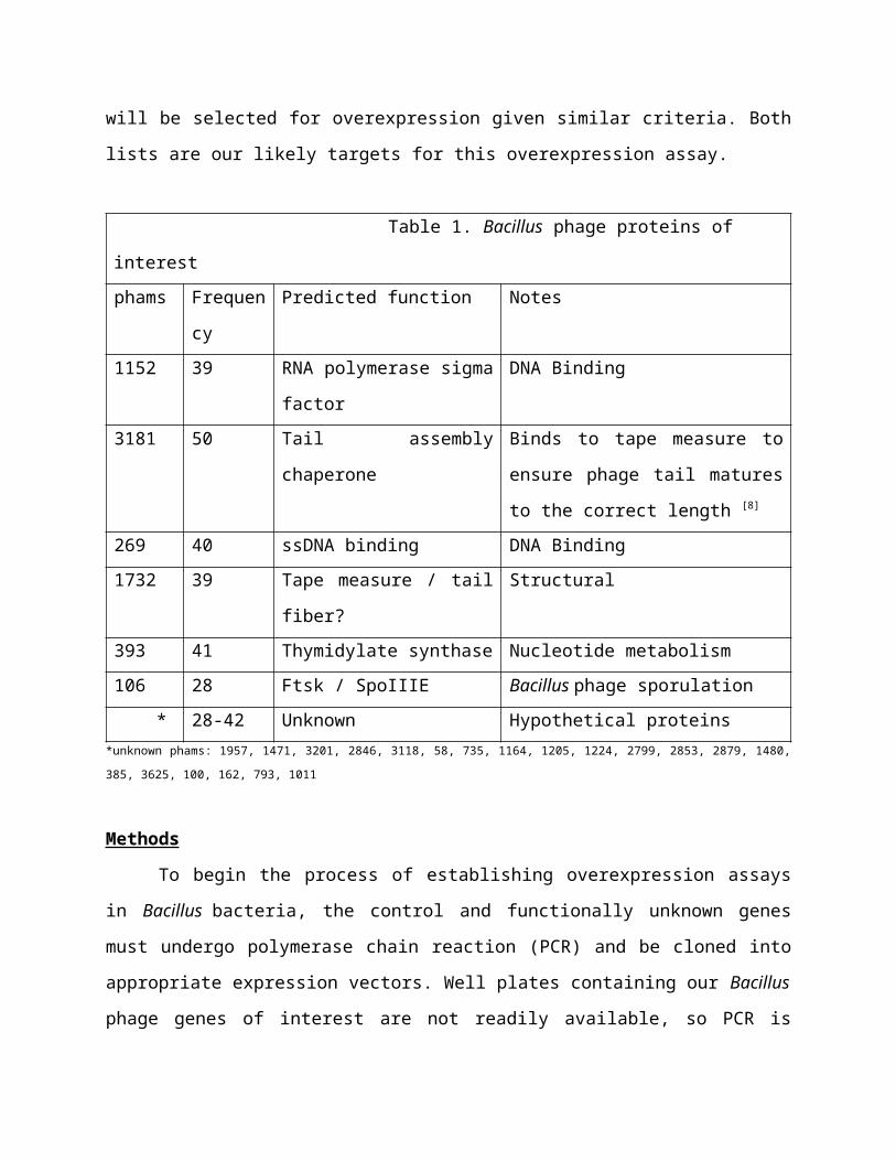
Of the 55 highly conserved genes 5-7 genes will be selected
as controls. Criteria for selecting controls will be based on its
presence in Phrodo, ability to undergo PCR with appropriate primers, and having a well-studied
protein- protein interaction or host interaction by the scientific community. A list has been
formulated for controls based on the criteria aforementioned (Table 1). In addition to a list of
Figure 5. TEM image of phage Phrodo.

controls, 15-20 unknown genes will be selected for overexpression given similar criteria. Both
lists are our likely targets for this overexpression assay.
Table 1. Bacillus phage proteins of interest
phams Frequenc
y
Predicted function Notes
1152 39 RNA polymerase sigma
factor
DNA Binding
3181 50 Tail assembly chaperone Binds to tape measure to ensure phage
tail matures to the correct length [8]
269 40 ssDNA binding DNA Binding
1732 39 Tape measure / tail fiber? Structural
393 41 Thymidylate synthase Nucleotide metabolism
106 28 Ftsk / SpoIIIE Bacillus phage sporulation
* 28-42 Unknown Hypothetical proteins*unknown phams: 1957, 1471, 3201, 2846, 3118, 58, 735, 1164, 1205, 1224, 2799, 2853, 2879, 1480, 385, 3625, 100, 162, 793, 1011
Methods
To begin the process of establishing overexpression assays in Bacillus bacteria, the
control and functionally unknown genes must undergo polymerase chain reaction (PCR) and be
cloned into appropriate expression vectors. Well plates containing our Bacillus phage genes of
interest are not readily available, so PCR is necessary in order to conduct this study. Phrodo will
be used for PCR as a DNA template.
The first step to PCR is primer design of each gene to be overexpressed. Primers are
currently being designed for control genes and will be ordered from New England BioLabs.
Guides and online tools for primer design used in this study can be found at New England
BioLabs and Integrated DNA technology websites. Requirements for primer design include a
section of the beginning and end of each gene (20-40bp in length) running 5’-3’ with a GC
content of ~50% and a melting point of ~55oC. Requirements may be flexible given the naturally
low GC content of Bacillus phages.
Once primers have been designed and delivered the PCR process can begin. For PCR,
phage DNA will be amplified using LongAmp Taq DNA polymerase from the New England
BioLabs. This experiment will complete two-step PCR using methods from the Heidelberg
European Molecular Biology Laboratory (EMBL). Step one of DNA amplification will involve
combining 12 bp long attB1 and attB2 sites to the forward and reverse ends of each primer
sequence with buffer and DNA polymerase and running 10 cycles of Denaturing, Annealing and
Extension after two minutes of denaturing prior to beginning the cycles. Step two combines step
one PCR product with 12 bp long attB1 and attB2 adapter primers with buffer and DNA
polymerase, then denaturing and running 5+ cycles. The adapter primers are added as an
extension to ensure the ORF is not cut short during reactions involving ligase. The product of
step two will be purified using a DNA clean-up kit. When genes have been successfully
amplified they will be cloned and transferred to entry vectors in E. coli bacteria.
After the PCR product has been purified, a BP Clonase reaction will be performed to
prepare entry vectors for transformation into chemically competent cells. PCR product is
combined with TE buffer, pDONR / Zeo entry vector and BP Clonase II enzyme. The following
mixture is incubated over night at 25oC. The next morning protein Kinase K is added and
incubated at 37oC briefly to stop the clonase reaction. When incubation is complete the clonase
product is transformed into chemically competent E. coli cells. The BP clonase reaction mimics
phage-host relations. Naturally phage DNA has attP sites and the host has attB sites, when these
sites combine the result is attL sites [9]. For BP clonase reactions the ORF product from PCR has
attP sites and the empty plasmid contains attB sites, when the sites are combined attL sites form.
In both situations the result is attL sites flanking the ORF and inserted into either a plasmid or
bacterial genome.
Once cells containing entry vectors have completed transformation into E. coli a mini
prep will be done using a Nucleic Acid and Protein Purification kit to isolate the plasmid DNA
containing our experimental ORF. The isolated plasmids are then transferred to an expression
vector by the LR Clonase II reaction. During this reaction the pDONR / Zeo entry vector is
combined with the pDG148 GW expression vector along with TE buffer and LR Clonase II
enzyme. The mixture is then incubated overnight at 25oC and the following morning briefly
incubated at 37oC after the addition of protein Kinase K to stop the clonase reaction. LR clonase,
similar to BP clonase, mimics phage – host interactions. When the LR clonase reaction occurs
the ORF containing attL sites isolated from an entry vector is combined with the attR sites in the

expression vector. Combining the attL and attR sites results in an expression clone with an ORF
flanked by attB sites. When this process of combining attL and attR sites occurs naturally
between phage and host the result is an attL site at the beginning of the ORF and an attR site at
the end. Once LR clonase is complete the product is transformed into chemically competent
Bacillus bacteria. For overexpression to occur the bacteria will be plated on media containing
IPTG to induce ORF translation.
Discussion
Wagemans and colleagues used a shuttle vector system to overexpress their experimental
proteins, just as we intend to do. However, their system results in a single copy of an ORF
integrated in the host bacteria. This experiment will use an
expression and entry vector system to overexpress Bacillus
phage proteins in Bacillus bacteria. The result is a cell
containing plasmids with an ORF instead of a single ORF
integrated in the cells genome. We chose to use expression and
entry vectors so we can control the rate of transcription with
confidence that the bacterial genome is not interfering with the
gene expression. By beginning with entry vectors we will have a
vector containing our ORF that is easily transferable to other
vectors for future experiments. We have chosen pDONR/ Zeo
(Fig.6) as our entry vector since it contains a phage T1/T2
promoter and is compatible with E. coli bacteria. Majority of the
prep work for this study will be done in E. coli bacteria because
they are easy to grow and well-studied. Chemically competent
E. coli cells have already been prepared for transformations
with plasmid DNA. Vector pDG148 GW (Fig.7) will be used as
our expression vector [10]. This vector is actively being requested
from the creator in preparation for this experiment. It is
compatible with both E. coli and Bacillus bacteria. Chemically
competent Bacillus will be made prior to beginning gene overexpressions. This vector contains
the Pspac promoter for Bacillus bacteria upstream from the experimental ORF. The promoter
allows us to induce and control the rate of ORF transcription in bacteria with the use of IPTG.
Figure 6. Visualization of pDONR/ Zeo vector and all the genes present. Figure 7. pDG148 GW vector showing all genes it contains. ccdB and CmR is where the ORF is inserted.
Media will contain various concentrations of IPTG to observe any effects the rate of ORF
transcription may have on the cell. IPTG works by binding to promoters upstream from
experimental ORFs, this allows RNA polymerase to bind to DNA and begin transcription. This
process is modeled after the Lac promoter in E. coli.
The study our experiment is modeled after provides an excellent example for results we
should expect from an overexpression assay. Of the 26 proteins they chose to over express, 6 of
them (protein 7, 8, 14, 15, 18, 30) resulted in either cell growth inhibition or interesting
phenotypes (Fig. 8). Wagemans and colleagues observed cells that could not properly divide
after one correct division when protein 7 was overexpressed. The resulting cells were elongated
with two nuclei. Protein 8, when over expressed, was observed to cause cells to begin cell
division correctly but promptly stop mid division, resulting in attached daughter cells that burst
several hours after division has halted. Both protein 14 and 18 induced in cells were observed to
exhibit a long filamentous phenotype. While 4 of the 6 proteins cause morphological effects in
cells, proteins 15 and 30 affect cells by inhibiting their growth. In our study we will screen for
Figure 8. Image extracted from Wagemans et al. showing the effects 6 of their 26 proteins had on their host cells when overexpressed. Our study will screen for similar results.
similar results when inducing proteins for overexpression. We aim to observe cell death,
inhibited cell growth or abnormal cell division.
The purpose of this experiment is to establish assays to screen for any phage protein-host
interactions that express phenotypes. Proteins that express interesting phenotypes will be selected
for further functional analysis. Experiments planned for the future on unknown proteins that
express phenotypes include Yeast 2 Hybrid assays. For Yeast 2 Hybrid assays majority of the
preparation will already be complete. This experiment provides amplified Phrodo DNA from
PCR, entry and expression vectors containing experimental ORFs and competent E. coli and
Bacillus cells.
Figure 9. Visualization of work plan to identify highly conserved Bacillus phage proteins of
unknown function for PCR and overexpression so that we may select candidates with interesting
observations for future functional analysis. This plan is designed to reach two aims using the
chronological plan of action shown above.

References
[1] Wommack, K. Eric, and Colwell, Rita R. "Virioplankton: Viruses in Aquatic
Ecosystems." Microbiology and Molecular Biology Reviews64.1 (2000): 69.
[2] Pope, Welkin H, Charles A Bowman, Daniel A Russell, Deborah Jacobs-Sera, David J Asai,
Steven G Cresawn, William R Jacobs, Roger W Hendrix, Jeffrey G Lawrence, and Graham F
[3] Maynard, Nathaniel D., Elsa W. Birch, Jayodita C. Sanghvi, Lu Chen, Miriam V. Gutschow,
Markus W. Covert, and Ivan Matic. "A Forward-Genetic Screen and Dynamic Analysis of
Lambda Phage Host-Dependencies Reveals an Extensive Interaction Network and a New Anti-
Viral Strategy (Host Genetic Requirements for Lambda Infection)."PLoS Genetics 6.7 (2010):
E1001017.
[4] Qimron, Udi, Boriana Marintcheva, Stanley Tabor, and Charles C. Richardson.
"Genomewide Screens for Escherichia Coli Genes Affecting Growth of T7
Bacteriophage." Proceedings of the National Academy of Sciences of the United States of
America 103.50 (2006): 19039-9044.
[5] Wagemans, Jeroen, Bob G. Blasdel, An Van Den Bossche, Birgit Uytterhoeven, Jeroen De
Smet, Jan Paeshuyse, William Cenens, Abram Aertsen, Peter Uetz, Anne-Sophie Delattre,
Pieter-Jan Ceyssens, and Rob Lavigne. "Functional Elucidation of Antibacterial Phage ORFans
Targeting P Seudomonas Aeruginosa." Cell Microbiol Cellular Microbiology 16.12 (2014):
1822-835. Web.
[6]Jakutytė, Lina et al. “Bacteriophage Infection in Rod-Shaped Gram-Positive Bacteria:
Evidence for a Preferential Polar Route for Phage SPP1 Entry in Bacillus Subtilis.” Journal of
Bacteriology 193.18 (2011): 4893–4903. PMC. Web. 23 Feb. 2016.
[7] Cresawn, Steven G., Matt Bogel, Nathan Day, Deborah Jacobs-Sera, Roger W. Hendrix, and
Graham F. Hatfull. "Phamerator: A Bioinformatic Tool for Comparative Bacteriophage
Genomics." BMC Bioinformatics 12.1 (2011): 395.

[8] Xu, Hendrix, and Duda. "Chaperone–Protein Interactions That Mediate Assembly of the
Bacteriophage Lambda Tail to the Correct Length."Journal of Molecular Biology 426.5 (2014):
1004-018. Web.
[9] "BxSeqTools Ultimate Molecular Cloning Guides - Gateway Cloning."BxSeqTools Ultimate
Molecular Cloning Guides - Gateway Cloning. BioInfoRx, 2016. Web. 08 May 2016.
<http://bioinforx.com/lims1/bxseqtools/ultimate-molecular-cloning-guides/
help_cloning_gw.php>.
[10] Dobrijevic, Dragana, Gaetana Di Liberto, Kosei Tanaka, Tomas De Wouters, Rozenn
Dervyn, Samira Boudebbouze, Johan Binesse, Hervé M. Blottière, Alexandre Jamet,
Emmanuelle Maguin, and Maarten Van De Guchte. "High-Throughput System for the
Presentation of Secreted and Surface-Exposed Proteins from Gram-Positive Bacteria in
Functional Metagenomics Studies." PLoS ONE 8.6 (2013): n. pag.