Weakness; Myopathy, Anterior horn cell disease, Neuropathies and ...
46
89 Spinal cord Weakness Weakness; Myopathy, Anterior horn cell disease, Neuropathies and Neuromuscular transmission defects In the neurological evaluation of weakness, we distinguish between upper motor neuron weakness, and lower motor neuron weakness. The differences are tabulated below. Lower motor neur on weakness (LMN) Upper motor neur on weakness (UMN) Flaccid Spasticity, including a Babinski response Decreased tone Increased tone Decreased muscle stretch reflexes Increased muscle stretch reflexes Profound muscle atrophy Minimal muscle atrophy Fasciculations present Fasciculations absent May have sensory disturbances May have associated sensory disturbances Fasciculations are irregular contractions of a group of muscle fibers innervated by one axon. Clinically this appears as a small muscle twitch. It is also customary, and very helpful, to classify LMN weakness on the basis of the anatomical station affected. These stations ar e: 1) The anterior (ventral) horn cell. 2) The peripheral nerve, (ventral and dorsal nerve roots i.e., radiculopathy or nerve i.e., neuropathy.) 3) The neuromuscular junction. 4) The muscle (i.e. myopathy).
Transcript of Weakness; Myopathy, Anterior horn cell disease, Neuropathies and ...

89 Spinal cordWeakness
Weakness; Myopathy, Anterior horn cell disease,Neuropathies and Neuromuscular transmission defects
In the neurological evaluation of weakness, we distinguish between upper motor neuronweakness, and lower motor neuron weakness. The differences are tabulated below.
Lower motor neuron weakness (LMN) Upper motor neuron weakness (UMN)Flaccid Spasticity, including a Babinski responseDecreased tone Increased toneDecreased muscle stretch reflexes Increased muscle stretch reflexesProfound muscle atrophy Minimal muscle atrophyFasciculations present Fasciculations absentMay have sensory disturbances May have associated sensory disturbances
Fasciculations are irregular contractions of a group of muscle fibers innervated by one axon.Clinically this appears as a small muscle twitch.
It is also customary, and very helpful, to classify LMN weakness on the basis of theanatomical station affected.
These stations are:1) The anterior (ventral) horn cell.2) The peripheral nerve, (ventral and dorsal nerve roots i.e., radiculopathy or nerve i.e.,
neuropathy.)3) The neuromuscular junction.4) The muscle (i.e. myopathy).

90Spinal cordWeakness
II. Anatomy of the lower motor neuron
Anterior (ventral) horn cells
The anterior horn cells are somatotopically organized in the spinal cord. That is, mediallylocated anterior horn cells innervate the proximal muscles, while laterally located ventral horn cellsinnervate more distal muscles. The arrangement at cervical segments is shown in figure 2. Thisorganization means that diseases that destroy anterior horn cells can result in highly selectiveweakness. Not only may a single muscle become weak, but only portions of the muscle may beaffected. As a rule however the adjacent anterior horn cells will also be affected with weakness of
adjacent muscles.Figure 2. The somatotopic arrangement of anterior horn cells at cervical and the first thoraciclevels. Because the anterior horn cells that innervate different muscles in the upper and lowerextremities are present at different segments of the spinal cord, a whole extremity is not presentedat a single level.
NervesA reminder on the classification of dorsal and ventral root fibers
The axons in the dorsal roots have been classified based upon their conduction velocitiesand their sizes. This has led to some confusion in the literature (and for medical students!!). Theclassifications scheme based upon fiber size uses Roman numerals. Thus, there are I, II, IIIand IV fiber types. You already have heard about the Ia fibers and that they are associated withmuscle spindles and are large and fast conducting. You also have heard that the Ib fibers areassociated with the Golgi tendon organs and are little smaller and slower conducting than the Ias.Also remember that II fibers are associated with muscle spindles but are slower conducting andsmaller that the Ias and Ibs. II fibers are also associated with receptors carrying information from

91 Spinal cordWeakness
encapsulated nerve endings used in two point discrimination, vibration and consciousproprioception. III fibers are smaller than Is and IIs and are only lightly myelinated and relativelyslow conducting. Such fibers are associated with cooling and first pain. Finally, IV fibers areunmyelinated and convey second pain and warming.
Now lets turn to the classification that uses letters versus Roman numerals. The largest andfastest conducting fibers are called A fibers. Aααααα (alpha) fibers are comparable to the Ias and Ibs. Aβββββ(alpha-beta) fibers are equivalent to II fibers in size and conduction velocities. Aδ (deltas) areequivalent to IIIs and associated with cooling and first pain. B fibers are smaller than A fibers, arelightly myelinated and are visceral afferents; they have no equivalent in the Roman numeral system.Finally, C fibers are unmyelinated and equivalent to IV fibers. In addition to carrying second painand warming such fibers are postganglionic autonomics (but these do not travel in the dorsal roots).
What about ventral root fibers. The processes of lower motor neurons that innervateextrafusal muscle fibers are Aαααααs (or just alpha motor neurons). The preganglionic autonomic axonsin the ventral root are B fibers. Finally, there are axons in the ventral roots that innervate theintrafusal (not extrafusal) fibers of the muscle spindles. These are called Aγγγγγ (gamma) motorneurons (no equivalent in Roman numerals).
Remember, A and B fibers are myelinated and Cs are not. In the Roman numeral system, justremember that only the IVs are not myelinated. This is important, since demyelinatingdiseases would affect the somatic and visceral afferents and efferent fibers in peripheralnerves, pain and temperature would not be affected.

92Spinal cordWeakness
Muscle
One anterior or ventral horn cell, and thus one axon, innervates a few hundred or even a fewthousand muscle fibers. The muscle fibers innervated by a single anterior horn cell are collectivelyknown as a motor unit. The “territory” of such a motor unit spans 10-15 mm in a muscle however itis rare that directly adjacent muscle fibers are innervated by the same anterior horn cell/axon. Thefigure below shows the seemingly random pattern of innervation of adjacent muscle fibers byindividual anterior horn cells. The clear muscle fibers below are innervated by a single anterior horncell and comprise a motor unit. The vertically oriented fibers are innervated by a different anteriorhorn cell constituting a second motor unit and the horizontally oriented represent yet another.
We also need to distinguish between type 1 (slow contracting) muscle fibers and type 2 (fastcontracting) muscle fibers. The type of muscle fiber is dependent on the type of anterior horn cellthat innervates it. Thus if a muscle fiber is innervated by a type 1 anterior horn cell, it will contractslowly. Certain histochemical reactions, amongst others myosin ATPase, distinguish between type 1and type 2 fibers. Thus muscle reacted with myosin ATPase will normally exhibit a checkerboardpattern as it is likely that the adjacent muscle fibers are innervated by another anterior horn cell of adifferent fiber type (figure 4).
Figure 4. The clear fibers in the figure above are myosin ATPase free and are all innervated by oneventral horn cell. The striped fibers are the ATPase rich and would look similar under a microscope.However, we want to illustrate that the ATPase rich fibers are innervated by two different ventralhorn cells (a and b; hence the different orientations of the stripes).
Neuromuscular junction
A muscle fiber is activated via a nerve impulse generated by an anterior horn cell. The impulse isconducted along the nerve fiber via saltatory conduction; that is an action potential is generated atone node of Ranvier and then jumps to the next node of Ranvier where another action potential isgenerated. Once the impulse reaches the neuromuscular junction, voltage sensitive Ca2+ channelsare opened which allow for the influx of Ca2+ into the nerve terminal. Ca2+ entry into the nerveterminal initiates the fusion of acetylcholine containing vesicles with the presynaptic membrane and

93 Spinal cordWeakness
the subsequent release of acetylcholine into the synaptic cleft. Acetylcholine binds to post-synapticacetylcholine receptors on the muscle membrane. This induces an end plate potential whichsubsequently results in the generation of an action potential in the muscle fiber membrane (figure 5).The end result of this reaction is muscle fiber contraction.
Figure 5. The neuromuscular junction.
III. Diagnosis of the different lower motor neuron subgroups
The diagnosis of a specific lower motor neuron syndrome starts with the localization of thedisease to one of the 4 anatomic stations. This can be accomplished by a combination of thefollowing investigations:
1) History and clinical examination
In recording the history it is of particular importance to document the following. The timeof disease onset, the presence or absence of a family history of other similarly affectedindividuals, consanguinity (patients born from parent related by blood), the pattern andprogression of muscle weakness, the presence or absence of sensory symptoms and the presenceof fatigability. The clinical examination serves to corroborate the clinical history, and todocument the patterns of weakness, sensory loss, fatigability and reflex changes.

94Spinal cordWeakness
2) Histological examination of muscle or nerve biopsy specimens
These will be dealt with in more detail during the neuropathology section of your Pathologycourse.
Muscle histology
Muscle is not too smart and can only react in a limited number of ways to insult. Thus mostprimary muscle diseases have non-specific features in common, such as muscle fiber necrosis,evidence for muscle fiber regeneration, structural abnormalities such as centrally located muscle fibernuclei and an increase in muscle connective tissue (figure 6). Some primary muscle diseases do showdiagnostic changes such as nemaline rod formations or central cores. Inflammation in muscle is
important as it may indicate a treatable disease.Figure 6. Typical, non-specific pathological findings in a primary myopathy. A necrotic fiber(asterix), and a hypercontracted muscle fiber (star), are shown. The entire muscle is shortened andthus, the hypercontracted fiber is thicker. The connective tissue between the muscle fibers isincreased.

95 Spinal cordWeakness
Muscle denervation
Anterior horn cell disease or a peripheral neuropathy result in exactly the same histologicalfindings in the muscle! The poor muscle can only interpret these events as “I am denervated.” Thepathological hallmarks of denervation are type grouping and group atrophy (figure 7). Becauseone anterior horn cell/motor axon innervates a number of muscle fibers, it follows that disease of ananterior horn cell or axon results in denervation of a number of muscle fibers. These muscle fibersthat have lost their innervation, may now be innervated by healthy axons that normally innervateadjacent muscle fibers. The end result is that now one axon innervates more muscle fibers thannormal, (a giant motor unit) and also the normal checkerboard pattern of innervation is lost. That is,a whole group of type 1 or 2 fibers can now be seen adjacent to one another (type grouping). Withprogression of the disease, the axon that sprouted to innervate previously denervated muscle fibersmay now also become diseased, resulting in an entire group of adjacent muscle fibers becomingatrophic (group atrophy).
Nerve histology
The nerve is equally unimaginative in its reaction to damage. In principle, only two pathologicalchanges are seen. Firstly axonal damage results in Wallerian degeneration, a bead-like disruptionof the peripheral nerve that involves both the axon cylinder and the surrounding myelin (Figure 8).This is seen in diseases affecting the axons in the peripheral nerve, or in anterior horn cell disease.

96Spinal cordWeakness
Figure 8. Wallerian degeneration seen in axonal damage.
Secondly demyelination results in peripheral nerves with shortened internodes or internodeswith thinner myelin (figure 9). Remember the axon cylinder in demyelinating diseases is fine andhealthy.
Figure 9. A nerve fiber with shortened internodes that are hypomyelinated; typical findings indemyelinating neuropathies.
3) Electromyographic (EMG) examination
This test consists of two parts:
a) Nerve conduction studies
Since there are few pure motor nerves to study, motor nerve conduction recordingelectrodes are placed over a distal muscle (i.e. thenar muscle group). The appropriate nerve(median) is then stimulated electrically and the evoked responses can be measured. These evokedresponses recorded from the surface of the muscle are called a compound muscle action potential(CMAP). The time it takes from stimulation to generation of the CMAP is the conduction speed.
The CMAP represents the action potentials of all muscle fibers activated by the nervestimulation and the measured response can be compared to a known standard for such stimulation.Reduction in the strength of this response indicates a loss in overall muscle mass or the loss of motorfibers and must further be investigated as to its cause.
For sensory nerve conduction studies, the recording electrodes are placed over superficialnerves (e.g. the sural nerve is a pure sensory nerve). Stimulation of a sensory nerve leads to actionpotentials in all of the fibers of that nerve and an electrode on the surface of such a nerve records thesensory nerve action potential (SNAP). Furthermore, by stimulating the same nerve over differentsegments the distances between stimulation sites can be measured and a conduction velocity for thenerve segment established.

97 Spinal cordWeakness

98Spinal cordWeakness
The conduction studies are followed by repetitive nerve stimulation studies. A routine motornerve conduction study is performed but the nerve is stimulated supramaximally at 2-3 Hz and theamplitude of the first 4 CMAPs recorded. In neuromuscular transmission defects the CMAPamplitude decreases with successive stimuli as some muscle fibers are not depolarized due to theneuromuscular transmission defect (figure 12). This is called a decremental response. (The exactmechanism of the decremental response is complex and beyond the scope of this course!! Don’tworry!)
Figure 12. Repetitive nerve stimulation study. Four CMAPs are shown in each tracing. Note that theamplitudes of the responses are the same in a normal muscle, but that a decremental response isrecorded in neuromuscular transmission defects.
Summary of nerve conduction findings in different disease groups
Station 1- Anterior (Ventral) Horn Cell disease: This results in low CMAP amplitudes in muscleinnervated by the dying anterior horn cells whose axons travel in the nerve being stimulated. Thereare fewer (than normal) axons that are able to “drive” action potentials in the muscle, the end resultbeing a smaller (than normal) CMAP. Since there is still a population of normal axons from otheranterior horn cells (non diseased) nerve conduction velocity is normal, i.e. the nerve (for instancethe median nerve) has normal axons that camouflage the dying ones. The sensory nerve conductionstudies are normal because ventral horn cells give rise to only motor fibers. Cell bodies of sensoryfibers lie in dorsal root or cranial nerve ganglia.
Station 2- Peripheral Nerve disease: The findings will depend on whether both the motor andsensory axons are affected. In most peripheral nerve diseases both become affected. If the changesresult in damage only to the axis cylinders, the nerve conduction velocities are normal (healthyaxons mask the defect), but both the CMAP and SNAP amplitudes will be reduced. If the peripheralnerve disease is predominantly demyelinating (i.e. all of the axons have demyelinated areas) the
REMEMBER: axonal or anterior horn cell diseases do not slow nerve conduction velocitiesappreciably as the remaining axons conduct at normal speed. There are however too few
normal axons and thus, the evoked potentials in the muscle (CMAPs) are small.

99 Spinal cordWeakness
findings are marked slowing in both the motor and sensory nerve conduction velocities andrelatively normal CMAP and SNAP amplitudes (the axis cylinders are OK).
Station 3- Neuromuscular Junction disease: Nerve conduction studies (motor and sensory) arenormal, but the hallmark of these diseases is a decremental CMAP response with repetitive nervestimulation.
Station 4- Muscle disease: Nerve conduction studies are normal, but the CMAP amplitudes will below, as there is loss of muscle fibers.
In addition to nerve conduction studies, the EMG also involves:
b) The needle examination
An electrode is introduced into the muscle and recordings are made with mild to moderateactivation of the muscle. This test is accompanied by some discomfort, but if performedappropriately should not be torture!
Depolarization of muscle fibers in close proximity to the needle electrode will be recorded asmotor unit potentials (MUPs; compare with CMAPs). A normal muscle and a normal MUP isshown in figure 13.
Figure 13. Normal muscle. The above muscle fibers are innervated by three different lower (alpha)motor neurons. Think of the MUP as representing the action potentials of the muscle fibersassociated with one of these motor neurons (a motor unit). For example, a slight contraction of themuscle during a movement will fire all of the “clear fibers” above, but neither of the “striped fiber”groups.
REMEMBER: demyelinating nerve diseases slow nerve conduction velocities, but the evokedpotentials are of relatively normal amplitudes.
REMEMBER: neuromuscular transmission defects result in decremental CMAP responseswith repetitive nerve stimulation.
REMEMBER: primary muscle diseases result in low CMAP amplitudes, similar to thefindings in ventral horn and axis cylinder lesions.

100Spinal cordWeakness
By analyzing the size of the MUP (mostly the amplitude and duration), we can make adistinction between diseases that are primarily myopathic (disease of the muscle) versus those whichresult from denervation (neurogenic). As described in the section on the anatomy of muscle fibers,the muscle fibers innervated by one anterior horn cell/motor axon are spread over 10-15mm of themuscle. Furthermore, the territory innervated by adjacent anterior horn cells overlap so that adjacentmuscle fibers are normally innervated by different anterior horn cells or motor axons. I know we havecovered this before, but other peoples children never listen! With damage to an anterior horn cellor a motor axon the denervated muscle fibers usually become reinnervated by another motor axonwith the result that more muscle fibers are innervated by the same anterior horn cell or motor axon inclose proximity to the EMG needle. This is seen histologically as type grouping as shown in figure7. In simplified terms this results in larger MUPs (“neurogenic” MUPs), figure 14. You mightwonder why, if there is reinnervation (or “sprouting”) and larger MUPs, why are the CMAPs smaller?Well, that is because muscle fibers are also dying (remember group atrophy?)Figure 14. Neurogenic atrophy with type grouping. A large MUP is recorded. Think about the
single active motor neuron in Figure 13 as innervating more muscle fibers. When it fires therewill be more muscle fiber action potentials and thus a larger MUP.
On the other hand in a primary muscle disease, there is loss of muscle fibers, ormuscle fibers have a smaller mean diameter than normal, resulting in small MUPs(“myopathic” MUPs), figure 15.
Figure 15. Myopathic muscle. A small MUP is recorded.

101 Spinal cordWeakness
Fasciculations are noted clinically as a contraction of a small group of muscle fibers. Theyresult from the spontaneous discharge of an anterior horn cell or a motor axon with the subsequentcontraction of all the muscle fibers innervated by that anterior horn cell or motor axon.Fasciculations can also be recorded with the needle electrode. Clinically, fasciculations are seen afterreinnervation of muscle fibers and they are particularly common in amyotrophic lateral sclerosis(motor neuron disease).
Fibrillation potentials on the other hand are not visible through the skin. They are the smallpotentials generated by single muscle fibers once the muscle fibers become denervated. When amuscle fiber is denervated, several pathological changes take place. The acetylcholine receptorsspread all across the muscle fiber instead of being grouped in a well-defined geographical area, theend-plate. This spread may play a role in attracting new innervation to the denervated muscle fiberfrom adjacent nerve sprouts. The muscle fiber becomes much more sensitive to free acetylcholinereleased spontaneously from adjacent nerve fibers and is depolarized and repolarized spontaneouslyas these molecules reach it. Each single depolarization is electrically detected as a single musclefiber action potential. If the muscle fiber reinnervates sucessfully, these potentials disappear again.Thus they are seen in axonal neuropathies provided reinnervation does not keep up with denervation.In active myopathies, portions of the muscle fiber also become denervated from its endplate becauseof focal muscle fiber necrosis. Fibrillation potentials can therefore also be seen in activemyopathies.
Figure 16. Fibrillation potentials. Each complex represents the spontaneous discharge of a singlemuscle fiber as recorded with a needle electrode in the muscle. Note that the discharge intervals areabsolutely constant.
4) Biochemical studies
Numerous studies are available but only neuromuscular transmission defects and primarymuscle diseases (myopathies) will be discussed.
Neuromuscular transmission defects. In myasthenia gravis, acetylcholine receptor antibodiesdestroy the post synaptic acetylcholine receptors and they are detectable in blood samples.
Primary muscle diseases - With muscle breakdown of any kind, creatine phosphokinase(CK) is released into the blood where it can be measured.
5) Genetic studies
The genetic defects of many neuromuscular diseases are now known and can be detected inperipheral blood or in muscle.

102Spinal cordWeakness
IV. Let us put this all together!
1. Anterior horn cell diseases
Common causes of anterior horn cell diseases are poliomyelitis, motor neuron disease andspinal muscular atrophy. Only spinal muscular atrophy will be discussed further. This is usuallyan autosomal recessively inherited disease with onset at any time from infancy to adulthood. Theprimary pathology is the progressive loss of anterior horn cells until the patients become so weakthat they die usually from an associated lung infection. The reason for the progressive loss ofanterior horn cells is not clear, but the disease is associated with an abnormality on chromosome 4.
EMG findings: Normal nerve conduction velocities, normal SNAP amplitudes, low CMAPamplitudes, large MUPs on needle examination, fasciculations.
Histology: Type grouping and group atrophy.Biochemistry: Defect on chromosome 4.
2. Peripheral nerve diseases
This encompasses a vast number of diseases and only a cursory overview will be attempted.
Clinical features
Damage to the peripheral nervous system results in motor, sensory and autonomicdysfunction. A neuropathy is any disease of the nerves. There are a number of different classesof neuropathies, but we will consider only one of them here.
Distal polyneuropathy: All the nerves are affected distally in the extremities. Clinically thepatients have sensory loss in a glove and stocking distribution, weakness and absent tendon reflexesin distal extremity muscles (e.g. ankle jerk). Longer nerves are affected more severely and thus thechanges predominate in the legs. Most distal polyneuropathies are purely sensory or affect thesensory and motor nerves together. Pure motor distal neuropathies are rare. Depending on theetiology, the neuropathies can be axonal (axis cylinder), demyelinating, or show features of both.Diseases that cause distal polyneuropathies include diabetes, toxins, and vitamin deficiency/alcoholabuse. Many of these neuropathies are familial.
EMG findings
a) Predominantly axonal disease: Normal motor and sensory nerve conduction velocities with lowor absent CMAP and SNAP amplitudes. Needle examination shows large MUPs that result fromdenervation and subsequent re-innervation. Fasciculations.
b) Predominantly demyelinating disease: Relatively normal CMAP and SNAP amplitudes withslowed nerve conduction velocities. Needle examination reveals normal MUPs as the axons are notdamaged and the muscle fibers are not denervated. In practice pure demyelination is rare and someassociated axonal damage is common.
Histology
Type grouping and group atrophy only if there is axonal (axis cylinder) damage.

103 Spinal cordWeakness
3. Neuromuscular transmission defects
Only myasthenia gravis will be discussed further. This disease is characterized byabnormal fatigue with exercise. Myasthenia gravis commonly affects young woman and has apredilection for ocular, facial, masticator and proximal upper extremity muscles. Typically thepatients recover to some degree after rest. Thus they feel much better in the morning, butbecome weaker as the day progresses. When the extraocular eye muscles are affected, diplopia(double vision) and ptosis (drooping of upper eyelid) are common and bothersome signs. Thisis an auto-immune disease with antibodies destroying the acetylcholine receptors (apostsynaptic defect).
Neuromuscular transmission defects
EMG findings:Normal nerve conduction velocities, CMAP and SNAP amplitudes.Decremental response on repetitive nerve stimulation.Needle examination: Relatively normal MUPs.Biochemistry:Acetylcholine receptor antibodies are present in blood.Histology:Usually normal.
4. Primary muscle diseases (myopathies)
Muscle dystrophies (dystrophy = faulty development) are genetically determined diseases withonset at any time after birth. They are diagnosed on the pattern of muscle involvement. Forexample Duchenne muscle dystrophy is characterized by large calves, proximal muscle weaknessand weakness of the latissimus dorsi muscles and pectoral muscles. Myotonic dystrophy patientsshow myotonia (an inability to relax a muscle after contraction) in addition to muscle weakness.
There also are congenital myopathies, metabolic myopathies and inflammatory myopathies thatare beyond the scope of this course.
EMG findings:Normal motor and sensory nerve conduction studies. The CMAPs are low because of loss in musclebulk. Needle examination show small MUPs.
Biochemical findings:All progressive myopathies have increased CK blood levels indicating the breakdown of muscle.
Histological findings:Non-specific myopathic features such as large fibers, necrotic fibers, and increased connectivetissue.

104Spinal cordWeakness
IN SUMMARY:
Anterior horn cell diseases. Clinically characterized by selective involvement of muscles. EMGfindings are those of low CMAP amplitudes (fewer axons firing muscle fibers), normal SNAP amplitudes(ventral horn cell axons are not sensory), relatively normal nerve conduction velocities (normal axonscamouflage dead ones), large “neurogenic” MUPs (normal axons take over muscle fibers of dead ones),muscle histology shows type grouping and group atrophy (normal axons take over muscle fibers of deadaxons [type grouping] and then eventually die [group atrophy] and blood CK is normal (CK levelsbecome elevated if a muscle fiber breaks down, or if the muscle membrane becomes porous. Bothconditions allow CK to leak from the muscle fiber into the blood. In neurogenic atrophy (of anteriorhorn cell or peripheral [axonal] nerve origin) the muscle membranes remain intact and thus CK levelsremain normal). Fasciculations (grossly) and fibrillations (only upon needle exam) are present.
Peripheral nerve diseases. Clinically characterized by the associated findings of sensory and autonomicabnormalities. EMG findings depend on whether it is primarily an axonal (axon cylinder) ordemyelinating neuropathy. Axonal EMG findings are those of low CMAP amplitudes (fewer axiscylinders in a nerve means fewer muscle fiber firing), lower SNAP amplitudes if axons in a sensorynerve, relatively normal nerve conduction velocities (normal axons camouflage dead ones), large“neurogenic” MUPs (normal axons take over muscle fibers of dead ones), muscle histology shows typegrouping and group atrophy (normal axons take over muscle fibers of dead axons [type grouping] andthen eventually die [group atrophy] and blood CK is normal. Fasciculations and fibrillations are present.
105 Spinal cordWeakness
Demyelination The EMG findings are those of relatively normal CMAP amplitudes (axis cylindersare fine, so normal number of muscle fibers are activated), normal SNAP amplitudes (axis cylindersare OK), reduced nerve conduction velocities (myelin loss=slowing), normal MUPs (axis cylindersare OK), muscle histology is normal (axis cylinders are OK) and CK is normal (no muscle fibers aredead!). No fasciculations or fibrillations.
You might wonder why normal myelinated fibers don’t camouflage the diseased fibers. In reality,demyelinating neuropathies affect multiple focal areas of every nerve and you have normal segmentsin between. Thus, you do not find “normal” and “abnormal fibers”, all the fibers are affected tosome degree. In axonal neuropathies some fibers are affected, others not. The normal fibersconduct normally and thus you do not see significant slowing of nerve conduction velocities, but yousee drop in the SNAP and CMAP amplitudes.
Neuromuscular transmission defects. Clinically characterized by abnormal fatigability; EMGshows normal nerve conduction velocities, normal CMAP and SNAP amplitudes, decrementalCMAP responses to repetitive nerve stimulation; muscle histology is relatively normal; blood CK isnormal. No fasciculations or fibrillations.
Primary muscle diseases. Clinically specific patterns of muscle weakness may be noted; EMG showsnormal nerve conduction velocities with low CMAP amplitudes (fewer muscle fibers per motorunit), normal SNAP amplitudes, smaller “myopathic” MUPs, muscle histology shows myopathicchanges; blood CK is elevated. Fibrillations are seen.
“SPEED PLAY”
Increased reflexes in a symptomatic limb suggest a central lesion, while reduced reflexessuggest a peripheral lesion.
Bilateral sensory and motor deficits throughtout the body below a roughly horizontal level inthe trunk, with normal function above that level, indicates a spinal cord lesion

106Spinal cordRadiculopathy
RADICULOPATHIES
Radiculopathy (L. radicula = little root; pathos = disease) is an irritation of one or more ofthe many nerve roots that exit the spinal cord along its length. It usually presents as a combinationof pain, weakness and numbness along the body region that the root supplies. Lateral spinal discprotrusion and bony spurs are common causes of radiculopathy. The motor, sensory and sundryother tracts, the spinal grey matter, and the spinal nerve roots should no longer be strangers to you.The terms and symptoms that will be discussed are used daily by clinical neurologists.
The peripheral nervous system begins at the nerve roots. Each segment of the spinal cordgives rise to a ventral or anterior motor and a dorsal or posterior sensory nerve root. The spinalnerve roots can be damaged as they traverse the spinal (vertebral) canal, but are especiallyvulnerable in the intervertebral foramina, where the ventral and dorsal spinal roots join to form thespinal nerves. Spinal nerves divide into dorsal and ventral rami. Dorsal rami supply the true backmuscles and skin of the back, whereas ventral rami supply everything else except the head of course.Dorsal and ventral rami contain three types of fibers; sensory, somatomotor and visceromotor.

107 Spinal cordRadiculopathy
The distribution of sensory fibers in each spinal nerve is called a dermatome.

108Spinal cordRadiculopathy
A group of muscles primarily innervated by the motor fibers in a single spinal nerve is called a myotome.
Root compression
The hallmark of acute or chronic nerve root compression is PAIN. Pain due to nerve rootcompression has certain characteristics:
* it tends to follow a dermatomal distribution* it may be accompanied by paresthesia (para, abnormal + aisthesis, sensation, abnormal sensation such
as pricking or tingling; heightened sensitivity) or sensory loss in a dermatomal distribution*dull pain is usually more proximal and difficult to localize*sharp pain usually localizes around the dermatomal borders
There is also a loss of power in the muscles innervated by the root.
One of the primary causes of radiculopathy is intervertebral disc disease.

109 Spinal cord
The lumbar roots emerge from below their respectivevertebrae. These roots are vulnerable just above their exitforamina, as they are then the most ventral (anterior) and mostlateral root in the vertebral canal and lie in the immediate pathof a lateral disc herniation (L5 in the drawing on the rightbelow). The intervertebral disc lying between vertebrae L4 andL5 is called the L4/5 disc. The disc between the L5 vertebraeand the sacrum is the L5/S1 disc. Since the L4 root emergesabove the L4/5 disc, a lateral herniation of the L4/5 discdamages the L5 root. Moreover, a lateral herniation of the L5/S1 disc damages the S1 root. KNOW THIS!!
Intervertebral disc herniation and degeneration is themost common cause of compressive radiculopathy.
The natural history of most disc herniation is self-limitedand does not require surgical therapy. One fifth of pain freepeople under the age of 60 have evidence of a herniated disc onMRI and 50% have evidence of a bulging disc. Hence a properneurologic evaluation is required to define symptoms anddeficits that can be linked to MRI findings. Current practice is todevelop a rehabilitation program for back pain and to investigatestructural causes and surgical therapies only when there is anobjective neurologic deficit or pain that does not improve.
Lumbar intervertebral disc herniation occurs mostcommonly at L4/5 (L5 root; 50%) and at L5/S1 (S1 root; 46.3%) interspace. Consequently,compression of the 5th lumbar nerve root is most common, with the first sacral nerve rootsa close second.
Radiculopathy
110Spinal cordRadiculopathy
A reason for the frequent compression of the L5 root may be the tight fit of the L5 root in itsforamen since this root has the largest diameter and its intervertebral foramen is narrower than anyother lumbar intervertebral foramen.
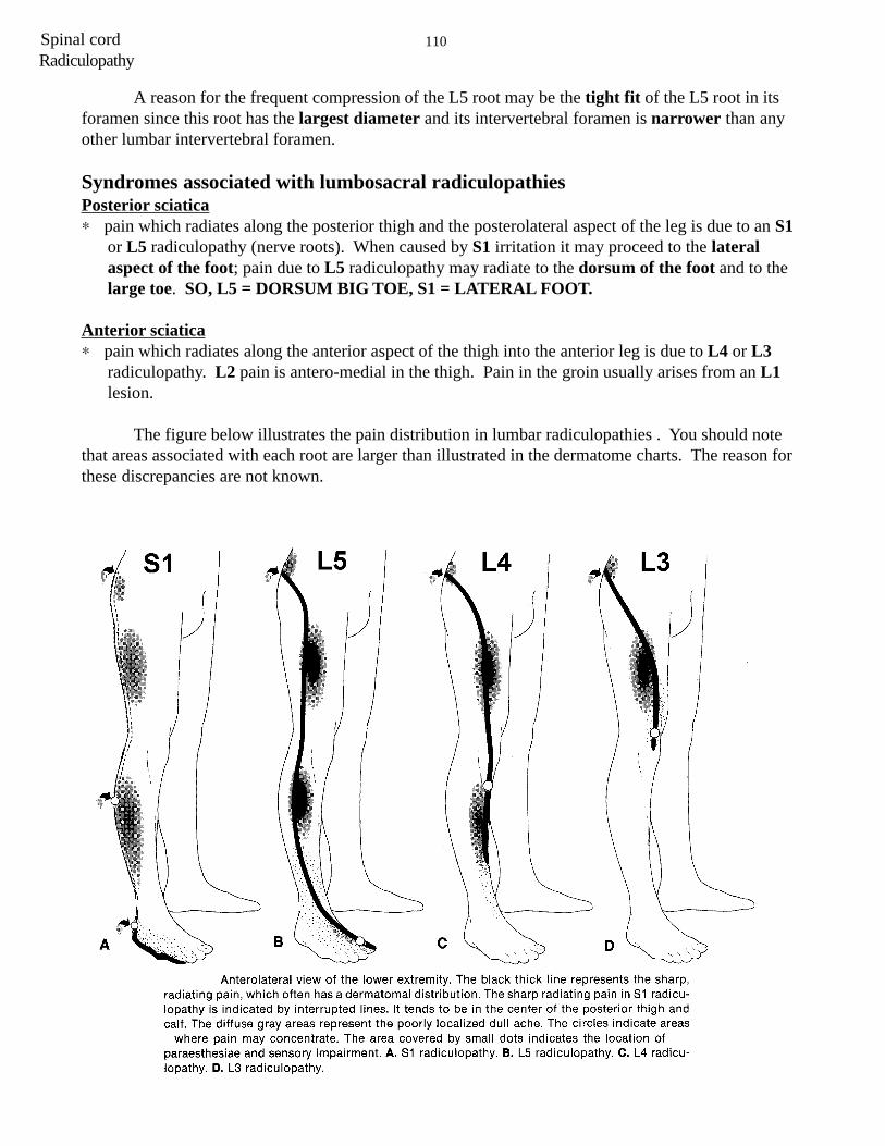
Syndromes associated with lumbosacral radiculopathiesPosterior sciatica∗ pain which radiates along the posterior thigh and the posterolateral aspect of the leg is due to an S1
or L5 radiculopathy (nerve roots). When caused by S1 irritation it may proceed to the lateralaspect of the foot; pain due to L5 radiculopathy may radiate to the dorsum of the foot and to thelarge toe. SO, L5 = DORSUM BIG TOE, S1 = LATERAL FOOT.
Anterior sciatica∗ pain which radiates along the anterior aspect of the thigh into the anterior leg is due to L4 or L3
radiculopathy. L2 pain is antero-medial in the thigh. Pain in the groin usually arises from an L1lesion.
The figure below illustrates the pain distribution in lumbar radiculopathies . You should notethat areas associated with each root are larger than illustrated in the dermatome charts. The reason forthese discrepancies are not known.

111 Spinal cord
Cervical radiculopathies (brachialgia = dull “achy” feeling in the arm and numbnessand tingling in the hand)
Because there are only 7 cervical vertebrae despite 8 cervical roots, the root numberexiting between two vertebrae is always the number of the lower vertebra. For example, theC5 root exits between the C4-C5 vertebrae and would be effected by a C4/5 disc herniation;the C8 root exits between C7-T1 vertebrae and would be compressed by a C7/T1 disc.
Pain due to a C6 and C7 (the most common) radiculopathy radiates from the neck and fromaround the shoulder into outer aspect of the arm and forearm. C6 radiculopathy may cause pain andnumbness along the dorsal aspect of the thumb and index finger, C7 pain and paresthesia mayradiate into the middle finger.
Radiculopathy

112Spinal cordRadiculopathy
The term cauda equina (horse’s tail)refers to the peripheral nerve roots which haveleft the spinal cord at approximately the level ofthe first lumbar vertebra. At that level, thestructure of the spinal cord itself ends and thenerves going to the pelvis and the lowerextremities continue through the spinal canal,leaving the spinal canal in pairs to the right andto the left as they pass to the pelvis and the lowerextremities. Significant pressure obstructing thespinal canal at any level from L1 downward cancause cauda equina syndrome (CES).

113 Spinal cord
MICTURITION
Normal bladder function depends on the coordinated activity of the bladder detrussor(smooth muscle) and the sphincter muscles (internal and external sphincters and muscles of pelvicfloor). The actual act of voiding is under the control of higher cortical centers that develops ascontinence (continere = to hold together) and is achieved in early childhood. Incontinence (lack ofcontrol of urinating) occurs when neuroanatomic pathways that innervate the bladder areinterrupted or when there are physical problems with the pelvic floor and sphincter muscles. Whendysfunction of the nervous system causes incontinence we use the term neurogenic bladder.Lesions of either upper or lower motor systems involved with micturition cause neurogenicbladders. Incontinence is an important symptom, and if it occurs in association with otherneurological deficits that localize to the spinal cord, needs tobe investigated aggressively.
The important points that you need to know in thiscourse are that the bladder is controlled by areas of the brainthat send axons down the spinal cord, traveling just medial tothe LCST. These bilateral projections terminate onpreganglionic parasympathetic neurons at S2, S3 and S4.The preganglionic parasympathetic neurons send their axonsout the ventral roots of S2, S3 and S4 to synapse onpostganglionic parasympathetic cells in ganglia near thebladder. These postganglionic parasympathetic cells in turninnervate the detrussor (smooth) muscle of the bladder forvoiding.
There are muscle spindles in the detrussor musclewhose cell bodies lie in dorsal root ganglia at S2, S3 and S4(all are not shown in the diagram). When the bladder fills,the muscle spindles are stretched and increase their firing.This information enhances neuronal firing of thepreganglionic parasympathetics at S2, S3 and S4 resulting incontraction of the bladder (voiding). This voiding reflex isnormally controlled by the voluntary descending inputs fromcortex.
AUTONOMIC DYSFUNCTION IN SPINAL CORD DISEASES
Autonomic dysfunction

114Spinal cordAutonomic dysfunction
You should be able to identify upper or lower motor dysfunction of the bladder. Both resultin clinically divergent neurogenic bladders.
Lesion 1 - Upper Motor Neuron Lesion:
Think about what happens right after a lesionof the LCST. Spinal shock and flaccidity, right? Thisis similar to what happens when the descendingpathways involved in bladder control are cut, only ithas to be a bilateral lesion. For example, following abilateral lesion of the entire spinal cord at C2 thedetrussor initially becomes flaccid (like arm and legmuscles following a lesion of the LCST) and thisresults in urinary retention. The bag fills as there isno tone. There may be overflow incontinence when thebladder cannot physically hold any more urine. Withtime spasticity develops and the bladder contracts withsmall degrees of stretch (analogous to an increasedmuscle stretch reflex in the arm and leg following alesion of the LCST). This causes urinary frequencyand urgency (there are some sensory pathways intact);whenever the bladder fills a little, the increased stretchcarried by afferents activate the parasympathetic motorneurons that control the detrussor and thus intermittentvoiding. The bladder is spastic (also called UMN,autonomic or reflexive). Therefore, in acute lesions ofthe spinal cord rostral to the sacral cord (UMNL), twothings occur. First there is a flaccid bladder (acute),then later there is a spastic bladder (chronic).
Lesion 2 - Lower Motor Neuron Lesion: (This is easier.)
When the parasympathetic lower motor neurons are injured or their axons compressed ordisrupted, then the lesion results in weakness, atrophy, and hyporeflexia. The bladder does notcontract and, if the sensory afferents are affected, no sensation of a full bladder will be perceived. Ifsensation is intact, but the motor efferents are affected, then there is an urge to void but gooddetrussor contraction is not possible. Lower motor neuron lesions can occur anywhere from thepreganglionic parasympatetic neurons at S2, S3 and S4 (located in the conus medullaris), the sacralroots in the cauda equina, the pelvic nerve, the pelvic plexus, or the second order, postganglionicparasympathetic neurons that innervate the detrussor.

115 Spinal cordAutonomic dysfunction
Remember, lesions of the spinal cord rostral to thesacral cord result first in a flaccid (atonic; acute) bladder,followed by a spastic ( chronic, automatic) bladder. Lesionsfrom S1 down, and involving all of the various nerves, result inONLY a flaccid bladder (also called atonic, autonomous, orLMN).
We have already discussed deficits that result fromlesions of the cauda equina. It is important to understand howthese deficits differ from those following lesions of the conusmedullaris.
In understanding the pathological basis of any diseaseinvolving the conus medullaris, keep in mind that this structureconstitutes part of the spinal cord (the distal part of the cord)and is in proximity to the nerve roots. Thus, injuries to this areaoften yield a combination of upper motor neuron (UMN) andlower motor neuron (LMN) symptoms and signs in thedermatomes and myotomes of the affected segments. On theother hand, a cauda equina lesion is a LMN lesion because thenerve roots are part of the peripheral nervous system (PNS).
Let’s compare these lesions

116Spinal cord
RESPIRATION
There are respiratory centers in the medulla and pons that will be discussed during theRespiratory section of your Physiology course. What is important in this Neuroscience course is thatsome spinal cord lesions have effects on respiration. Respiratory centers in the medulla and ponscontrol respiration via pathways to the spinal cord. These pathways influence the:
Diaphragm-the primary muscle for breathing. When this dome-shaped muscle contracts, itflattens, descending into the abdominal cavity, causing the lungs to inflate.
Intercostal muscles-They connect the ribs. When they contract, the chest wall is lifted up andoutwards.
Accessory muscles-Are located in the neck and shoulders. When they contract, the first tworibs are elevated and the sternum is raised.
Abdominal muscles-Push the diaphragm up, causing the alveoli to be squeezed into a smallerspace. These are muscles you use when you cough or sneeze.
The way your breathing is effected following spinal cord injury will depend on the level ofyour injury, whether the injury is complete or incomplete and how much improvement or recovery thepatient may get. Everyone knows that there is voluntarily control of breathing. This voluntary controlpathway for breathing travels via the corticospinal tract and is bilateral. In addition to the voluntarypathways for control of breathing there are involuntary pathways from the medulla and pons. Themedullary and pontine pathways travel in the ventral funiculus and are also bilateral. Interestingly,Ondine’s curse is a condition where the involuntary descending pathways are damaged (or theircenters in the medulla and pons) while the voluntary pathway is OK. Thus, the patient can breathvoluntarily but not involuntarily.
Keep in mind that the descending pathways from the medulla and pons are bilateral. Thus,unilateral lesions are not going to give signs of major respiratory failure.
Quadraplegia
If you have a very high injury in your neck (C1-3) all the LMNs of your breathing muscles areisolated from their UMN control centers. This causes paralysis of all the muscles that you need tobreathe, including the diaphragm. This is rare but it may mean that you need a ventilator (respirator) tohelp you to breathe. If your injury is lower in the neck (C4 - C8), your diaphragm will be working andtherefore you should be able to breathe on your own. However, your abdominal and intercostalmuscles will all be paralysed and you will not be able to breathe as well as you did before your injury.You will need help to cough to clear your sputum and are more likely to have problems with chestinfections from time to time. The higher the injury in your neck, the more difficulty you may find withyour breathing. What would happen if the lesion was in the ventral horn of C3, 4 and 5?
Autonomic dysfunction

117 Spinal cord
Paraplegia
If you have a high paraplegia (above T6), some of the/intercostal muscles and all of theabdominal muscles will be paralysed and therefore breathing may still not be as good as before andyou may need assistance to be able to cough well. The lower the level of your paraplegia (T6-T12) themore intercostal and abdominal muscles you will have working and the better your breathing will be.If your injury is below T12 all of the respiratory muscles will be working and your breathing should beclose to as good as it was before your spinal cord injury.
Ondine’s Curse (This is purely informational and is not meant to be sexist in any way)There is a myth about the water nymph Ondine. Like all her sisters, she was magically
beautiful. Free and independent, Ondine was very wary of men, since they are the only threat to anymph’s immortality. If a nymph ever falls in love with a mortal and bears his child, she loses her giftof everlasting life; she will start to age and will die after the span of a normal life. Despite all this,when Ondine saw the handsome young knight Sir Lawrence near her pond, she was impressed by him.When Lawrence saw her, he was, as they used to say, smitten by her beauty. He longed to know herbetter and came back many times to try to see her again. Ondine soon found herself looking forwardto the knight’s visits. In time they met, they spoke, and they fell in love. As happens in most fairytales, these two attractive and special beings married. When they exchanged vows, Sir Lawrence said,“My every waking breath shall be my pledge of love and faithfulness to you.” Ondine in turnpromised, “As long as our love is true, my magic will serve as your shield and will never be turnedagainst you.” Unfortunately, however, this tale is not one in which the couple lives happily ever after.
A year after their marriage Ondine gave birth to Lawrence’s son. From that moment on shebegan to age. Her body became susceptible to the weathering effects of sun, wind, and time, and herspectacular beauty began to slowly fade. Sir Lawrence, it turns out, seems to have been driven moreby passion than by love. As Ondine’s physical attractiveness diminished, he began to develop awandering eye, with particular interest in some of the younger, prettier women living nearby.
One afternoon Ondine was walking near the stables when she heard the familiar and distinctivesnoring of her husband. Amused by the fact that he had apparently fallen asleep in the middle of theday in this odd place, she decided to wake him up and take him home to finish his nap. When sheentered the stable, however, she saw Sir Lawrence lying on a pile of hay in the arms of some woman.Items of clothing strewn around the stable told the story. Ondine’s sacrifice of her immortality for thisman, who had now betrayed her, demanded retribution. Still retaining enough magic to achieve hervengeance, Ondine kicked her husband awake, pointed her finger at him, and uttered her curse: “Youswore faithfulness to me with every waking breath, and I accepted your oath. So be it. As long as youare awake, you shall have your breath, but should you ever fall asleep, then that breath will be takenfrom you and you will die!” The tale ends with the favorite line of many old story tellers: “And so itwas.”
Autonomic dysfunction

118Spinal cordAutonomic dysfunction
Syndromes and Anatomic Localization
The basic principle of neurology is to define the anatomy of where the nervous system is affected andthe etiology for what is going wrong. The anatomy is defined by symptoms, patterns of neurologic loss,and exam findings. Combinations and patterns of sensory and motor loss help define many anatomicsites. Without some concept of where the lesion is, appropriate evaluation with modern imagingtechniques cannot be directed. MRI scans are amazing in revealing abnormalities, but cannot help ifyou do not know where to look. The etiology for lesions of the nervous system relate more to the onsetor progression of deficit and often require confirmation using laboratory screening for specificconditions, e.g. B12 deficiency.
So far in this course you have been exposed to consequences of neurologic deficits located below theforamen magnum. You should be familiar with the following anatomic patterns of neurologic loss.
Myopathy
Affects specific muscles, usually proximal muscles giving weakness.No sensory loss.
Myopathies may be inherited and then termed dystrophies. Othercommon causes of myopathy are inflammation (polymyositis),endocrine abnormalities or drugs/toxins.
An
119 Spinal cord
Neuromuscular Junction
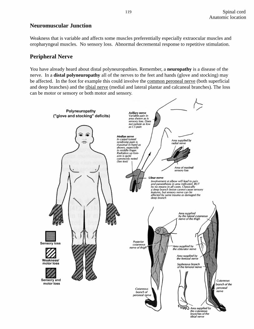
Weakness that is variable and affects some muscles preferentially especially extraocular muscles andoropharyngeal muscles. No sensory loss. Abnormal decremental response to repetitive stimulation.
Peripheral Nerve
You have already heard about distal polyneuropathies. Remember, a neuropathy is a disease of thenerve. In a distal polyneuropathy all of the nerves to the feet and hands (glove and stocking) maybe affected. In the foot for example this could involve the common peroneal nerve (both superficialand deep branches) and the tibial nerve (medial and lateral plantar and calcaneal branches). The losscan be motor or sensory or both motor and sensory.
Anatomic location
Anatomic location

120Spinal cord
There are also diseases of specific nerves either from compression or vascular disease (usuallyvasculitis or small infarctions associated with diabetes). Common nerves to be compressed are themedian at the wrist (carpal tunnel), ulnar at the elbow, peroneal at the fibular head, lateral cutaneousnerve of the thigh at the inguinal ligament. Diabetes often is associated with femoral nerve or cranialnerve lesions. When multiple nerves are affected the term mononeuropathy multiplex is used.
Anatomic location

121 Spinal cord
Radiculopathy
Pain, sensory, and motor loss. Referable to a dermatome and weakness in muscles innervated by thesame root. Lower motor neuron.
Anatomic location

122Spinal cord
Spinal Cord
Central cord e.g. syringomyelia: bilateralsensory with a cape distribution. Uppermotor neuron. Lower motor neuron at thelevel of the lesion. Bowel bladderfunction may be involved.
Transverse lesion: bilateral sensory andmotor loss with a level corresponding to thelesion. Bowel and bladder dysfunction.
Anatomic location

123 Spinal cordCase histories
CASE PRESENTATION
This 6 year old boy was brought to your office by his parents who were complaining that the boy had hadprogressive difficulty walking, climbing stairs and appeared “clumsy”. The child’s teacher also felt thatthe boy was always behind his peers in any physical activity. Academically he did well at school. Thechild was a product of normal pregnancy and normal delivery. His early developmental milestones werenormal. He was able to walk independently at the age of 14 months.
The family history was noncontributory.
General physical examination was unremarkable except for slightly exaggerated lumbar lordosis (forwardcurvature).
Neurological examination showed normal mental status and cranial nerves. Motor examination showedmarked enlargement of both calves. There was evidence of contractures in his Achilles tendons. He hadprominent hyperlordosis of his lumbar spine. Muscle tone was slightly decreased. He had mild to moderateproximal weakness, especially in his legs but also to some extent in his arms. The boy had markeddifficulty rising from the floor and did it by climbing up his thighs (positive Gowers’ sign). Sensation wasnormal. Tendon reflexes were decreased. Plantar reflexes were flexor. His gait was waddling.
QUESTIONS CASE HISTORY I
1) Where is the lesion/defect that might explain the findings on your clinical exam. Is the weaknesscaused by involvement of the corticospinal tract, anterior horn cells, roots, peripheral nerves, muscle,neuromuscular junction?
CASE HISTORY I

124Spinal cordCase histories
2) What could be the possible etiology?
3) What diagnostic tests would you order and why?
125 Spinal cord
A 25 year old woman came to your office complaining of intermittent double vision for the last threeweeks. She also has complained of fatigue. She has felt best during the early morning hours, but later,during the course of the day, she gradually develops double vision and diffuse weakness. Her boy-friendhas observed that her right eyelid has been drooping frequently. She used to play competitive basketballwhile in college, but now she has been short of breath after climbing only 2 flights of stairs. Also, on afew occasions, she choked on food and her friends noted that her speech was slurred or thick.
On examination her mental status was normal. She had marked ptosis on the right side. Eye movementexamination showed decreased movement in all directions in the right eye. There was also slightlydecreased abduction in the left eye. On repetitive blinking she developed ptosis of the left eye lid and herright sided ptosis got much worse. Motor examination showed normal muscle bulk and tone. Muscletesting revealed that she was initially strong, but rapidly became “tired” or weak with repeated effort. Shewas unable to hold her arms abducted at 90 degrees for more than 30 seconds. All sensory modalities,reflexes, coordination and gait examination were normal. Plantar reflexes were flexor.
QUESTIONS CASE HISTORY II
1) Where is the lesion and why? Is it in the corticospinal tract, anterior horn cells, peripheral nerves,neuromuscular junction or muscle?
CASE HISTORY II
Case histories

126Spinal cordCase histories
2) What is the possible etiology?
3) What diagnostic tests would you order and why? What results would you expect?

127 Spinal cord
This 20 year old college student came to the emergency room complaining of tingling in her feet andfingers. She appeared anxious. Neurological examination showed no abnormality and she was dischargedto home with a diagnosis of anxiety and hyperventilation. However, she returned to the emergency roomthe next day complaining of fatigue, weakness and shortness of breath. On specific questioning the patientadmitted to having “flu like” illness 10 days before.
Her examination at this time showed mild diffuse weakness, decreased muscle tone and absent tendonreflexes. Plantar reflexes were flexor. Sensation to pain was slightly decreased in the feet. There was nosensory level. A sensory level is a region of the body below which a sensation(s) is lost and above whichsensation is normal (for a lesion of the spinothalamic tract at T1, the level is T3). Her respiration rate was26 (normal adult rate is 10-15). Mental status and cranial nerve examination was normal. She wasadmitted to the hospital for observation. Over the next two days her weakness dramatically increased andshe developed respiratory failure and had to be intubated and placed on a ventilator.
During the first week of her illness she had frequent fluctuations of the heart rate and blood pressure.
QUESTIONS CASE HISTORY III
1) Where is the lesion and why? Is it in the brain, spinal cord, nerve roots, peripheral nerves, neuromuscularjunction or muscle ?
CASE HISTORY III
Case histories

128Spinal cordCase histories
2) What are the possible etiologies?
3) What diagnostic tests would you order and why? What results would you expect?

129 Spinal cord
A 45 year old delivery man comes to see you complaining of low back pain that has beenintermittent for the past 6 months. The pain is in the middle of the lower back and usually radiatesinto the left buttock. The pain is made worse by sneezing, coughing, or when he hits a pot holewhile driving. In some cases these maneuvers cause the pain to radiate down the back of his left leginto the bottom lateral aspect of his foot. Over the past 6 weeks, he has noted that it is difficult forhim to stand on his tip toes and that this is primarily because of weakness in his left foot.
On exam he has a normal neurological exam except that his left ankle jerk is absent and he hasweakness of his left gastrocnemius. There is abnormal sensation over the lateral aspect of the leftfoot. He cannot stand on his toes of his left foot. When you have him lying down, you cannotelevate his left leg above 35 degrees because of shooting pain into his left buttock and down theback of his left leg.
QUESTIONS CASE HISTORY IV
1) Where is the lesion?
2) What would an EMG/NCV study show?
CASE HISTORY IV
Case histories

130Spinal cordCase histories
3) What diagnostic testing would you order?
4) How should this patient be treated and what is his prognosis?

131 Spinal cord
A 65 year old man presents with a six month history of progressive fatigue, weakness and legcramps. On a few occasions he choked on food. His wife noted diffuse twitching of muscles on his chestand upper back. Two months ago he developed a foot drop in his left leg. He has not complained of anysensory symptoms. There has been no cognitive decline. He has no difficulty with bowel or bladderfunction. His family history is noncontributory.
Examination showed that the patient had normal mental status. Motor examination showed severe,bilateral diffuse muscle wasting in both upper and lower extremities. The most atrophied were the deltoid,triceps, biceps, hand muscles and quadriceps on either side and the left anterior tibialis. There wereprominent fasciculations in all muscle groups. The muscle tone was increased, (spastic) in both upperand lower extremities. There was diffuse weakness in all 4 extremities with complete left foot drop. Neckextensors were profoundly weak so that the patient was barely able to keep his head up.
The tendon reflexes were hyperactive in all four extremities. The plantar reflexes were extensor (Babinskisign).
Sensory examination and coordination were normal.
His gait was characterized by decreased arm swing and limping of the left leg.
QUESTIONS CASE HISTORY V
1) Where is the lesion and why? Does it involve the corticospinal tracts, anterior horn cells, nerve roots,peripheral nerves, neuromuscular junction or muscle?
CASE HISTORY V
Case histories

132Spinal cordCase histories
2) What are the possible etiologies and why?
3) What diagnostic tests would you order and why? What results would you expect?

133 Spinal cord
CASE HISTORY VI
A healthy 25 year old woman is brought to the emergency room after being stabbed in the neck.
Examination shows:
1) left ptosis and meiosis (small pupil)2) weakness of the left upper and lower extremities3) absent left biceps stretch reflex4) other left sided muscle stretch reflexes diminished5) loss of left finger and toe joint position sense6) loss of left finger and toe vibratory sensation7) loss of pain and temperature sensation below C7 on right
1) Where is the location of the lesion?
2) Explain why each of the findings are present.
1.
2.
3.
4.
5.
6.
7.
Case histories

134Spinal cord
3) Why might atrophy of the upper arm develop over time?