Water Resources Center Annual Technical Report FY 2004 · Water Resources Center Annual Technical...
59
Water Resources Center Annual Technical Report FY 2004 Introduction The Minnesota WRRI program is a component of the University of Minnesotas Water Resources Center (WRC). The WRC is a collaborative enterprise involving several colleges across the University, including the College of Natural Resources (CNR), the College of Agriculture, Food, and Environmental Sciences (COAFES), the Minnesota Extension Service (MES), and the University of Minnesota Graduate School. The WRC reports to the Dean of CNR. In addition to its research and outreach programs, the WRC is also home to the Water Resources Sciences graduate major. The WRC has two co-directors, Professor Deborah Swackhamer and Professor James Anderson, who share the activities and responsibilities of administering its programs. The WRC funds 3-4 research projects each year, and the summaries of the current projects are found in the rest of this report. Research Program
Transcript of Water Resources Center Annual Technical Report FY 2004 · Water Resources Center Annual Technical...
Water Resources Center Annual Technical Report
FY 2004
IntroductionThe Minnesota WRRI program is a component of the University of Minnesotas Water Resources Center(WRC). The WRC is a collaborative enterprise involving several colleges across the University, includingthe College of Natural Resources (CNR), the College of Agriculture, Food, and Environmental Sciences(COAFES), the Minnesota Extension Service (MES), and the University of Minnesota Graduate School.The WRC reports to the Dean of CNR. In addition to its research and outreach programs, the WRC is alsohome to the Water Resources Sciences graduate major. The WRC has two co-directors, Professor DeborahSwackhamer and Professor James Anderson, who share the activities and responsibilities of administeringits programs.
The WRC funds 3-4 research projects each year, and the summaries of the current projects are found in therest of this report.
Research Program
Effects of Riparian Forest Harvest on Instream Habitat and Fishand Invertebrate Communities
Basic Information
Title: Effects of Riparian Forest Harvest on Instream Habitat and Fish andInvertebrate Communities
Project Number: 2002MN2B
Start Date: 3/1/2002
End Date: 5/31/2005
Funding Source: 104B
Congressional District: 7 and 8
Research Category: Not Applicable
Focus Category: Water Quality, Management and Planning, None
Descriptors:
Principal Investigators: Raymond Newman, Jim Perry, Bruce Vondracek
Publication1. N. Schlesser, D. Atuke, R. Newman and B. Vondracek. 2004. Effects of riparian forest harvest on
habitat and fish assemblages in Northern Minnesota. 65th Annual Midwest Fish and WildlifeConference, 14 December 2004, Indianapolis, IN.
Effects of Riparian Forest Harvest on Instream Habitat, and Fish and Invertebrate Communities Principal Investigators R. M. Newman and J.A. Perry, Department of Fisheries, Wildlife and Conservation Biology, University of Minnesota and Bruce Vondracek, USGS, Minnesota Cooperative Fish and Wildlife Research Unit Research Assistants D.M. Atuke, Department of Fisheries, Wildlife and Conservation Biology Start date: March 1, 2004 End date: February 28, 2005 Executive summary Stream riparian zones are critical to the health of stream fish and invertebrate communities. Forest harvest within the riparian zone may thus impact stream fish and macroinvertebrate communities and the determination of the level of acceptable harvest within the riparian zone is important to balance forestry needs with stream biotic integrity. This is an ongoing manipulative experiment focused on determining the effects of no, low and high levels of riparian harvest on stream habitat and fish and invertebrate communities. This report provides a summary of the findings of the first year post-harvest data collection, conducted in summer 2004. Total number of fish species sampled was similar for 2003 and 2004. Although the total number of individuals was higher in 2004, this is a reflection of large increases in a few streams rather than a general trend. Index of Biological Integrity (IBI) scores were comparable and similar in 2003 and 2004. Macroinvertebrate community indices indicate within-site and between-site variability but none were significantly different (p>0.05). The qualitative habitat evaluation index (QHEI) scores exhibited variability between reaches and between treatments and none were significantly different (p>0.05). Dissolved oxygen and pH exhibited similar trends in both pre- and first year post-harvest data. In contrast nitrate, alkalinity and conductivity showed considerable variability in 2004 in comparison to 2003 at all sites. These year-to-year differences between sites and between treatments indicate the need to continue monitoring for longer time to define the effects of riparian forest harvest. Second year post-harvest sampling will occur in summer 2005. Introduction Forest products are an important natural resource in the upper Midwest. In Minnesota, timber harvest has been increasing and will continue to increase in the near future (Anonymous 2001). Timber harvest activities have the potential to degrade water quality and aquatic resources and for this reason, best management practices (BMPs) or site-level forest management guidelines have been adopted to protect riparian and aquatic resources in Minnesota (MFRC 1999, Anonymous 2001). Although these BMPs are based on the best available scientific information, and implementation monitoring is being conducted (Anonymous 2001), they have not been evaluated for effectiveness at protecting aquatic resources. Most research on the effects of forest harvest on streams and the effectiveness of forest harvest BMPs has been conducted in more mountainous regions such as
Tasmania (Davies and Nelson 1994), the Sierra Nevada, the Pacific Northwest and Appalachia (e.g., Castelle and Johnson 2000). Results from these areas may not be directly applicable to the midwest (Perry et al. 1992). Riparian zones provide many protective services to streams (Castelle and Johnson 2000). Determination of the necessary width of riparian buffers (e.g., Castelle and Johnson 2000) or the permissible level of harvest within a buffer is essential to adequately protect stream resources without removing a large portion of the basin from harvest. Most studies on the effectiveness of riparian buffers at protecting streams from upslope harvest have focused on the width of the buffer and have not considered harvest within the buffer zone (e.g., Castelle and Johnson 2000). Current Minnesota BMPs allow varying degrees of harvest within the riparian management zone (RMZ). Harvest within the RMZ may be used to promote regeneration of shade intolerant species. Thus. it is important to know the level of harvest that reduces it’s the effectiveness of the RMZ in maintaining stream quality. The objective of this project was to experimentally determine the effectiveness of various levels of riparian forest harvest on in-stream resources. We examine site-based effects associated with high, low and no riparian harvest (30m Riparian Management Zone, upland clearcuts) on aquatic habitat, macroinvertebrates and fish. Specifically, we evaluate effects on fish and invertebrate habitat (temperature, sediment composition, embeddedness, depth, width, cover, bank stability, canopy coverage, and woody debris, etc.), and benthic macroinvertebrate and fish communities. Methods The study sites range across northern Minnesota and are located in Beltrami, Carlton, Cook, Lake, and St. Louis counties. Eight pairs of treatment sites (streams) were located and harvest plots marked in 2003. Within each pair, a riparian control (no riparian harvest with upland clearcut) and one riparian management treatment (low or high residual basal area with upland clearcut) were established to compare the effects of different residual basal area levels (e.g., 4 high basal area and 4 low basal area replicates). We were also able to establish a non-harvested control (both upland and riparian zone not harvested) at seven of the eight plots (beaver activity preclude a non-harvested control plot at one site). Target riparian harvest treatments in winter 2004 were high residual (11.9 m2 basal area/ha remaining) or low residual (6.3 m2 basal area/ha). During harvesting, the target residual basal area was not always met and actual values varied by ± 0.9 m2 basal area/ha. All sites were sampled for habitat, fish and invertebrates in summer 2004 (post-harvest). This includes the one high residual basal area plot (Reservation River Tributary) that was not harvested in winter 2003-2004. Harvesting on this plot was completed in winter 2004-2005. Sampling in 2004 was done on the same reaches that were established in the no-harvest control, riparian control and riparian harvest plots in 2003. Within each plot, we sampled 100-meter reaches above the plot (upstream), within the plot (downstream most 100m) and below the plot (100m downstream of plot) – this design provides internal upstream controls and allows for assessment of downstream effects. Ideally, at a given site, we
would generally sample nine 100-m reaches; up-, within and below at the non-harvested control, the riparian control and the harvest treatment. Due to spatial and habitat constraints, up and below reaches were not always feasible for some sites. Temperature monitoring: Temperature loggers (Optic StowAway®, Onset Computer, Pocasset, MA) were placed in all reaches at each site in May 2003 and 2004. Temperature was recorded at 30 min intervals until removal in October or November. Water quality: Water quality was recorded in the within reaches at each site in spring and fall: in the field, conductivity, dissolved oxygen, and pH were recorded with a Quanta Water Quality Monitoring System® (Hydrolab Corporation); alkalinity (methyl orange; mg CaCO3) was determined by titration, and orthophosphate was determined by the PhosVer 3 (Ascorbic Acid) method with a Hach model DR/2000 spectrophotometer. Nitrate was determined spectrophotometrically (APHA 1989) on samples preserved in HCL with a Spectronic 1201 Dual Beam spectrophotometer in the laboratory. Instream habitat: In July, each 100-m reach was sampled for habitat characteristics following the methods of Merten (1999) that are modifications of methods given by Bailey et al. (1993). Variables measured include visual estimates of bank cover, channel stability, cover, woody debris, percent riffles, runs and pools, and aquatic plant coverage. Canopy coverage was determined in each reach with a spherical densiometer. Streambed sediment and substrate type and size (e.g., percent silt, sand, gravel, cobble, etc.) and percent embeddedness were characterized along 14 transects placed at regular intervals in each reach with a maximum total of 56 measurements per reach. Mean depth, velocity and discharge were measured at the fourteen transects within each reach. A qualitative habitat evaluation index (QHEI) was calculated from these data. Blow-down trees were also recorded in each reach. Benthic macroinvertebrates: Benthic macroinvertebrates were assessed in July following the family-level, composited, multi-habitat rapid bioassessment protocol (Barbour et al. 1999) in each of the upstream (internal control) and within-plot reaches for the control, riparian control and riparian harvest plots. Two composited samples of 20 kicks / net (each sample representing 50 m) were collected with a D-net in each 100-m reach. Samples were sorted and macroinvertebrates identified to family in the laboratory. Fish assemblages: Fish assemblages were sampled in August. Sampling was conducted in the up- (internal control), within- and downstream reaches at each treatment plot (including the control sites) with pulsed DC electrofishing (Wisconsin AbP-3 backpack shocker) following the protocol of Simonson and Lyons (1995). Fish were identified to species, measured (total length), weighed and returned to the stream. Cold-water Index of Biotic Integrity (IBI) values were calculated according to Mundahl and Simon (1998), and warm-water IBI values according to Karr et al. (1986) and Lyons (1992) to assess the environmental health of the stream fish communities. Species richness, species abundances and IBI scores (normalized to 100) were analyzed to determine the effects of harvest treatment.
Results to date Instream habitat: There was substantial variation in habitat characteristics between sites. Water temperatures varied among sites and overall, temperatures were below normal in August and above normal in September. However, the trout streams (Reservation River Tributary, West Split Rock River, East Branch Beaver River, and East Baptism River) maintained temperatures ≤ 19 ˚C throughout the summer (range from 12-19˚C), whereas other streams had summer maxima up to 25 ˚C. Conductivity and alkalinity ranged from 32 µS/cm and 20 mg CaCO3/L, respectively at the Cloquet River Tributary to 228 µS/cm and 127 mg CaCO3/L at Shotley Brook. Dissolved oxygen was above 7.5 mg/L at all sites and the pH was > 7.5 at all sites, except the Cloquet River Tributary where pH was 7.2. Orthophosphate ranged from 5 µg-P/L to 170 µg-P/L. However, during both seasons, most sites had less than 50 µg-P/L. Spring nitrate concentrations were comparable to 2003 and ranged from 0.36 mg-N/L to 0.97 mg-N/L. However, nitrate concentrations in fall 2004 were higher and ranged from 0.95 mg-N/L to 1.80 mg-N/L. Qualitative habitat evaluation index scores ranged from 45-78. There were no significant differences in QHEI between sites and treatments. However, in general the smaller intermittent flowing streams had lower QHEI scores compared to the larger perennial streams. Macroinvertebrate communities: Macroinvertebrate indices indicated both within-site and between-site variations. In the low RBA sites, mean number of individuals per net varied from a minimum of 298 to a maximum of 1598, species richness had a range of 6-21 families, percent EPT taxa ranged from 0-25%, while percent Chironomidae varied from 10-60%. In the high RBA sites, mean number of individuals per net varied from 356-2164, species richness had a range of 14-20 families, percent EPT taxa ranged from 16-44%, and percent Chironomidae had a range of 34-75%. Fish assemblages: Seventeen species of fish were found among over 2600 fish collected. Total number of individuals was higher in 2004, but reflected large increases in a few sites (Reservation River Tributary and East Branch Beaver River) rather than a general trend. We observed a reduction in the percentage of brook trout sampled in 2004 in West Split Rock River and East Branch Beaver River. Indices of biotic integrity were computed using the appropriate warm or coldwater IBIs. The IBI scores ranged from 15-95 (out of 120) in the trout streams and 22-45 (out of 100) in the mud minnow dominated streams. Ongoing work Second year post-harvest data will be collected in summer 2005. Habitat and invertebrate samples will be collected in July and fish will be sampled in August. Summary of findings Significant variability was observed in the number of individuals and species of fish and macroinvertebrates between 2003 and 2004, but there was no obvious trend that could be discerned in relation to harvest. QHEI and IBI scores between years were not
significantly different although year-to-year variation was observed. Water quality attributes such as temperature, conductivity, alkalinity, phosphorus and nitrates also indicate seasonal and annual variability. Further monitoring will occur in the next two years. Literature cited APHA. 1989. Standard methods for the examination of water and wastewater. 17th ed.
APHA-AWA-WPCF, Washington, DC. Anonymous. 2001. Minnesota's nonpoint source management program plan 2001.
Minnesota Pollution Control Agency, St. Paul, MN. Bailey, P.A., J.W. Enblom, S.K. Hanson, P.A. Renard, and K.S. Schmidt. 1993. Fish
Community Analysis in the Minnesota River Basin. Minnesota Pollution Control Agency, St. Paul.
Barbour, M.T., J. Gerritsen, B.D. Snyder, and J.B. Stribling. 1999. Rapid Bioassessment Protocols for Use in Streams and Wadeable Rivers: Periphyton, Benthic Macroinvertebrates and Fish, Second Edition. EPA 841-B-99-002. U.S. Environmental Protection Agency; Office of Water; Washington, D.C.
Castelle, A. J. and A. W. Johnson. 2000. Riparian vegetation effectiveness. National Council for Air and Stream Improvement, Technical Bulletin 799, Research Triangle Park, NC.
Davies, P. E., and M. Nelson. 1994. Relationships between riparian buffer widths and the effects of logging on stream habitat, invertebrate community composition and fish abundance. Australian Journal of Marine and Freshwater Research 45: 1289-1305.
Karr, J. R., K. D. Fausch, P. L. Angermeier, P. R. Yant, and I. J. Schlosser. 1986. Assessing biological integrity in running waters: a method and its rationale. Illinois Natural History Survey, Special Publication 5, 28 pp.
Lyons, J. 1992. Using the Index of Biotic Integrity (IBI) to measure environmental quality in warmwater streams of Wisconsin. USDA Forest Service, North Central Experiment Station, Gen. Tech. Rep. NC-149, St. Paul, MN.
Merten, E.C. 1999. Effects of forest harvest on coldwater stream fish and habitat in northern Minnesota. M.S. Thesis, University of Minnesota, St. Paul, MN. Also available online at: http://www.fw.umn.edu/Research/riparian/Merten.htm
Minnesota Forest Resources Council (MFRC). 1999. Sustaining Minnesota forest resources: voluntary site-level forest management guidelines for landowners, loggers and resource managers. Minnesota Forest Resources Council, St. Paul, MN.
Mundahl, N. D. and Simon, T.P. 1998. Development and application of an index of biotic integrity for coldwater streams of the upper midwestern United States. In Thomas P. Simon (ed.) Assessing the Sustainability and Biological Integrity of Water Resources Using Fish Communities. CRC Press, Boca Raton, FL, pp 383-415.
Perry, J., K. Brooks, W. Olsen, T. Geier, W. Johnson, R. Newman, L. Mizner and N. Troelstrup. 1992. Water quality and fisheries: a technical paper for a Generic Environmental Impact Statement on Timber Harvesting and Forest Management in Minnesota. Report to the Minnesota Environmental Quality Board by Jaakko Pöyry Consulting. 325 pgs.
Simonson, T. D., and J. Lyons. 1995. Comparison of catch per effort and removal procedures for sampling stream fish assemblages. North American Journal of Fisheries Management 15:419-425.
Related grants submitted or funded as a result of this project The Legislative Commission on Minnesota Resources funded the initial manipulation, travel, supplies and field assistance and the Minnesota Forest Resources Council provided $10,000 for some supplies and field assistance. Vondracek, B. and R.M. Newman. Effects of riparian forest harvest on instream habitat and fish and invertebrate communities. Minnesota Department of Natural Resources, 6/15/04-6/30/05. $37,500: funded travel, supplies, field assistance and one additional graduate student. A proposal for continuation of this project (2005-2007) has been recommended for funding ($97,700) by the Legislative Commission on Minnesota Resources. Description of student training provided by project: Directly funded: Name: Dickson Atuke Program: Fisheries and Aquatic Biology Track in Conservation Biology Degree being sought: PhD Funded by other sources (Fellowships, MN DNR and LCMR grants): Name: Nicholas Schlesser Program: Fisheries and Aquatic Biology Track in Conservation Biology Degree being sought: MS Name: Nathaniel Hemstad Program: Water Resources Science Degree being sought: PhD Name: Matt Ihnken Program: Department of Fisheries, Wildlife and Conservation Biology Degree being sought: BS
Photochemistry of Antibiotics and Estrogens in Surface Waters:Persistence and Potency
Basic Information
Title: Photochemistry of Antibiotics and Estrogens in Surface Waters: Persistenceand Potency
Project Number: 2003MN32G
Start Date: 9/1/2003
End Date: 8/31/2005
Funding Source: 104G
Congressional District: Minnesota District 5
Research Category: Not Applicable
Focus Category: Non Point Pollution, Surface Water, Ecology
Descriptors:
Principal Investigators: Kristopher McNeill, Deborah L. Swackhamer
Publication1. A.L. Boreen, W. A. Arnold, K. McNeill, Triplet-sensitized photodegradation of sulfa drugs
containing six-membered heterocyclic groups: Identification of an SO2 extrusion photoproduct,Environ. Sci. Technol. 2005, 39, 3630-3638.
2. J.J. Werner, K. McNeill, W.A. Arnold, Environmental photodegradation of mefenamic acid.Chemosphere 2005, 58, 1339-1346.
3. D.E. Latch, J.L. Packer, B.L. Stender, J. VanOverbeke, W.A. Arnold, K. McNeill, Aqueousphotochemistry of triclosan: Formation of 2,4-dichlorophenol, 2,8-dichlorodibenzo-p-dioxin andoligomerization products, Environ. Toxicol. Chem. 2005, 24 (3), 517-525.
4. A.L. Boreen, W.A. Arnold, K. McNeill, Photochemical fate of sulfa drugs in the aquaticenvironment: Sulfa drugs containing five-membered heterocyclic groups, Environ. Sci. Technol.,2004, 38, 3933-3940.
5. J.J. Werner, A.L. Boreen, B. Edhlund, K.H. Wammer, E. Matzen, K. McNeill, W.A. Arnold,Photochemical transformation of antibiotics in Minnesota waters, CURA Reporter 2005, 35(2), 1-5.
6. D. E. Latch, Environmental photochemistry: Studies on the degradation of pharmaceutical pollutantsand the microheterogeneous distribution of singlet oxygen. Ph.D., University of Minnesota,Minneapolis, MN, 2005, 256 pp.
7. K. McNeill and W.A. Arnold, Photo-generated Reactive Species and the Degradation of
Pharmaceutical Pollutants, Society for Environmental Toxicology and Chemistry (SETAC) NationalMeeting Symposium: Beyond Occurrence: Fate and Effects of Pharmaceutical and Other EmergingWastewater Contaminants in Aquatic Systems, November 13-17, 2005, Forthcoming.
8. K. McNeill, Photochemical approaches to environmental pharmaceutical pollutants, AmericanChemical Society (ACS) National Meeting Symposium: Strategies and Molecular Mechanisms ofContaminant Degradation Chemistry, Washington, D.C., August 28-Sept. 1, 2005, Forthcoming
9. K. McNeill, Photo-generated Reactive Species and the Degradation of Pharmaceutical Pollutants,Stanford University, Civil and Environmental Engineering Student Seminar Series, May 20, 2005,Forthcoming.
10. K. McNeill, Mechanistic Environmental Chemistry: Photo-generated Reactive Species and PollutantDegradation, Cornell University, Department of Chemistry, March 7, 2005.
11. K. McNeill, Mechanistic Environmental Chemistry: Photo-generated Reactive Species and PollutantDegradation, University of Rochester, Department of Chemistry, March 4, 2005.
12. K. McNeill, Mechanistic Environmental Chemistry: Photo-generated Reactive Species and PollutantDegradation, University of California, Berkeley, Department of Chemistry, February 25, 2005.
13. K. McNeill, Mechanistic Environmental Chemistry: Photo-generated Reactive Species and PollutantDegradation, Northwestern University, Department of Chemistry, February 18, 2005.
14. K. McNeill, Mechanistic Environmental Chemistry: Photo-generated Reactive Species and PollutantDegradation, 3M, November 12, 2004.
15. A.L. Boreen, W.A. Arnold, K. McNeill, Photochemical fate of sulfa drugs in the aquaticenvironment. Physics and Chemistry Colloquium, Concordia College, Moorhead, MN, November, 2004.
16. A.L. Boreen, W.A. Arnold, K. McNeill, Photochemical fate of sulfa drugs in the aquaticenvironment. Celebrating Women Chemists Luncheon, University of Minnesota, Minneapolis, MN,September, 2004.
17. K. McNeill. Photochemical fate of pharmaceutical pollutants. Gordon Research Conference,Environmental Sciences: Water, June 27 - July 2, 2004.
18. A.L. Boreen, W.A. Arnold, K. McNeill. Photochemical fate of pharmaceuticals in the environment:Sulfa drugs. Oral presentation. 9th Biennial MN Water Conference, Minneapolis, MN, March 23, 2004.
19. J.J. Werner, K. McNeill, W.A. Arnold, Speciation-dependent photochemistry of tetracyclineantibiotics: acid-base speciation and metal-binding effects. Oral Presentation. Midwest EnvironmentalChemistry Workshop, Madison, WI, October 16-17, 2004.
20. K. McNeill, W.A. Arnold. Contribution of photochemistry to the fate of pharmaceuticals andpersonal care products in surface waters. Oral Presentation. ENVR, Presented at the special symposium onEnvironmental aspects of pharmaceuticals and personal care products at the 228th ACS National Meeting,Philadelphia, PA, August 2004.
21. A.L. Boreen, W.A. Arnold, K. McNeill. Photochemical fate of sulfa drugs in the aquaticenvironment. Oral Presentation. ENVR, Presented at the special symposium on Environmental aspects ofpharmaceuticals and personal care products at the 228th ACS National Meeting, Philadelphia, PA, August2004.
22. K.H. Wammer, K. McNeill, T.M. LaPara, W.A. Arnold, D.L. Swackhamer. Changes in potency ofantibacterials in the environment due to photochemical transformations. Poster Presentation. ENVR,Presented at the special symposium on Environmental aspects of pharmaceuticals and personal careproducts at the 228th ACS National Meeting, Philadelphia, PA, August 2004.
23. J.J. Werner, K. McNeill, W.A. Arnold, Kinetics of the environmental photodegradation of mefenamicacid. Poster Presentation. ENVR, Presented at the special symposium on Environmental aspects ofpharmaceuticals and personal care products at the 228th ACS National Meeting, Philadelphia, PA, August
2004 24. W.A. Arnold, J.J. Werner, K. McNeill, Kinetics of the environmental photodegradation of mefenamic
acid. Poster presentation. Environmental Sciences: Water Gordon Research Conference, Plymouth, NH,June 27-July 2, 2004.
25. K.H. Wammer, D.L. Swackhamer, W.A. Arnold, K. McNeill, Photochemical transformations ofantibacterial compounds. Poster presentation. Environmental Sciences: Water Gordon ResearchConference, Plymouth, NH, June 27-July 2, 2004.
26. J.J. Werner, K. McNeill, W.A. Arnold, Photochemical fate of pharmaceuticals in the environment.Poster presentation. 9th Biennial MN Water Conference, Minneapolis, MN, March 23, 2004.
Photochemistry of Antibiotics and Estrogens in Surface Waters: Persistence and Potency Principal Investigators K. McNeill, Assistant Professor and PI, Department of Chemistry; W.A. Arnold, Assistant Professor and Co-PI, Department of Civil Engineering; D.L. Swackhamer, Professor and Co-PI, Division of Environmental Health Sciences, University of Minnesota. Postdoctoral Fellow K.H. Wammer, Departments of Chemistry, Civil Engineering, and Environmental Health Sciences, University of Minnesota. (Funded through a Dreyfus Environmental Chemistry Postdoctoral Fellowship) Research Assistants A.L. Boreen, B.L. Edhlund, and D.E. Latch, Department of Chemistry; J.J. Werner, Water Resources Sciences Program, University of Minnesota. Funding Source: USGS-WRRI 104G National Grants Competition Project Duration: 9/01/2003-8/31/2005 Report Duration: 3/1/2004-2/28/2005 Summary Antibiotics and estrogens are two classes of wastewater contaminants that have been detected in US surface waters. The potentially adverse effects of these pollutants on water quality are unknown, but will be determined, in part, by their persistence and the biological activity of both the parent compound as well as the degradates. Photolysis is one possible loss process, and the direct and indirect photolysis of five sulfa drug antibiotics, four nitrofuran antibiotics, four fluoroquinolones, and tetracyline has been investigated. The structure of the R-substituent on the sulfa drugs controls the reactivity; those containing six-membered substituents degrade through both direct photolysis and reaction with triplet dissolved organic matter. Both processes result in SO2 extrusion. The photochemical kinetic rate constants for the loss of tetracycline under natural sunlight are a function of its various environmentally-relevant aqueous chemical species, including acid-base equilibria and metal-binding. Direct photolysis has been found to be the major photochemical degradation pathway for the nitrofuran antibiotics, with the formation of a photostationary state between the syn and anti isomers occurring in the first several minutes of light exposure. All antibacterial compounds tested, three sulfa drugs and triclosan (an antimicrobial agent), photodegraded to products with no observable antibacterial activity.
Introduction Reports of pharmaceuticals and personal care products (PPCPs) in natural waters have recently appeared with increasing frequency.1-5 Two important subclasses of these emerging contaminants are particularly worrisome due to their potential to adversely affect surface waters: antibiotics and environmental estrogens. Estrogenic compounds have a demonstrated ability to interfere with the development of aquatic organisms,5, 6 while there is concern that the presence of antibiotics in natural waters will lead to an increase of antibiotic resistant bacteria.7, 8 These compounds are released into surface waters as a result of human use, through discharge of
treated and untreated wastewater. An additional, major source of antibiotics comes from their wide use in the production of food animals and in fish farming. 1-5 The magnitude of the effects and potential threat to water quality due to antibiotics and hormones is, in part, determined by the compounds’ persistence in aquatic systems. The principle goal of this proposed study is to understand one aspect of their persistence—their degradation by photochemical processes. Based upon our work9-15 and that of others,11, 16-22 we believe that photodegradation may be a major loss process for these compounds in sunlit waters. Thus, it is important to understand the photochemical processes that degrade these chemicals in surface waters, to identify intermediates and products that are formed, and to assess the biological activity of these products. Methods Direct and natural water photolysis experiments Photolysis experiments were performed outdoors under natural sunlight or indoors under medium pressure Hg-vapor lamps or a Suntest CPS+ solar simulator equipped with a Xe-arc lamp and a UV Special Glass filter to mimic the solar spectrum. Sample solutions were contained in quartz test tubes (OD = 13 mm, ID = 11 mm, V = 10 mL). For kinetic analyses approximately 0.5 mL samples were withdrawn from the quartz tubes at predetermined intervals and analyzed on an 1100 Series Hewlett Packard HPLC equipped with UV-absorbance detection and a computer driven data acquisition system. In experiments designed to probe for pH effects, various buffer solutions were employed to set the pH values. Solar quantum yields were calculated by comparing the rate constant for the disappearance of the PPCPs under either natural sunlight or the Suntest CPS+ solar simulator with the rate constant for the disappearance of a p-nitroanisole actinometer. For toxicity experiments, test tubes were sacrificed at pre-selected time intervals and saved for HPLC analysis of remaining antibiotic concentration and subsequent antibacterial activity testing. The wavelength dependence of the direct photolysis of the nitrofuran antibiotics was probed using a series of cut-off filter tubes (absorbing λ < 320 nm, 280 nm, and 220 nm). Quartz test tubes containing the photolysis solutions were placed inside the filter tubes during photolysis. Natural water photolysis experiments were performed in 0.2 µm filtered Lake Josephine (LJW) water or Lake Superior (LSW) water. To determine which pathways were responsible for the photodegradation, various quenchers were added to or removed from the water samples (sodium azide or DABCO for 1O2, isopropanol for radicals, oxygen and isoprene for triplet DOM) and the substrate was also photolyzed in DI water in a separate tube. Speciation dependent behavior of tetracycline Association constants which determine the speciation of calcium- and magnesium-tetracycline complex formation were measured by pH titrations (pH 3 to 11) performed at various constant metal concentrations and the collection of UV-vis spectral data. The first order rate constant for the loss of tetracycline under simulated sunlight (Suntest CPS+ photosimulator, Atlas) was observed at various pH, calcium, and magnesium concentrations. Kinetic experiments were performed as detailed above. The concentration-dependent initial rate of photochemical degradation was monitored for various initial tetracycline concentrations and extrapolated to
infinite dilution to determine the first-order rate constant for the loss of tetracycline in the absence of self-sensitization. Singlet oxygen Singlet oxygen reaction kinetics were measured in one of three ways, directly by laser flash photolysis (LFP), or indirectly by either steady-state photolysis (SSP) or thermal generation of 1O2. In both LFP and SSP experiments the substrate (typically at micromolar concentrations) and 100 µM perinaphthenone, a well-defined singlet oxygen sensitizer, were dissolved in aqueous buffer solutions. In the LFP experiments, a pulse of laser light excites the sensitizer, which then produces singlet oxygen after the excited-state sensitizer is quenched by dissolved molecular oxygen. A sensitive Ge-photodiode detector then monitors the phosphorescence emission from singlet oxygen. The rate of disappearance of the singlet oxygen phosphorescence signal is a measure of a substrate’s activity toward singlet oxygen. The resulting total quenching rate constant (ktot) is the sum of the chemical reaction and physical quenching rate constants. In SSP experiments, the samples were photolyzed continuously and small aliquots were removed for analysis by HPLC. In this case, the disappearance of the PPCP was monitored (as decreases in peak area), rather than the singlet oxygen signal. This allows for determination of the chemical reaction rate constant (krxn) for the PPCP with singlet oxygen. To avoid any competing photochemical reaction occurring in SSP, thermal generation of 1O2 was used. In these experiments, 1O2 was generated through the reaction of hydrogen peroxide (H2O2) and molybdate (MoO4
2-).23-25 H2O2 (1 M) was added to a buffered solution containing MoO42- (1
mM), a reference compound of known krxn (FFA; 100 µM), and substrate (100 µM). Aliquots of the reaction solutions were added to an aqueous solution of sodium azide (507 mM) at a series of time points to quench the reaction. Samples were then analyzed for both reference compound and substrate degradation via HPLC. Product identification Since large volumes of photolysate were required for product identification, photolyses were executed using a higher intensity light source (450 W medium pressure Hg-vapor lamp) which was completely immersed in the photolysis solution (100 µM substrate, 300 mL). After photolysis, the solution was concentrated to a total volume of 2 mL and the desired photoproduct was isolated using preparative HPLC. Sufficient amounts of product for analysis were obtained by combining the collected fraction from multiple injections of the photolysate on the preparative HPLC column. Following isolation, the product was identified using an array of analyses including mass spectrometry, infrared spectroscopy, and nuclear magnetic resonance (NMR). Mass spectral data were obtained for both the raw photolysate and the isolated products using a Bruker BioTOF ESI-TOF mass spectrometer. High resolution mass spectra were obtained using an internal standard of poly(ethylene glycol). Infrared absorbance spectra were acquired using a MIDAC Corporation M-Series FT-IR by placing a solution of the isolated photoproduct in methanol-d4 between two NaCl plates. The 1H-NMR and 13C-NMR spectra of isolated photoproducts were obtained on a Varian Inova 300 MHz spectrometer. A quantitative 1H-NMR spectrum of the same sample was acquired using an internal standard.
Biological activity The ability of the antibacterial compounds and their photolysis products to inhibit bacterial growth was tested using E. coli DH5α. The bacteria were maintained on agar plates and grown up overnight on Iso-Sensitest broth (ISB) (Oxoid, Inc.) prior to testing. One mL of antibacterial compound or photolysis product and 100 µL of E. coli were added to test tubes containing nine mL of ISB prepared in a pH 7 phosphate buffer (9.7 g KH2PO4 and 19.4 g Na2PO4 per liter deionized water). The solutions were incubated in the dark at 37 °C while being shaken. Bacterial growth was assessed after 8 hours by measuring optical density at 600 nm (OD600).
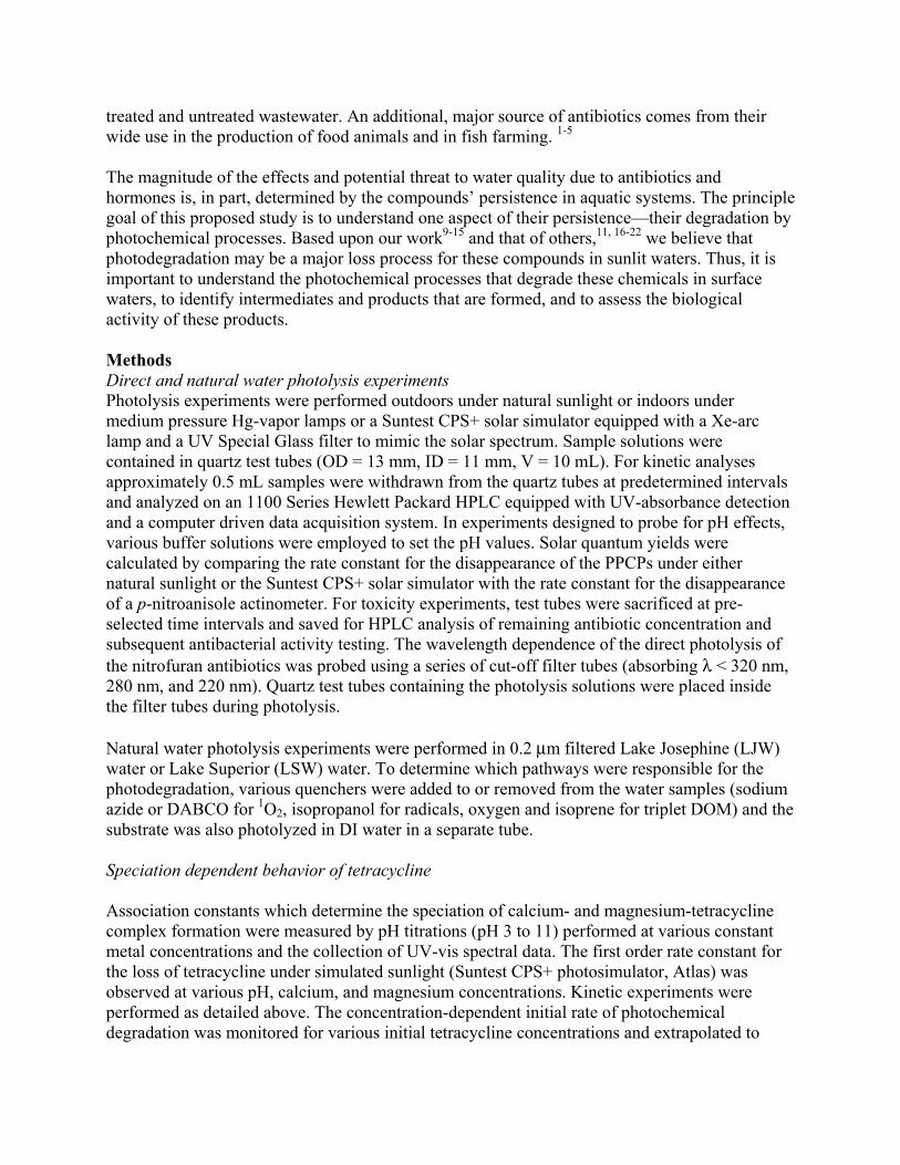
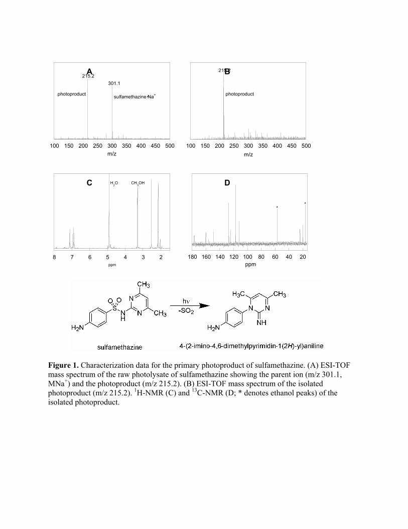
The antibacterial compounds and their photolysis products were also tested for their ability to inhibit bacterial respiration. The respiration assay used was based on the ability of the bacteria to reduce iodonitrotetrazolium chloride. E. coli (400µL) was added to 40 mL of ISB and incubated at 37 ºC. Once the OD600 of this solution had reached 0.4 (in the exponential phase of the growth curve), 1 mL aliquots were centrifuged at 19,000g for five minutes. The supernatant was decanted, and 0.5 mL of antibiotic or photolyzed antibiotic was added. The bacterial pellet was resuspended, and the tubes were then incubated in the dark at 37 °C while being shaken. After one hour of incubation (approximately one generation time), 0.5 mL of a 5 mM solution of the tetrazolium salt was added and the tubes were incubated for an additional hour. The tubes were then centrifuged, the supernatant decanted, and 1 mL of an organic solution (1:1 dimethylformamide: ethanol) was added to the bacterial pellet to extract the formazan. The pellet was resuspended, and the tubes were incubated in the dark at room temperature for one hour. After centrifuging, the absorbance of the supernatant was measured at 464 nm to quantify the amount of formazan formed. Results to date Photodegradation of the Sulfa drugs The photolysis rates of the sulfa drugs containing six-membered heterocyclic substituents (sulfachloropyridazine, sulfadiazine, sulfamerazine, and sulfamethazine) in Lake Josephine (DOC = 5.9 mg/L) water were enhanced by a factor of 1.4-2.6 relative to the photodegradation rates in DI H2O. The enhancement in the natural water has been attributed to reaction of the sulfa drugs with excited triplet dissolved organic matter (3DOM). Verification that the reaction is sensitized by 3DOM was provided by the characteristic enhancement of the degradation upon eliminating oxygen from the system and suppression of the degradation upon addition of isoprene, quenching of triplet-excited state perinaphthenone during LFP experiments, and the lack of reaction between the sulfa drugs and 1O2 as measured using thermal generation methods. The natural water photodegradation of sulfadimethoxine matched the degradation in DI H2O, and the degradation was thus attributed solely to direct photolysis. The direct photolysis of sulfadimethoxine is pH dependent, and is explained by differing reactivity of the protonation states. The remaining sulfa drugs’ direct photolysis and triplet-sensitized degradations are not pH dependent over the pH range 6-9. The primary product of both direct photolysis and triplet-sensitized degradation was identified as an SO2 extrusion product (Figure 1). The yield of this product from sulfamethazine was found to be 64%.
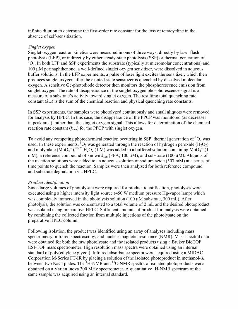
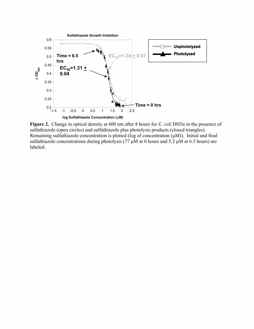
Tetracyline The pseudo-first-order rate constant for the photochemical loss of tetracycline was observed, under environmentally-relevant conditions, to be dependent on pH and both calcium and magnesium concentration. For each of the four acidic protons in tetracycline, deprotonation leads to both increased solar action spectrum and increased rate constant for photochemical degradation. The binding of tetracycline species to either calcium or magnesium leads to a further increase in the action spectrum for solar absorption. In the laboratory, the high tetracycline concentrations (1 to 10 µM) led to significant self-sensitization, especially at higher pH values. For example, at a pH of 7.5, the observed pseudo-first-order rate constant appeared to double when increasing the initial tetracycline concentration from 1 to 15 µM, with a linear dependence on initial tetracycline within the concentration range. As an example of the rapid kinetics, the half-life of tetracycline extrapolated to infinite dilution at pH 7.5 was 9.9 minutes, where the experimental light intensity was approximately the same as that of a clear summer day, noon, 45° latitude. Photochemical behavior of the nitrofuran antibiotics The photodegradation of the nitrofuran antibiotics (Table 1) occurs in two steps; the first involves formation of a photostationary state within the first several minutes of exposure to irradiation and the second is the subsequent direct photodegradation. The photostationary state forms in response to the reversible photo-induced isomerization that occurs at the carbon-nitrogen double bond of the nitrofurans. The photoequilibrium constant for this photostationary state has been calculated to be 0.95 for furazolidone and 0.63 for nitrofurantoin. The photoequilibrium constant for furazolidone was found to be irradiation wavelength dependent. When the sample was irradiated with wavelengths longer than 320 nm, the photoequilibrium lies towards a higher concentration of the photo-induced isomer. The photoequilibrium constant for nitrofurantoin (pKa 7.7) was determined to be pH dependent, and is larger in solutions buffered to a pH below the pKa and lower in solutions at a pH greater than the pKa. The direct photodegradation of the nitrofurans has been investigated under artificial sunlight, and the quantum yields of direct degradation and environmentally relevant half-lives for furazolidone and nitrofurantoin have been determined (Table 2). The products of the photodegradation have been studied through the use of HPLC and comparison with authentic standards of suspected products. The production of nitrofuraldehyde has been ruled out based on HPLC retention time and the rate at which it undergoes direct photolysis. Biological Activity Comparing the growth of E. coli DH5α in the presence of unphotolyzed sulfathiazole (Figure 2, open circles) versus in the presence of partially photolyzed sulfathiazole (Figure 2, closed triangles) revealed little difference in the inhibition of bacterial growth as a function of sulfathiazole concentration. Any photolysis products generated at a given point along the curve and present in the samples in the photolyzed series in addition to the sulfathiazole would be responsible for deviations from the unphotolyzed sulfathiazole series. The concentration at which sulfathiazole has reached half of its maximum effective concentration (EC50 values) for these two curves were statistically similar. This suggests that the products of the photolysis do not retain any significant ability to inhibit bacterial growth; that is, the antibacterial activity of the

photolyzed solution only comes from the unreacted sulfathiazole. Similar results were observed for sulfamethoxazole, sulfachloropyridazine, and triclosan. Ongoing work Ongoing work on tetracyline will first involve further in-depth data analysis using mathematical software to determine the values and certainty of the metal-binding constants of interest. Once the aqueous speciation is known explicitly, photolysis experiments will be performed under additional conditions to elucidate species-dependent quantum yields for the loss of tetracycline under natural sunlight. The goal is to determine the physical constants necessary to predict the pseudo-first-order photochemical loss rate constant of tetracycline in any given system with knowledge of pH, calcium, magnesium, and sunlight distribution. Ongoing investigation of the nitrofuran antibiotics includes examining reaction with singlet oxygen and additional product identification using mass spectrometry, preparative LC, and NMR. Finally, work is being conducted to characterize photodegradation of the fluoroquinolone antibiotics in natural waters including analysis of the antibacterial activity of the photolysis products. Summary of findings The photodegradation mechanism for the sulfa drugs containing six-membered substituents involves both direct photolysis and reaction with triplet dissolved organic matter generating an SO2 extrusion photoproduct. Comparison of these results with those obtained for the sulfa drugs containing five-membered substituents reveals that minor structural changes can give rise to disparate environmental loss mechanisms. The photochemical kinetic constants for the loss of tetracycline under natural sunlight are a function of its various environmentally-relevant aqueous chemical species, including acid-base and metal-bound forms. Direct photolysis has been found to be the major photochemical degradation pathway for the nitrofuran antibiotics, with the formation of a photostationary state between the syn and anti isomers occurring in the first several minutes of light exposure. All antibacterial compounds tested, three sulfa drugs and triclosan, photodegraded to products with no observable antibacterial activity. References 1. Daughton, C.G. and T.A. Ternes, Pharmaceuticals and personal care products in the
environment: agents of subtle change? Environ. Health Perspect. Suppl., 1999. 107(6): p. 907-938.
2. Halling-Sorensen, B., et al., Occurrence, fate and effects of pharmaceutical substances in the environment - a review. Chemosphere, 1997. 36(2): p. 357-393.
3. Kolpin, D.W., et al., Pharmaceuticals, Hormones, and Other Organic Wastewater Contaminants in U.S. Streams, 1999-2000: A National Reconnaissance. Environ. Sci. Technol., 2002. 36(6): p. 1202-1211.
4. Jorgensen, S.E. and B. Halling-Sorensen, Drugs in the environment. Chemosphere, 2000. 40(7): p. 691-699.
5. Ternes, T., Pharmaceuticals and metabolites as contaminants of the aquatic environment, in Pharmaceuticals and Personal Care Products in the Environment, C.G. Daughton and T.L. Jones-Lepp, Editors. 2001, American Chemical Society: Washington, D. C. p. 39-54.
6. Stuer-Lauridsen, F., et al., Environmental risk assessment of human pharmaceuticals in Denmark after normal therapeutic use. Chemosphere, 2000. 40(7): p. 783-793.
7. Kumpel, T., R. Alexy, and K. Kummerer, What do we know about antibiotics in the environment? Pharmaceuticals in the Environment, 2001: p. 67-76.
8. Hirsch, R., et al., Occurrence of antibiotics in the aquatic environment. Sci. Total Environ., 1999. 225(1,2): p. 109-118.
9. Latch, D.E., et al., Photochemical Conversion of Triclosan to 2,8-Dichlorodibenzo-p-dioxin in aqueous solution. J. Photochem. Photobiol. A, 2003. 158(1): p. 63-66.
10. Latch, D.E., et al., Photochemical Fate of Pharmaceuticals in the Environment: Cimetidine and Ranitidine. Environ. Sci. Technol., 2003. 37: p. 3342-3350.
11. Boreen, A., W.A. Arnold, and K. McNeill, Photodegradation of pharmaceuticals in the aquatic environment: A review. Aquatic Sciences, 2003. 65: p. 317-338.
12. Packer, J.L., et al., Photochemical fate of pharmaceuticals in the environment: naproxen, diclofenac, clofibric acid, and ibuprofen. Aquatic Sciences, 2003. 65: p. 1-10.
13. Boreen, A.L., W.A. Arnold, and K. McNeill, Photochemical Fate of Sulfa Drugs in the Aquatic Environment: Sulfa Drugs Containing Five-Membered Heterocyclic Groups. Environ. Sci. Technol., 2004. 38(14): p. 3933-3940.
14. Werner, J.J., K. McNeill, and W.A. Arnold, Environmental photodegradation of mefenamic acid. Chemosphere, 2005. 58(10): p. 1339-1346.
15. Boreen, A.L., W.A. Arnold, and K. McNeill, Triplet-Sensitized Photodegradation of Sulfa Drugs Containing Six-Membered Heterocyclic Groups: Identification of an SO2 Extrusion Photoproduct. Environ. Sci. Technol., 2005. 39: p. 3630-3638.
16. Buser, H.-R., T. Poiger, and M.D. Mueller, Occurrence and Fate of the Pharmaceutical Drug Diclofenac in Surface Waters: Rapid Photodegradation in a Lake. Environ. Sci. Technol., 1998. 32(22): p. 3449-3456.
17. Lindström, A., et al., Occurrence and Environmental Behavior of the Bactericide Triclosan and Its Methyl Derivative in Surface Waters and in Wastewater. Environ. Sci. Technol., 2002. 36(11): p. 2322-2329.
18. Moore, D.E. and W. Zhou, Photodegradation of sulfamethoxazole: a chemical system capable of monitoring seasonal changes in UVB intensity. Photochem. Photobiol., 1994. 59(5): p. 497-502.
19. Oka, H., et al., Photodecomposition products of tetracycline in aqueous solution. J. Agric. Food Chem., 1989. 37(1): p. 226-31.
20. Poiger, T., H.-R. Buser, and M.D. Muller, Photodegradation of the pharmaceutical drug diclofenac in a lake: pathway, field measurements, and mathematical modeling. Environmental Toxicology and Chemistry, 2001. 20(2): p. 256-263.
21. Singer, H., et al., Triclosan: Occurrence and Fate of a Widely Used Biocide in the Aquatic Environment: Field Measurements in Wastewater Treatment Plants, Surface Waters, and Lake Sediments. Environ. Sci. Technol., 2002. 36(23): p. 4998-5004.
22. Tixier, C., et al., Phototransformation of Triclosan in Surface Waters: A Relevant Elimination Process for This Widely Used Biocide - Laboratory Studies, Field Measurements, and Modeling. Environ. Sci. Technol., 2002. 36(16): p. 3482-3489.
23. Aubry, J.M., Search for singlet oxygen in the decomposition of hydrogen peroxide by mineral compounds in aqueous solutions. J. Am. Chem. Soc., 1985. 107(21): p. 5844-9.
24. Aubry, J.M. and B. Cazin, Chemical sources of singlet oxygen. 2. Quantitative generation of singlet oxygen from hydrogen peroxide disproportionation catalyzed by molybdate ions. Inorg. Chem., 1988. 27(12): p. 2013-14.
25. Boehme, K. and H.D. Brauer, Generation of singlet oxygen from hydrogen peroxide disproportionation catalyzed by molybdate ions. Inorg. Chem., 1992. 31(16): p. 3468-71.
Description of student training provided by project: Name: Anne L. Boreen Program: Department of Chemistry, University of Minnesota Degree being sought: Ph.D. Name: Betsy L. Edhlund Program: Department of Chemistry, University of Minnesota Degree being sought: Ph.D. Name: Douglas E. Latch Program: Department of Chemistry, University of Minnesota Degree earned: Ph.D. (2005) Name: Jeffrey J. Werner Program: Water Resources Science, University of Minnesota Degree earned: M.S. (2004) Degree being sought: Ph.D.
100 150 200 250 300 350 400 450 500m/z
215.2301.1
photoproduct sulfamethazine Na+.
100 150 200 250 300 350 400 450 500
m/z
215.2
photoproduct
2345678ppm
H2O CH
3OH
20406080100120140160180
ppm
**
Figure 1. Characterization data for the primary photoproduct of sulfamethazine. (A) ESI-TOF mass spectrum of the raw photolysate of sulfamethazine showing the parent ion (m/z 301.1, MNa+) and the photoproduct (m/z 215.2). (B) ESI-TOF mass spectrum of the isolated photoproduct (m/z 215.2). 1H-NMR (C) and 13C-NMR (D; * denotes ethanol peaks) of the isolated photoproduct.
C D
A B
Unphotolyzed
Photolyzed
Unphotolyzed
Photolyzed
0.2
0.25
0.3
0.35
0.4
0.45
0.5
0.55
0.6
-1.5 -1 -0.5 0 0.5 1 1.5 2 2.5
Sulfathiazole Growth Inhibition∆
OD
600
log Sulfathiazole Concentration (µM)
EC50=1.31 +0.04
EC50=1.30 + 0.07
Time = 0 hrs
Time = 6.5 hrs
Figure 2. Change in optical density at 600 nm after 8 hours for E. coli DH5α in the presence of sulfathiazole (open circles) and sulfathiazole plus photolysis products (closed triangles). Remaining sulfathiazole concentration is plotted (log of concentration (µM)). Initial and final sulfathiazole concentrations during photolysis (77 µM at 0 hours and 5.2 µM at 6.5 hours) are labeled.
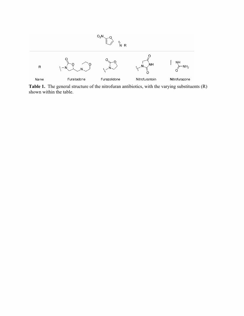
Table 1. The general structure of the nitrofuran antibiotics, with the varying substituents (R) shown within the table.

Nitrofuran Φ Mid-summer t1/2 Mid-winter t1/2Furazolidone 0.003 ± 0.001 7 ± 5 min 27 ± 23 min Nitrofurantoin 0.0014 ± 0.0002 13 ± 4 min 52 ± 15 min
Table 2. Quantum yields and environmentally relevant half-lives for two nitrofuran antibiotics in DI H2O adjusted to pH 7.6. Half-lives are calculated based on noon, 45° latitude, mid-summer (August 6) or mid-winter (February 5) solar radiation.
Phyto-enhanced Remediation: A Wetland Treatment System forSurface Water Protection
Basic Information
Title: Phyto-enhanced Remediation: A Wetland Treatment System for Surface Water Protection
Project Number: 2004MN48B
Start Date: 3/1/2004
End Date: 2/28/2006
Funding Source: 104B
Congressional District: 5
Research Category: Not Applicable
Focus Category: Wetlands, Non Point Pollution, Treatment
Descriptors:
Principal Investigators: William Alan Arnold, Timothy Michael LaPara
Publication1. DeJournett, T., Fritsch, J., McNeil, K., and Arnold, B., 2005. Preparation of
14C-cis-1,2-dichloroethylene from 14C-trichloroethylene using a cobalt porphyrin catalyst. Journalof Labelled Compounds and Radiopharmeceuticals, 48(5): p. 353-357.
2. DeJournett, T.D; Arnold, W.A.; LaPara, T.M., 2005. The Effect of Vegetation on MethanotrophicBacterial Populations in a Constructed Wetland, Applied Soil Ecology, in review.
3. Characterization of methanotrophic bacterial populations in the rhizosphere of emergent wetlandplants. Poster N-096 American Society for Microbiology 104th Annual Meeting, New Orleans, LA,May 23-27 2004.
4. Stimulation of methanotrophic bacteria in a wetland treatment system. Environmental EngineeringSeminar, University of Missouri-Rolla November 7, 2003.
5. Phyto-Enhanced Remediation: A constructed wetland for removal of chlorinated ethylenes fromgroundwater Poster Presentation, Frontiers in Assessment Methods for the Envrironment (FAME),Minneapolis, MN August 10-13 2003.
6. Phyto-Enhanced Remediation: A constructed wetland for removal of chlorinated ethylenes fromgroundwater Poster Presentation, CSWEA/AWMA Conference on the Environment, Bloomington,MN, November 20, 2003.
Phyto-enhanced Remediation: A wetland Treatment System for Surface Water Protection Principal investigators W.A. Arnold, Ph.D., Department of Civil Engineering; T.M. LaPara, Ph.D., Department of Civil Engineering, University of Minnesota. Research Assistants T.D. DeJournett, Department of Civil Engineering, University of Minnesota Start date: 3/01/2004 End date: 2/28/2006 Executive summary Halogenated solvents, such as dichloroethylene (DCE), present a challenging remediation problem due to their prevalence and persistence in the environment. In groundwater contamination scenarios where the source pools cannot be located/removed, there is great demand for a long-term cost effective alternative to treat the contaminant plume. Wetland treatment is an attractive alternative because of its passive nature and low operation/maintenance costs. A wetland treatment system was implemented as a remedial action to protect Lake Minnetonka from a DCE plume emanating from a former manufacturing facility in Mound, Minnesota. This work was initiated to address a lack of data regarding the role of wetland vegetation in the removal of DCE by the constructed wetland. Work conducted to date suggests that wetland vegetation did not affect the size or structure of methanotrophic bacterial communities in the field, and cometabolic oxidation of DCE by methanotrophs was not a significant fate mechanism in laboratory microcosm studies. In the case of cattails, transport from the subsurface to the atmosphere via plant tissues is the primary fate mechanism for DCE in laboratory microcosms. The transpiration stream concentration factor, the primary metric for vascular uptake of contaminants by plants, was significantly higher (~7-fold) for cattails than predicted by previously published models. This phenomenon may be attributed to volatilization/gas-phase diffusion of DCE through gas-filled voids (aerenchyma tissue) in wetland plants. Previously published models are based on terrestrial plants, such as hybrid poplar trees, which lack aerenchyma tissue. Cattails also prevented the accumulation of vinyl chloride, an anaerobic biodegradation product of DCE. Because DCE removal by cattails is strongly influenced by transpiration rate, it may be possible to adapt wetland management practices to enhance DCE removal or to moderate DCE efflux to the atmosphere if necessary. Introduction Halogenated solvents, such as chlorinated methanes, ethanes, and ethylenes, are among the most prevalent pollutants at contaminated sites on the National Priorities List as well as sites owned by the Department of Defense and Department of Energy. Contamination is also often observed at dry-cleaning and degreasing operations. Halogenated solvents pose an extremely difficult remediation problem. These compounds generally have low aqueous solubility and collect at impermeable layers forming pools of non-aqueous phase liquid (1). While several remediation techniques are currently available for the removal or degradation of chlorinated compounds at contaminated sites, these techniques are subject to significant technical
and economic limitations. Phytoremediation is a burgeoning technology that utilizes living plants to help remove contaminants from the environment. Phyto- and phyto-enhanced remediation are potentially low cost and aesthetically pleasing remediation alternatives. One example of phytoremediation is a wetland treatment system, in which wetland plants facilitate the removal of contaminants from water as it flows through the wetland. Wetland treatment systems are becoming widely used to treat municipal wastewater (2-5) as well as numerous other waste streams including landfill leachate and acid mine drainage (6,7). Wetlands offer a unique remediation environment, as shown by wastewater treatment applications that take advantage of the ability of the diverse microbial population supported by the wetland environment to degrade a variety of contaminants. The root zone, or rhizosphere, of wetland plants may play an important role in supporting essential, waste-degrading microbes. Wetland treatment systems also have great potential for removing chlorinated solvents from groundwater (8). Wetlands have been shown to support microbes, such as methanotrophic bacteria, capable of degrading chlorinated solvents (8-11). Wetland plants may also have the capability to take up and transpire/mineralize chlorinated solvents (12,13), although this has yet to be specifically demonstrated for most wetland plants. Root systems of wetland plants may also enhance the bacterial mineralization of chlorinated solvents in the rhizosphere through the excretion of root exudates and oxygen (14,15). The objective of this research is to determine the specific roles of the soil and plants and the impact of plant-microbial interactions in the removal of chlorinated ethylenes in a constructed wetland. Additionally, this study will elucidate the effect of wetland vegetation on the growth of methanotrophic bacteria in wetland sediment. Methods Field Mesocosms. Three field mesocosms (one unvegetated, two planted with a mixture of cattails, giant bur-reed, bottlebrush sedge, and bulrush) were observed from April-October (the growing season for Minnesota). Porewater samples were collected from the mesocosms via stainless steel microwells embedded in the sediment at 13-cm intervals. Samples were drawn from the microwells via a glass gas-tight syringe and Teflon-lined tubing. Porewater samples were analyzed for chlorinated ethenes and methane via gas chromatography. Dissolved oxygen, sulfate, and sulfide were measured using a handheld colorimetric test kit (CHEMetrics Company Vacu-VialsTM).
The effect of the root systems of wetland plants on methanotrophic biomass levels was evaluated via sampling of mesocosm sediment. Soil cores were taken from each mesocosm cell in November 2002, May 2003, and July 2003. Soil cores were split in half along the longitudinal axis, and 2-gram composite soil samples were taken at 13-cm intervals along the length of the core. These samples were stored on ice for transport to the laboratory and immediately frozen at -20°C upon their arrival. DNA was extracted from soil samples using a FastDNA spin kit for soil (Qbiogene) and methanotrophic biomass was quantified via competitive polymerase chain reaction (cPCR) focusing on 16S rRNA genes for Type I and Type II methanotrophs. Competitor DNA was prepared using 16S rRNA material from M. methanica (Type I) and M. trichosporium (Type II). Additionally, methanotrophic community structure was evaluated using nested PCR and denaturing gradient gel electrophoresis.
Laboratory Microcosms. Laboratory microcosm studies were conducted to evaluate the fate of DCE and the effect of wetland plants on methantrophic bacterial populations in a controlled system. Experimental treatments applied include: wetland plants growing in hydroponic solution and wetland plants growing in sediment from the site. Controls consisting of a glass rod in place of the wetland plant stem were included to account for any leakage through the plug. Experiments were conducted in triplicate using microcosms consisting of a root compartment and shoot compartment separated by a wax/clay composite seal. Either ¼-strength Hoagland’s solution (hydroponic experiments) or synthetic groundwater with methane (plants with soil) were fed to the root compartment via flexible carboys under constant hydrostatic pressure. The air in the shoot compartment was exchanged continuously using a vacuum system. Exhaust air from the shoot compartments was passed through an activated carbon trap and two sequential potassium hydroxide traps. Replicate microcosms were spiked with a mixture of unlabeled and 14C-cis-DCE. A new method for converting 14C-TCE to 14C-cis-DCE using Ti(III) citrate and a cobalt-porphyrin catalyst was developed as an economical alternative to purchasing commercially-synthesized 14C-cis-DCE.
Aqueous samples (1-mL) were collected from the root compartments and analyzed for methane as well as cis-DCE, vinyl chloride, and ethylene were monitored using headspace analysis on a GC equipped with a flame ionization detector. A separate 0.5-mL aqueous sample was collected from the root compartment and added to a sealed 10-mL vial containing 3 mL of hexane and 1 mL of 1 M KOH solution. The vials were equilibrated overnight and the two phases were sampled separately and analyzed for 14C via liquid scintillation counting (LSC) in order to determine the relative amounts of 14VOC and 14CO2 present in the root compartment solution. The activated carbon traps were extracted in hexane, and this extract was analyzed by LSC to determine the amount of 14VOC transported through plant tissues. KOH traps were sampled and analyzed via LSC to determine the amount of 14CO2 transported through the plant tissues. Transpiration was tracked by weighing the flexible carboys.
At the end of each experiment, the microcosms were dismantled, and soil was sampled for PCR and 14C analysis. The plant roots were gently rinsed with DI water, and blotted dry. The plants were then divided into root, submerged shoot, and emergent shoot sections. Each section was weighed, flash-frozen in liquid nitrogen, and stored in Teflon-capped glass jars at -20 °C. DNA from triplicate soil samples was extracted as previously described and subjected to the aforementioned cPCR and nested PCR analyses. Additionally, DNA was extracted from the frozen/pulverized root tissue and subjected to the PCR analyses. Data Analysis. To characterize the transport of chlorinated VOCs through the plants, the transpiration stream concentration factor (TSCF) was computed for each of the vegetated microcosms. TSCF is defined as follows (6): TSCF = Concentration in the transpiration stream/Concentration in bulk solution TSCF was determined with a finite difference model utilizing the following equation: Uptaket1-t2 = TSCF × Transt1-t2 × (Cbulk solution, t1 – Cbulk solution, t2)/2 Variables were defined as follows:
Uptaket1-t2 = the amount of 14C trapped on the activated carbon over a specific time period Transt1-t2 = the volume of water transpired over a specific time period Cbulk solution, t1 = the concentration of 14C VOCs in bulk solution at the beginning of the time
period Cbulk solution, t2 = the concentration of 14C VOCs in bulk solution at the end of the time period Uptake values were plotted versus the corresponding value of Transt1-t2 × (Cbulk solution, t1 – Cbulk
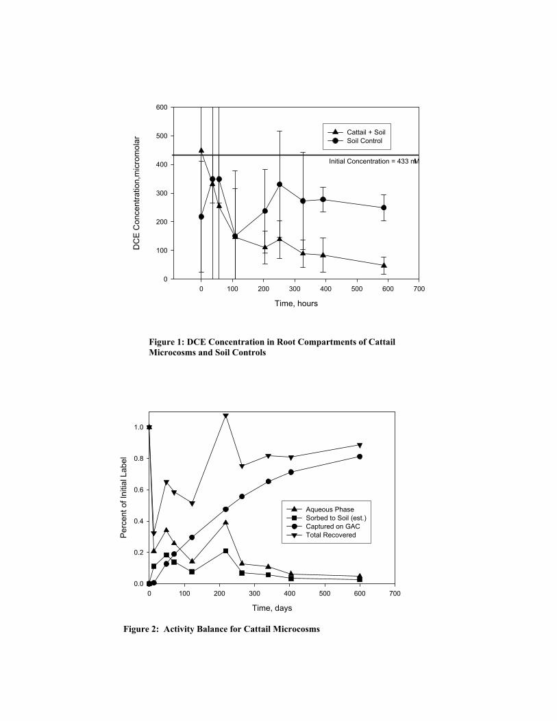
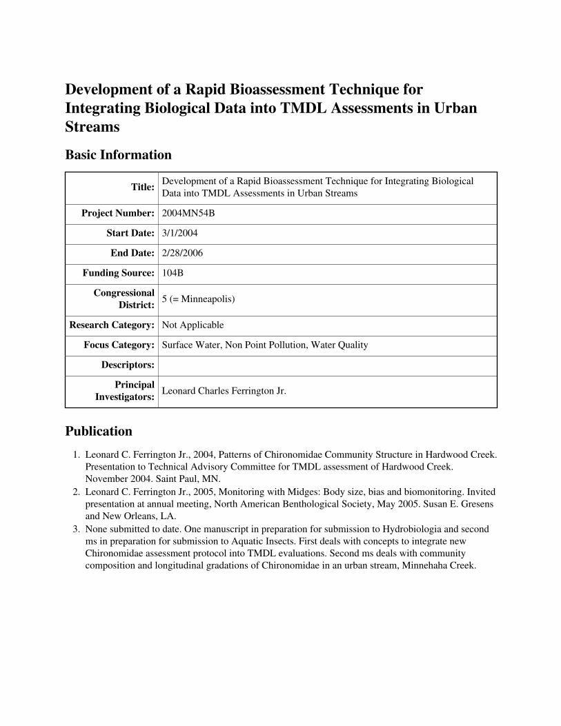
solution, t2)/2 and a linear regression was performed on the data. The slope of the best fit line corresponds to the TSCF. Results to date Field Mesocosms. Chlorinated ethylenes were not detected at any depth in any of the mesocosms. Data provided by Barr Engineering Company indicated that both DCE and vinyl chloride were present in substantial amounts in the deeper aquifer (15-20 ft). Additionally, the vertical groundwater gradient in the vicinity of the mesocosms was neutral, indicating minimal influence of groundwater discharge on subsurface conditions in the mesocosms. Large amounts of methane were detected in all three mesocosms, with methane concentration increasing with depth in each mesocosm. While methane concentration profiles were similar for the vegetated and unvegetated mesocosms during the Spring and Fall, vegetated mesocosms exhibited depressed methane concentrations in the upper 30 cm during the height of the growing season. While large quantities of both Type I and Type II methanotrophic bacteria were detected in all three mesocosms, no trends in population size were observed with respect to time of year, depth, or presence/absence of vegetation. The qualitative analysis of methanotroph population structure revealed the following seasonal population shifts for Type I methanotrophs: appearance of Methylocaldum sp. in Fall, appearance of Methylobacter sp. in Spring, and appearance of Methylomonas sp. in Summer. No trends in Type I methanotrophic population structure were observed with respect to depth or presence/absence of vegetation. No trends in Type II methanotrophic population structure was observed with respect to time of year, depth, or presence/absence of vegetation. Laboratory Microcosms. DCE disappeared in the root compartments of microcosms with cattails under hydroponic conditions and in microcosms with soil (Figure 1). While the unvegetated hydroponic controls showed minimal loss of DCE, some DCE loss was observed in soil controls. Most of the radiolabel (60%-80%) was recovered on the activated carbon (Figure 2), indicating that transport through plant tissues was the most important fate mechanism. No 14CO2 was observed in either the root compartment or the KOH traps. While the extent to which 14CO2 may have been sequestered by the plant during photosynthesis is unknown, the radiation balance suggests that cometabolic oxidation of DCE by methanotrophic bacteria could account for no more than 10% of the DCE removed. Reductive dechlorination, indicated by the appearance of vinyl chloride in the root compartment, was observed in microcosms with soil. While significant amounts of vinyl chloride accumulated in the unvegetated soil controls, vinyl chloride appearance was transient in the microcosms with cattails. It is unclear whether this difference in vinyl chloride concentration is the result of
transport of vinyl chloride through plant tissues, modification of sediment redox conditions by the plant, or cometabolic oxidation of vinyl chloride by methanotrophic bacteria. TSCF values computed for the cattail microcosms were similar for hydroponic and soil-filled root compartments. TSCF values ranged from 2.7 to 5.1. The predicted TSCF for DCE based on log Kow is 0.75 (6), much lower than observed in this work. This suggests that another mechanism in addition to uptake in the transpiration stream is involved in translocation of DCE through plant tissues. This mechanism is likely volatilization/gas phase diffusion through gas-filled voids in the plant (aerenchyma tissue). Ongoing work Current work is focusing on evaluating effect of cattails on the size and structure of methanotrophic bacterial communities in the sediment from the microcosms. Additionally, analysis of plant tissues for 14C content has yet to be conducted. The microcosm studies are also being repeated with giant bur-reed to determine if a different plant species will exhibit a different effect on the fate of DCE and methanotrophic bacterial populations in the wetland microcosms. Summary of findings While wetland plants do not appear to significantly affect the size or structure of the methanotrophic bacterial populations in a constructed wetland, they can play a significant role in removal of DCE from groundwater via vascular uptake/volatilization through tissues. Wetland plants can also prevent accumulation of the undesirable daughter product vinyl chloride. Removal of DCE by wetland plants is strongly influenced by transpiration rate, suggesting that management practices could be adapted to balance DCE removal with efflux to the atmosphere. References 1. McGuire, J.T., Smith, E.W., Long, D.T., Hyndman, D.W., Haack, S.K., Klug, M.J., and
Velbel, M.A., 2000. Temporal variations in parameters reflecting terminal-electron-accepting processes in an aquifer contaminated with waste fuel and chlorinated solvents. Chemical Geology, 169: p. 471-785.
2. Holmes, A.J., Costello, A., Lindstrom, M.E. and Murrell, J.C., 1995. Evidence that particulate monooxygenase and ammonia monooxygenase may be evolutionarily related. FEMS Microbiology Letters, 132: p. 203-208.
3. Wise, M.G., McArthur, J.V., and Shimkets, L.J., 1999. Methanotroph diversity in landfill soil: isolation of novel Type I and Type II methanotrophs whose presence was suggested by culture-independent 16S ribosomal DNA analysis. Applied and Environmental Microbiology, 65(11): p. 4887-4897.
4. Horz, H.P., Yimga, M.T., and Liesack, W., 2001. Detection of methanotroph diversity on roots of submerged rice plants by molecular retrieval of pmoA, mmoX, mxaF, and 16SrRNA and ribosomal DNA, including pmoA-based terminal restriction fragment length polymorphism profiling. Applied and Environmental Microbiology, 67: p. 4177-4185.
5. Baker, P.W., Futamata, H., Shigeaki, H., and Watanabe, K., 2001. Molecular diversity of pMMO and sMMO in a TCE-contaminated aquifer during bioremediation. FEMS Microbiology Ecology, 38: p. 161-167.
6. Burken, J.G. and Schoor, J.L., 1998. Predictive relationships for uptake of organic contaminants by hybrid poplar trees. Environmental Science and Technology, 32: p. 3379-3385.
7. Bankston, J.L., Sola, D.L., Komor, A.T., and Dwyer, D.F., 2002. Degradation of trichloroethylene in wetland microcosms containing broad-leaved cattail and eastern cottonwood. Water Research, 36: p. 1539-1546.
8. Anderson, J.E. and McCarty, P.L., 1997. Effect of Chlorinated Ethenes on Smin for a Methanotrophic Mixed Culture. Environmental Science and Technology, 31(8): p. 2204-2210.
9. Anderson, J.E. and McCarty, P.L., 1997. Transformation yields of chlorinated ethenes by a methanotrophic mixed culture expressing particulate methane monooxygenase. Applied and Environmental Microbiology, 63(2): p. 687-693.
10. Chang, H.-L. and Alvarez-Cohen, L., 1995. Model for the cometabolic biodegradation of chlorinated organics. Environmental Science and Technology, 29: p. 2357-2367.
11. Chang, H.-L. and Alvarez-Cohen, L., 1996. Biodegradation of individual and multiple chlorinated aliphatic hydrocarbons by methane-oxidizing cultures. Applied and Environmental Microbiology, 62(9): p. 3371-3377.
12. Dolan, M.E. and McCarty, P.L., 1995. Methanotrophic Chloroethene Transformation Capacities And 1,1-dichloroethene Transformation Product Toxicity. Environmental Science and Technology, 29(11): p. 2741-7.
13. Marsman, E.H., van Veen, W.W., Appelman, J.J.M., and Urlings, L.G.C.M., 1995. Biodegradation of chlorinated solvents under cometabolic conditions - full-scale experiments. Soil Environ., 5(Contaminated Soil 95, Vol. 2): p. 1075-1082.
14. Lontoh, S., Zahn, J. A., DiSpirito, A.A., and Semrau, J. D., 2000. Identification of intermediates of in vivo trichloroethylene oxidation by the membrane-associated methane monooxygenase. FEMS Microbiology Letters, 186: p. 109-113.
15. Chang, H., and Alvarez-Cohen, L., 1997. Two-stage methanotrophic bioreactor for the treatment of chlorinated organic wastewater. Water Research, 31(8): p. 2026-2036.
. Statement of related grants submitted or funded as a result of this project None. Description of student training provided by project: Name: Todd D. DeJournett Program: Department of Civil Engineering, University of Minnesota Degree being sought: Ph.D.
Time, hours
0 100 200 300 400 500 600 700
DC
E C
once
ntra
tion,
mic
rom
olar
0
100
200
300
400
500
600
Cattail + SoilSoil Control
Initial Concentration = 433 mM
Per
cent
of I
nitia
l Lab
el
0.0
0.2
0.4
0.6
0.8
1.0
Figur
Figure 1: DCE Concentration in Root Compartments of Cattail Microcosms and Soil Controls
Time, days
0 100 200 300 400 500 600 700
Aqueous PhaseSorbed to Soil (est.)Captured on GACTotal Recovered
e 2: Activity Balance for Cattail Microcosms
Development of a Rapid Bioassessment Technique forIntegrating Biological Data into TMDL Assessments in Urban Streams
Basic Information
Title: Development of a Rapid Bioassessment Technique for Integrating BiologicalData into TMDL Assessments in Urban Streams
Project Number: 2004MN54B
Start Date: 3/1/2004
End Date: 2/28/2006
Funding Source: 104B
Congressional District: 5 (= Minneapolis)
Research Category: Not Applicable
Focus Category: Surface Water, Non Point Pollution, Water Quality
Descriptors:
Principal Investigators: Leonard Charles Ferrington Jr.
Publication1. Leonard C. Ferrington Jr., 2004, Patterns of Chironomidae Community Structure in Hardwood Creek.
Presentation to Technical Advisory Committee for TMDL assessment of Hardwood Creek.November 2004. Saint Paul, MN.
2. Leonard C. Ferrington Jr., 2005, Monitoring with Midges: Body size, bias and biomonitoring. Invitedpresentation at annual meeting, North American Benthological Society, May 2005. Susan E. Gresensand New Orleans, LA.
3. None submitted to date. One manuscript in preparation for submission to Hydrobiologia and secondms in preparation for submission to Aquatic Insects. First deals with concepts to integrate newChironomidae assessment protocol into TMDL evaluations. Second ms deals with communitycomposition and longitudinal gradations of Chironomidae in an urban stream, Minnehaha Creek.
Development of a Rapid Bioassessment Technique for Integrating Biological Data into TMDL Assessments in Urban Streams Principal investigators Leonard C. Ferrington, Jr., Department of Entomology, University of Minnesota Start date: 3/1/2004 End date: 2/28/2006 Research objectives The goal of the project was to develop and refine a rapid bioassessment technique to generate biological data to be integrated into a current TMDL study of the Minnehaha Creek Watershed in Carver and Hennepin counties, Minnesota. The original project was leveraged with two additional small grants that enabled the field work to be expanded to two additional urban streams in the Minneapolis/Saint Paul Metropolitan area that are also candidates for TMDL development--- Shingle Creek (Hennepin County, MN) and Hardwood Creek (Washington and Anoka counties). Shingle Creek is being investigated for elevated conductivity potentially resulting from road salt applications during winter and Hardwood Creek is being investigated for low dissolved Oxygen during summer. In all three streams, EPA protocols for stressor identification have been employed to identify potential “stressors” that are contributing to patterns of Chironomidae community structure. Research objective 1 was to build a data base of empirical tolerances for each identified stressor for each chironomid species encountered in the three streams. Research objective 2 consists of developing conceptual models to predict changes in community structure that should occur if a variety of TMDL targets are met in the future. Research objective 3 is to develop a model of community composition that is characteristic of a “best attainable” water quality condition for local streams. The “best attainable” approach is similar to using a RIVPACS model to define a hypothetical community assemblage. I am using composition and phenology data for Chironomidae that have been generated for streams that are considered as regional reference candidates. Improving water quality conditions that are anticipated in the urban streams over the near-term future will provide for tests of the models’ accuracies, and will allow for fine-tuning or validation of them. The protocols developed from this project will be tested independently in urban streams of Baltimore, Maryland. Based on results of the Baltimore study, the protocol can be fine tuned as a generalized method for generating biological data that can be integrated into TMDL assessments of urban streams in major metropolitan areas. Methodology In this project collections of surface floating pupal exuviae (SFPE) have been used to generate information about chironomid communities at an array of sites within the Minnehaha Creek, Shingle Creek and Hardwood Creek watersheds. This monitoring
technique is not well understood by water quality managers in the United States. Consequently, a detailed description of the technique in the following paragraphs. Although not widely used in water quality investigations in the United States, collecting SFPE is not a new approach for gathering information about Chironomidae communities. It was first suggested by Thienemann (1910), but only occasionally used in taxonomic and biogeographic studies (Thienemann 1954, Brundin 1966) or ecological studies (Humphries 1938) until more recently. During the last 35 years, however, there has been increasing use of pupal exuviae collections in chironomid studies. Reiss (1968) and Lehmann (1971) used collections SFPE to supplement their larval collections when investigating Chironomidae community composition. In Western Europe and England collections of SFPE have been used extensively for surface water quality monitoring (McGill et al. 1979, Ruse 1995a, b; Ruse & Wilson 1984, Wilson 1977, 1980, 1987, 1989; Wilson & Bright 1973, Wilson & McGill 1977, Wilson & Wilson 1983). In North America the methodology has been successfully used in studies of phenology (Coffman 1973, Boerger 1981, Wartinbee & Coffman 1976), diel emergence patterns (Coffman 1974), ecology and community composition (Blackwood et al. 1995, Chou et al. 1999, Ferrington 1998, 2000, Ferrington et al. 1995, Kavanaugh 1988), microbial decomposition (Kavanaugh 1988), assessment of effects of point sources of enrichment (Coler 1984, Ferrington & Crisp 1989), non-point pesticide effects (Wright & Ferrington 1996), and effects of agricultural practices (Barton et al. 1995). In England and the United States SFPE collections have been used to study water and sediment quality (Ruse & Wilson 1984, Ruse et al. 2000, Ferrington 1993b), and used in Australia for measuring phenology (Hardwick et al. 1995) and effects of stream acidification on Chironomidae (Cranston et al. 1997). The following paragraphs briefly describe aspects of the methodology common to most of the above applications. Chironomid larvae live in soft sediments or on rocks and interstitial materials in stream beds, where they generally attain densities of 1000 or more larvae per square meter in healthy stream systems (Coffman & Ferrington 1995), and often more than 30,000 larvae per square meter in organically enriched streams (Ferrington 1990). Upon completion of the larval life they attach themselves with silken secretions to the surrounding substrates and pupation occurs. When the developing adult matures the pupa frees itself from the silken chamber and swims to the surface of the water where the adult emerges from within the pupal skin (or exuvia). The exuvia fills with air and by virtue of an outer waxy layer of the cuticle (which has non-wettable properties) it remains floating on the water surface until bacteria begin to decompose the wax layer. Floating exuviae are concentrated by stream currents into eddy areas or into regions such as slack water areas downstream of rocks or points where riparian vegetation or fallen trees contact the water surface. By collecting exuviae from these "natural" collection points, one can rapidly evaluate the emergence of Chironomidae from a broad spectrum of microhabitats in the stream. Emergence frequencies are then calculated for all species in the SFPE sample. Field collection of SFPE is accomplished by dipping an enameled pan into the water downstream of areas where pupal exuviae accumulate. Water, detritus and floating pupal exuviae flow in as one edge of the pan is dipped beneath the surface of the water. After
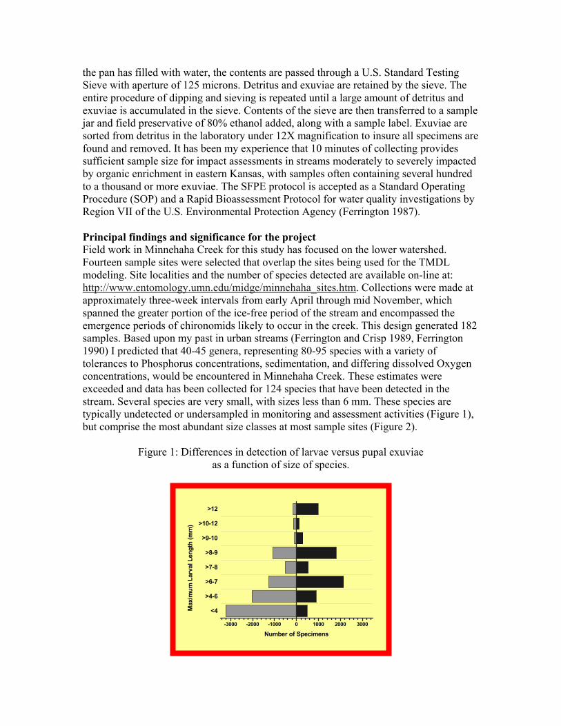
the pan has filled with water, the contents are passed through a U.S. Standard Testing Sieve with aperture of 125 microns. Detritus and exuviae are retained by the sieve. The entire procedure of dipping and sieving is repeated until a large amount of detritus and exuviae is accumulated in the sieve. Contents of the sieve are then transferred to a sample jar and field preservative of 80% ethanol added, along with a sample label. Exuviae are sorted from detritus in the laboratory under 12X magnification to insure all specimens are found and removed. It has been my experience that 10 minutes of collecting provides sufficient sample size for impact assessments in streams moderately to severely impacted by organic enrichment in eastern Kansas, with samples often containing several hundred to a thousand or more exuviae. The SFPE protocol is accepted as a Standard Operating Procedure (SOP) and a Rapid Bioassessment Protocol for water quality investigations by Region VII of the U.S. Environmental Protection Agency (Ferrington 1987). Principal findings and significance for the project Field work in Minnehaha Creek for this study has focused on the lower watershed. Fourteen sample sites were selected that overlap the sites being used for the TMDL modeling. Site localities and the number of species detected are available on-line at: http://www.entomology.umn.edu/midge/minnehaha_sites.htm. Collections were made at approximately three-week intervals from early April through mid November, which spanned the greater portion of the ice-free period of the stream and encompassed the emergence periods of chironomids likely to occur in the creek. This design generated 182 samples. Based upon my past in urban streams (Ferrington and Crisp 1989, Ferrington 1990) I predicted that 40-45 genera, representing 80-95 species with a variety of tolerances to Phosphorus concentrations, sedimentation, and differing dissolved Oxygen concentrations, would be encountered in Minnehaha Creek. These estimates were exceeded and data has been collected for 124 species that have been detected in the stream. Several species are very small, with sizes less than 6 mm. These species are typically undetected or undersampled in monitoring and assessment activities (Figure 1), but comprise the most abundant size classes at most sample sites (Figure 2).
Figure 1: Differences in detection of larvae versus pupal exuviae as a function of size of species.
<4
>4-6
>6-7
>7-8
>8-9
>9-10
>10-12
>12
-3000 -2000 -1000 0 1000 2000 3000
Max
imum
Lar
val L
engt
h (m
m)
Number of Specimens
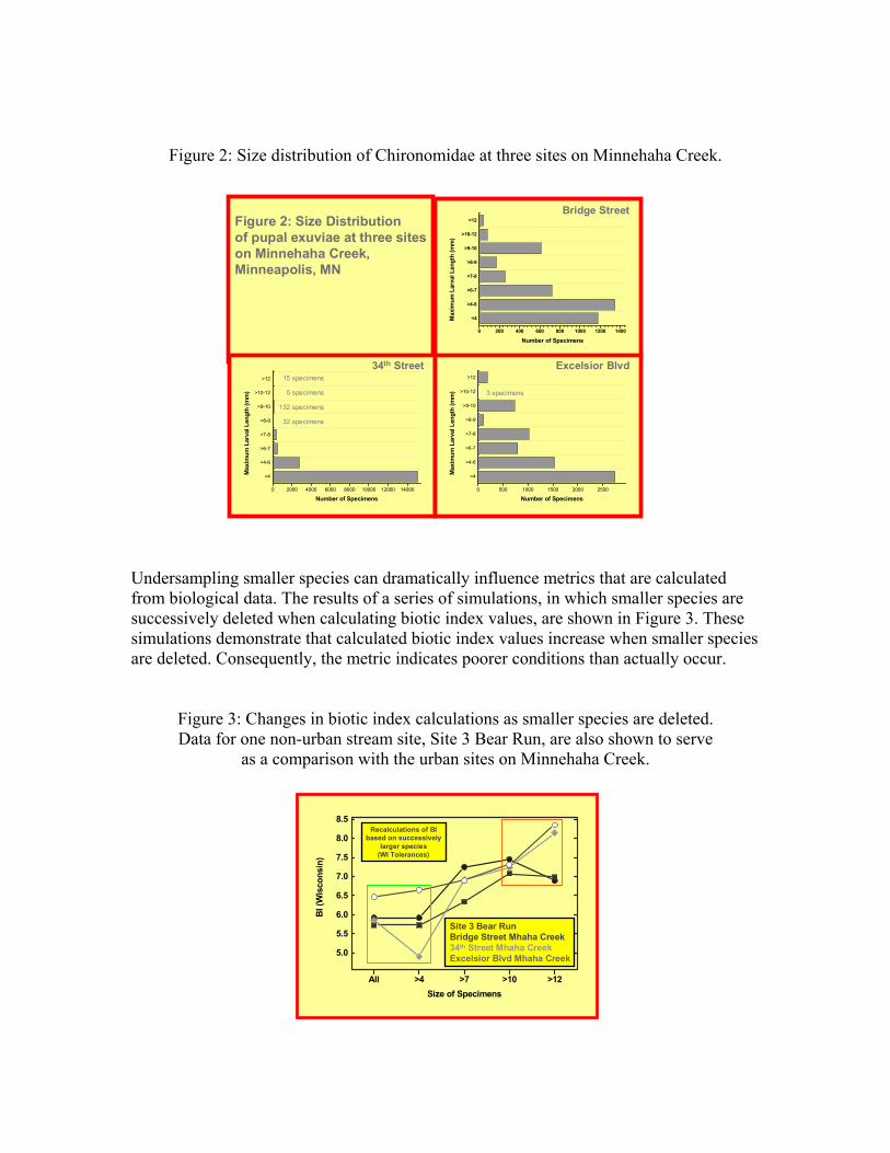
Figure 2: Size distribution of Chironomidae at three sites on Minnehaha Creek.
<4
>4-6
>6-7
>7-8
>8-9
>9-10
>10-12
>12
0 200 400 600 800 1000 1200 1400
Max
imum
Lar
val L
engt
h (m
m)
Number of Specimens
Figure 2: Size Distributionof pupal exuviae at three sites on Minnehaha Creek,Minneapolis, MN
<4
>4-6
>6-7
>7-8
>8-9
>9-10
>10-12
>12
0 2000 4000 6000 8000 10000 12000 14000
Max
imum
Lar
val L
engt
h (m
m)
Number of Specimens
<4
>4-6
>6-7
>7-8
>8-9
>9-10
>10-12
>12
0 500 1000 1500 2000 2500
Max
imum
Lar
val L
engt
h (m
m)
Number of Specimens
15 specimens
5 specimens
132 specimens
32 specimens
3 specimens
Bridge Street
34th Street Excelsior Blvd
Undersampling smaller species can dramatically influence metrics that are calculated from biological data. The results of a series of simulations, in which smaller species are successively deleted when calculating biotic index values, are shown in Figure 3. These simulations demonstrate that calculated biotic index values increase when smaller species are deleted. Consequently, the metric indicates poorer conditions than actually occur.
Figure 3: Changes in biotic index calculations as smaller species are deleted. Data for one non-urban stream site, Site 3 Bear Run, are also shown to serve
as a comparison with the urban sites on Minnehaha Creek.
All >4 >7 >10 >12
5.0
5.5
6.0
6.5
7.0
7.5
8.0
8.5
BI (
Wis
cons
in)
Size of Specimens
Site 3 Bear RunBridge Street Mhaha Creek34th Street Mhaha CreekExcelsior Blvd Mhaha Creek
Recalculations of BIbased on successively
larger species(WI Tolerances)

Publications associated with this project or previous projects funded by NIWR/WRC – None submitted to date. One manuscript in preparation for submission to Hydrobiologia and second ms in preparation for submission to Aquatic Insects. First deals with concepts to integrate new Chironomidae assessment protocol into TMDL evaluations. Second ms deals with community composition and longitudinal gradations of Chironomidae in an urban stream, Minnehaha Creek. Students supported by the project – Mr. Adam Sealock, Graduate Student in Water Resources Program, enrolled in MS degree program.
The Effects of Long-Term Low-Level Antibiotic Exposure on theDevelopment of Antibiotic Resistance
Basic Information
Title: The Effects of Long-Term Low-Level Antibiotic Exposure on the Developmentof Antibiotic Resistance
Project Number: 2004MN65B
Start Date: 3/1/2004
End Date: 2/29/2005
Funding Source: 104B
Congressional District: 5
Research Category: None
Focus Category: Non Point Pollution, Ecology, Water Quality
Descriptors: None
Principal Investigators: Kristine Wammer
Publication1. K.H. Wammer, T.M. LaPara, L.J. Onan, The effects of long-term low-level antibiotic exposure on the
development of antibiotic resistance. Abstract submitted for consideration as a poster presentation,Minnesota Water 2005 and Annual Water Resources Joint Conference, Brooklyn Center, MN,October 2005.
The Effects of Long-Term Low-Level Antibiotic Exposure on the Development of Antibiotic Resistance Principal Investigator K.H. Wammer, Postdoctoral Associate, Departments of Environmental Health Sciences, Chemistry, and Civil Engineering, University of Minnesota Undergraduate Research Assistants C.G. Klatt and L.J. Onan, Department of Civil Engineering, University of Minnesota. Funding Source: USGS-WRRI 104B/CAIWQ Competitive Grants Program Project Duration: 3/01/2004-2/28/2005 Summary Antibacterial compounds have been detected in the environment at low, subtherapeutic levels. Here, we examined whether the presence of antibacterials at these levels would lead to an increase in antibacterial resistance among exposed bacteria. Three chemostats were operated with identical enrichment cultures from Mississippi River water. One chemostat contained no antibacterials, a second contained four antibacterials (sulfamethoxazole, norfloxacin, trimethoprim, and tylosin) as a mixture at very low concentrations (≤ 1 µg/L each), and a third contained norfloxacin at a 1 µg/L concentration. Enumeration of the proportion of bacteria from each chemostat exhibiting antibiotic resistance was performed approximately weekly using heterotrophic plate counts on nutrient media supplemented with elevated levels of each antibiotic. Ability of the bacteria to grow on liquid media supplemented with each antibiotic was also tested periodically. Polymerase chain reaction followed by denaturing gradient gel electrophoresis (PCR-DGGE) was used to track changes in the community structure over time. No significant differences were observed for the bacterial populations from the three chemostats, which suggests that very low antibiotic concentrations may not select for antibiotic resistant bacteria in the environment. Introduction The presence of pharmaceutical and personal care products (PPCPs) in natural waters has gained attention due to recent reports (1-5) including a national reconnaissance of United States streams conducted by the U.S. Geological Survey in which pharmaceuticals were detected at 80% of the sites sampled (6). The widespread occurrence of antibacterial compounds is especially alarming for several reasons. First, the presence of antibiotics in natural waters may lead to the proliferation of antibiotic resistance among exposed microorganisms, and this resistance may be transferable to human pathogens (7-10). Alternatively, if present at high enough concentrations, antibacterial compounds may adversely affect native microbial communities. Antibacterial compounds have been detected thus far in natural waters at concentrations far below therapeutic levels. While there is a concern that long-term exposure at these low levels may result in an increase in antibiotic resistance among environmental bacteria, this relationship has not yet been conclusively established through studies in environmental systems (11-13). This project was designed to explicitly study the suspected link between low-level antibacterial exposure and proliferation of resistant bacteria by exposing environmental bacteria to
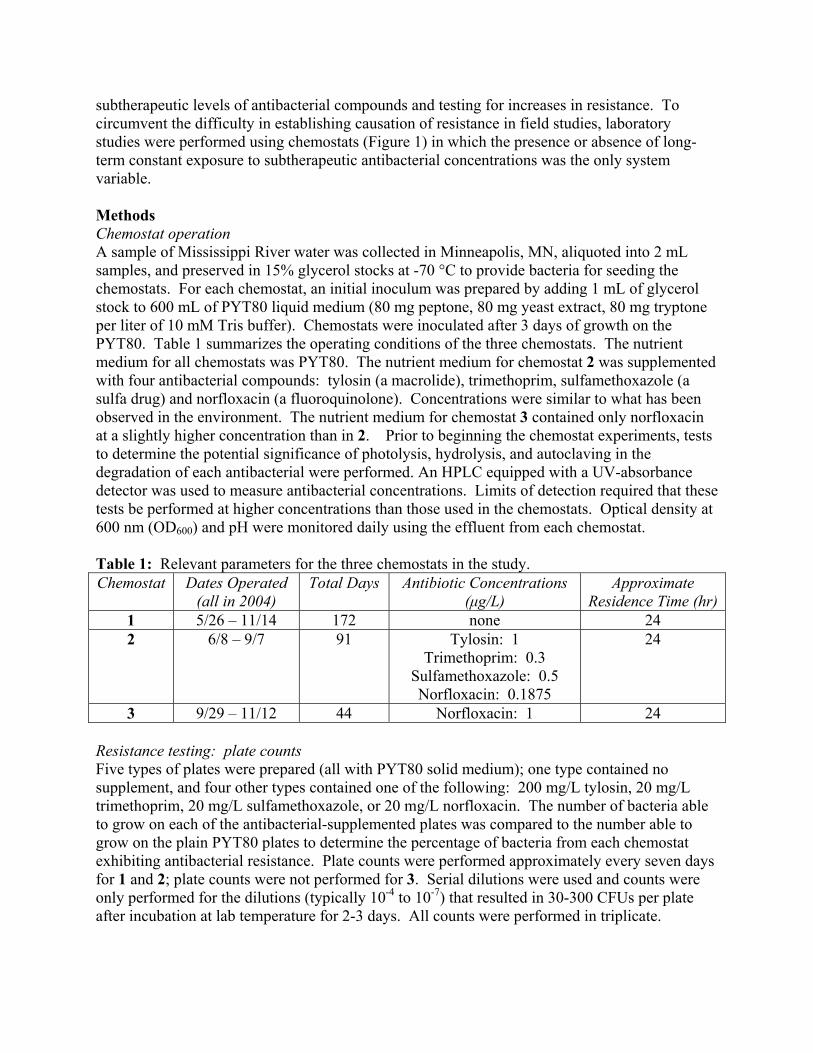
subtherapeutic levels of antibacterial compounds and testing for increases in resistance. To circumvent the difficulty in establishing causation of resistance in field studies, laboratory studies were performed using chemostats (Figure 1) in which the presence or absence of long-term constant exposure to subtherapeutic antibacterial concentrations was the only system variable. Methods Chemostat operation A sample of Mississippi River water was collected in Minneapolis, MN, aliquoted into 2 mL samples, and preserved in 15% glycerol stocks at -70 °C to provide bacteria for seeding the chemostats. For each chemostat, an initial inoculum was prepared by adding 1 mL of glycerol stock to 600 mL of PYT80 liquid medium (80 mg peptone, 80 mg yeast extract, 80 mg tryptone per liter of 10 mM Tris buffer). Chemostats were inoculated after 3 days of growth on the PYT80. Table 1 summarizes the operating conditions of the three chemostats. The nutrient medium for all chemostats was PYT80. The nutrient medium for chemostat 2 was supplemented with four antibacterial compounds: tylosin (a macrolide), trimethoprim, sulfamethoxazole (a sulfa drug) and norfloxacin (a fluoroquinolone). Concentrations were similar to what has been observed in the environment. The nutrient medium for chemostat 3 contained only norfloxacin at a slightly higher concentration than in 2. Prior to beginning the chemostat experiments, tests to determine the potential significance of photolysis, hydrolysis, and autoclaving in the degradation of each antibacterial were performed. An HPLC equipped with a UV-absorbance detector was used to measure antibacterial concentrations. Limits of detection required that these tests be performed at higher concentrations than those used in the chemostats. Optical density at 600 nm (OD600) and pH were monitored daily using the effluent from each chemostat. Table 1: Relevant parameters for the three chemostats in the study. Chemostat Dates Operated
(all in 2004) Total Days Antibiotic Concentrations
(µg/L) Approximate
Residence Time (hr)1 5/26 – 11/14 172 none 24 2 6/8 – 9/7 91 Tylosin: 1
Trimethoprim: 0.3 Sulfamethoxazole: 0.5 Norfloxacin: 0.1875
24
3 9/29 – 11/12 44 Norfloxacin: 1 24 Resistance testing: plate counts Five types of plates were prepared (all with PYT80 solid medium); one type contained no supplement, and four other types contained one of the following: 200 mg/L tylosin, 20 mg/L trimethoprim, 20 mg/L sulfamethoxazole, or 20 mg/L norfloxacin. The number of bacteria able to grow on each of the antibacterial-supplemented plates was compared to the number able to grow on the plain PYT80 plates to determine the percentage of bacteria from each chemostat exhibiting antibacterial resistance. Plate counts were performed approximately every seven days for 1 and 2; plate counts were not performed for 3. Serial dilutions were used and counts were only performed for the dilutions (typically 10-4 to 10-7) that resulted in 30-300 CFUs per plate after incubation at lab temperature for 2-3 days. All counts were performed in triplicate.
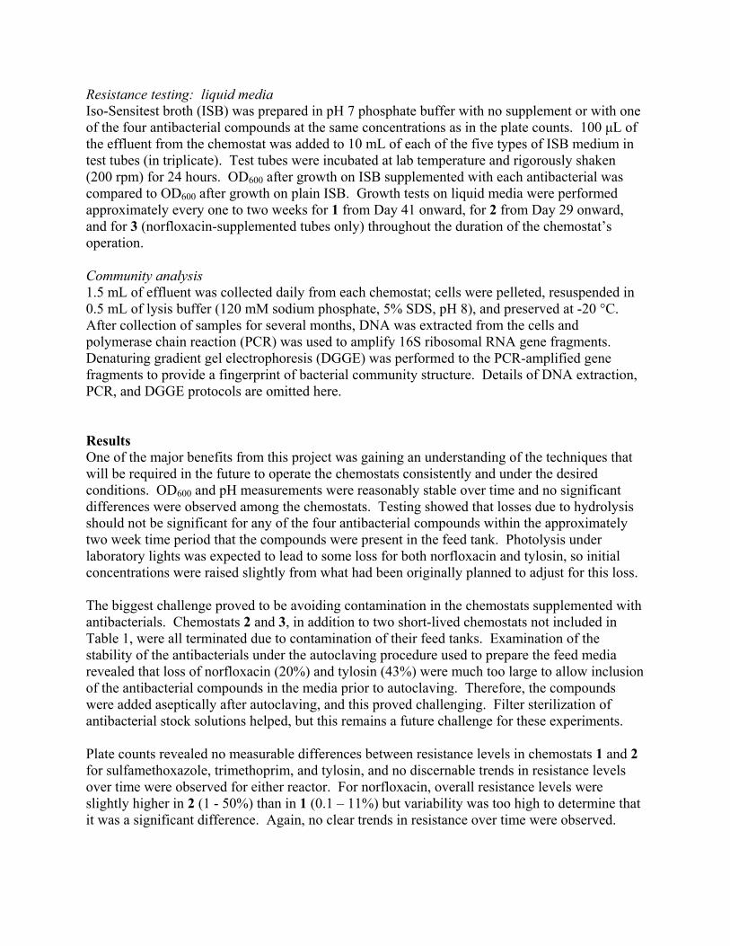
Resistance testing: liquid media Iso-Sensitest broth (ISB) was prepared in pH 7 phosphate buffer with no supplement or with one of the four antibacterial compounds at the same concentrations as in the plate counts. 100 µL of the effluent from the chemostat was added to 10 mL of each of the five types of ISB medium in test tubes (in triplicate). Test tubes were incubated at lab temperature and rigorously shaken (200 rpm) for 24 hours. OD600 after growth on ISB supplemented with each antibacterial was compared to OD600 after growth on plain ISB. Growth tests on liquid media were performed approximately every one to two weeks for 1 from Day 41 onward, for 2 from Day 29 onward, and for 3 (norfloxacin-supplemented tubes only) throughout the duration of the chemostat’s operation. Community analysis 1.5 mL of effluent was collected daily from each chemostat; cells were pelleted, resuspended in 0.5 mL of lysis buffer (120 mM sodium phosphate, 5% SDS, pH 8), and preserved at -20 °C. After collection of samples for several months, DNA was extracted from the cells and polymerase chain reaction (PCR) was used to amplify 16S ribosomal RNA gene fragments. Denaturing gradient gel electrophoresis (DGGE) was performed to the PCR-amplified gene fragments to provide a fingerprint of bacterial community structure. Details of DNA extraction, PCR, and DGGE protocols are omitted here. Results One of the major benefits from this project was gaining an understanding of the techniques that will be required in the future to operate the chemostats consistently and under the desired conditions. OD600 and pH measurements were reasonably stable over time and no significant differences were observed among the chemostats. Testing showed that losses due to hydrolysis should not be significant for any of the four antibacterial compounds within the approximately two week time period that the compounds were present in the feed tank. Photolysis under laboratory lights was expected to lead to some loss for both norfloxacin and tylosin, so initial concentrations were raised slightly from what had been originally planned to adjust for this loss. The biggest challenge proved to be avoiding contamination in the chemostats supplemented with antibacterials. Chemostats 2 and 3, in addition to two short-lived chemostats not included in Table 1, were all terminated due to contamination of their feed tanks. Examination of the stability of the antibacterials under the autoclaving procedure used to prepare the feed media revealed that loss of norfloxacin (20%) and tylosin (43%) were much too large to allow inclusion of the antibacterial compounds in the media prior to autoclaving. Therefore, the compounds were added aseptically after autoclaving, and this proved challenging. Filter sterilization of antibacterial stock solutions helped, but this remains a future challenge for these experiments. Plate counts revealed no measurable differences between resistance levels in chemostats 1 and 2 for sulfamethoxazole, trimethoprim, and tylosin, and no discernable trends in resistance levels over time were observed for either reactor. For norfloxacin, overall resistance levels were slightly higher in 2 (1 - 50%) than in 1 (0.1 – 11%) but variability was too high to determine that it was a significant difference. Again, no clear trends in resistance over time were observed.
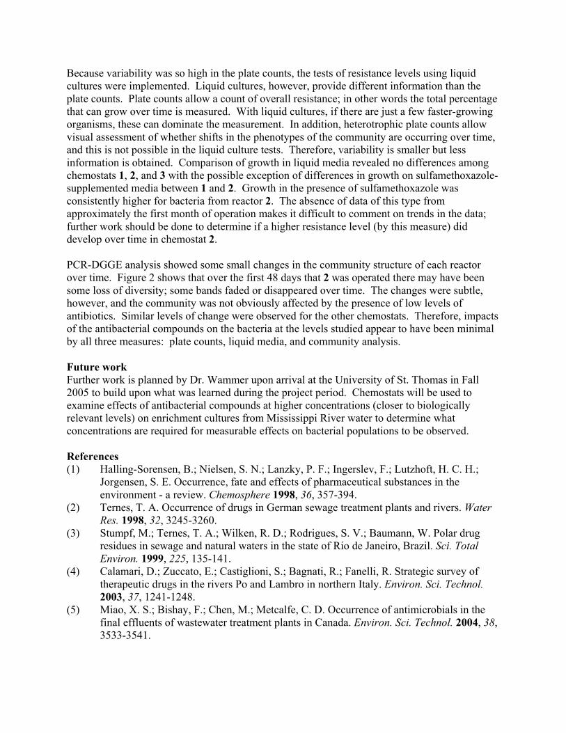
Because variability was so high in the plate counts, the tests of resistance levels using liquid cultures were implemented. Liquid cultures, however, provide different information than the plate counts. Plate counts allow a count of overall resistance; in other words the total percentage that can grow over time is measured. With liquid cultures, if there are just a few faster-growing organisms, these can dominate the measurement. In addition, heterotrophic plate counts allow visual assessment of whether shifts in the phenotypes of the community are occurring over time, and this is not possible in the liquid culture tests. Therefore, variability is smaller but less information is obtained. Comparison of growth in liquid media revealed no differences among chemostats 1, 2, and 3 with the possible exception of differences in growth on sulfamethoxazole-supplemented media between 1 and 2. Growth in the presence of sulfamethoxazole was consistently higher for bacteria from reactor 2. The absence of data of this type from approximately the first month of operation makes it difficult to comment on trends in the data; further work should be done to determine if a higher resistance level (by this measure) did develop over time in chemostat 2. PCR-DGGE analysis showed some small changes in the community structure of each reactor over time. Figure 2 shows that over the first 48 days that 2 was operated there may have been some loss of diversity; some bands faded or disappeared over time. The changes were subtle, however, and the community was not obviously affected by the presence of low levels of antibiotics. Similar levels of change were observed for the other chemostats. Therefore, impacts of the antibacterial compounds on the bacteria at the levels studied appear to have been minimal by all three measures: plate counts, liquid media, and community analysis. Future work Further work is planned by Dr. Wammer upon arrival at the University of St. Thomas in Fall 2005 to build upon what was learned during the project period. Chemostats will be used to examine effects of antibacterial compounds at higher concentrations (closer to biologically relevant levels) on enrichment cultures from Mississippi River water to determine what concentrations are required for measurable effects on bacterial populations to be observed. References (1) Halling-Sorensen, B.; Nielsen, S. N.; Lanzky, P. F.; Ingerslev, F.; Lutzhoft, H. C. H.;
Jorgensen, S. E. Occurrence, fate and effects of pharmaceutical substances in the environment - a review. Chemosphere 1998, 36, 357-394.
(2) Ternes, T. A. Occurrence of drugs in German sewage treatment plants and rivers. Water Res. 1998, 32, 3245-3260.
(3) Stumpf, M.; Ternes, T. A.; Wilken, R. D.; Rodrigues, S. V.; Baumann, W. Polar drug residues in sewage and natural waters in the state of Rio de Janeiro, Brazil. Sci. Total Environ. 1999, 225, 135-141.
(4) Calamari, D.; Zuccato, E.; Castiglioni, S.; Bagnati, R.; Fanelli, R. Strategic survey of therapeutic drugs in the rivers Po and Lambro in northern Italy. Environ. Sci. Technol. 2003, 37, 1241-1248.
(5) Miao, X. S.; Bishay, F.; Chen, M.; Metcalfe, C. D. Occurrence of antimicrobials in the final effluents of wastewater treatment plants in Canada. Environ. Sci. Technol. 2004, 38, 3533-3541.
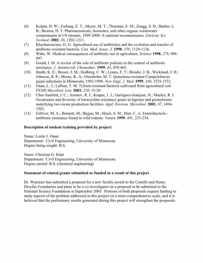
(6) Kolpin, D. W.; Furlong, E. T.; Meyer, M. T.; Thurman, E. M.; Zaugg, S. D.; Barber, L. B.; Buxton, H. T. Pharmaceuticals, hormones, and other organic wastewater contaminants in US streams, 1999-2000: A national reconnaissance. Environ. Sci. Technol. 2002, 36, 1202-1211.
(7) Khachatourians, G. G. Agricultural use of antibiotics and the evolution and transfer of antibiotic-resistant bacteria. Can. Med. Assoc. J. 1998, 159, 1129-1136.
(8) Witte, W. Medical consequences of antibiotic use in agriculture. Science 1998, 279, 996-997.
(9) Gould, I. M. A review of the role of antibiotic policies in the control of antibiotic resistance. J. Antimicrob. Chemother. 1999, 43, 459-465.
(10) Smith, K. E.; Besser, J. M.; Hedberg, C. W.; Leano, F. T.; Bender, J. B.; Wicklund, J. H.; Johnson, B. P.; Moore, K. A.; Osterholm, M. T. Quinolone-resistant Campylobacter jejuni infections in Minnesota, 1992-1998. New Engl. J. Med. 1999, 340, 1525-1532.
(11) Onan, L. J.; LaPara, T. M. Tylosin-resistant bacteria cultivated from agricultural soil. FEMS Microbiol. Lett. 2003, 220, 15-20.
(12) Chee-Sanford, J. C.; Aminov, R. I.; Krapac, I. J.; Garrigues-Jeanjean, N.; Mackie, R. I. Occurrence and diversity of tetracycline resistance genes in lagoons and groundwater underlying two swine production facilities. Appl. Environ. Microbiol. 2001, 67, 1494-1502.
(13) Gilliver, M. A.; Bennett, M.; Begon, M.; Hazel, S. M.; Hart, C. A. Enterobacteria - antibiotic resistance found in wild rodents. Nature 1999, 401, 233-234.
Description of student training provided by project: Name: Leslie J. Onan Department: Civil Engineering, University of Minnesota Degree being sought: B.S. Name: Christian G. Klatt Department: Civil Engineering, University of Minnesota Degree earned: B.S. (chemical engineering) Statement of related grants submitted or funded as a result of this project Dr. Wammer has submitted a proposal for a new faculty award to the Camille and Henry Dreyfus Foundation and plans to be a co-investigator on a proposal to be submitted to the National Science Foundation in September 2005. Portions of both proposals request funding to study aspects of the problem addressed in this project on a more comprehensive scale, and it is believed that the preliminary results generated during this project will strengthen the proposals.
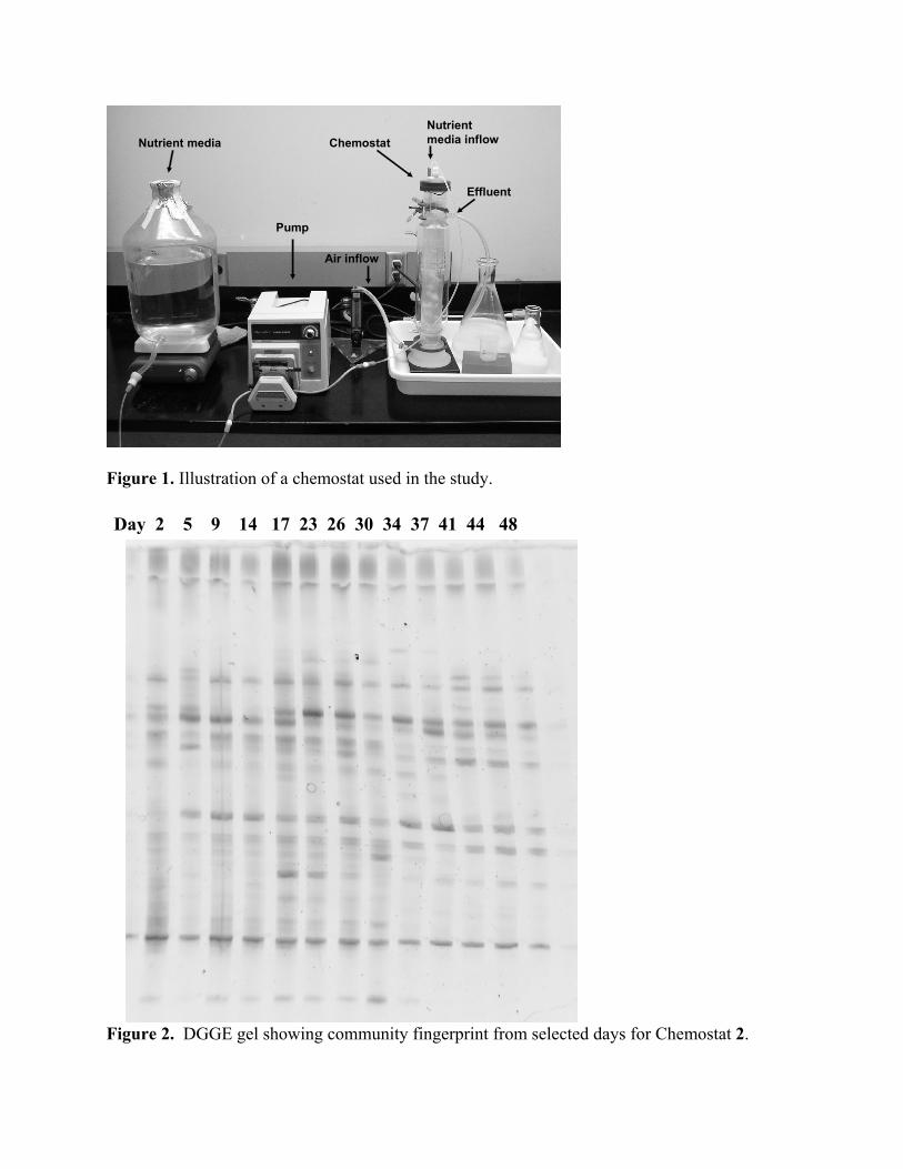
Pump
Nutrient media Chemostat
Effluent
Nutrient media inflow
Air inflow
Figure 1. Illustration of a chemostat used in the study. Day 2 5 9 14 17 23 26 30 34 37 41 44 48
Figure 2. DGGE gel showing community fingerprint from selected days for Chemostat 2.
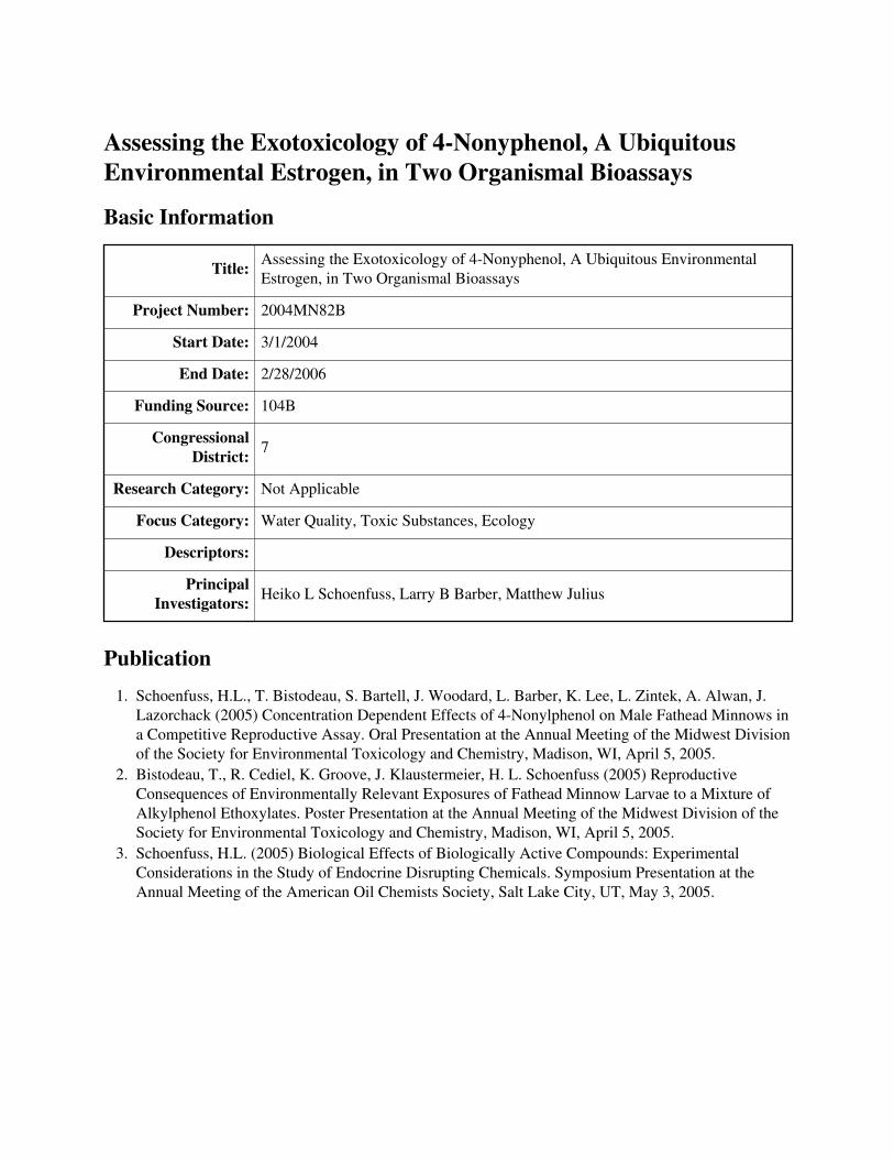
Assessing the Exotoxicology of 4-Nonyphenol, A UbiquitousEnvironmental Estrogen, in Two Organismal Bioassays
Basic Information
Title: Assessing the Exotoxicology of 4-Nonyphenol, A Ubiquitous EnvironmentalEstrogen, in Two Organismal Bioassays
Project Number: 2004MN82B
Start Date: 3/1/2004
End Date: 2/28/2006
Funding Source: 104B
Congressional District: 7
Research Category: Not Applicable
Focus Category: Water Quality, Toxic Substances, Ecology
Descriptors:
Principal Investigators: Heiko L Schoenfuss, Larry B Barber, Matthew Julius
Publication1. Schoenfuss, H.L., T. Bistodeau, S. Bartell, J. Woodard, L. Barber, K. Lee, L. Zintek, A. Alwan, J.
Lazorchack (2005) Concentration Dependent Effects of 4-Nonylphenol on Male Fathead Minnows ina Competitive Reproductive Assay. Oral Presentation at the Annual Meeting of the Midwest Divisionof the Society for Environmental Toxicology and Chemistry, Madison, WI, April 5, 2005.
2. Bistodeau, T., R. Cediel, K. Groove, J. Klaustermeier, H. L. Schoenfuss (2005) ReproductiveConsequences of Environmentally Relevant Exposures of Fathead Minnow Larvae to a Mixture ofAlkylphenol Ethoxylates. Poster Presentation at the Annual Meeting of the Midwest Division of theSociety for Environmental Toxicology and Chemistry, Madison, WI, April 5, 2005.
3. Schoenfuss, H.L. (2005) Biological Effects of Biologically Active Compounds: ExperimentalConsiderations in the Study of Endocrine Disrupting Chemicals. Symposium Presentation at theAnnual Meeting of the American Oil Chemists Society, Salt Lake City, UT, May 3, 2005.
Assessing the Ecotoxicology of 4-Nonylphenol, A Ubiquitous Environmental Estrogen, in Two Organismal Bioassays Principal Investigators H.L. Schoenfuss, PhD., Department Biological Sciences, St. Cloud State University; L.B. Barber, PhD, US Geological Survey, Boulder, CO, M.L. Julius, PhD., Department Biological Sciences, St. Cloud State University, St. Cloud, Minnesota. Research Assistants T. Bistodeau, Graduate Students, J. Koch, Graduate Student, K. Groove, Undergraduate, R. Cediel, Undergraduate, St. Cloud State University, St. Cloud, Minnesota Start date: 3/21/2004 End date: 2/28/2006 Executive summary Alkylphenols (APs) were recently discovered in many surface water samples and even in some drinking waters in Europe and North America. Wastewater effluent was identified as a major source of contamination and found to revert much of the metabolic products of these biologically active compounds back to their most potent form, 4-nonylphenol. Alkylphenols are used in large quantities (thousands of tons annually in North America) as surfactants in industrial and domestic settings and are know to bind to the estrogen receptor of mammalian cells. Environmental estrogens such as APs are known to disrupt normal endocrine hormone that are central to maturation and reproduction in fishes, and the ubiquitous presence of these biologically active compounds in surface waters should be of environmental and human health concern. In this study we propose to examine the effects of 4-nonylphenol on two organismal bioassays, representing the base and apex of the aquatic food chain. At the base of the food chain, diatoms, a group of photosynthetically active organisms, serve as a preferred food source for larval fish. Near the top, fathead minnows represent an important link in the food chain as a consumer of diatoms and as a food source for game fish. Previously we have demonstrated that both of these organisms are sensitive to aquatic pollution. Diatoms will reduce their lipid content, which makes them a lower quality food source for developing fish larvae, while fathead minnows exposed to pollutants will be less likely to reproduce. Our preliminary results indicate that the quality of diatoms as food source will diminish rapidly if they are exposed to 4-nonylphenol at concentrations frequently measured in rivers below treated sewage effluent outfalls. Male fathead minnows exposed to similar, environmentally relevant concentrations of 4-nonylphenol also appear to be less likely to reproduce than unexposed males. We are currently attempting to repeat these results in a second series of exposures and link the two assays by feeding exposed diatoms to fish larvae. If our preliminary results are confirmed in these experiments, then 4-nonylphenol might represent a substantial pollution source in many riverine systems in Minnesota and the US and might partially account for the decrease in some fish populations.
Introduction This study set out to determine whether 4-nonylphenol, the most biologically potent alkylphenol (APs), has significant effects on reproduction and health of two model organisms. Alkylphenols, a well recognized class of “environmental estrogens”, contribute significantly to the estrogenicity of wastewater effluents (Field 1996; Barber 2000; Farre 2002), however, the effects of these compounds on exposed organisms is largely unknown. The few studies that have exposed aquatic vertebrates to APs in the laboratory have focused on sub-organismal endpoints such as gene expression of zona radiata protein (Arukwe 2001; Ackermann 2002), MCF-7 breast tumor cell proliferation (E-screen) (Gutendorf 2001; Folmar 2002), rainbow trout hepatocyte cultures (Madigou 2001), insulin-like growth factor (Le Gac 2001), and yeast based estrogen receptor assays (Gutendorf 2001; Madigou 2001; Folmar 2002). By far the most commonly used endpoints relate to the synthesis of the egg-yolk protein vitellogenin (VTG) (Foran 2000; Arukwe 2001; Hemmer 2001; Nichols 2001; Ackermann 2002; Folmar 2002; Villeneuve 2002) and the induction of the hepatic VTG mRNA (Hemmer 2001). The aforementioned endpoints have merits in determining acute exposure of oviparous vertebrates to compounds (“environmental estrogens”) binding to the estrogen receptors; however, their value in assessing the reproductive consequences for exposed organisms is limited. Several studies have, therefore, employed biomarkers more closely related to the reproductive competence of exposed organisms. As environmental estrogens, including APs, affect the hypothalamic-pituitary-gonadal steroidal axis, it seems intuitive to analyze hormones of these endocrine pathways. Estradiol (E2) concentrations increased in male and female fathead minnows exposed to nonylphenol in studies by Giesy and colleagues (2000) but did not exhibit a similar response in fathead minnow studies by Nichols and colleagues (2001) or in carp exposed by Villeneuve and colleagues (2002). The latter two studies also analyzed testosterone concentrations in the exposed organisms and did not report any significant differences from the respective control treatments (Nichols 2001; Villeneuve 2002). Harris and colleagues (2001) exposed female rainbow trout to nonylphenol and reported a decrease follicle stimulating and luteinizing hormones, which are both central to the fecundity of female fishes. Gametogenesis, the production of eggs and/or sperm, was measured directly in fathead minnows (Nichols 2001) and trout (Le Gac 2001). Several studies have also assessed sex ratios and the induction of intersex, the presence of female reproductive tissues in the testis of exposed male organisms (Jobling 1997; Gray 1999; Ackermann 2002). However, none of these endpoints allows for a direct assessment of the reproductive fitness of AP exposed animals.
The present study investigated two objectives to establish the effects of 4-nonyphenol through the development of two organismal bioassays. These objectives are:
(1) Determining the impacts of 4-nonyphenol exposure on the physiological development of a ubiquitous diatom species. Diatoms appear to be a particularly satisfactory food source for many aquatic animals, including fingerling fish (Volkman 1989, Ahlgren 1990). In addition, they are well known as highly sensitive indicators of environmental change (see Stoermer 2000). A laboratory experiment is proposed, examining the effects of 4-nonyphenol concentrations on the gross morphology and physiology of the diatom species Cyclotella meneghiniana. Ultimately, this experiment will lay the foundation for determining the impacts of pharmaceutical contamination on the development and food quality of species in the primary production community. If effects are confirmed, this study will also serve as the impetus for developing protocols for rapidly and inexpensively triaging other alkyphenol effects using certain diatom
species as model organisms.
(2) Determine the effects of 4-nonyphenol exposure on reproductive success in laboratory fishes. The fathead minnow was chosen for this study as it is a tier one screening organism for endocrine- disrupting compounds (Ankley 1998), is easily maintained in the laboratory, and reproduces year round in the laboratory. Fathead minnows are particularly well suited for a competitive assay as they establish a dominance hierarchy and compete for nest sites in which females then deposit eggs (Unger 1983, 1988; Sargent 1988, 1989). Thus, reproductive success of male fathead minnows is linked to its ability to acquire and defend a nest site until larvae hatch. In this study we introduced direct competition between males by limiting the number of available nest sites. By exposing male fathead minnows to APs and allowing these males to compete directly with control males we assessed the effects of this exposure on their reproductive competence. Methods Two bioassays will be utilized to meet the objectives of this study. To examine the effects of 4-nonyphenol on the gross morphology and physiological development of the diatom species Cyclotella meneghiniana we compared cell density, lipid composition, fatty acid concentrations, and the electron microscope ultra-structure in control and 4- nonyphenol exposed cultures. This diatom was selected because it commonly occurs in most freshwater environments and has been the subject of other toxicological studies. This provides a framework for structuring this project’s experimental design. An adequate literature base also exists for evaluating results of this experiment. The species grows rapidly and is easy to maintain in culture. The use of a phytoplankton species as a test organism complements the fish portion of the study by considering effects at the apex and base of the food chain.
In a second bioassay we attempted to determine whether 4-nonyphenol exposure has adverse effects on the reproductive success of male fathead minnows. The fathead minnow was chosen as model species for this assay, as it is a widely used model organism for toxicological studies and has been named a tier one screening organism for endocrine disrupters by the US EPA (Ankley 1998). Furthermore, detailed protocols for handling fathead minnows for experimental testing exist (Denny 1987) and were incorporated in our previous studies of endocrine disrupters (Schoenfuss 2001; Schoenfuss 2003). Finally, we have developed DNA microsatellite primers to allow for paternity determination of offspring in the reproductive assay.
The fathead minnow is an attractive model for studies of the effects of aquatic contaminants because of the reproductive strategy of this species. Fathead minnows are nest breeders, with the male establishing the nest site and defending it until larvae hatch (Unger 1983; Unger 1988; Sargent 1989). The aggressive behavior of the male during nest holding is directly controlled by the endocrine system through testosterone release and is therefore vulnerable to endocrine disruption. Furthermore, reproductive success relies on the ability of the male to defend the nest site until larvae hatch. Any weakening of the male due to contaminant exposure may prevent him from defending the nest site and will result in reproductive failure (Sargent 1988). Fathead minnows in the reproductive assay were exposed to low and reasonable concentrations of 4-nonyphenol, which have been identified as a ubiquitous contaminant in European and North American surface waters. Dosage of 4-nonyphenol in this study was adjusted to bracket values found by the USGS in an ongoing survey of alkyphenol contamination in Minnesota.
Determine the impacts of 4-nonyphenol exposure on the physiological development of the ubiquitous diatom species Cyclotella meneghiniana. Cyclotella meneghiniana were isolated into culture during the spring of 2002 from Mississippi River collections obtained at a sampling location on SCSU’s campus. An individual specimen was extracted from the algal sample to initiate the cultures, ensuring the cultures are unialgal and homozygous. The stock culture were maintained in WC media (Guillard 1975) at 20°C on a 16:8 light/ dark cycle at 200 µE·m-2·s-1, achieving an approximate density of 100,000 cells/ml. For experimental purposes, stock cultures were divided among six 2-L acid washed flasks. Two flasks were used as a control and two were exposed to one of two concentrations of 4-nonyphenol. Exposure to 4- nonyphenol concentrations were initiated at 2pm each afternoon, corresponding to the 8th hour of the light period. The experiment ran for 10 days and culture flasks were sampled on day 0, 1, 3, 5, 7, and 10. This 10-day experiment was repeated once. Sub samples were enumerated to evaluate changes in cell density over time. Lipid concentrations were quantified for the sub samples using an Iatroscan Mark IV (Sicko-Goad 1993). Additionally, fatty acid concentrations were quantified for sub samples using a gas chromatograph (Sicko-Goad 1988).
Determine the effects of 4-nonyphenol exposure on reproductive success in the fathead minnow (Pimephales promelas). Juvenile fathead minnows (3 months old) were purchased from Kurt’s Fisheries, PA in the spring of the first year of this study. Fish were held in flow-through 500L stock tanks under constant conditions (16:8 light:dark, 25-27ºC, fed ad libitum, 2L water/fish density) and reared to maturity (approx. 5-6 months old) in our fish holding facility. Water was supplied through an in-house well, avoiding premature exposure of fathead minnows to alkyphenols in surface waters. Upon maturation fish were separated by sex, and density was lowered to avoid changes in hormone concentrations due to male/male and male/female interactions. Fish were then randomly placed into groups of 8 males and exposed (in duplicate) to one of four concentrations of 4-nonylphenol (0.19; 0.25; 0.84; 3.2 µg/L NP) or a well water control for 28 days.
After exposure, male fathead minnows were marked with fin clips to allow for identification of individual fish throughout the reproductive assay. Each exposed fish was matched with a control fish and placed into the competitive spawning scenarios for seven days. Fish were observed twice daily during this period to determine which male was defending the nest site. Eggs were counted once daily. After seven days, just prior to hatching of the first larvae, the experiment was terminated. All male fish were processed for secondary sexual characters (tubercle distribution and prominence of the dorsal pad), relative size of testis (GSI) and liver (HSI), plasma vitellogenin concentrations, and gonadal histology.
Results to date We were able to complete a full round of exposures of diatoms to a mixture of APs and to expose the fathead minnow to a series of 4-nonylphenol concentrations. Experimental protocols for both experiments were strengthened and subsequent exposure experiments are underway.
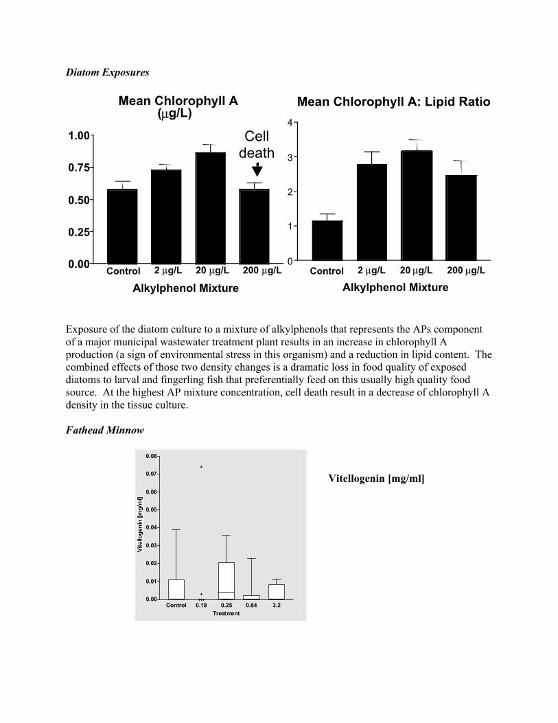
Diatom Exposures
Exposure of the diatom culture to a mixture of alkylphenols that represents the APs component of a major municipal wastewater treatment plant results in an increase in chlorophyll A production (a sign of environmental stress in this organism) and a reduction in lipid content. The combined effects of those two density changes is a dramatic loss in food quality of exposed diatoms to larval and fingerling fish that preferentially feed on this usually high quality food source. At the highest AP mixture concentration, cell death result in a decrease of chlorophyll A density in the tissue culture. Fathead Minnow
Control 0.19 0.25 0.84 3.20.00
0.01
0.02
0.03
0.04
0.05
0.06
0.07
0.08
Treatment
Vitellogenin [mg/ml]
Control 2 µg/L 20 µg/L 200 µg/L
Mean Chlorophyll A: Lipid Ratio
0
1
2
3
4
Alkylphenol Mixture
Mean Chlorophyll A(µg/L)
0.00
0.25
0.50
0.75
1.00
Alkylphenol MixtureControl 2 µg/L 20 µg/L 200 µg/L
Celldeath
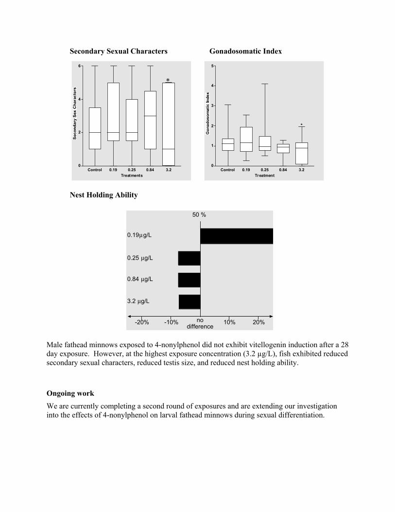
Gonadosomatic Index
Male fathead miday exposure. However, at the highesecondary sexual characters, reduced
Ongoing work We are currently compinto the effects of 4-nonylphe
-20%
0.19µg/L
0.25 µg/L
0.84 µg/L
3.2 µg/L
Control 0.19 0.25 0.840
2
4
6
Treatments
4
5
nnows exposed to 4-n
leting a secondnol on l
Nest Holding Ability
Secondary Sexual Characters
*
nin induction after a 28
st exposure testis
during sexual differentiation.
50 %
-10% 10% 20%nodifference
3.2 Control 0.19 0.25 0.84 3.20
1
2
3
*
Treatment
onylphenol did not exhibit vitellogeconcentration (3.2 µg/L), fish exhibited reduced
size, and reduced nest holding ability.
round of exposures and are extending our investigation arval fathead minnows
Summary of findings To date it appears that a mixture of APs, including 4-nonylphenol at concentrations well below the US EPA proposed criterion for chronic exposure (5.9µg/L) results in diminished lipid content in exposed diatoms. As a result larval and fingerling fish, which feed preferentially on this food source, will be faced with a lower quality food source. Nonylphenol also exhibits adverse effects on exposed male fathead minnows. These effects are more subtle, however at the highest concentration used in this experiment (3.2µg/L), fish exhibited consistent effects, including diminished secondary sexual characters, lower GSI, and reduced nest holding ability. The combination of these factors is indicative of reduced reproductive competency in exposed male fathead minnows. Further experiments are under way to ensure the repeatability of our results. References Ackermann, G., Schwaiger J, Negele RD, Fent K (2002). “Effects of long-term nonylphenol
exposure on gonadal development and biomarkers of estrogenicity in juvenile rainbow trout (Oncorhynchus mykiss).” Aquatic Toxicology 60: 203-221.
Ahlgren, G., L. Lundstedt, M. Brett, and C. Forsberg (1990) Lipid composition and food quality of some freshwater phytoplankton for cladoceran zooplankters. Journal of Plankton Research 12: 809-818.
Ankley, G. T., and J. P. Giesy (1998). Endocrine disruptors in wildlife: a weight-of-evidence perspective. Principles and Processes for Evaluating Endocrine Disruption in Wildlife. R. Kendall, R. Dickerson, J. P. Giesy, and W. Suk. Pensacola, FL, SETAC Press: 349-367.
Arukwe, A., Kullman SW, Hinton DE (2001). “Differential biomarker gene and protein expressions in nonylphenol and estradiol-17b treated juvenile rainbow trout (Oncorhynchus mykiss).” Comparative Biochemistry and Physiology Part C 129: 1-10.
Barber, L. B., Browb, G. K., Zaugg S. D. (2000). Potential endocrine disrupting organic chemicals in treated municipal wastewater and river water. Analysis of environmental endocrine disruptors: American Chemical Society Symposium Series.
Bell, A. (2001). “Effects of an endocrine disrupter on courtship and aggressive behaviour of male threespined stickleback, Gasterosteus acleatus.” Animal Behaviour 62: 775-780.
Denny, J. S. (1987). Guidelines for the culture of fathead minnows Pimephales promelas for the use in toxicity tests. Duluth, MN, Environmental Protection Agency: 1-41.
Farre, M., Kloter G, Petrovic M, Alonso MC, Lopez de Alda MJ, Barcelo D (2002). “Indentification of toxic compounds in wastewater treatment plants during a field experiment.” Analytica Chimica Acta 456: 19-30.
Field, J., Reed RL (1996). “Nonylphenol polyethoxy carboxylate metabolites of nonionic surfactants in US paper mill effluents, municipal sewage treatment plant effluents, and river waters.” Environmental Science Technology 30: 3544-3550.
Folmar, L. C., N. D. Denslow, V. Rao, M. Chow, A. Crain, J. Enblom, J. Marcino, and L. J. Guillette Jr. (1996). “Vitellogenin introduction and reduced serum testosterone concentrations in feral male carp (Cyprinus carpio) captured near a major metropolitan sewage treatment plant.” Environmental Health Perspectives 104: 1096-1100.
Folmar, L., Hemmer MJ, Denslow ND, Kroll K, Chen J, Cheek A, Richman H, Meredith H, Gordon-Grau E (2002). “A comparison of the estrogenic potencies of estradiol, ethynylestradiol, diethylstilbestrol, nonylphenol and methoxychlor in vivo and in vitro.” Aquatic Toxicology 60: 101-110.
Folmar, L., ND Denslow, K Kroll, EF Orlando, J Enblom, J Marcino, C Metcalfe, and LJ Guilette Jr (2001). “Altered serum sex steroids and vitellogenin induction in walleye (Stizostedion vitreum) collected near a metropolitan sewage treatment plant.” Arch Environ Contam Toxicol 40: 392-398.
Foran, C., Bennett ER, Benson WH (2000). “Exposure to environmentally relevant concentrations of different nonylphenol formulations in Japanese medaka.” Marine Environmental Research 50: 135-139.
Giesy, J., Pierens SL, Snyder EM, Miles-Richardson S, Kramer VJ, Snyder SA, Nichols KM, Villeneuve DA (2000). “Effects of 4-nonylphenol on fecundity and biomarkers of estrogenicity in fathead minnows (Pimephales promelas).” Environmental Toxicology and Chemistry 19: 1368-1377.
Gray, M. A., A. J. Niimi, and C. D. Metcalfe (1999). “Factors affecting the development of testis-ova in medaka, Orysias latipes, exposed to octylphenol.” Environ Toxicology and Chemistry 18(8): 1835-1842.
Gutendorf, B., Westendorf J (2001). “Comparison of an array of in vitro assays for the assessment of the estrogenic potential of natural and synthetic estrogens, phytoestrogens and xenoestrogens.” Toxicol 166: 79-89.
Harris, C., EM Santos, A Janbakhsh, TG Pottinger, CR Tyler, JP Sumpter (2001). “Nonylphenol affects gonadotropin levels in the pituitary gland and plasma of female rainbow trout.” Environ Sci Technol In Press
Hemmer, M., Hemmer BL, Bowman CJ, Kroll KJ, Folmar LC, Marcovich D, Hoglund MD, Denslow ND (2001). “Effects of p-nonylphenol, methoxychlor, and endosulfan on vitellogenin induction and expression in sheepshead minnow (Cyprinodon variegatus).” Environmental Toxicology and Chemistry 20: 336-343.
Jobling, S., M. Nolan, C. R. Tyler, G. Brighty, and J.P. Sumpter (1998). “Widespread sexual disruption in wild fish.” Environmental Science and Technology 32(17): 2498-2506.
Lazinsky, D. and L. Sicko-Goad (1990). Morphometric analysis of phosphate and chromium interactions in Cyclotella meneghiniana. Aquatic Toxicology 16: 127-140.
Lazorchak, J., Hemming JM, Allen HJ, Huggett D, Brooks BW, Lattier D, Smith ME, Weichman B, Reddy TV, Gordon D (2001). Evaluation of a wastewater discharge using vitellogenin gene expression and plasma protein levels in male fathead minnows. Baltimore, MA, Setac.
Le Gac, F., Luc Thomas J, Mourot B, Loir M (2001). “In vivo and in vitro effects of prochloraz and nonylphenol ethoxylates on trout spermatogenesis.” Aqua Toxicol 53: 187-200.
Madigou, T., Le Goff P, Salbert G, Cravedi JP, Segner H, Pakdel F, Valotaire Y (2001). “Effects of nonylphenol on estrogen receptor conformation, transcriptional activity and sexual reversion in rainbow trout (Oncorhynchus mykiss).” Aqua Toxicol 53: 173-186.
Nichols, K. M., S. R. Miles-Richardson, E. M. Snyder, and J. P. Giesy (1998). “Effects of exposure to municipal wastewater in situ on the reproductive physiology of the fathead minnow (Pimephales promelas).” Environmental Toxicology and Chemistry 18(9): 2001-2012.
Nichols, K., Snyder EM, Snyder SA, Pierens SL, Miles-Richardson SR, Giesy JP (2001). “Effects of nonylphenol ethoxylate exposure on reproductive output and bioindicators of environmental estrogen exposure in fathead minnowsk, Pimephales promelas.” Environmental Toxicology and Chemistry 20: 510-522.
Sargent, R. C. (1988). “Paternal care and egg survival both increase with clutch size in the
fathead minnow, Pimephales promelas.” Behav Ecol Sociobiol 23: 33-37. Sargent, R. C. (1989). “Allopaternal care in the fathead minnow, Pimephales promelas:
stepfathers discriminate against their adopted eggs.” Behav Ecol Sociobiol 25: 379-385. Schelske, C.L. and L. Sicko-Goad (1990). Effect of chelated trace metals on phosphorus uptake
and storage in natural assemblages of Lake Michigan phytoplankton. Journal of Great Lakes Research 16: 82-89.
Schoenfuss, H. L. (2003). The need for novel approaches in assessing the biological impact of biologically active compounds. Third International Conference on Pharmaceuticals and Endocrine Disrupting Chemicals in Water, Minneapolis, USA, National Groundwater Association.
Schoenfuss, H. L., D Martinovic, P W Sorensen (2001). “Effects of exposure to low levels of water-borne 7b-estradiol on nest holding ability and sperm quality in fathead minnows.” Water Resources Update 120(9): 49-55.
Schoenfuss, H., JT Levitt, G Van Der Kraak, PW Sorensen (2002). “Ten week exposure to treated sewage effluent discharge has relatively minor, variable effects on reproductive behavior and sperm production in goldfish.” Environmental Toxicology and Chemistry 21(10): 2185-2190.
Sicko-Goad, L. and E.F. Stoermer (1979). A morphometric study of lead and copper effects on Diatoma tenue var. elongatum (Bacillariophyta). Journal of Phycology 15: 316-321.
Sicko-Goad, L. and N.A. Andresen (1991). Effect of growth and light/ dark cycles on diatom lipid content and composition. Journal of Phycology 27: 710-718.
Sicko-Goad, L. and N.A. Andresen (1993a). Effect of lipid composition on the toxicity of trichlorobenzene isomers to diatoms. I. Short-term effects of 1,3,5-trichlorobenze. Arichives of Environmental Contamination and Toxicology 24: 236-242.
Sicko-Goad, L. and N.A. Andresen (1993b). Effect of lipid composition on the toxicity of trichlorobenzene isomers to diatoms. II. Long-term effects of 1,2,3-trichlorobenze. Arichives of Environmental Contamination and Toxicology 24: 243-248.
Sicko-Goad, L. M.S. Evans, D. Lazinsky, J. Hall, and M.S. Simmons (1989b) Effects of chlorinated benzenes on diatom fatty acid composition and quantitative morphology. IV. Pentachlorobenzene and comparison with trichlorobenze isomers. Arichives of Environmental Contamination and Toxicology 18: 656-668.
Sicko-Goad, L., B.G. Ladewski, and D. Lazinsky (1986). Synergistic effects of nutrients and lead on the quantitative ultrastructure of Cyclotella (Bacillariophyceae). Archives of Environmental Contamination and Toxicology 15: 291-300.
Sicko-Goad, L., J. Hall, D. Lazinsky, and M.S. Simmons (1989a). effects of chlorinated benzenes on diatom fatty acid composition and quantitative morphology. III. 1,2,3-trichlorobenzene. Archives of Environmental Contamination and Toxicology 18: 647-655.
Sicko-Goad, L., M.S. Simmons, D.L. Lazinsky, and J. Hall (1988). Effect of light cycle on diatom fatty acid composition and quantitative morphology. Journal of Phycology 24: 1-7.
Stoermer, E.F. and J. Smol (2000). The Diatoms: Applications for Earth and Environmental Sciences. Cambridge University Press, 482 pp.
Trudeau, V., Wade MG, Van Der Kraak G, Peter RE (1993). “Effects of 17b-estradiol on pituitary and testicular function in male goldfish.” Can. J. Zool. 71: 1131-1135
Unger, L. M. (1983). “Nest defense by deceit in the fathead minnow, Pimephales promelas.”
Behav Ecol Sociobiol 13: 125-130. Unger, L. M., and R. C. Sargent (1988). “Allopaternal care in the fathead minnow, Pimephales
promelas: females prefer males with eggs.” Behav Ecol Sociobiol 23: 27-32. Villeneuve, D., Villalobos SA, Keith TL, Snyder EM, Fitzgerald SD, Giesy JP (2002). “Effects
of waterborne exposure to 4-nonylphenol on plasma sex steroid and vitellogenin concentrations in sexually mature male carp (Cyprinus carpio).” Chemosphere 47: 15-28.
Volkman, J.K., S.W. Jeffery, P.D. Nichols, G.I. Rogers, and C.D. Garland (1989). Fatty acid and lipid composition of 10 species of microalgae used in mariculture. Journal of Experimental Marine Biology and Ecology 128: 219-240.
Winkler, M., J. R. Lawrence, et al. (2001). “Selective degradation of ibuprofen and clofibric acid in two model river biofilm systems.” Water Research 35(13): 3197-3205.
Zapolska-Downar, D. a. M. N. (2001). “A pleiotropic antiatherogenic action of ibuprofen.” Med Sci Monit 7(4): 837-841.
Statement of related grants submitted or funded as a result of this project Drs. Schoenfuss, Barber, Julius, and Norris have submitted a US EPA STAR grant application to further the investigation into the effects of alkylphenols on aquatic vertebrates and to develop rapid assessment tools for their detection. Drs. Schoenfuss, Barber and Lee received a grant from the MN Pollution Control Agency to continue our work on the effects of 4-nonylphenol on aquatic vertebrates in Minnesota. Description of student training provided by project: Name: Travis Bistodeau Program: Department of Biological Sciences, St. Cloud State University, St. Cloud, MN Degree being sought: Masters of Science Name: Jason Koch Program: Department of Biological Sciences, St. Cloud State University, St. Cloud, MN Degree being sought: Masters of Science Name: Kent Groove Program: Department of Biological Sciences, St. Cloud State University, St. Cloud, MN Degree being sought: B.S., and Masters of Science

Information Transfer ProgramN/A
Student SupportNone
Notable Awards and AchievementsMcNeill, Kristopher. 2004. McKnight Land-Grant Professor. Recipients are honored with this title, aspecial award they will hold for two years. The award consists of a $25,000 research grant in each of twoyears, summer support, and a research leave in the second year. The winners were chosen for theirpotential for important contribution to their field; the degree to which their past achievements and currentideas demonstrate originality, imagination, and innovation; the potential for attracting outstandingstudents; and the significance of the research and the clarify with which it is conveyed to thenon-specialist.
LaPara, Timothy. 2004. Awarded research grant from CURA for his project titled "The Potential Role ofMunicipal Wastewater Treatment Facilities in the Proliferation of Antibiotic Resistant Bacteria."
LaPara, Timothy. 2004. Received research grant from NASA for his project titled "Advanced LifeSupport: Development of the M2BR for biological treatment of wastewater generated during long-termspace missions."
Leonard C. Ferrington, Jr., asked to serve on Technical Advisory Committee by Emmons & OlivierResources, Inc. Technical Advisory Committee assists in developing field design and interpreting resultsof studies to aid in TMDL development for Hardwood Creek in Washington County, MN. Invitation toserve on committee was due to experience derived from this project and others designed to integratebiological data into TMDL assessment protocols for use by state of Minnesota.
Travel grant from graduate school awarded to Mr. Adam Sealock (Graduate Student in Water ResourcesProgram, enrolled in MS degree program) to attend annual meeting of the North American BenthologicalSociety in New Orleans, May 2005. The title of his presentation was "Bottle Traps and Dipnetting:Evaluation of two Sampling Techniques for Assessing Macroinvertebrate Biodiversity in DepressionalWetland." Mr. Sealock is a student supported with Dr. Ferrington’s grant.
Travel grant from graduate school awarded to Mr. Jeffrey Werner (Graduate Student in Water ResourcesProgram, enrolled in PhD degree program) to attend the 28th American Chemical Society NationalMeeting, in Philadelphia, PA , August 8-13, 2004. The title of his presentation was "Kinetics of theEnvironment Photodegradation of Mefenamic Acid" Mr. Werner is a student supported with Drs. WilliamArnold and Kris McNeill’s grants.
Publications from Prior Projects1. 1999MNB1 ("Investigation of Factors Affecting the Removal of Cryptosporidium parvum Ocycts in
Porous Media") - Conference Proceedings - Dai, X., and Hozalski, R. M. 2001. "Effect of nom andbiofilm on the removal of cryptosporidium parvum oocysts in bench-scale granular media filters."Proceedings - Water Quality Technology Conference, 812-819.
2. 1999MNB1 ("Investigation of Factors Affecting the Removal of Cryptosporidium parvum Ocycts inPorous Media") - Articles in Refereed Scientific Journals - Dai, X., and Hozalski, R. M. 2003.
"Evaluation of microspheres as surrogates for cryptosporidium parvum oocysts in filtrationexperiments." Environmental Science and Technology, 37(5), 1037-1042.
3. 2001MN281G ("Photochemical Fate of Pharmaceutical Compounds Discharged and Detected inNatural Waters") - Articles in Refereed Scientific Journals - Boreen, A. L., Arnold, W. A., and McNeill,K. 2003a. "Photodegradation of pharmaceuticals in the aquatic environment: A review." Aquatic Sciences,65(4), 320-341.
4. 2001MN281G ("Photochemical Fate of Pharmaceutical Compounds Discharged and Detected inNatural Waters") - Conference Proceedings - Latch, D. E., Arnold, W. A., and McNeill, K. 2002a. "Singletoxygen and the photochemical fate of triclosan." Preprints of Extended Abstracts presented at the ACSNational Meeting, American Chemical Society, Division of Environmental Chemistry, 42(1), 593-597.
5. 2001MN281G ("Photochemical Fate of Pharmaceutical Compounds Discharged and Detected inNatural Waters") - Conference Proceedings - Latch, D. E., Arnold, W. A., and McNeill, K. 2002b."Singlet oxygen and the photochemical fate of triclosan." Abstracts of Papers, 223rd ACS NationalMeeting, Orlando, FL, United States, April 7-11, 2002, ENVR-039.
6. 2001MN281G ("Photochemical Fate of Pharmaceutical Compounds Discharged and Detected inNatural Waters") - Articles in Refereed Scientific Journals - Latch, D. E., Packer, J. L., Arnold, W. A., andMcNeill, K. 2003a. "Photochemical conversion of triclosan to 2,8-dichlorodibenzo-p-dioxin in aqueoussolution." Journal of Photochemistry and Photobiology, A: Chemistry, 158(1), 63-66.
7. 2001MN281G ("Photochemical Fate of Pharmaceutical Compounds Discharged and Detected inNatural Waters") - Conference Proceedings - Latch, D. E., Stender, B. L., and McNeill, K. 2001a. "Singletoxygen and the photochemical fate of pharmaceuticals." Abstracts of Papers, 222nd ACS NationalMeeting, Chicago, IL, United States, August 26-30, 2001, ENVR-135.
8. 2001MN281G ("Photochemical Fate of Pharmaceutical Compounds Discharged and Detected inNatural Waters") - Conference Proceedings - Latch, D. E., Stender, B. L., and McNeill, K. 2001b. "Singletoxygen and the photochemical fate of pharmaceuticals." Preprints of Extended Abstracts presented at theACS National Meeting, American Chemical Society, Division of Environmental Chemistry, 41(2), 57-60.
9. 2001MN281G ("Photochemical Fate of Pharmaceutical Compounds Discharged and Detected inNatural Waters") - Conference Proceedings - Packer, J. L., Latch, D. E., McNeill, K., and Arnold, W. A.2003a. "Photochemical fate of pharmaceuticals in the environment: Naproxen, ibuprofen, diclofenac, andclofibric acid." Preprints of Extended Abstracts presented at the ACS National Meeting, AmericanChemical Society, Division of Environmental Chemistry, 43(1), 396-400.
10. 2001MN281G ("Photochemical Fate of Pharmaceutical Compounds Discharged and Detected inNatural Waters") - Conference Proceedings - Packer, J. L., Latch, D. E., McNeill, K., and Arnold, W. A.2003b. "Photochemical fate of pharmaceuticals in the environment: Naproxen, ibuprofen, diclofenac, andclofibric acid." Abstracts of Papers, 225th ACS National Meeting, New Orleans, LA, United States, March23-27, 2003, ENVR-218.
11. 2001MN281G ("Photochemical Fate of Pharmaceutical Compounds Discharged and Detected inNatural Waters") - Articles in Refereed Scientific Journals - Packer, J. L., Werner, J. J., Latch, D. E.,McNeill, K., and Arnold, W. A. 2003c. "Photochemical fate of pharmaceuticals in the environment:Naproxen, diclofenac, clofibric acid, and ibuprofen." Aquatic Sciences, 65(4), 342-351.
12. 2001MN1041G ("Antibiotic Losses in Runoff and Drainage from Manure-Applied Fields") - OtherPublications - 2001MN1041G Chander, Y., Kumar, K., Gupta, S. C., and Goyal, S. M. 2003."Antimicrobial activity of soil bound antibiotics." Agronomy Abstract (S11-Chander 633887).
13. 2003MN29B ("Arsenic in Minnesota Groundwater and its Impact on the Drinking Water Supply") -Other Publications - Erickson, M. L. B., R.J. 2004a. "Arsenic in ground water: Recent research andimplications for minnesota." CURA Reporter, 34(2).
14. 2003MN29B ("Arsenic in Minnesota Groundwater and its Impact on the Drinking Water Supply") -Other Publications - Erickson, M. L. B., R.J. 2004b. "Arsenic in ground water: Recent research andimplications for minnesota." Minnesota Ground Water Association Newsletter, 23(1).
15. 2003MN32G ("Photochemistry of Antibiotics and Estrogens in Surface Waters: Persistence andPotency") - Conference Proceedings - Boreen, A. L., Arnold, W. A., and McNeill, K. 2003b."Photochemical fate of pharmaceuticals in the environment: Sulfa drugs." Abstracts of Papers, 226th ACSNational Meeting, New York, NY, United States, September 7-11, 2003, ENVR-180.
16. 2003MN32G ("Photochemistry of Antibiotics and Estrogens in Surface Waters: Persistence andPotency") - Conference Proceedings - Boreen, A. L., Arnold, W. A., and McNeill, K. 2003c."Photochemical fate of pharmaceuticals in the environment: Sulfa drugs." Preprints of Extended Abstractspresented at the ACS National Meeting, American Chemical Society, Division of EnvironmentalChemistry, 43(2), 193-199.
17. 2003MN32G ("Photochemistry of Antibiotics and Estrogens in Surface Waters: Persistence andPotency") - Conference Proceedings - Boreen, A. L., Arnold, W. A., and McNeill, K. Photochemical fateof sulfa drugs in the aquatic environment. Abstracts of Papers, 228th ACS National Meeting, Philadelphia,PA, United States, August 22-26, 2004, ENVR-287.
18. 2003MN32G ("Photochemistry of Antibiotics and Estrogens in Surface Waters: Persistence andPotency") - Other Publications - Boreen, A. L., Arnold, W. A., and McNeill, K. 2004c. "Photochemicalfate of sulfa drugs in the aquatic environment." Preprints of Extended Abstracts presented at the ACSNational Meeting, American Chemical Society, Division of Environmental Chemistry, 44(2), 1402-1407.
19. 2003MN32G ("Photochemistry of Antibiotics and Estrogens in Surface Waters: Persistence andPotency") - Articles in Refereed Scientific Journals - Boreen, A. L., Arnold, W. A., and McNeill, K.2004d. "Photochemical fate of sulfa drugs in the aquatic environment: Sulfa drugs containingfive-membered heterocyclic groups." Environmental Science and Technology, 38(14), 3933-3940.
20. 2003MN32G ("Photochemistry of Antibiotics and Estrogens in Surface Waters: Persistence andPotency") - Articles in Refereed Scientific Journals - Latch, D., E., Stender, B., L., Packer, J., L., Arnold,W., A., and McNeill, K. 2003b. "Photochemical fate of pharmaceuticals in the environment: Cimetidineand ranitidine." Environmental science & technology, 37(15), 3342-50.
21. 2003MN32G ("Photochemistry of Antibiotics and Estrogens in Surface Waters: Persistence andPotency") - Conference Proceedings - Latch, D. E., Packer, J., Arnold, W. A., and McNeill, K. 2003c."The photochemical fate of cimetidine and ranitidine hydrochloride." Preprints of Extended Abstractspresented at the ACS National Meeting, American Chemical Society, Division of EnvironmentalChemistry, 43(1), 266-271.
22. 2003MN32G ("Photochemistry of Antibiotics and Estrogens in Surface Waters: Persistence andPotency") - Conference Proceedings - Latch, D. E., Packer, J. L., Arnold, W. A., and McNeill, K. 2003d."The photochemical fate of cimetidine and ranitidine hydrochloride." Abstracts of Papers, 225th ACSNational Meeting, New Orleans, LA, United States, March 23-27, 2003, ENVR-006.
23. 2003MN32G ("Photochemistry of Antibiotics and Estrogens in Surface Waters: Persistence andPotency") - Articles in Refereed Scientific Journals - Latch, D. E. P., J.L., Stender, B.L.; VanOverbeke, J.;Arnold, W.A.; McNeill, K. submitted. "Aqueous photochemistry of triclosan: Formation of2,4-dichlorodibenzo-p-dioxin and oligomerization products." Environ. Toxicol. Chem.