VTE PrEVEnTion and TrEaTmEnT - Clot Connectfiles. · 2012-02-06 · VTE PrEVEnTion . and TrEaTmEnT:...
22
This activity is co-provided by Global Education Group and SCEPTER. vte treatment VTE PREVENTION and TREATMENT: A Practical “How To” DATE OF RELEASE: July 6, 2010 DATE OF EXPIRATION: July 6, 2011 This activity is jointly sponsored by SCEPTER TM and Quintiles Medical Education. This activity is supported by an educational grant from sanofi-aventis U.S. A CME/CE CERTIFIED EDUCATIONAL PAMPHLET
Transcript of VTE PrEVEnTion and TrEaTmEnT - Clot Connectfiles. · 2012-02-06 · VTE PrEVEnTion . and TrEaTmEnT:...
This activity is co-provided by Global Education Group and SCEPTER.
vtetreatm
ent VTE PrEVEnTion and TrEaTmEnT:A Practical “How To”
DaTE of rElEasE: July 6, 2010 DaTE of ExPiraTion: July 6, 2011
This activity is jointly sponsored by SCEPTERTM and Quintiles Medical Education.
This activity is supported by an educational grant from sanofi-aventis U.S.
A CME/CE CERTifiEd EdUCATionAl PAMPHlET

1
Program sTaTEmEnT of nEEDsA recent multinational survey based on acute care hospital chart reviews utilized American College of Chest Physicians (ACCP) guidelines to assess the risk of developing venous thromboembolism (VTE). Survey results showed that less than half of at-risk medical patients received ACCP-recom-mended VTE prophylaxis, highlighting the important need for proactive identification of at-risk medical patients in acute care through stratification and institution of guideline- recommended preventive measures. in addition to inconsistent recognition and underutilization of appropriate preventive strategies, there are also important clinical needs concerning the treatment of VTE. for instance, the transition from the hospital environment to the home or other outpatient setting is a time when suboptimal management or gaps in communication can adversely impact outcomes. Recognizing these clinical gaps in patient care, the Joint Commission on Accreditation of Healthcare organizations (JCAHo) has established prophylaxis and treatment of VTE as a safety priority in their currently published national Patient Safety Goals (nPSGs).
This Educational Pamphlet will address methods to assess risk for deep-vein thrombosis (dVT), duration of VTE prophylaxis and treatment, and other important issues relevant to the contemporary treatment of VTE.
Agenda• front Matter: 5 minutes • Pamphlet Presentation: 45 minutes • Evaluation forms: 5 minutes• Posttest: 5 minutes• Total Estimated Time to Complete This Activity: 1 hour
CmE informaTionEducational Objectives• identify hospitalized patients who are at risk for venous
thromboembolic events • describe appropriate VTE prophylaxis and treatment based
on current evidence-based recommendations • Review JCAHo standards concerning VTE management and
assess their clinical applications
AudienceThis activity is intended for hospitalists, orthopedic sur-geons, hematologists, intensivists, general internal medicine and family physicians, and nurses who treat patients with or at risk for dVT.

CrEDiT DEsignaTion sTaTEmEnTPhysiciansSCEPTER designates this educational activity for a maximum of 1.0 AMA PRA Category 1 CreditTM. Physicians should only claim credit commensurate with the extent of their participa-tion in the activity.
This activity, VTE Prevention and Treatment: A Practical ‘How To”, has been reviewed and is acceptable for up to 1 Prescribed credits(s) by the American Academy of family Physicians. AAfP accreditation begins July 6, 2010. Terms of approval is for one year(s) from this date, with option for yearly renewal.
NursesThis educational activity for 1.0 contact hour is provided by Global Education Group.
for information about the nursing accreditation of this program, please contact Global at 303-395-1782 or [email protected].
aCCrEDiTaTion sTaTEmEnTSCEPTER is accredited by the Accreditation Council for Continu-ing Medical Education (ACCME) to provide continuing medical education (CME) for physicians.
Global Education Group is an approved provider of continu-ing nursing education by the Colorado nurses Association, an accredited approver by the American nurses Credentialing Center’s Commission on Accreditation.
CmE ConTEnT rEViEwThis activity has been reviewed for relevance, accuracy of content, and fair balance by CME Peer Review, llC.
fEE informaTionThere is no fee to participate in this educational activity.
CErTifiCaTE of CrEDiT fulfillmEnT insTruCTionsParticipants should read the educational objectives and review the pamphlet in its entirety. After reviewing the pamphlet, complete the posttest and evaluation in the back of the pamphlet and fax to 866-463-1693. You can also visit www.sceptercme.com/VTEHowTo to complete the posttest and evaluation online.
2
3
faCulTyThis activity has been prepared under the direction ofStephan Moll, MdAssociate Professordivision of Hematology-oncologydepartment of MedicineUniversity of north Carolina School of MedicineChapel Hill, north Carolina
aafP rEViEwErMichael E. Cobble, Md, AAfP, fnlAdirector, Canyons Medical CenterSandy, Utah Adjunct faculty, University of Utah School of MedicineSalt lake City, UtahChief Medical officer, Atherotech, inc.Birmingham, Alabama
DisClosurE informaTionPolicy on Disclosure SCEPTER and Quintiles Medical Education adhere to the ACCME Essential Areas and Policies, including the Standards for Commercial Support, regarding industry support of CME. disclosure information is provided during the planning process to ensure resolution of any identified conflicts. disclosure of faculty and commercial relationships, as well as the discussion of unlabeled or unapproved use of any drug, device, or procedure by the faculty, will be disclosed to learners.
Global Education Group (Global) requires instructors, planners, managers, and other individuals who are in a position to control the content of this activity to disclose any real or apparent conflict of interest they may have as related to the content of this activity. All identified conflicts of interest are thoroughly vetted by Global for fair balance, scientific objectivity of studies mentioned in the materials or used as the basis for content, and appropriate-ness of patient care recommendations.
The planners and managers reported the following financial relationships or relationships to products or devices they or their spouse/life partner have with commercial interests related to the content of this CME activity:Michael Perlmutter, MS, Pharmd, has no significant financial relationships to disclose.Jackie dawson, MSn, has no significant financial relationships to disclose.
Amanda Glazer, Phd, has no significant financial relationships to disclose.
4
Faculty DisclosureStephan Moll, Md, has received honoraria from ortho-Mcneil and international Technidyne Corporation. He has received research support from international Technidyne Corporation.
dr. Moll may discuss unlabeled/unapproved uses of dabiga-tran and rivaroxaban.
AAFP Reviewer DisclosureMichael Cobble, Md, has served on the speakers bureaus for Abbott, AstraZeneca, Eli lilly, GlaxoSmithKline, forest and novo nordisk. He has served as a consultant for Atherotech, inc.
CME Peer Review, LLC, Conflict of Interest ResolutionReviewers are required to disclose all relevant relationships with any commercial interest to CME Peer Review, llC, prior to being accepted as a reviewer. Reviewers are required to update such information, are vetted and determined to be free of conflict of interest prior to conducting each activity content evaluation.
Disclosure of Unlabeled UseThis educational activity may contain discussion of pub-lished and/or investigational uses of agents that are not indicated by the fdA. Global Education Group (Global) and SCEPTER do not recommend the use of any agent outside of the labeled indications.
The opinions expressed in the educational activity are those of the faculty and do not necessarily represent the views of any organization associated with this activity. Please refer to the official prescribing information for each product for discussion of approved indications, contraindications, and warnings.
DisClaimErThe opinions expressed herein are those of the authors and do not necessarily represent the views of the sponsor, the educational partner, or the supporter. Please review complete prescribing information for specific drugs or com-bination of drugs, including indications, contraindications, warnings, and adverse effects before administering pharmacologic therapy to patients.

5
VEnous ThromboEmbolism:30 Clinical highlights and management aids
stephan moll, mDdepartment of Medicine, division of Hematology-oncologyUniversity of north Carolina School of MedicineChapel Hill, nC, USA
a. VTE PrEVEnTion1. VTE prophylaxis: Upon admission to the hospital, individualized
VTE risk assessment of each patient is necessary. VTE prophylaxis should be given based on VTE risk, unless absolute or relative contraindications exist. Hospital-wide VTE prophylaxis guidelines should be in place at each institution.1-2 detailed, evidence-based recommendations of VTE prophylaxis are discussed in the American College of Chest Physicians (ACCP) 2008 guidelines.2 discrepant rec-ommendations for VTE prophylaxis for patients undergoing hip and knee arthroplasty exist between the ACCP and American Academy of orthopedic Surgeons (AAoS).3 VTE risk categories are as follows:
• Highest risk (These patients should receive pharmacologic VTE prophylaxis PlUS sequential compression device):
- Hip and knee arthroplasties
- Hip and pelvic/acetabular fractures
- Acute spinal injury with paresis
- Major trauma
- High risk neurosurgery
• Moderate to high risk (These patients should receive pharmacologic VTE prophylaxis; sequential compression device is optional):
- Medical and surgical patients, not low or highest risk
• low risk (These patients do not require pharmacologic VTE prophy-laxis or sequential compression device. Early ambulation is sufficient):
- Medical patients, fully mobile
- Minor surgery, fully mobile patients, no risk factors
- Pregnancy, no other risk factors
- Arthroscopy, no risk factors
2. Reassessment of VTE risk: The risk of VTE and the indication for VTE prophylaxis should be re-assessed daily in hospitalized patients.
3. Length of VTE prophylaxis: Extended VTE prophylaxis for up to 4-5 weeks should be considered in high risk orthopedic and surgical patients, such as those undergoing total hip and knee replacement,
5
66
hip fracture surgery, general and gynecologic cancer surgery, or those with a previous history of VTE.
b. Diagnosis4. VTE risk factors: Recognition of a patient’s VTE risk factors is
important when assessing the etiology of a patient’s extrem-ity or respiratory symptoms. The Well’s score can be used for outpatients.4-5 if the pretest probability for dVT or PE based on the Well’s criteria is low, a d-dimer can be obtained. A negative d-dimer in patients with low pretest probability of VTE rules out VTE. A positive d-dimer needs to be followed up with an imaging study. A d-dimer or guide imaging work-up should not be obtained in the patient with an intermediate or high pretest probability for VTE.
5. Computed Tomography Angiography: CTA findings may be false positive or false negative6: a negative CTA in a patient with a high pretest probability for PE does not rule out PE and should be followed by another imaging study, such as a ventilation-perfusion (VQ) scan. And, vice versa, a positive CTA in a patient with a low pretest probability for PE does not necessarily mean that the patient has a PE. Another imaging study, such as a VQ scan, should be obtained.
6. VQ scan: A VQ scan can not differentiate between acute or chronic PE because both present as a perfusion-ventilation mismatch. VQ scan abnormalities frequently persist for at least several months.7 Thus, in a patient with previous PE and new respiratory symptoms, an abnormal VQ scan is not diagnostic for recurrent PE unless an old VQ scan is available for compari-son.
C. TrEaTmEnT7. ACCP Guidelines: The 2008 American College of Chest Physicians
Practice Guidelines on Antithrombotic and Thrombolytic Therapy offer detailed, evidence-based recommendations on a multitude of anticoagulation issues regarding venous thromboembolism prevention and treatment. They are collectively available as a Pdf file on the web.8 They are an extremely valuable tool.
a) Acute Management:8. Outpatient management: A subcutaneous drug such as lMWH or
fondaparinux is the preferred treatment for patients with acute dVT. if the patient needs to be admitted, lMWH is the pre-ferred choice over unfractionated heparin (UfH).9 Alternatively, fondaparinux can be used. if UfH is used, a heparin nomogram should be administered: a bolus of 80 U/kg should be followed by a continuous infusion of 18 U/kg/hour.9 for outpatient treatment of PE, lMWH, fondaparinux or UfH are good options, but, for inpatients lMWH is preferred over UfH.9
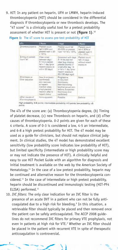
9. HIT: in any patient on heparin, UfH or lMWH, heparin-induced thrombocytopenia (HiT) should be considered in the differential diagnosis if thrombocytopenia or new thrombosis develops. The “4T score” is a clinically useful tool for a pretest probability assessment of whether HiT is present or not (figure 1).10 The 4Ts of the score are: (a) Thrombocytopenia degree, (b) Timing of platelet decrease, (c) new Thrombosis on heparin, and (d) oTher causes of thrombocytopenia. 0-2 points are given for each of these 4 criteria. A score of 0-3 is considered a low, 4-5 an intermediate, and 6-8 a high pretest probability for HiT. The 4T model may be used as a guide for clinicians, but should not replace clinical judg-ment. in clinical studies, the 4T model has demonstrated excellent sensitivity (low probability score indicates low probability of HiT), but limited specificity (intermediate or high probability score may or may not indicate the presence of HiT). A clinically helpful and easy to use HiT Pocket Guide with an algorithm for diagnosis and initial treatment is available on the web by the American Society of Hematology.11 in the case of a low pretest probability, heparin may be continued and alternative reason for the thrombocytopenia con-sidered.11 in the case of intermediate or high pretest probability, heparin should be discontinued and immunologic testing (HiT-Pf4 EliSA) performed.11
10. IVC filters: The only clear indication for an iVC filter is the presence of an acute dVT in a patient who can not be fully anti-coagulated due to a high risk for bleeding.9 in this situation, a retrievable filter should typically be placed and then removed once the patient can be safely anticoagulated. The ACCP 2008 guide-lines do not recommend iVC filters for primary VTE prophylaxis, not even in patients at high risk for VTE.2 Whether an iVC filter should be placed in the patient with recurrent VTE in spite of therapeutic anticoagulation is controversial.
7
figure 1: The 4T score to assess pre-test probability of HiT

11. Thrombolytic therapy ± mechanical thrombectomy: The role of thrombolytic therapy and mechanical thrombectomy in the treatment of dVT and in preventing PTS has not yet been appropriately studied. Thus, their effectiveness and safety in different types of patients is not yet clear. However, this is presently being studied in the ATTRACT trial and patients with dVT symptoms of ≤2 weeks should be encouraged to find an ATTRACT trial site for consideration of enrollment.12 Thrombo-lytic therapy in PE is indicated in the patient with hemody-namic compromise. it can be considered in selected patients without hypotension, but at high risk for adverse clinical outcomes, i.e. patients with right ventricular dysfunction. its benefits in this situation have not yet been appropriately studied, but results of an ongoing trial (PEiTHo) are expected in 2011-2012.
12. Genotyping for warfarin sensitivity: Certain polymorphisms in the CYP2C9 enzyme metabolizing warfarin and in the VKoR enzyme metabolizing vitamin K help predict what warfarin dose a patient may need to prolong the inR into the therapeutic range.13-14 However, this method has not yet been shown to improve clinical outcomes or to be cost- effective.13-14 Accordingly, genotyping should not be considered for routine clinical use at this time.
13. Warfarin nomogram: A nomogram should be used when initi-ating warfarin therapy (figure 2).15 The starting dose should be decreased if indicators of a lower warfarin dose are pres-ent, for example age >75 years, malnutrition, serum albumin <3.0 g/dl, recent bowel resection, antibiotic therapy, liver dysfunction, baseline inR elevation, liver disease, alcoholism, congestive heart failure, diarrhea, body weight <50 kg, or drugs known to interfere with warfarin metabolism. A warfa-rin initiation dose can also be calculated by using the non-profit website www.warfarindosing.org.
8
figure 1: The 4T score to assess pre-test probability of HiT
figure 2. nomogram for warfarin initiation15
9
14. Overlapping warfarin with parenteral anticoagulant: The patient with acute VTE who is initiated on warfarin needs to receive a parenteral anticoagulant overlapping with warfarin for at least 5 days and until the inR is above 2.0 in order to minimize risk of progression of thrombosis and of warfarin skin necrosis.9
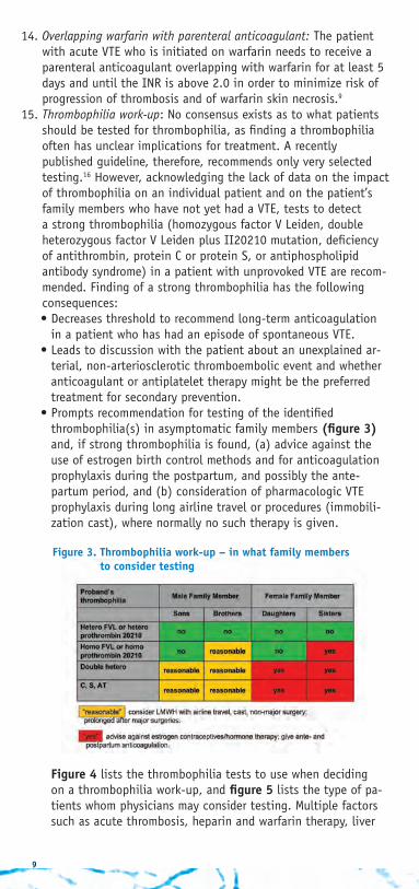
15. Thrombophilia work-up: no consensus exists as to what patients should be tested for thrombophilia, as finding a thrombophilia often has unclear implications for treatment. A recently published guideline, therefore, recommends only very selected testing.16 However, acknowledging the lack of data on the impact of thrombophilia on an individual patient and on the patient’s family members who have not yet had a VTE, tests to detect a strong thrombophilia (homozygous factor V leiden, double heterozygous factor V leiden plus ii20210 mutation, deficiency of antithrombin, protein C or protein S, or antiphospholipid antibody syndrome) in a patient with unprovoked VTE are recom-mended. finding of a strong thrombophilia has the following consequences:
• decreases threshold to recommend long-term anticoagulation in a patient who has had an episode of spontaneous VTE.
• leads to discussion with the patient about an unexplained ar-terial, non-arteriosclerotic thromboembolic event and whether anticoagulant or antiplatelet therapy might be the preferred treatment for secondary prevention.
• Prompts recommendation for testing of the identified thrombophilia(s) in asymptomatic family members (figure 3) and, if strong thrombophilia is found, (a) advice against the use of estrogen birth control methods and for anticoagulation prophylaxis during the postpartum, and possibly the ante-partum period, and (b) consideration of pharmacologic VTE prophylaxis during long airline travel or procedures (immobili-zation cast), where normally no such therapy is given. figure 4 lists the thrombophilia tests to use when deciding on a thrombophilia work-up, and figure 5 lists the type of pa-tients whom physicians may consider testing. Multiple factors such as acute thrombosis, heparin and warfarin therapy, liver
figure 3. Thrombophilia work-up – in what family members to consider testing
10
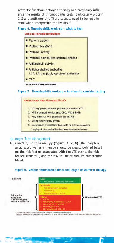
synthetic function, estrogen therapy and pregnancy influ-ence the results of thrombophilia tests, particularly protein C, S and antithrombin. These caveats need to be kept in mind when interpreting the results.17
b) longer-Term Management16. Length of warfarin therapy (figures 6, 7, 8): The length of
anticipated warfarin therapy should be clearly defined based on the risk factors associated with the VTE event, the risk for recurrent VTE, and the risk for major and life-threatening bleed.
figure 4. Thrombophilia work-up – what to test
figure 5. Thrombophilia work-up – in whom to consider testing
figure 6. Venous thromboembolism and length of warfarin therapy
11
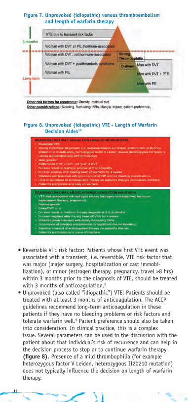
• Reversible VTE risk factor: Patients whose first VTE event was associated with a transient, i.e. reversible, VTE risk factor that was major (major surgery, hospitalization or cast immobi- lization), or minor (estrogen therapy, pregnancy, travel >8 hrs) within 3 months prior to the diagnosis of VTE, should be treated with 3 months of anticoagulation.9
• Unprovoked (also called “idiopathic”) VTE: Patients should be treated with at least 3 months of anticoagulation. The ACCP guidelines recommend long-term anticoagulation in these patients if they have no bleeding problems or risk factors and tolerate warfarin well.9 Patient preference should also be taken into consideration. in clinical practice, this is a complex issue. Several parameters can be used in the discussion with the patient about that individual’s risk of recurrence and can help in the decision process to stop or to continue warfarin therapy (figure 8). Presence of a mild thrombophilia (for example heterozygous factor V leiden, heterozygous ii20210 mutation) does not typically influence the decision on length of warfarin therapy.
figure 8. unprovoked (idiopathic) VTE - length of warfarin Decision aides15
figure 7. unprovoked (idiopathic) venous thromboembolism and length of warfarin therapy
.

12
• Cancer and dVT or PE: lMWH for 3-6 months, thereafter lMWH or warfarin, inR 2-3 indefinitely or until the cancer has resolved.9
• distal dVT, symptomatic: Anticoagulation for 3 months, then stop.
• Superficial thrombophlebitis: Spontaneous, i.e. not related to phlebotomy or iV catheter: at least 4 weeks of anti-coagulation, such as intermediate dose lMWH or UfH, or warfarin (target inR 2-3) overlapped with lMWH or UfH for 5 days.9
17. Anatomy/Terminology: Confusion as to which veins are superficial and which are deep can lead to misclass- ification of superficial thrombophlebitis and dVT and, thus, to incorrect treatment decisions. The key terminology:
• Arm: Basilic and cephalic veins are superficial veins; brachial vein is a deep vein.
• Leg: Greater and lesser saphenous veins are superficial veins; popliteal vein and anything proximal to it are proxi-mal veins; gastrocnemius and soleal veins are intramuscular calf veins and part of the deep venous system, and, together with the peroneal and tibial veins, make up the deep veins of the distal leg. The superficial femoral vein has been renamed femoral vein; it is a deep vein.
• doppler ultrasound of the legs can only visualize the veins distal to the inguinal ligament and therefore can not assess dVT in the iliac veins. CT or MRi venogram is needed to examine the iliac veins for dVT.
18. Anticoagulation clinics: Patients on warfarin need to be followed in a systematic way to optimize safety and efficacy.18 While smaller volume physician practices may well have these criteria in place, structured anticoagula-tion clinics often have the expertise and resources for optimal warfarin management. information about the location of anticoagulation clinics can be found at www.acforum.org, the website of the non-profit Anticoagulation forum.
19. INR patient self-testing: in appropriately selected patients on warfarin, self-testing is reliable, safe and effective.19 it is reimbursable for the patient by Medicare, Medicaid and many other insurance carriers. instructions on how patients and physicians can obtain a device are given in detail in a newsletter from the non-profit patient organization nBCA.20
20. Managing elevated INRs and bleeding on warfarin: figure 9 summarizes management as recommended by the ACCP 2008 guidelines.14
21. Surgical procedures on warfarin: if anticoagulation needs to be discontinued in a patient because of a surgical procedure, the decision of whether pre- and post-procedural anticoagulation bridging with a parenteral anticoagulant (lMWH, UfH, or fondaparinux) is needed

13
figure 9. management of elevated inrs or bleeding on warfarin
figure 10. Perisurgical anticoagulation management in patients on warfarin (= bridging therapy)21-22
depends on a patient’s thrombotic risk when not on antico-agulants, as well as the bleeding risk when on the bridging anticoagulant. The ACCP 2008 guidelines give guidance as to whom to bridge and what anticoagulant dose to use (prophy-lactic or therapeutic dose), summarized in figure 10.21-22
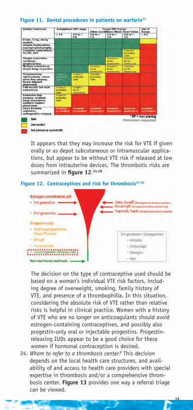
22. Dental procedures on warfarin: Many dental procedures can be done while on warfarin with therapeutic inRs (figure 11).23 More extensive procedures may require warfarin dose reduction or interruption.23 dentists can also use local measures (extra stitches, gauze packing, use of topical hemostatic agents) to minimize bleeding risk.
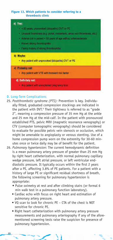
23. Safe birth control options: The risk of VTE with contraceptives is determined by the estrogen component, but also the type of progestin in combination pills. limited data are available on the risk for VTE with progestins taken alone for contraceptive purposes.
14
it appears that they may increase the risk for VTE if given
orally or as depot subcutaneous or intramuscular applica-tions, but appear to be without VTE risk if released at low doses from intrauterine devices. The thrombotic risks are summarized in figure 12.24-28 The decision on the type of contraceptive used should be based on a woman’s individual VTE risk factors, includ-ing degree of overweight, smoking, family history of VTE, and presence of a thrombophilia. in this situation, considering the absolute risk of VTE rather than relative risks is helpful in clinical practice. Women with a history of VTE who are no longer on anticoagulants should avoid estrogen-containing contraceptives, and possibly also progestin-only oral or injectable progestins. Progestin- releasing iUds appear to be a good choice for these women if hormonal contraception is desired.
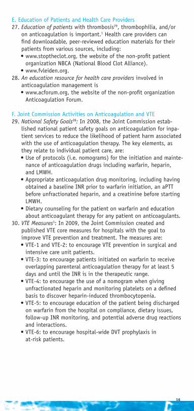
24. Whom to refer to a thrombosis center? This decision depends on the local health care structures, and avail-ability of and access to health care providers with special expertise in thrombosis and/or a comprehensive throm- bosis center. figure 13 provides one way a referral triage can be viewed.
figure 11. Dental procedures in patients on warfarin23
figure 12. Contraceptives and risk for thrombosis24-28
Permission requested.
15
d. long-Term Complications25. Postthrombotic syndrome (PTS): Prevention is key. individu-
ally fitted, graduated compression stockings are indicated in the patient with dVT.9 Their tightness is referred to as “grade 2”, meaning a compression pressure of 35 mm Hg at the ankle and 25 mm Hg at the mid-calf. in the patient with pronounced established PTS, pelvic MRV (magnetic resonance venography) or CTV (computer tomographic venography) should be considered to evaluate for possible pelvic vein stenosis or occlusion, which might be amenable to angioplasty or venous stenting. Use of a home compression pump worn on the extremity for 30-60 min-utes once or twice daily may be of benefit for the patient.
26. Pulmonary hypertension: The current hemodynamic definition is a mean pulmonary artery pressure of greater than 25 mm Hg by right heart catheterization, with normal pulmonary capillary wedge pressure, left atrial pressure, or left ventricular end- diastolic pressure. it typically occurs within the first 2 years after a PE, affecting 3.8% of PE patients. for a patient with a history of large PE or significant residual shortness of breath, the following screening for pulmonary hypertension is appropriate: • Pulse oximetry at rest and after climbing stairs (or formal 6
min walk test in a pulmonary function laboratory). • Cardiac echo with focus on right heart and estimation of
pulmonary artery pressure. • VQ scan to look for chronic PE – CTA of the chest is noT
sensitive for chronic PE. • Right heart catheterization with pulmonary artery pressure
measurements and pulmonary arteriography if any of the afore-mentioned screening tests raise the suspicion for presence of pulmonary hypertension.
figure 13. which patients to consider referring to a thrombosis clinic
16
E. Education of Patients and Health Care Providers27. Education of patients with thrombosis29, thrombophilia, and/or
on anticoagulation is important.1 Health care providers can find downloadable, peer-reviewed education materials for their patients from various sources, including:
• www.stoptheclot.org, the website of the non-profit patient organization nBCA (national Blood Clot Alliance).
• www.fvleiden.org.28. An education resource for health care providers involved in
anticoagulation management is • www.acforum.org, the website of the non-profit organization
Anticoagulation forum.
f. Joint Commission Activities on Anticoagulation and VTE29. National Safety Goals28: in 2008, the Joint Commission estab-
lished national patient safety goals on anticoagulation for inpa-tient services to reduce the likelihood of patient harm associated with the use of anticoagulation therapy. The key elements, as they relate to individual patient care, are:
• Use of protocols (i.e. nomograms) for the initiation and mainte-nance of anticoagulation drugs including warfarin, heparin, and lMWH.
• Appropriate anticoagulation drug monitoring, including having obtained a baseline inR prior to warfarin initiation, an aPTT before unfractionated heparin, and a creatinine before starting lMWH.
• dietary counseling for the patient on warfarin and education about anticoagulant therapy for any patient on anticoagulants.
30. VTE Measures1: in 2009, the Joint Commission created and published VTE core measures for hospitals with the goal to improve VTE prevention and treatment. The measures are:
• VTE-1 and VTE-2: to encourage VTE prevention in surgical and intensive care unit patients.
• VTE-3: to encourage patients initiated on warfarin to receive overlapping parenteral anticoagulation therapy for at least 5 days and until the inR is in the therapeutic range.
• VTE-4: to encourage the use of a nomogram when giving unfractionated heparin and monitoring platelets on a defined basis to discover heparin-induced thrombocytopenia.
• VTE-5: to encourage education of the patient being discharged on warfarin from the hospital on compliance, dietary issues, follow-up inR monitoring, and potential adverse drug reactions and interactions.
• VTE-6: to encourage hospital-wide dVT prophylaxis in at-risk patients.
abbrEbViaTionsAAoS = American Academy of orthopedic SurgeonsACA = anticardiolipin antibodyACCP = American College of Chest PhysiciansAfib = atrial fibrillationAT = antithrombinAPlA = antiphospholipid antibodiesaPTT = activated partial thromboplastin time CBC = complete blood count CHAdS2 = congestive heart failure–hypertension–age–
diabetes–strokeCT = computed tomographyCTA = computer tomographic angiographyCTV = computer tomographic venographydVT = deep vein thrombosisfVl = factor V leidenHg = mercuryHiT = heparin-induced thrombocytopeniainR = international normalized ratioiUd = intrauterine deviceiVC = inferior vena cava lA = lupus anticoagulantlMWH = low molecular weight heparin MTHfR = methylene tetrahydrofolate reductaseMRi = magnetic resonance imagingMRV = magnetic resonance venographynBCA = national Blood Clot AlliancePE = pulmonary embolism PnH = paroxysmal nocturnal hemoglobinuriaPTS = postthrombotic syndromeUfH = unfractionated heparinVKA = vitamin K antagonistsVKoR = vitamin k epoxide reductoseVQ = ventilation-perfusionVTE = venous thromboembolism
rEfErEnCEs1. Joint Commission on Accreditation of Healthcare organizations.
Joint Commission Perspectives. 2009;29:4-6. Available at: www.jointcommission.org/nR/rdonlyres/Ad24B9CB-57f6-4BdC-86dd-5dA0576AC15E/0/S4JCP0409.pdf. Accessed June 5, 2010.
2. Geerts WH, Bergqvist d, Pineo Gf, et al. Prevention of venous throm-boembolism. American College of Chest Physicians evidence-based clinical practice guidelines (8th edition). Chest. 2008;133:381S-453S.
3. American Academy of orthopaedic Surgeons. Clinical guideline on prevention of pulmonary embolism in patients undergoing total hip or knee arthroplasty. Available at: www.aaos.org/Research/guidelines/PE_guideline.pdf. Accessed June 17, 2010.
17

4. Wells PS, Anderson dR, Rodger M, et al. Evaluation of d-dimer in the diagnosis of suspected deep-vein thrombosis. N Engl J Med. 2003;349:1227-1235.
5. Wells PS, Anderson dR, Rodger M, et al. derivation of a simple clinical model to categorize patients probability of pulmonary embolism: increasing the models utility with the SimpliREd d-dimer. Thromb Haemost. 2000;83:416-420.
6. Stein Pd, fowler SE, Goodman lR, et al. Multidetector computed tomography for acute pulmonary embolism. N Engl J Med. 2006; 354:2317-2327.
7. Wartski M, Collignon MA. incomplete recovery of lung perfusion after 3 months in patients with acute pulmonary embolism treated with antithrombotic agents. THESEE Study Group. Tinzaparin ou Heparin Standard: Evaluation dans l’Embolie Pulmonaire Study. J Nucl Med. 2000;41:1043-1048.
8. American College of Chest Physicians. Antithrombotic and thrombolytic therapy, 8th Ed: ACCP guidelines. Chest. 2008;133: (suppl 6). Available at: http://www.chestnet.org/accp/guidelines/antithrombotic-and-thrombolytic-therapy-8th-edition. Accessed June 17, 2010.
9. Kearon C, Kahn SR, Agnelli G, Goldhaber SZ, Raskob GE, Comerota AJ. Antithrombotic therapy for venous thromboembolic disease: American College of Chest Physicians evidence-based clinical practice guidelines (8th edition). Chest. 2008;133:454S-545S.
10. lo GK, Juhl d, Warkentin TE, Sigouin CS, Eichler P, Greinacher A. Evaluation of pretest clinical score (4 T’s) for the diagnosis of heparin-induced thrombocytopenia in two clinical settings. J Thromb Haemost. 2006;4:759-765.
11. American Society of Hematology. 2009 Clinical practice guideline on the evaluation and management of heparin-induced thrombocytope-nia (HiT). Available at: http://www.hematology.org/SearchResults.aspx?searchtext=HiT. Accessed June 17, 2010.
12. The Attract Study Web site. Available at: http://www.attract.wustl. edu/default.aspx. Accessed June 17, 2010.13. Moyer TP, o’Kane dJ, Baudhuin lM, et al. Warfarin sensitivity geno-
typing: a review of the literature and summary of patient experience. Mayo Clin Proc. 2009;84:1079-1094.
14. Ansell J, Hirsh J, Hylek E, Jacobson A, Crowther M, Palareti G. Pharmacology and management of the vitamin K antagonists: American College of Chest Physicians evidence-based clinical practice guidelines (8th Edition). Chest. 2008;133:160S-198S.
15. Crowther MA, Harrison l, Hirsh J. Warfarin: less may be better. Ann Intern Med. 1997;127:332-333.
16. Baglin T, Gray E, Greaves M, et al. Clinical guidelines for testing for heritable thrombophilia. Br J Haematol. 2010;149:209-220.
17. foy P, Moll S. Thrombophilia: 2009 update. Curr Treat Options Cardiovasc Med. 2009;11:114-128.
18. Garcia dA, Witt dM, Hylek E, et al. delivery of optimized anticoagu-lant therapy: consensus statement from the Anticoagulation forum. Ann Pharmacother. 2008;42:979-988.
19. Heneghan C, Alonso-Coello P, Garcia-Alamino JM, Perera R, Meats E, Glasziou P. Self-monitoring of oral anticoagulation: a systematic review and meta-analysis. Lancet. 2006;367:404-411.
20. national Alliance for Thrombosis & Thrombophilia. Winter 2007/2008 newsletter. Available at: http://www.stoptheclot.org/newsletters/nATT_Winter_07_newsletter_final.pdf. Accessed June 15, 2010.
21. Hirsh J, Bauer KA, donati MB, Gould M, Samama MM, Weitz Ji. Parenteral anticoagulants: American College of Chest Physicians evidence-based clinical practice guidelines (8th edition). Chest. 2008;133:141S-159S.
18

19
22. Kraai EP, lopes Rd, Alexander JH, Garcia d. Perioperative management of anticoagulation: guidelines translated for the clinician. J Thromb Thrombolysis. 2009;28:16-22.
23. Herman WW, Konzelman Jl, Sutley SH. Current Perspectives on dental patients receiving coumarin anticoagulant therapy. J Am Dent Assoc. 1997;128:327-335.
24. Weiss G. Risk of venous thromboembolism with third- generation oral contraceptives: a review. Am J Obstet Gynecol. 1999;180:295-301.
25. Rosendaal fR, Helmerhorst fM, Vandenbroucke JP. oral contraceptives, hormone replacement therapy and thrombosis. Thromb Haemost. 2001;86:112-123.
26. Van Hylckama Vlieg A, Helmerhorst fM, Vandenbroucke JP, et al. The venous thrombotic risk of oral contraceptives, ef-fects of oestrogen dose and progestogen type: results of the MEGA case-control study. BMJ. 2009;339:b2921-b2929.
27. Bergendal A, odlind V, Persson i, et al. limited knowledge on progestogen-only contraception and risk of venous thromboembolism. Acta Obstet Gynecol Scand. 2009;88: 261-266.
28 Barsoum M, Heit JA, Ashrani AA, et al. is progesterone an independent risk factor for incident venous thromboembolism? A population-based case-control study. J Thromb Haemost.
2009;7(suppl 2): Abstract AS-TH-003.29. The Joint Commission. 2008 national Patient Safety Goals.
Available at: www.jointcommission.org/nR/rdonlyres/ EA60f1A1-74C4-4BfE-8965-2f54BCB7A774/0/08_lTC_nPSGs_ Master.pdf. Accessed June 15, 2010.

20
PosTTEsT anD EValuaTionParticipants should read the pamphlet in its entirety, complete the following posttest and evaluation, and fax the completed forms to 866-463-1693. To complete online, please visit www.sceptercme.com/vtehowto
jeffecks
Highlight
jeffecks
Highlight
jeffecks
Highlight
jeffecks
Highlight
jeffecks
Highlight
jeffecks
Highlight
jeffecks
Highlight