VTE IN HOSPITALISED PATIENTS DR AHLAM NAILA...
75
DR AHLAM NAILA KORI VTE IN HOSPITALISED PATIENTS
Transcript of VTE IN HOSPITALISED PATIENTS DR AHLAM NAILA...

DR AHLAM NAILA KORI
VTE IN HOSPITALISED PATIENTS

▪ “Clinicians often have greater apprehension about bleeding than clotting”
▪ “Coronary, cerebrovascular, and venous thromboses are the leading killers of adults in ICUs”
▪ Coagulopathy in Critically Ill Patients
▪ Todd W. Rice, MD, MSc; and Arthur P. Wheeler, MD, FCCP
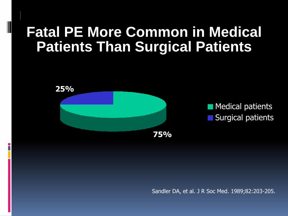
Fatal PE More Common in Medical Patients Than Surgical Patients
Sandler DA, et al. J R Soc Med. 1989;82:203-205.
75%
25%
Medical patients
Surgical patients

Hospital Associated VTE
▪ Age, hospital, surgery, prior VTE, cancer are the major risk factors
▪ 60% of incident VTE associated with recent hospitalization
▪ Risk period often extends beyond hospital stay
Heit JA. The epidemiology of Venous Thromboembolism in the Community. Arteriosclerosis, Thrombosis, and Vascular Biology 2008; 28:370-372


PREVENTING VTE

CASE 1
▪ 38 Malay lady Obese
▪ Slipped an fall in her bathroom.
▪ Severe low back pain
▪ Exacerbated by sitting up or lying supine.
▪ Admitted to orthopaedic unit.

Lumbosacral Xray

▪ After 5 days of pain reliever and bed rest in ward
▪ Developed sudden onset of breathlessness upon returning from bathroom
▪ Intubated for respiratory failure
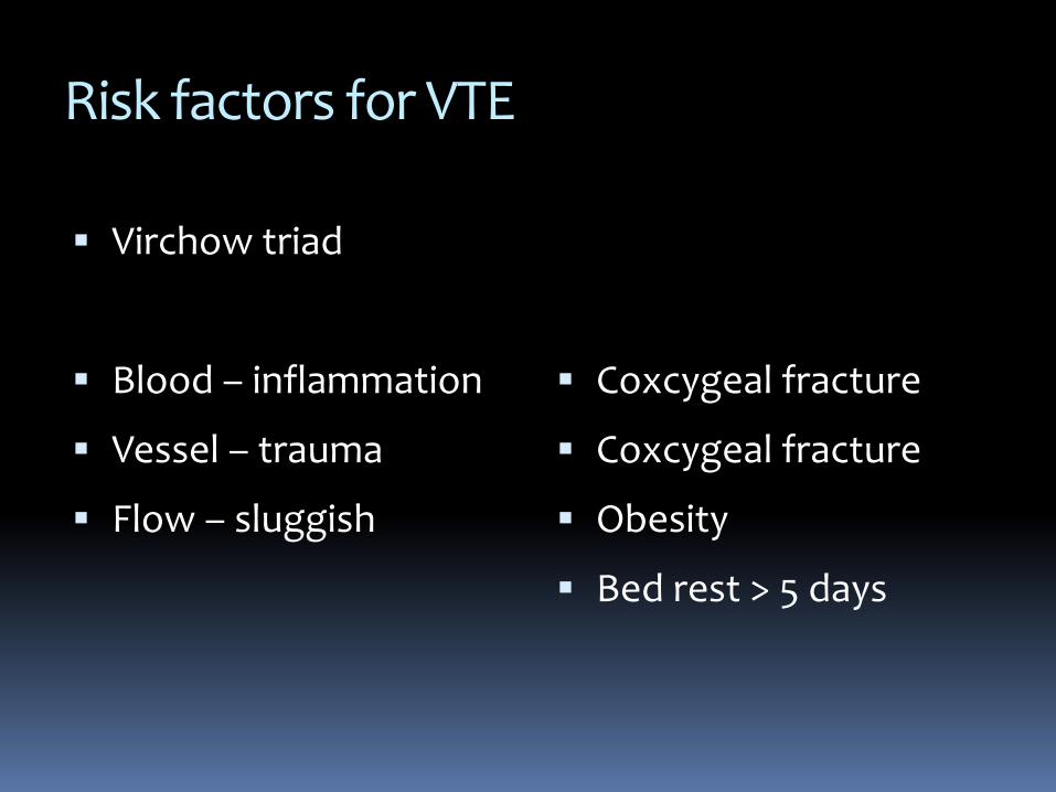
Risk factors for VTE
▪ Virchow triad
▪ Blood – inflammation
▪ Vessel – trauma
▪ Flow – sluggish
▪ Coxcygeal fracture
▪ Coxcygeal fracture
▪ Obesity
▪ Bed rest > 5 days

CTPA

AUDIT OF THROMBOPROPHYLAXIS PRACTICES IN HTAA
▪ 1ST FEB - 28TH FEB 2017
▪ 250 patients
▪ All medical/surgical/orthopaedic/ ICUs and O&G wards
▪ Randomised selections

THROMBOPROPHYLAXIS PRACTICES BY DEPARTMENT

VTE RISK

BLEEDING RISK

RECOGNISING VTE

CASE 2 KS JAN/2015
▪ 40 Indian Man
▪ Abd pain and distention x 8/7
▪ Vomiting- food particles and bilious x 2/7
▪ Similar complaints to HSAJB 3 yrs PTA. Treated conservatively
▪ Denies bleeding tendencies
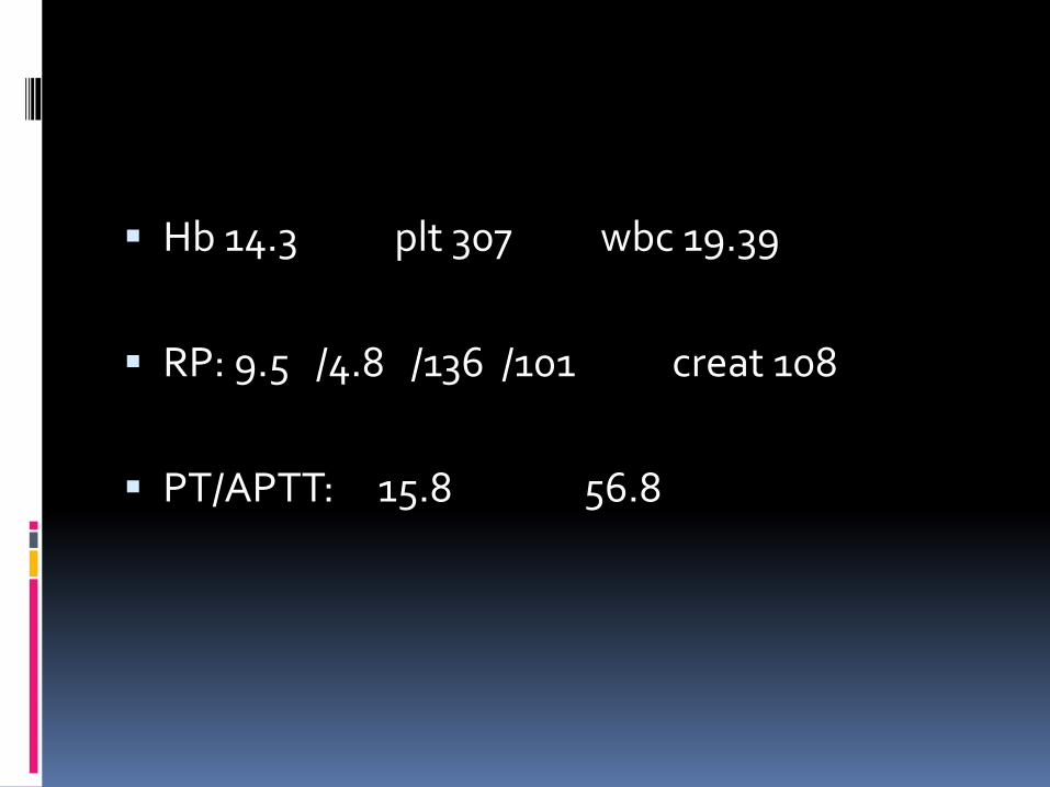
▪ Hb 14.3 plt 307 wbc 19.39
▪ RP: 9.5 /4.8 /136 /101 creat 108
▪ PT/APTT: 15.8 56.8


▪ Emergency explorative laparatomy was performed 15H after admission
▪ SB resection with primary anastomosis for SB gangrene 2^ mesenteric thrombosis
▪ No excessive bleeding

Courtesy of Mr WA HTAA 2015

Courtesy of Mr WA HTAA 2015

GROSS EXAMINATION
▪ Specimen consists of a segment of dilated and dusky colored small intestine, 540mm in length
▪ Cut opened shows gangrenous, flattened to edematous small intestinal mucosa with slough
▪ No tumour mass identified
Courtesy of DAS HTAA 2015

MICROSCOPIC EXAMINATION
Full thickness haemorrhagicnecrosis
Blood vessels thrombosis with fibrin and area of lamination
Courtesy of DAS HTAA 2015

▪ Mixing test: APTT 99.6s
Mix 1:1 54.1s at 1H or 2H inc
▪ IV cefoperazone/ metronidazole
▪ LMWH – sc clexane 60mg bd
▪ Discharged > 9 days total hospitalization

▪ First HC review: (4/52)
▪ Thrombophilia screen: Not validated hence verbal result via phone was not released
▪ Lupus anticoagulant (local lab) – positive
▪ Clinic review 16/2/2015. 9/3/2015. 11/5/2015
▪ Patient then defaulted

Readmitted 23/8/2015
▪ Abdo pain and distention 3/7
▪ vomitting food particles x 1/7
▪ Explorative laparatomy
▪ 40cm SB ischemia involving previous anastomotic site (80cm from ileocaecal jt , 60cm from dudenojejuno jt)
▪ Dense SB adhesions to surrounding organs
▪ Distal loop ileostomy slightly dusky
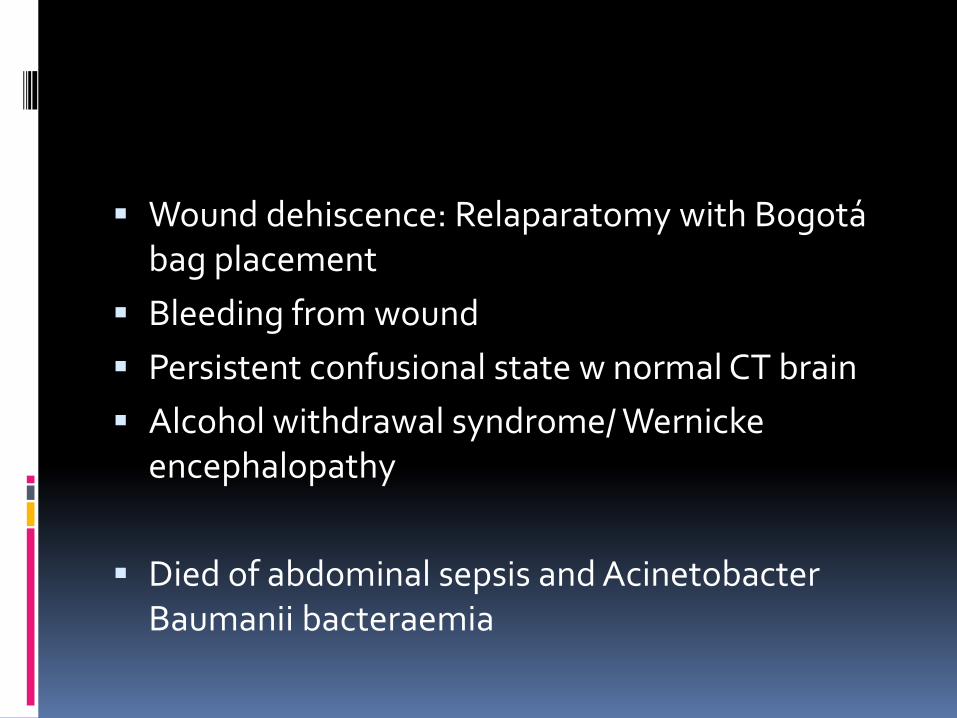
▪ Wound dehiscence: Relaparatomy with Bogotá bag placement
▪ Bleeding from wound
▪ Persistent confusional state w normal CT brain
▪ Alcohol withdrawal syndrome/ Wernickeencephalopathy
▪ Died of abdominal sepsis and AcinetobacterBaumanii bacteraemia

CASE 3 ASMA AUGUST/2015
▪ 40/Malay/ lady
▪ L lower limb intermittent claudication x 2/52
▪ HPT/HPL x 10 years on aspirin/perindopril/ hydrochlorothiazide and simvastatin
▪ L CVA with R hemiparesis in 2011. total neurological resolution

▪ Past Obst Hx: 5 pregnancies. 1 living child. 1 abortion. 3 premature deliveries
▪ Clinically:
▪ Left foot – cool, tender, slightly dusky
▪ Non palpable pulse of Left Popliteal Artery, L PTA & L DPA

CT ANGIO

▪ Hb 11.26 WBC 9.0 PLT 345
▪ PT 1.0 APTT 66.25 Mix 1:1 54.3
▪ Rheumatic factor negative
▪ C3: 1.51 C4: 0.49
▪ Thrombophilia screen 13/8/2015
▪ Lupus Anticoagulant positive x2

▪ Discharged after 3 days with ASA/ Tramadol/ Simvastatin and anti HPT
▪ Represented after 3 days with worsening pain
▪ Started on LMWH and warfarin upon discharge.
▪ Latest INR 2.5 on 6 mg OD


APLS is a THROMBOTIC DISORDER
▪ antiphospholipid antibody inhibits the phospholipid used in the APTT testing
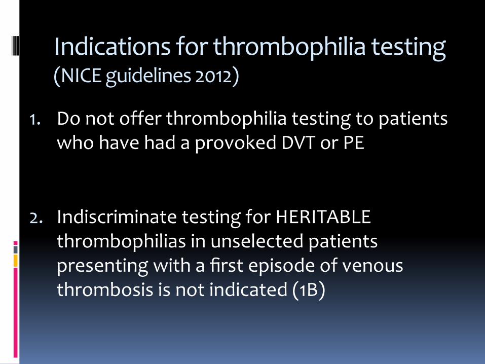
Indications for thrombophilia testing (NICE guidelines 2012)
1. Do not offer thrombophilia testing to patients who have had a provoked DVT or PE
2. Indiscriminate testing for HERITABLE thrombophilias in unselected patients presenting with a first episode of venous thrombosis is not indicated (1B)

In-Accuracy of thrombophilia testing
▪ The interpretation of thrombophilia test results is difficult and errors in interpretation are frequent, which results in both reduced sensitivity and specificity
▪ Thus genuine deficiencies and abnormalities may not be detected and false positive diagnoses are common
Jennings et al, 2005Bjh guidelines 2010

CONFIRMING VTE

Case 4 RI
▪ 45/ Malay lady
▪ abdominal distention x 3/52
▪ breathlessness x 1/52
▪ Clinically : Pale, afebrile. Tachypnoeic.
▪ Gross abd distention with bilateral LL oedema. Vague enlarged mass
▪ No calf tenderness nor dilated veins

▪ ABG: 7.46 /28.0 /69.0 /19.9
▪ Hb 11.9 WBC 11.67 PLT 54
▪ PT/APTT: 13s / 25s

CXR

DOPPLER

▪ D-Dimer sent
▪ If D-Dimer high >> CTPA
▪ D-Dimer 1.6-3.2 ug/mL


CTPA

▪ CA 125: 1088
▪ Pleural/Peritoneal Fluid: Metastatic Adenocarcinoma

Causes of a high D-Dimer
▪ DVT
▪ Cellulitis/ infection
▪ Haematoma or bleeding
▪ DIC
▪ Pregnancy
▪ Inflammation/ Fracture
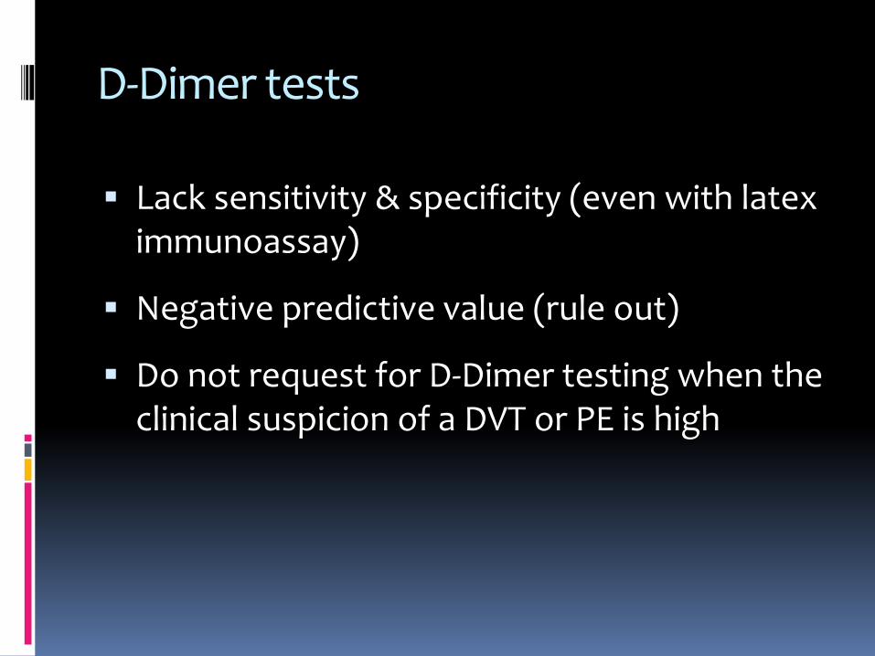
D-Dimer tests
▪ Lack sensitivity & specificity (even with latex immunoassay)
▪ Negative predictive value (rule out)
▪ Do not request for D-Dimer testing when the clinical suspicion of a DVT or PE is high

Confirming VTE
▪ CTPA
▪ Doppler US
▪ Venogram-

NICE guidelines Published date: June 2012
▪ Offer patients in whom PE is suspected and with a likely two-level PE Wells
▪ an immediate computed tomography pulmonary angiogram (CTPA) or
▪ immediate interim parenteral anticoagulant therapy followed by a CTPA, if a CTPA cannot be carried out immediately.

Diagnostic imaging is necessary
▪ To avoid risk, inconvenience and costs of inappropriate anticoagulation

CANCER RELATED THROMBOSIS

Armand Trousseau, 1860Trousseau’s sign of malignancy

What is the difference?
Cancer Patients Non-Cancer Patients
Ambulatory
8-19%
Ambulatory
1.4%


TREATING VTE

Case 1
▪ Assess risk of VTE
▪ VTE prophylaxis

Inpatient VTE Prophylaxis
ACCP NCCNNonsurgical:
High risk patients
Surgical:Depends upon surgical
site and patient risk
All cancer patients
*Note: These recommendations are all in the absence of contraindications to anticoagulation.
Active cancer PLUS
acute medical illness OR
reduced mobility
ASCO
Prophylactic doses UFH
LMWH Fondaparinux

Nonsurgical VTE Risk
Padua Prediction Score
Risk Factor Points
Active Cancer 3
Previous VTE (excluding SVT) 3
Reduced mobility* 2
Already known thrombophilic condition 1
Recent (≤ 1 mo.) trauma/surgery 1
Elderly age (≥ 70y) 1
Heart and/or respiratory failure 1
Acute MI or ischemic stroke 1
Obesity (BMI ≥ 30) 1
Ongoing hormonal treatment 1
*Anticipated bed rest with bathroom privileges for at least 3 days
High Risk≥ 4 points

Surgical VTE Risk
Roger
▪ Operation type
Thoracic area highest risk
▪ Cancer
Disseminated cancer
Chemo within 30 days
Caprini
▪ Recent Stroke (<1 mo.)
▪ History of VTE
▪ Age
▪ Malignancy
▪ BMI

CASE 2 AND 3
▪ LMWH
▪ Followed by oral anticoagulant
▪ VKA or NOAC
▪ Duration – life long

CASE 4
▪ Chemotherapy – cisplatin based
▪ Debulking surgery
▪ LMWH until disease in remission/ indefinite if risk factor persist
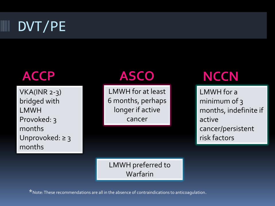
DVT/PE
ACCP NCCNVKA(INR 2-3) bridged with LMWH Provoked: 3 monthsUnprovoked: ≥ 3 months
LMWH for a minimum of 3 months, indefinite if active cancer/persistent risk factors
*Note: These recommendations are all in the absence of contraindications to anticoagulation.
LMWH for at least 6 months, perhaps
longer if active cancer
ASCO
LMWH preferred to Warfarin

5 to 10 days 3 to 6 months > 6 months
Vitamin K Antagonists LMWHOral XaI or dabigatran
Heparin LMWHFondaparinuxThrombolysis Thrombus Removal IVC filterRivaroxaban Apixaban
Initial (acute) treatment
Long term-treatment
Extended treatment
Vitamin K AntagonistsASA 100 mgOral XaI or dabigatran
TREATMENT OF VENOUS THROMBOEMBOLISM

THE BLEEDER AND THE CLOTTER
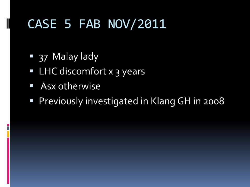
CASE 5 FAB NOV/2011
▪ 37 Malay lady
▪ LHC discomfort x 3 years
▪ Asx otherwise
▪ Previously investigated in Klang GH in 2008

▪ Clinically: massive splenomegaly
▪ FBC: Hb 17.5 PCV 55.2 MCV 79.9 PLT 419 WBC: 28.71

FBP
▪ Refused BMAT
▪ PB JAK2V6175 mutation detected
▪ USG: gross hepatosplenomegaly with portal HPT. Varices at porta hepatis. No intra abdominal lymphadenopathy.
▪ OGDS: Fundal varices
▪ HBs Ag and antiHCV - Negative
▪ Started on Hydrea and propanolol

MARCH 2015
▪ Defaulted follow up
▪ Represented with haematemesis, fatigue and weight loss
▪ Hb 11.1 PCV 30 MCV 80 PLT 297 wbc 20.8
▪ OGDS: grade II –III 3 columns OV
▪ USG: Portal HPT
▪ FBP: features of IDA

POST PV MYELOFIBROSIS
▪ PV, polycythaemic stage. Hypercellular bone marrow

TRIPHASE CT

▪ Started on LMWH prophylaxis dose.
▪ On Aldactone/ Propanolol and HU
▪ HLA (1+2): no matched siblings
▪ Still awaiting JAKAVI from ZAKAT Johor/ Pahang after 2 years
▪ On SC IFN 2MU 3X/week

Hospitalized Medical Patients
▪ ACCP Evidence-based Practice Guidelines 2008 (revised 2012)
LMWH, Unfractionated Heparin, or Fondaparinux (Grade 1A) Mechanical methods if above contraindicated
Patients with heart failure, sever respiratory disease, or confined to bed with1 or more risk factors (cancer, previous VTE, sepsis,acute neurologic disease, IBD)
Duration not specified, 4 to 14 days in clinical trials
▪ ACP Clinical Practice Guideline 2011
Risk assessment for VTE and bleeding, heparin or related drug unless bleeding risk outweighs benefit
Geerts et al. Prevention of venous thromboembolism. American College of Chest Physicians Evidence Based Practice Guidelines(8th edition). CHEST 2008;133: 381-453.
Qaseem A et al. Venous thromboembolism prophylaxis in hospitalizedpatients: A Clinical Practice Guideline from the American College of Physicians. Annals of Internal Medicine 2011; 155: 625-632.

STOP BLOOD CLOTS AND SAVES LIVES