Voluntary Movement From Ch. 38 “Principles of Neural Science”, 4 th Ed. Kandel et al.
29
Voluntary Movement From Ch. 38 “Principles of Neural Science”, 4 th Ed. Kandel et al
-
Upload
sherman-roy-cobb -
Category
Documents
-
view
220 -
download
0
Transcript of Voluntary Movement From Ch. 38 “Principles of Neural Science”, 4 th Ed. Kandel et al.
Voluntary Movement
From Ch. 38
“Principles of Neural Science”, 4th Ed.
Kandel et al
Voluntary movement
• Voluntary movements are organized in cortex
• Sensory feed back– Visual information– Proprioceptive information– Sounds and somatosensory information
• Goal of movement– Vary in response to the same stimulus depending on behavioral task (precision
vs. power grip)
• Improves with learning/ experience
• Can be generated in response to external stimuli or internally
Cortical organization• Hierarchical organization of motor control and task features
– Populations of neurons encode motor parameters e.g. force, direction, spatial patterns
– The summed activity in a population determines kinematic details of movement
– Voluntary movement is highly adaptable• Novel behavior requires processing in several motor and parietal areas as it is continuously monitored
for errors and then modified
– Primary motor cortex • Fires shortly before and during movement• Fires only with certain tasks and patterns of muscle activation
– Premotor areas encode global features of movement• Set-related neurons
– Sensorimotor transformations (external environment integrated into motor programs)– Delayed response
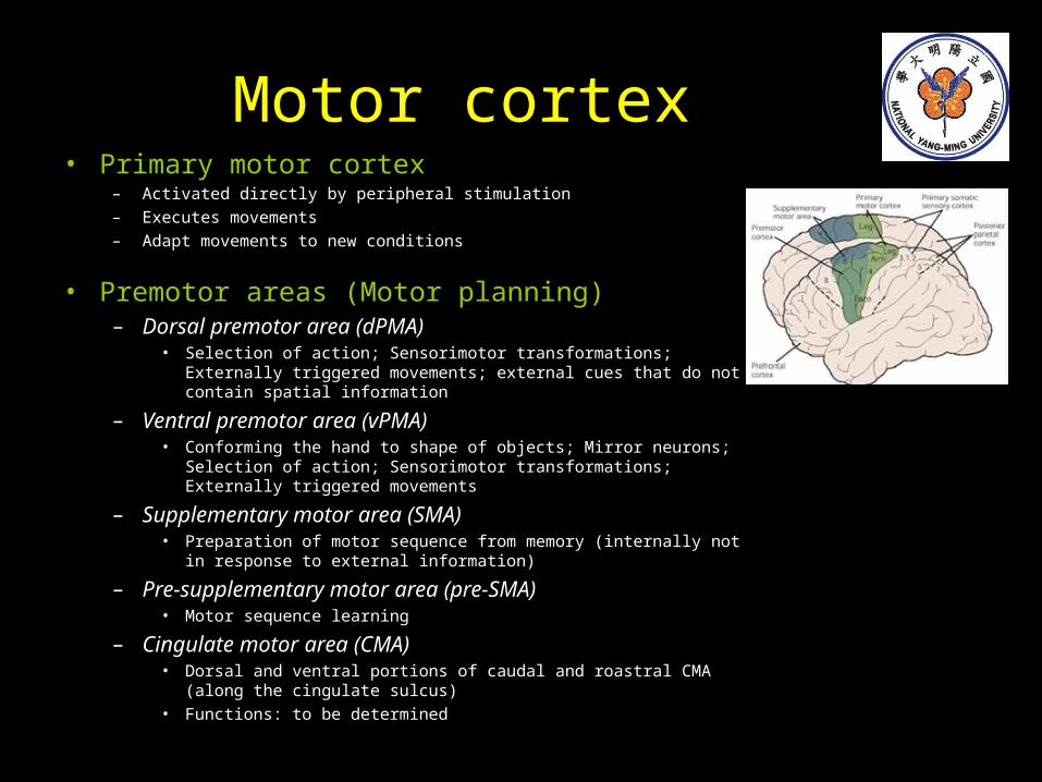
Motor cortex• Primary motor cortex
– Activated directly by peripheral stimulation– Executes movements– Adapt movements to new conditions
• Premotor areas (Motor planning)– Dorsal premotor area (dPMA)
• Selection of action; Sensorimotor transformations; Externally triggered movements; external cues that do not contain spatial information
– Ventral premotor area (vPMA)• Conforming the hand to shape of objects; Mirror neurons; Selection of
action; Sensorimotor transformations; Externally triggered movements
– Supplementary motor area (SMA)• Preparation of motor sequence from memory (internally not in response
to external information)
– Pre-supplementary motor area (pre-SMA)• Motor sequence learning
– Cingulate motor area (CMA)• Dorsal and ventral portions of caudal and roastral CMA (along the
cingulate sulcus)• Functions: to be determined
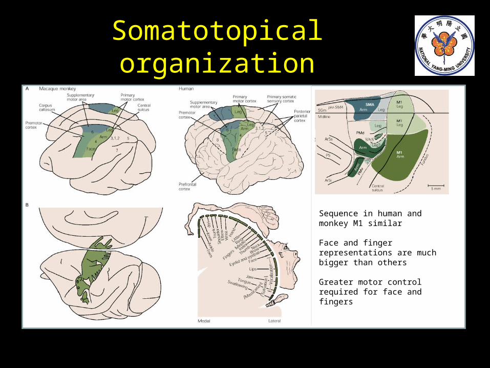
Somatotopical organization
Sequence in human and monkey M1 similar
Face and finger representations are much bigger than others
Greater motor control required for face and fingers
Motor cortex stimulation
Historical perspective• 1870 Discovery of electrical excitability of cortex in the dog;
first brain maps (Fritsh and Hitzig)
• 1875 First motor map of the primate brain (Ferrier)
• 1926 Recording of extracellular spike activity of a nerve fiber (Adrian)
• 1937 First experimentally derived human motor map (Penfield and Boldrey)
• 1957 Microelectrode recordings to map primary somatosensory area (Mountcastle et al.)
• 1958 First recordings from neurons in awake monkeys (Jasper)
• 1967 Intracortical microstimulation for mapping of cortical motor output (Asanuma)
• 1985 TMS is used to activate motor cortex noninvasively (Barker et al.)
Transcranial stimulation• TES – transcranial electrical stimulation (Merton and Morton 1980)
– High voltage (1-2kV), short duration pulses (10-50us), low resistance electrodes.– Direct stimulation occurs at the anode– Current passes through skin and scalp (resistance) before reaching cortex.
• TMS – transcranial magnetic stimulation (Barker 1985)– Discharge of large capacitive currents (5-10kA, 2-300us) through a coil producing high magnetic field (1-2T). – Stimulus site depends on coil design, coil orientation and stimulus intensity
• Non-invasive techniques to study– Structure-function relationship (e.g. rTMS virtual lesion)– Map brain motor output (typically averaged EMG as output =MEP)– Measure conduction velocity
• TMS has advantages over TES– No discomfort (no current passes through skin and high current densities can be avoided)– No attenuation of field when passing through tissue– No skin preparation (conduction gel)
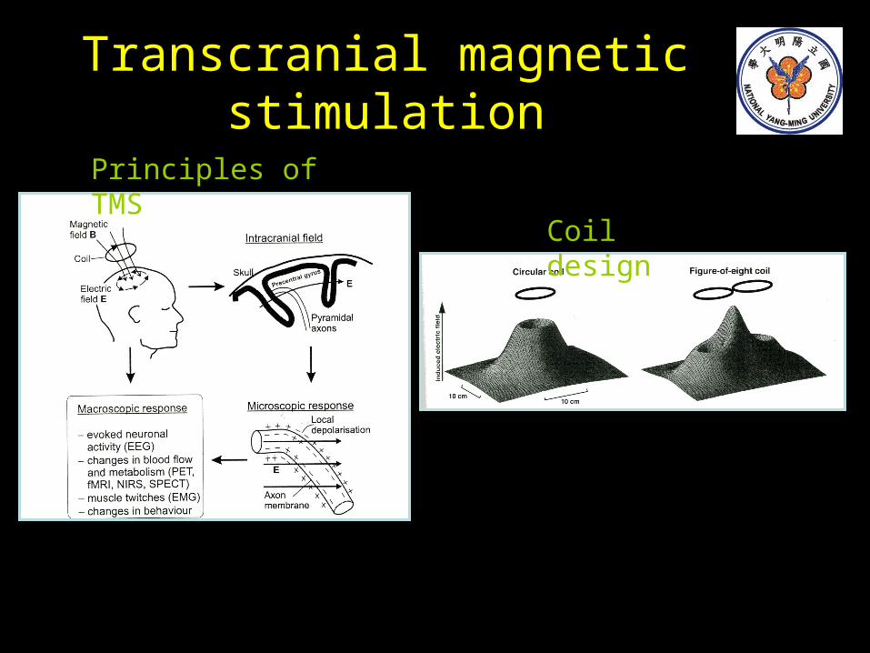
Transcranial magnetic stimulation
Principles of TMS
Coil design
Motor cortex stimulation
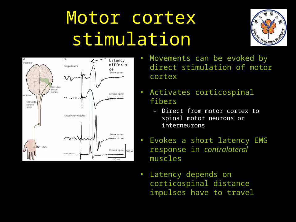
• Movements can be evoked by direct stimulation of motor cortex
• Activates corticospinal fibers– Direct from motor cortex to spinal
motor neurons or interneurons
• Evokes a short latency EMG response in contralateral muscles
• Latency depends on corticospinal distance impulses have to travel
Latency difference
Cortex-muscle connections
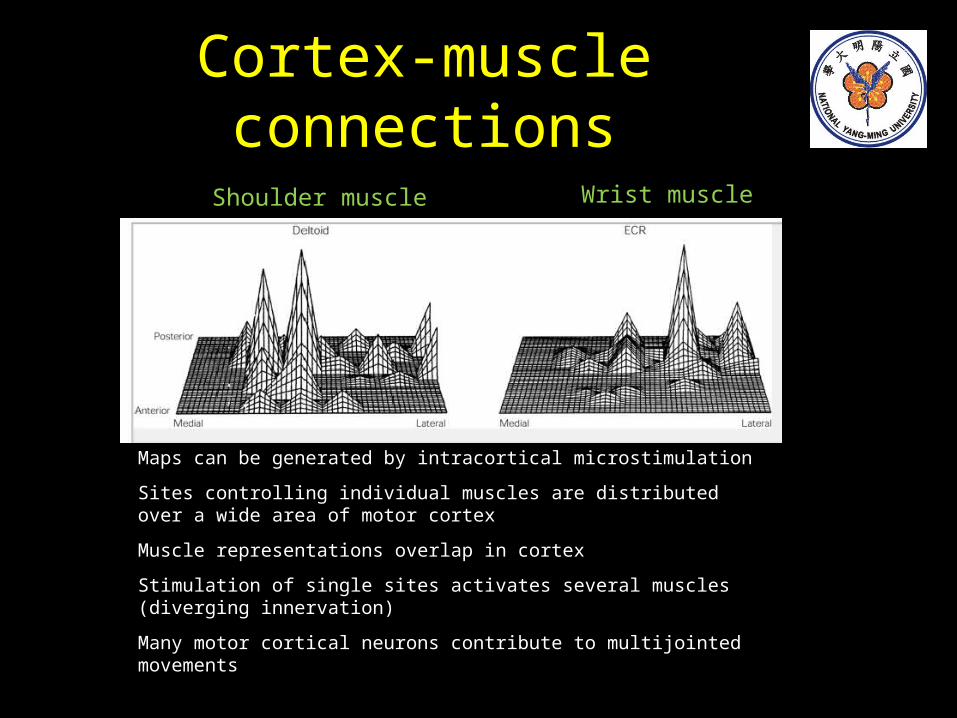
Shoulder muscle Wrist muscle
Maps can be generated by intracortical microstimulation
Sites controlling individual muscles are distributed over a wide area of motor cortex
Muscle representations overlap in cortex
Stimulation of single sites activates several muscles (diverging innervation)
Many motor cortical neurons contribute to multijointed movements
Cortical projections
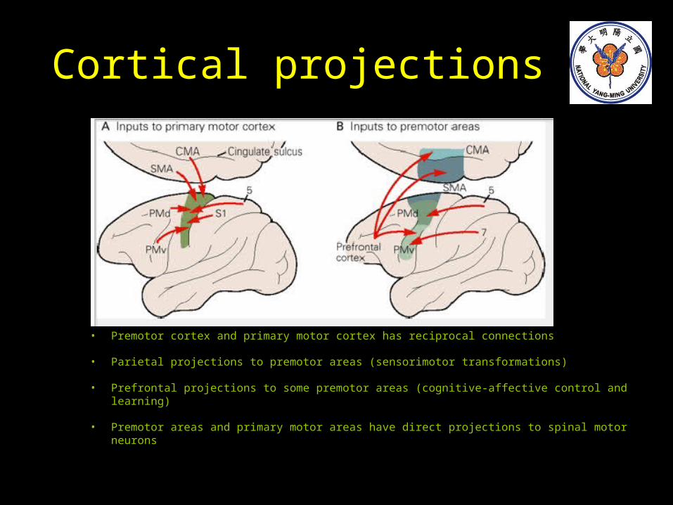
• Premotor cortex and primary motor cortex has reciprocal connections
• Parietal projections to premotor areas (sensorimotor transformations)
• Prefrontal projections to some premotor areas (cognitive-affective control and learning)
• Premotor areas and primary motor areas have direct projections to spinal motor neurons
Other projections
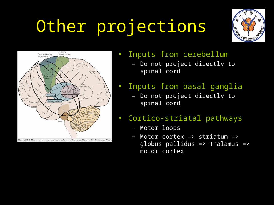
• Inputs from cerebellum– Do not project directly to spinal cord
• Inputs from basal ganglia– Do not project directly to spinal cord
• Cortico-striatal pathways– Motor loops– Motor cortex => striatum => globus pallidus
=> Thalamus => motor cortex
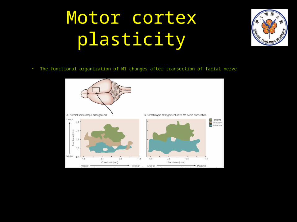
Motor cortex plasticity
• The functional organization of M1 changes after transection of facial nerve
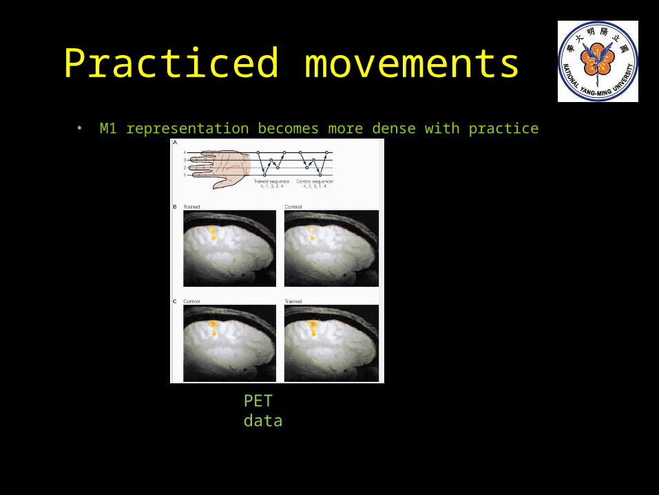
Practiced movements• M1 representation becomes more dense with practice
PET data
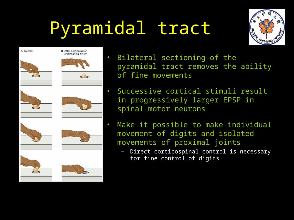
Pyramidal tract
• Bilateral sectioning of the pyramidal tract removes the ability of fine movements
• Successive cortical stimuli result in progressively larger EPSP in spinal motor neurons
• Make it possible to make individual movement of digits and isolated movements of proximal joints
– Direct corticospinal control is necessary for fine control of digits
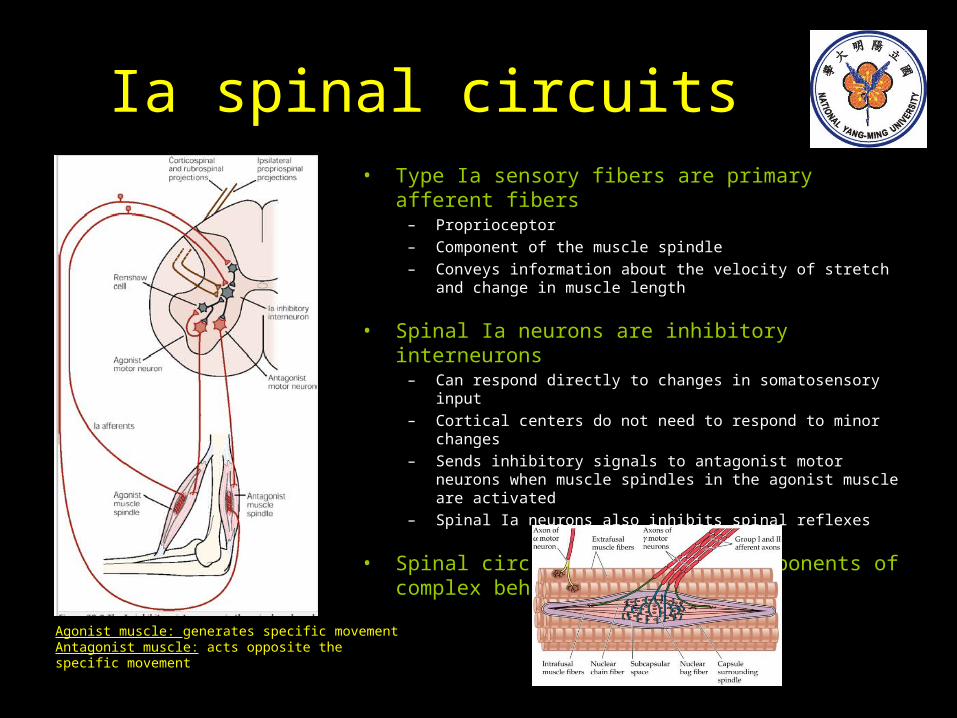
Ia spinal circuits• Type Ia sensory fibers are primary afferent fibers
– Proprioceptor– Component of the muscle spindle– Conveys information about the velocity of stretch and change in
muscle length
• Spinal Ia neurons are inhibitory interneurons– Can respond directly to changes in somatosensory input– Cortical centers do not need to respond to minor changes– Sends inhibitory signals to antagonist motor neurons when
muscle spindles in the agonist muscle are activated– Spinal Ia neurons also inhibits spinal reflexes
• Spinal circuits are used as components of complex behaviors
Agonist muscle: generates specific movementAntagonist muscle: acts opposite the specific movement
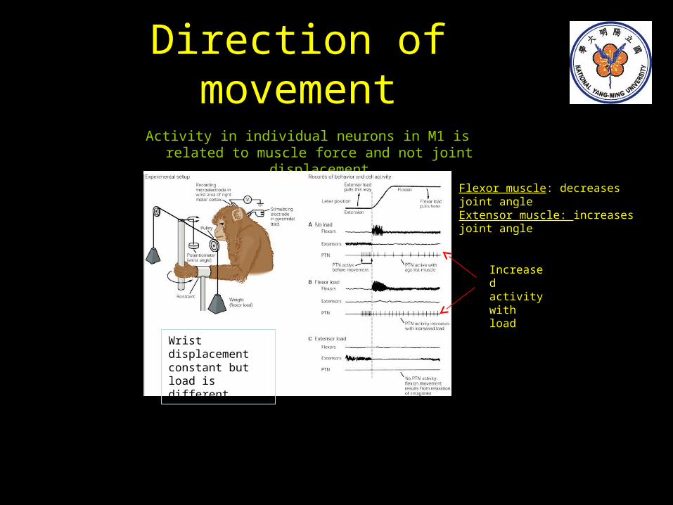
Direction of movement
Activity in individual neurons in M1 is related to muscle force and not joint displacement
Increased activity with load
Wrist displacement constant but load is different
Flexor muscle: decreases joint angle Extensor muscle: increases joint angle
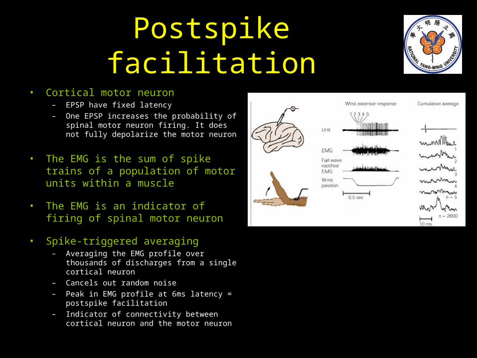
Postspike facilitation• Cortical motor neuron
– EPSP have fixed latency– One EPSP increases the probability of spinal
motor neuron firing. It does not fully depolarize the motor neuron
• The EMG is the sum of spike trains of a population of motor units within a muscle
• The EMG is an indicator of firing of spinal motor neuron
• Spike-triggered averaging– Averaging the EMG profile over thousands of
discharges from a single cortical neuron– Cancels out random noise– Peak in EMG profile at 6ms latency = postspike
facilitation– Indicator of connectivity between cortical neuron
and the motor neuron
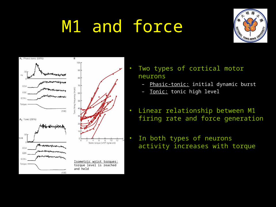
M1 and force
• Two types of cortical motor neurons– Phasic-tonic: initial dynamic burst
– Tonic: tonic high level
• Linear relationship between M1 firing rate and force generation
• In both types of neurons activity increases with torque
Isometric wrist torques: torque level is reached and held
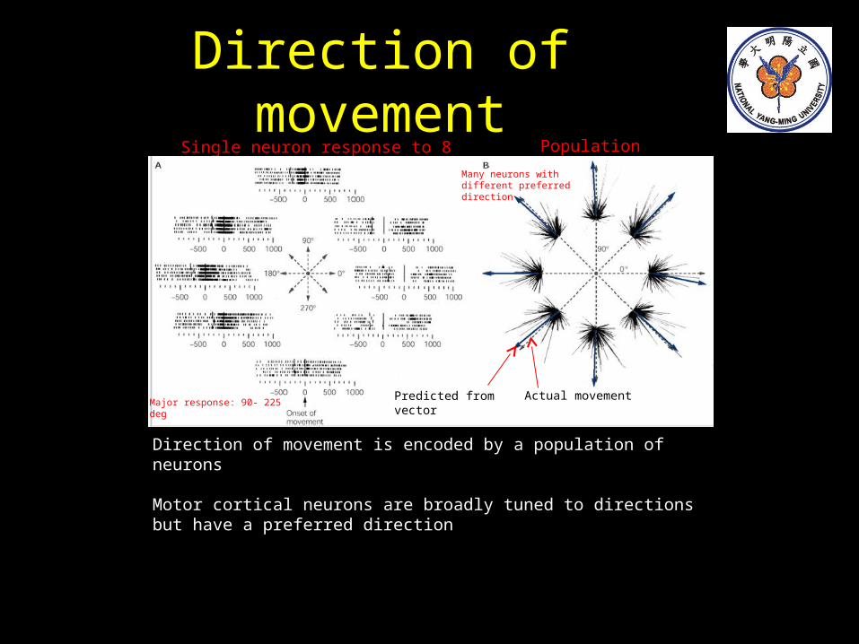
Direction of movement
Direction of movement is encoded by a population of neurons
Motor cortical neurons are broadly tuned to directions but have a preferred direction
Single neuron response to 8 directions Population vector
Predicted from vector Actual movementMajor response: 90- 225 deg
Many neurons with different preferred direction
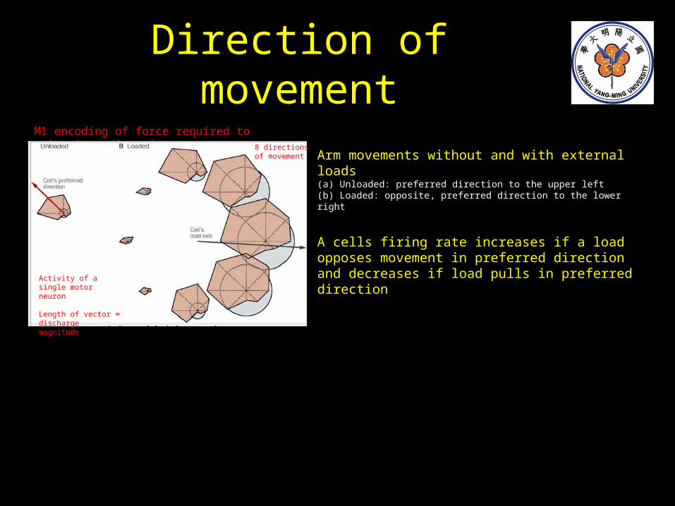
Direction of movement
SingleArm movements without and with external loads(a) Unloaded: preferred direction to the upper left(b) Loaded: opposite, preferred direction to the lower right
A cells firing rate increases if a load opposes movement in preferred direction and decreases if load pulls in preferred direction
M1 encoding of force required to maintain a direction
Activity of a single motor neuron
Length of vector = discharge magnitude
8 directions of movement
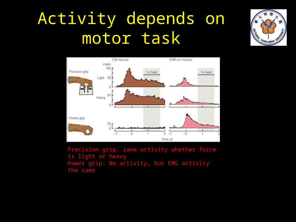
Activity depends on motor task
Precision grip: same activity whether force is light or heavyPower grip: No activity, but EMG activity the same
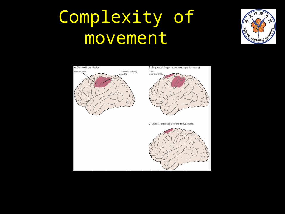
Complexity of movement
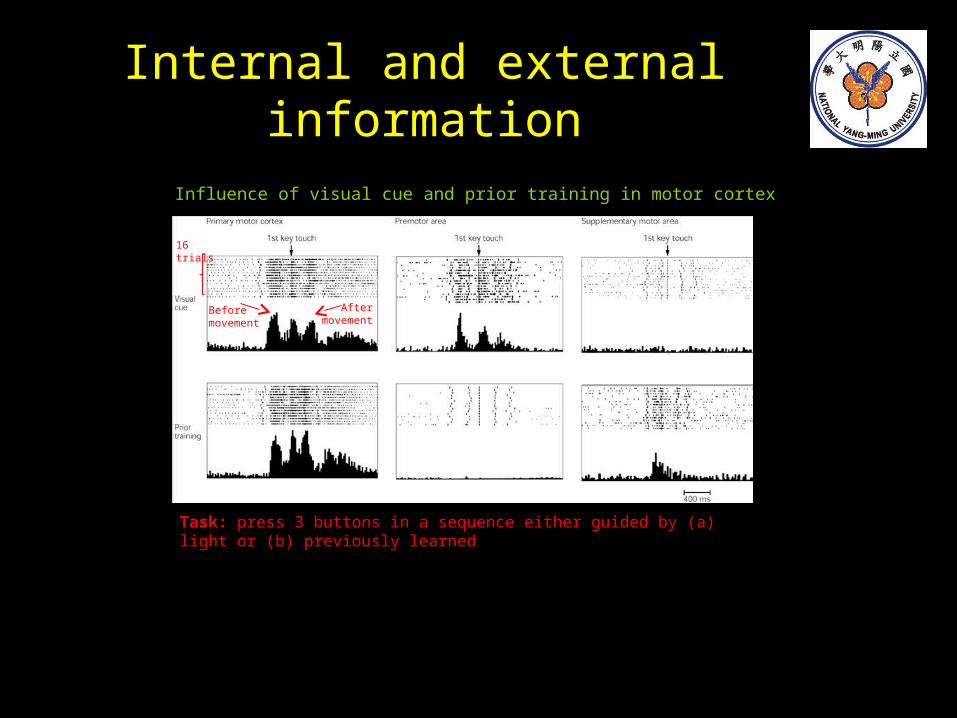
Internal and external information
Influence of visual cue and prior training in motor cortex
Task: press 3 buttons in a sequence either guided by (a) light or (b) previously learned
Before movement
After movement
16 trials
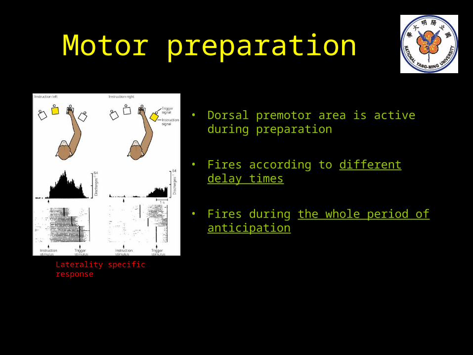
Motor preparation
• Dorsal premotor area is active during preparation
• Fires according to different delay times
• Fires during the whole period of anticipation
Laterality specific response
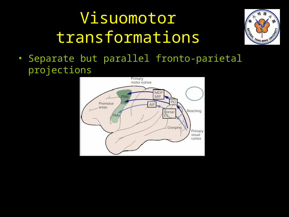
Visuomotor transformations
• Separate but parallel fronto-parietal projections
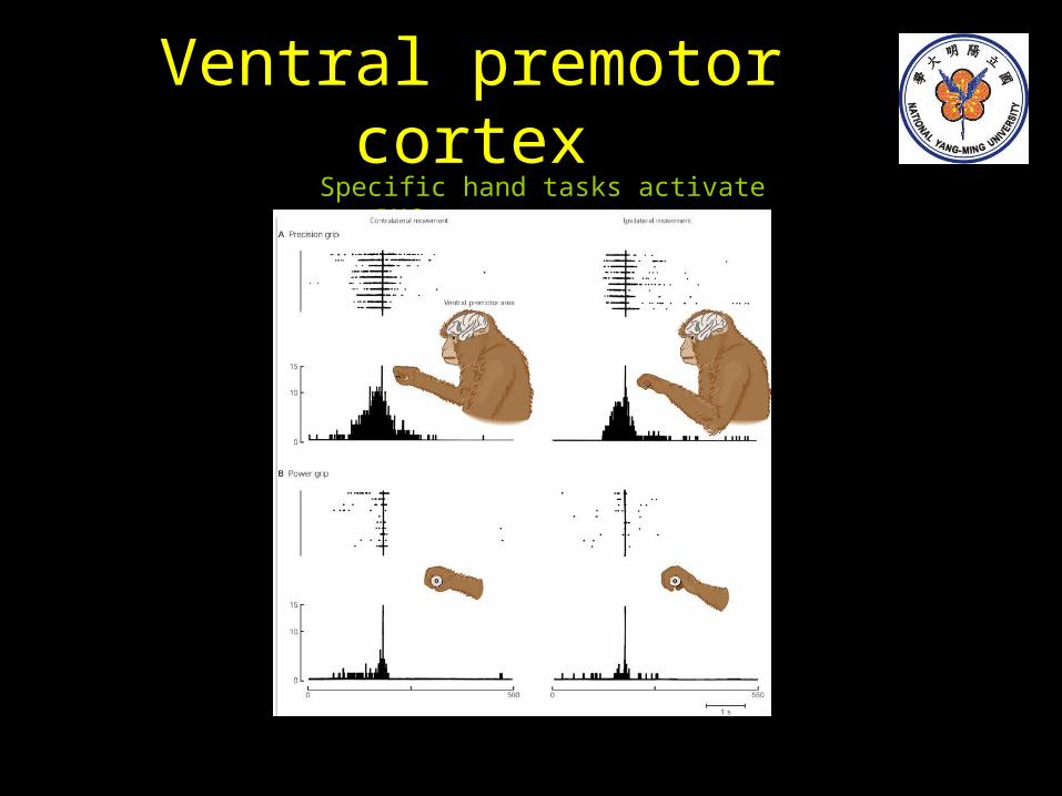
Ventral premotor cortexSpecific hand tasks activate vPMC
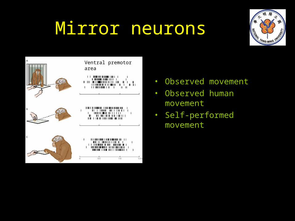
Mirror neurons
• Observed movement• Observed human movement• Self-performed movement
Ventral premotor area
Summary• Hierarchical organization of motor control and task
features– Populations of neurons encode motor parameters e.g. force, direction, spatial patterns
– The summed activity in a population determines kinematic details of movement
– Voluntary movement is highly adaptable• Novel behavior requires processing in several motor and parietal areas as it is continuously monitored for errors and
then modified
– Primary motor cortex • Fires shortly before and during movement• Fires only with certain tasks and patterns of muscle activation
– Premotor areas encode global features of movement
• Set-related neurons– Sensorimotor transformations (external environment integrated into motor programs)– Delayed response