VOLUM DE REZUMATE ABSTRACT BOOK · apparent that the sharp improvement brought about by the new...
39
VOLUM DE REZUMATE / ABSTRACT BOOK NUCLEAR MEDICINE DAYS/ZILELE MEDICINEI NUCLEARE 5 TH NATIONAL CONFERENCE OF NUCLEAR MEDICINE / A 5a CONFERINȚĂ NAȚIONALĂ DE MEDICINĂ NUCLEARĂ Coordonatori: Dr. Raluca Mititelu Prof. Dr. Cipriana Ștefănescu ISSN 2668-3717, ISSN-L 2668-3717 7-9 iunie, Sinaia, România
Transcript of VOLUM DE REZUMATE ABSTRACT BOOK · apparent that the sharp improvement brought about by the new...
VOLUM DE REZUMATE
/
ABSTRACT BOOK NUCLEAR MEDICINE DAYS/ZILELE MEDICINEI NUCLEARE
5TH NATIONAL CONFERENCE OF NUCLEAR MEDICINE /
A 5a CONFERINȚĂ NAȚIONALĂ DE MEDICINĂ NUCLEARĂ
Coordonatori:
Dr. Raluca Mititelu
Prof. Dr. Cipriana Ștefănescu
ISSN 2668-3717, ISSN-L 2668-3717
7-9 iunie, Sinaia, România
2
CUPRINS
PREZENTĂRI INVITATE ...................................... Error! Bookmark not defined. 1. FROM STATIC TO DYNAMIC NUCLEAR IMAGING: HOW NOVEL TECHNOLOGIES AND
RADIOPHARMACEUTICALS ARE CHANGING THE FIELD .............................................................................. 5
Adriano Duatti............................................................................................................................................................ 5
2. WORKING WITH PROTOCOLS IN NUCLEAR MEDICINE .......................................................................... 5
G. Andries1, A. Pavel2, G. Crisan3 ............................................................................................................................. 5
3. OPTIMIZATION OF PREPARATION OF GA68 RADIOPHARMACEUTICALS: PRIVATE PRACTICE
EXPERIENCE .............................................................................................................................................................. 6
Masha Maharaj ........................................................................................................................................................... 6
4. ADVANCES IN MOLECULAR IMAGING ........................................................................................................ 6
Simona Ben Haim ...................................................................................................................................................... 6
5. QUANTITATIVE MYOCARDIAL BLOOD FLOW MEASUREMENTS WITH EMPHASIS ON SPECT .... 6
Simona Ben Haim ...................................................................................................................................................... 6
6. NUCLEAR MEDICINE IN TRANSTHYRETIN AMYLOIDOSIS - PERSONAL EXPERIENCE ................. 7
Stan C. A.1, Chirion C. 1, Stanescu D. A. 1, Badelita S. 2, Jurcut R. 3, Beladan C. 3 .................................................... 7
7. BIOMARKERS IN THYROID TUMORS ........................................................................................................... 7
Iulia Andreea Chiriac, A.L. Goldstein ....................................................................................................................... 7
8. CONTROVERSIES, CONSENSUS AND COLLABORATION IN THE USE OF I-131 THERAPY IN
DIFFERENTIATED THYROID CANCER “THE MARTINIQUE PRINCIPLES” ................................................ 8
G. Kermoison, L. Hîţu, G. Rusu, H. Haba, A. Alexiev, E. Spinu, K. Gabora, E. Olariu, A. Piciu, D. Piciu; ............ 8
9. MANAGEMENT OF I131 REFRACTORY THYROID CANCER: A MULTIMODALITY APPROACH ......... 9
Partha S Choudhury. .................................................................................................................................................. 9
10. CZT IMAGING AND TECHNOLOGY ............................................................................................................. 10
Alex Frenkel ............................................................................................................................................................. 10
11. THE ROLE OF CHOLINE PETCT IN PROSTATE CANCER. REVIEW OF BIBLIOGRAPHY ................ 10
Mariela Agolti .......................................................................................................................................................... 10
12. LU-PSMA PRIVATE PRACTICE CLINICAL EXPERIENCE: THE WINS AND WOES ............................ 11
Masha Maharaj ......................................................................................................................................................... 11
13. PSMA PRLT – AUGMENTED AND INDIVIDUALIZED TREATMENT (PRLT 2.0) .................................. 11
Same Ezziddin.......................................................................................................................................................... 11
14. FDG PET/CT IN COLORECTAL MALIGNANT DISEASES. PRACTICAL ISSUES IN OUR CLINICAL
EXPERIENCE. ........................................................................................................................................................... 11
Dragos Cuzino, Magda Iriciuc, Raluca Mititelu, Mirela Oancea, Carmen Tipar, Catalin Mazilu ........................... 11
15. RADIOPROTECTIA IN CAZUL UNUI INCIDENT CU SUBSTANTE RADIOACTIVE ............................. 12
Fizician medical Maria-Alina Gherman ................................................................................................................... 12
E-POSTERE ......................................................................................................... 13 16. IDENTIFICAREA DE BIOMARKERI IMAGISTICI IN SINDROMUL CARDIORENAL PE MODEL
ANIMAL PRIN 18F-FDG PET/MRI -REZULTATE PRELIMINARE-................................................................... 14
L. Agrigoroaie1, D.M. Furcea1, Cristina Uritu2, Gabriela Stanciu2, C.T. Mihai2, I. Gardikiotis2, R. Iliescu1,2,
Cipriana Stefanescu1,2, M.M. Gutu1 ......................................................................................................................... 14
17. HEPATIC UPTAKE ON 99mTc HYDROXYMETHYLENE DIPHOSPHONATE BONE SCAN IMAGING:
A CASE REPORT....................................................................................................................................................... 15
Iulia Andreea Chiriac1, Gabriela Voicu2, Mariana Purice2, Diana Neagu2, F. Alexiu2, A.L. Goldstein1 .................. 15
18. BONE SCINTIGRAPHY AS PRELIMINARY STEP IN IDENTIFYING SCHNITZLER'S SYNDROME
VS. ERDHEIM-CHESTER DISEASE ...................................................................................................................... 15
Polixenia-Laura Grecu2, Daniela Chețan1, Roxana Iacob3, Ș. Bîlha1, Cipriana Ștefănescu2,3 , A.G. Naum1,3 .......... 15
19. LEZIUNE MUSCULARĂ ACTIVĂ METABOLIC LA EXAMINAREA PET-CT F18-FDG, LA O
PACIENTĂ CU CANCER TIROIDIAN PAPILAR .................................................................................................. 16
H. Haba, G. Rusu, L. Hitu, G. Kermoison, A. Alexiev, E. Spinu, K. Gabora, E. Olariu, D. Piciu .......................... 16
20. UN SIMPLU CARCINOM TIROIDIAN PAPILAR CE S-A TRANSFORMAT INTR-O DILEMĂ
TERAPEUTICĂ ......................................................................................................................................................... 17
L.Hîţu, G. Kermoison, G. Rusu, A. Alexiev, E. Spînu, K. Gabora, E. Olariu, A. Piciu, ........................................... 17
21. HORSESHOE KIDNEY VISUALISATION: 99MTC-DMSA RENAL SCAN VS ULTRASONOGRAPHY .. 19
Wael Jalloul1, Alexandru Tarca1, Mihai Gutu1,2, Russu Radu3, Irina Cristina Grierosu1,2, Cipriana Ștefănescu1,2 ... 19
22. TRANSPLANTUL AUTOLOG DE CELULE STEM MEDULARE AMELIOREAZĂ FUNCȚIA CINETICĂ
DIN ARIA INFARCTULUI MIOCARDIC LA ECG-GATED SPECT .................................................................... 19

3
Miruna Mihaela Micheu1, Gabriela Nicula1, Nicoleta Monica Popa-Fotea1, Ileana Monica Stoian1, Nicoleta
Oprescu1, C.A. Mihai1, V. Bataila1, L. Calmac1, Magdalena Stoica1, Elena Luminita Stanciulescu1, V. Ploscaru1,
Alina Ioana Scarlatescu1, Alexandru Scafa-Udriste1,2, Maria Dorobantu1,2.............................................................. 19
23. PACIENȚII CU CANCER TIROIDIAN DIFERENȚIAT TRATAȚI CU I-131: RECOMANDĂRI
INDIVIDUALIZATE DE RADIOPROTECȚIE ....................................................................................................... 21
Moisescu-Goia Cristina1, Sabo Alexandrina1, Cecan Iulia1, Pestean Claudiu1,2, Olariu Elena1, Piciu Doina1,2 ....... 21
24. MIELOM MULTIPLU CU METASTAZE MUSCULARE ŞI CUTANATE ADEVARAT SAU FALS? ....... 22
E. Olariu1, M.I. Larg1,2, K.Gabora1,2, L.Hîţu1, E.Spȋnu1, B. Grădinaru1, G.Kermoison1, G.Rusu1, A. Alexiev1, C.
Peştean1,2, D.Piciu1,2 ................................................................................................................................................ 22
25. EVALUAREA DOZIMETRICĂ POSTRADIOIODOTERAPIE LA PACIENŢII CU HIPERTIROIDISM .. 23
Sabo Alexandrina1, Moisescu-Goia Cristina1, Cecan Iulia1, Pestean Claudiu1,2, Crişan Monica1, Olariu Elena1,
Piciu Doina1,2 ........................................................................................................................................................... 23
26. THE ROLE OF 99mTc-TEKTROTYDE SOMATOSTATIN RECEPTOR IMAGING IN THE
MEDULLARY THYROID CARCINOMA PROGRESSION .................................................................................... 23
Stolniceanu CR1,2, Ungureanu CM5, Preda C5, Statescu AM2, ................................................................................. 23
Gutu M1,2, Ionescu TM2, Matovic M3,4 Stefanescu C1,2 ............................................................................................ 23
27. IMAGINEA ANULUI 2018 ÎN MEDICINA NUCLEARĂ............................................................................... 24
RC Ciobanca, G Cobzac, C Sfrângeu, GL Andrieş. ................................................................................................. 24
28. ARTEFACTELE: O PARTE ESENTIALA IN VIATA UNUI MEDIC DE MEDICINA NUCLEARA ......... 25
AC Țârcă1, TM Ionescu1, CR Stolniceanu1,2, AM Stătescu1, IC Grierosu1,2, MM Guțu1,2, C Ștefănescu1,2 ............. 25
29. ROLUL DIAGNOSTIC AL IMAGISTICII FUNCȚIONALE PENTRU O FORMAȚIUNE SOLITARĂ
ADRENALĂ ASOCIATĂ CANCERULUI DE SÂN – PREZENTARE DE CAZ .................................................... 26
Laura Teodoriu1, Roxana Iacob1, Bianca Ioan2, Ana-Maria Stătescu3, Irena Grierosu1,3, Mihai Guțu1,3, Cristina
Preda1, 2, Cipriana Ștefănescu1,3 ................................................................................................................................ 26
TEACHING FILES ............................................................................................. 28 30. ASPECT FALS POZITIV DE DETERMINĂRI SECUNDARE OSOASE ÎNTR-UN CAZ DE
GLUCAGONOM PANCREATIC. PREZENTARE DE CAZ ................................................................................... 29
M. G. Alexa, Irina Maria Strugari, Mirela Gherghe ................................................................................................. 29
31. DIAGNOSTICUL DIFERENȚIAL ȘI STABILIREA CONDUITEI TERAPEUTICE PENTRU O TUMORĂ
OSOASĂ SCAPULARĂ SUPRASPINOASĂ PRIN SCINTIGRAFIA OSOASĂ ÎN TREI FAZE VERSUS ALTE
TEHNICI IMAGISTICE ............................................................................................................................................ 30
M. Dragoteanu1, Cecilia Pîgleșan1, Ioana Duca2 ...................................................................................................... 30
32. TUMORI PRIMARE ASOCIATE CANCERULUI TIROIDIAN ..................................................................... 30
K. Gabora1,2, B. Grădinaru2, E. Spînu2, G. Rusu2, L. Hîțu2, A. Alexiev2, H. Haba2, G. Kermoison2, E. Olariu1,2, A.
Piciu1,2, D. Piciu1,2 .................................................................................................................................................... 30
33. STRUCTURAL AND FUNCTIONAL IMAGING ASPECTS OF NEUROENDOCRINE CARCINOMA OF
THE CERVIX : A CASE REPORT ........................................................................................................................... 33
Wael Jalloul1, Ruxandra Tibu1, Ana-Maria Statescu1, Cipriana Ștefănescu1, 2 ......................................................... 33
34. 18F-FDG PET/CT UNUSUAL UPTAKE IN CUTANEOUS METASTASIS DEBUT LUNG CANCER ........ 34
Polixenia-Laura Grecu2, Daniela Chețan1, Roxana Iacob3, Ș. Bîlha1, Cipriana Ștefănescu2,3, A.G. Naum1,3 .......... 34
35. DILATAREA ISCHEMICĂ TRANZITORIE DE EFORT A VENTRICULULUI STÂNG: PREZENTARE
DE CAZ ....................................................................................................................................................................... 35
I.Muntea³, A. Chirilă1,2, R. Hagiu1, G. Andries2,3 ..................................................................................................... 35
36. EXAMINAREA PET/CT CU F18-FDG VERSUS SCINTIGRAFIA RECEPTORILOR DE
SOMATOSTATINA IN CAZUL UNEI TUMORI NEUROENDOCRINE ILEALE G2 ......................................... 36
M. Mutuleanu, Romina Costache, Mirela Gherghe .................................................................................................. 36
37. DUPLICAȚIA RENALĂ: 99mTc –DTPA ȘI 99mTc-DMSA ÎȘI UNESC FORȚELE PENTRU A
DETERMINA FUNCȚIA ȘI VIABILITATEA ......................................................................................................... 37
Ruxandra Țibu1, T.M. Ionescu1, Irena Cristina Grierosu1,3, A.C. Țârcă1, M.M. Guțu1,3, Magdalena Stârcea2,
Cipriana Ștefănescu1,3 .............................................................................................................................................. 37
38. THE ROLE OF F18-FDG PET/CT BRAIN SCANNING IN PATIENTS WITH ONCOLOGICAL
PATHOLOGY FOR ASSESSMENT OF PITUITARY METABOLISM: PRIMARY TUMORS, METASTASIS
OR PHYSIOLOGICAL UPTAKE AND SUBSEQUENT MANAGEMENT ............................................................ 38
E.Spinu*; B. Gradinaru*; D.Piciu; E.Olariu; L.Hitu; G.Rusu; H. Haba; G. Kermoison. A.Piciu; K. Gabora;
I.Cecan. .................................................................................................................................................................... 38

4
PREZENTĂRI
INVITATE
5
1. FROM STATIC TO DYNAMIC NUCLEAR IMAGING: HOW NOVEL
TECHNOLOGIES AND RADIOPHARMACEUTICALS ARE CHANGING
THE FIELD
Adriano Duatti
University of Ferrara and Legnaro National Laboratories (LNL-INFN), Italy
The imaging technologies currently in use for both SPECT and PET make hard to collect dynamic images during the course
of a routine clinical study. The possibility of achieving a temporal follow-up of the accumulation and clearance of the
radiopharmaceutical at the target site may disclose fundamental diagnostic information that are now average out because of
the long acquisition time.
This scenario is doomed to change after the introduction of new, ultrafast SPECT and PET scanners that can shorten the
acquisition time to less than one minute while attaining a spatial resolution below the one-millimeter scale. When these new
tools will become widely available, a sharp improvement of the conventional nuclear imaging approach has to be expected,
particularly if combined with a new generation of target-specific diagnostic agents characterized by pharmacokinetic
properties suitable for ultrafast imaging. As an example, although in the past myocardial perfusion agents showing rapid
washout were abandoned despite their superior kinetic properties such as linearity with blood flow, they may now become
the most suitable markers of perfusion when employed in combination with ultrafast imaging. In this perspective, it is
apparent that the sharp improvement brought about by the new high-speed, high-resolution imaging modalities will also
determine a radical change of the conceptual design of radiopharmaceuticals for better matching the characteristics of the
new instrumentation. Presumably, this will lead to rethink the diagnostic use of old radiopharmaceuticals and to give rise to
a new generation of imaging agents.
In this lecture, an overview of the new imaging technologies will be briefly outlined and the question about how they could
potentially impact the development of a more genuine approach to molecular imaging with novel classes of
radiopharmaceuticals will be shortly discussed.
2. WORKING WITH PROTOCOLS IN NUCLEAR MEDICINE
G. Andries1, A. Pavel2, G. Crisan3 1Clinical County Emergency Hospital Cluj-Napoca
2Gamma Medical Cluj-Napoca 3Clinical County Emergency Hospital Cluj-Napoca
Clinical imaging protocols play an important role in the provision of high-quality care in nuclear medicine. The development
of protocols is intended to standardize technical factors, timing of imaging and the views obtained during imaging to provide
the best information from which the scan may be reported. It is imperative that all nuclear medicine facilities have site
specific protocols for every procedure performed.
A protocol is defined as a detailed plan for a medical experiment, treatment, or procedure. The goal of any protocol is to
provide detailed structure for how to manage the patient and how to perform the procedure.
It is a challenge to write the best protocols, to make the most of the advantages and minimise the disadvantages. It rises a lot
of questions: Why do we need protocols – legally and practically? Are they altogether a good thing? Do they change the need
for staff training and experience? Who writes and who approves the protocols? What should be included in them?
When a procedure is performed in a standardized, reproducible manner, inter- and intra-operator variability is reduced,
ensuring that each patient study is of optimal quality and every patient receives the same quality of service.
Protocols are also important for other reasons. They provide an outline for technologist training on performing procedures
along with a mechanism to assess competency in performing procedures. A protocol provides a written record of the expected
care to be provided to a patient. As long as the protocol adheres to best patient practices and complies with all federal, state,
and local laws and regulations, it may provide a measure of protection in medical malpractice negligence claims.
Finally, protocols play an important role in accreditation, as the submission and evaluation of clinical protocols is an essential
consideration in the accreditation process

6
3. OPTIMIZATION OF PREPARATION OF GA68
RADIOPHARMACEUTICALS: PRIVATE PRACTICE EXPERIENCE
Masha Maharaj
Director, Department of Nuclear Medicine, Molecular Imaging and therapy centres of Excellence, Umhlanga
Netcare Hospital, Kwa-Zulu Natal, South Africa.
The era of newer targeting tracers and radionuclides prompted a need for enhanced radiopharmaceuticals providing improved
resolution, sensitivity and specificity. PET/CT hybrid modality resulted in the optimum “one-stop-shop” imaging technique
for both localizing the metabolic and anatomical variations within a tumour. Gallium-68 a positron emitter radionuclide, has
greatly impacted the nuclear medicine community, it is now widely used in positron emission tomography (PET) diagnosis
of various malignancies especially in neuroendocrine tumors (NETs) and prostate cancer. We will discuss the optimization
of preparation of Ga68 radiopharmaceuticals with reference to our centre experience.
4. ADVANCES IN MOLECULAR IMAGING
Simona Ben Haim
Israel
Molecular Imaging is the visualization, characterization and measurement of biological processes at the molecular and
cellular levels in humans and other living systems and typically includes two- or three-dimentional imaging as well as
quantification over time. The talk will include an overview of some of the recent developments in the field of Molecular
Imaging and a discussion how these developments are driving and will drive in future towards personalized medicine and
precision medicine, mainly in the field of Oncology.
5. QUANTITATIVE MYOCARDIAL BLOOD FLOW MEASUREMENTS
WITH EMPHASIS ON SPECT
Simona Ben Haim
Israel
Myocardial blood flow (MBF) and myocardial flow reserve (MFR) are important physiologic parameters for the detection
of hemodynamically significant coronary artery disease (CAD) and have been shown to improve diagnostic accuracy and
risk stratification of myocardial perfusion imaging (MPI) beyond that provided by relative perfusion abnormalities alone.
Quantitative assessment of MBF can be obtained from cardiac PET, well validated and reproducible. However, PET is
infrequently used in clinical practice due to the limited availability of PET scanners, suitable radiotracers and dedicated
software as compared to the widely used SPECT. MPI SPECT with conventional Anger technology has been traditionally
limited to visual analysis or semi-quantitative perfusion analysis. SPECT quantification of MBF requires fast acquisition of
dynamic data in 5-10 sec, as well as corrections, mainly for attenuation and scatter, which enable absolute measurement as
well as a suitable radiotracer. Conventional SPECT can be used for quantitation with the microsphere method; however,
results are not as accurate as PET. Dynamic SPECT images can be obtained with a rapidly rotating conventional SPECT
camera, but the development of non-rotating cardiac systems has made this simpler. Studies are showing good correlation
with microspheres in a porcine model, and in human’s good correlations with coronary angiography, as well as 13N-ammonia
and 15O-H2O. More studies are ongoing to assess the incremental diagnostic and prognostic value of SPECT MBF and MFR
measurements.

7
6. NUCLEAR MEDICINE IN TRANSTHYRETIN AMYLOIDOSIS -
PERSONAL EXPERIENCE
Stan C. A.1, Chirion C. 1, Stanescu D. A. 1, Badelita S. 2, Jurcut R. 3, Beladan C. 3 1Fundeni Clinical Institute, Dept. of Nuclear Medicine,
2Fundeni Clinical Institute, Dept. of. Hematology 3“Prof.dr.C.C.Iliescu” Cardiovascular Institute
Background of the study. Amyloidosis is a heterogeneous group of diseases caused by extracellular deposition of insoluble
fibrils. Amyloid depositions can occur in multiple organs (heart, liver, kidney, skin, eyes, lungs, nervous system) resulting in
a variety of clinical manifestations. Several types of amyloid can infiltrate the heart resulting in a restrictive cardiomyopathy,
heart failure, and atrial and ventricular arrhythmias. The most clinically relevant cardiac involvement occurs in primary light-
chain (AL) amyloidosis, familial transthyretin amyloidosis (mutant transthyretin, ATTRm), and senile transthyretin
amyloidosis (wild-type transthyretin, ATTRwt). Endomyocardial biopsy (EMB) is the gold standard for diagnosis of cardiac
amyloid, but is performed only in specialized centers. Instead of highly sensitive, EMB does not provide sufficient
information about extent and progression of disease, prognostic information, nor response to treatment. The purpose of the
study was to evaluate bone seeking tracer (99mTechnetium - hydroxydiphosphonate, 99mTc-HDP) scintigraphy (BSTS) for
the detection of cardiac amyloidosis.
Methodology. Ten subjects (5 female/5 male) between 47- 83 years, underwent 99mTc-HDP cardiac planar or whole body
imaging and single photon emission computed tomography (SPECT) for detection of cardiac involvement of suspected or
confirmed systemic amyloidosis.
We correlated cardiac nuclear imaging with: cardiac ultrasound, ECG, neurological exam and EMG (electromyography),
FNA (fine needle aspiration) for abdominal fat pad with Congo red staining and bone marrow or renal biopsy. We calculated
sensitivity (Se), specificity (Sp), positive and negative predictive values (PPV, NPV). Cardiac retention was assessed with
both a semi quantitative visual score (VS) (0 = no uptake, 1 = mild uptake, 2 = moderate uptake, 3 = strong uptake) and by
quantitative analysis by drawing a region of interest over the heart corrected for contralateral counts and calculating a heart-
to-contralateral ratio (H/CL).
Results. We found: Se 100%, Sp 100%, PPV 100% and NPV 100% for detection of ATTR with cardiac involvement. Five
of them (50%) were confirmed by biopsy for ATTRm and were positive for cardiac amyloid deposition, on scintigraphy. One
of the patients (10%) (83y.) had a negative biopsy for ATTRm or AL, but a positive scintigraphy test is suggestive for a senile
amyloidosis (ATTRwt). Three patients (30%) with negative biopsy for ATTR were also negative on scintigraphy. Another
patient (10%) with AL (62y.) and suspected cardiac involvement was negative at scintigraphy. We obtained a cardiac retention
assessed by VS=2 at 4 patients and VS=1 in 2 cases. H/CL score varied from 1.68 to 2.7. From 12 patients send in Nuclear
Medicine Department, from Jan to May 2019 with suspicion of systemic amyloidosis, two of them (16.67%) were positive
for ATTR, one for ATTRwt, the other for ATTRm.
Conclusion. EMB remains the gold standard for detection of cardiac amyloidosis. However, EMBs, does not provide
sufficient information about extent and progression of disease, prognostic information, nor response to treatment.
Scintigraphy with bone seeking radiotracer is a noninvasive method that may facilitate early diagnosis, distinguish various
forms of cardiac amyloid and may be useful in following disease burden.
7. BIOMARKERS IN THYROID TUMORS
Iulia Andreea Chiriac, A.L. Goldstein
Compartimentul Endocrinologie - Terapie Izotopică, Institutul Național de Endocrinologie “C.I. Parhon”,
București
Obiective: Să prezentăm o descriere generală a biomarkerilor tumorilor tiroidiene, utilizarea și limitările lor în practica
clinică curentă pentru un diagnostic și o utilizare adecvată.
Metode: Folosind probe tisulare și de sânge, markerii biologici pot fi separați și măsurați obiectiv la nivel celular, biochimic
sau molecular, pentru a identifica statusul fiziologic sau patofiziologic, sau pentru a evalua răspunsul terapeutic al cancerelor
tiroidiene diferențiate din epiteliu folicular, medulare și agresive.
Rezultate: Biomarkerii standard în cancerele tiroidiene sunt furnizați de examinarea imunohistochimică, pe lângă aceștia
biomarkerii proteici specifici, ca de exemplu tiroglobulina și calcitonina, sunt utilizați pentru detectarea și monitorizarea
bolii neoplazice. Soluții noi, cum ar fi conceptul de "biopsie lichidă", celulele tumorale circulante, modificari genetice și

8
microARN, ajută la identificarea sau orintarea patologiei; acestea sunt studiate și pot fi utilizate ca teste-biomarker pentru
cancerele tiroidiene.
Concluzii: Necesitatea de a găsi markeri bilogici cu specificitate si sensibilitate înalte a implicat diferite discipline precum
endocrinologia, medicina de laborator, medicina nucleară, anatomia patologică și oncologia în abordarea multidisciplinară a
tumorilor tiroidiene.
Cuvinte cheie: biomarkeri, “biopsie lichidă”, celulele tumorale circulante, modificări genetice, microARN
BIOMARKERS IN THYROID TUMORS
Iulia Andreea Chiriac1, A.L. Goldstein1
Endocrinology and Isotopic Therapy Department, National Institute of Endocrinology “C.I. Parhon”,
Bucharest
Objective: To present a general review of the thyroid tumor biomarkers, to describe their current usage and limitations in
clinical practice for the proper diagnosis and management.
Methods: Using tissue and blood samples, biological markers can be separated and be objectively measured at a celluar,
biochimal or molecular level in order to identify the physiological or pathophysiological status, or to evaluate the therapeutic
response of differentiated, medullary and aggressive thyroid cancers.
Results: The standard biomarkers in thyroid cancer are provided by the histological examination, besides these serum
disease-specific protein biomarkers, as per example thyroglobulin and calcitonin, are used to detect and monitor the illness.
Novel solutions, as the “liquid biopsy” concept, circulating tumor cells, gene transcripts and microRNA, which help
identifying the disease status, are being studied and can be used as cancer biomarker assays.
Conclusions: The need to find biomarkers with high-level accuracy has involved different disciplines like endocrinology,
laboratory medicine, nuclear medicine, pathology, and oncology in the multidisciplinary approach of thyroid tumors.
Key words: biomarkers, “liquid biopsy”, circulating tumor cells, gene transcripts, microRNA
8. CONTROVERSIES, CONSENSUS AND COLLABORATION IN THE USE
OF I-131 THERAPY IN DIFFERENTIATED THYROID CANCER “THE
MARTINIQUE PRINCIPLES”
G. Kermoison, L. Hîţu, G. Rusu, H. Haba, A. Alexiev, E. Spinu, K. Gabora, E. Olariu, A. Piciu, D. Piciu;
“Prof. Dr. Ion Chiricuta” Institute of Oncology, Cluj-Napoca, Romania.
Introduction. Publication of the 2015 American Thyroid Association (ATA) management guidelines for adult patients with
thyroid nodules and differentiated thyroid cancer was met with disagreement by the extended nuclear medicine community
related to the diagnostic and therapeutic utilization of radioiodine (I-131). European Association of Nuclear Medicine
(EANM) and Society of Nuclear Medicine and Molecular Imaging (SNMMI) declined to support the guidelines, considering
this ATA, EANM, SNMMI, and the European thyroid association (ETA) worked together to define the format and scope of
a meeting in Martinique. After 2 days (13-14 January 2018) of consideration, debate, and collegial exchange of concepts, the
conference participants agreed on a set of nine principles: The Martinique principles.
Meeting report:
➢ Principle 1: Advancing our understanding of optimal thyroid cancer management requires a commitment by
clinicians, researchers, patients and organizations to engage in proactive, purposeful, and inclusive inter-disciplinary
cooperation.
➢ Principle 2: The goal of I-131 therapy should be characterized as remnant ablation, adjuvant treatment, or treatment
of known disease using standardized definitions.
➢ Principle 3: Evaluation of post-operative disease status is required to optimize proper patient selection for I-131
therapy (remnant ablation, adjuvant treatment, or treatment of known disease).
9
➢ Principle 4: Post-operative disease status evaluations should be standardized and integrated into routine clinical
care.
➢ Principle 5: Optimal patient selection for I-131 adjuvant treatment requires consideration and evaluation of multiple
factors beyond post-operative disease status and risk stratification.
➢ Principle 6. The optimal administered I-131 activity for adjuvant treatment cannot be definitely determined from
published literature. Evaluation of the role of adjuvant treatment by a review of the literature is difficult as most
studies have examined relatively small cohorts followed for suboptimal time periods.
➢ Principle 7: Characteristics used to classify patients as I-131 refractory should be used to risk stratify patients with
regard to the likelihood that a tumor will respond to I-131 therapy and not necessarily as definitive criteria to
mandate whether or not I-131 therapy should be recommended. Common clinical scenarios that suggest a patient
may have I-131 refractory thyroid cancer: 1) No radioiodine uptake is present on a diagnostic radioiodine scan 2)
No radioiodine uptake is present on a radioiodine scan performed several days after I-131 therapy 3) Radioiodine
uptake is only present in some but not other tumor foci 4) DTC metastasis(es) progress despite radioiodine uptake
5) DTC metastasis(es) progress despite a cumulative I-131 activity of >22.2 Gbq(600mCi).
➢ Principle 8: I-131 refractory criteria will continue to evolve as a) additional studies address important limitations
and technical issues confounding the current literature, b) techniques for radioiodine imaging are optimized and
standardized, and c) redifferentiation therapies who improve the efficacy of I-131 therapy.
➢ Principle 9: Major gaps in knowledge and evidence regarding optimal use of I-131 therapy should be addressed
with properly designed prospective studies.
Conclusion: The Martinique meeting restored trust, confidence and a sense of collegiality between individuals and
organizations that are committed to optimal management for patients with thyroid disease.
9. MANAGEMENT OF I131 REFRACTORY THYROID CANCER: A
MULTIMODALITY APPROACH
Partha S Choudhury.
Rajiv Gandhi Cancer Institute & Research Centre, Delhi, India
Differentiated Thyroid Cancer (DTC) is best treated with the use of initial thyroidectomy followed by radio-iodine therapy
(RAI). In a metastatic setting RAI can also be effectively used for treatment when the sodium iodide symporter (NIS) are
intact leading to cure in a significant number of patients. NIS is found to be expressed more in differentiated thyroid cancer
tissue and is usually negative in poorly differentiated tissues like in oxyphilic change and anaplastic transformation. GLUT
-1 expression was more observed in NIS negative cases and vice versa leading to the concept of personalized treatment by
the theranostic approach. NIS expression may also be helpful in predicting response and enhance patient management. The
aggressive variety of thyroid cancers generally over express other receptors like GLUT, GLUT1, hexokinase 1 or HIF-1α
with or without NIS. RAI refractory DTC is best imaged by Positron Emission Tomography-Computed Tomography (PET-
CT) either with 18F FDG or more recently with 68Ga labelled DOTA compounds. RAI refractory DTC needs a multimodality
approach of treatment depending on the site and volume of disease or whether there are NIS expression at some sites or such
expression is totally absent. Therefore the treatment can be local or systemic or a combination of both. Many genetic
alterations have been seen in the molecular pathogenesis of DTC, most commonly RET/PTC translocations and BRAFV600E
point mutations in PTC and RAS point mutations in FTC & poorly DTC. Elevated expression of vascular endothelial growth
factor (VGEF) and its receptors (VGEFR) may play a role in thyroid carcinoma. Studies of the tumor biology of DTC has
led to the development of targeted therapies based on the theranostic concepts. Currently based on the findings of multicentre,
randomized, double blind placebo-controlled phase III studies, two agents, Sorafenib and Lenvatinib has been approved in
US and Europe for the treatment of this group of thyroid cancer. The DECISION & SELECT trials showed that Sorafenib &
Lenvatinib has significant benefit in terms of progression free survival over placebo. Patients of RAI refractory disease a
negative RAI scan and a positive FDG PET-CT scan are the candidates for this treatment and FDG PET can also be used for
objective response evaluation by PERCIST. So far these drugs have shown promising results in terms of disease control,
regression or stable disease. It must be however remembered that these agents are associated with side effects most commonly
being hand / foot skin reactions, rash, fatigue, mucositis, hypertension diarrhoea, ECG changes and weight loss. The severity
of the symptoms of side effects and the potential benefit needs to be critically evaluated before starting this form of treatment
PTC with BRAF mutations are associated with significantly reduced expression of genes involved in the metabolism of
iodine, including genes for NIS, Tg & thyroperoxidase (TPO). On the other hand, BRAF mutated tumors exhibit higher
GLUT-1 receptors levels. These play an important part in the tumor dedifferentiation reducing efficacy of RAI. Another
10
treatment option which has been recently proposed for RAI refractory thyroid cancer is peptide receptor radionuclide therapy
(PRRT) based on the theranostic concept. PRRT is a unique way of targeting somatostatin receptors over expression on
tumor cells in many cancers including thyroid. At this point of time and with the available literature PRRT shows variable
results in the form of partial remission or stable disease in approx. 50% of the treated patients. However, the currently
available results cannot be treated as sufficient evidence for recommending this form of treatment in iodine refractory DTC.
Further long-term studies will be needed before PRRT can be established as an option in the treatment of RAI refractory
metastatic DTC. In this presentation the investigational and therapeutic approach in a patient of RAI refractory DTC will be
discussed.
10. CZT IMAGING AND TECHNOLOGY
Alex Frenkel
Israel
A new integrated cadmium zinc telluride (CZT) nuclear-imaging detector has been designed to enable a new generation of
high-resolution, low-dose nuclear-medicine cameras.
Those detectors uses direct transfer of energy and location information that results in better energy and spatial resolution than
conventional SPECT.
From organ dedicated imaging, the CZT technology, evolved in general purpose whole body cameras with much better
performance than the conventional NaI(Tl) camera.
These cameras provide better spatial resolution and superior patient positioning options. Better energy resolution
accommodates dual isotope scans and scatter rejection giving better contrast.
Increasing sensitivity of the CZT camera allowed reducing the dose injected or the time of the study.
This issue is well demonstrated in the clinical studies.
11. THE ROLE OF CHOLINE PETCT IN PROSTATE CANCER. REVIEW
OF BIBLIOGRAPHY
Mariela Agolti
Argentina
Choline PET/CT imaging has been proposed for the detection of primary intraprostatic cancer (PC) , for staging of the
disease, and for detection of tumour recurrence in patients with biochemical relapse. Regards the detection of primary PC
patients with high PSA values and repetitive negative biopsies , In 25% of patients, 18F-choline PET/CT allowed the
identification of neoplastic prostatic zones, however the main article regards this , considered focal uptake and there was no
important difference in suv between hyperplasia and carcinoma, there are other articles that says it is useful although with
low specificity. In my country we only have 18 FCH PET/CT and most articles refer to 11CHOLINE , so it s important to
set up that there is solid evidence regards both protocols performed equally for early recurrent PC staging show an overall
excellent concordance on a perpatient and a per-lesion basis(1) Considering initial diagnosis, there is clear evidence that
prostate cancer was identified in all the patients on PET/CT. Local disease was seen in 62%; loco-regional node involvement
in 21% and metastatic disease in 17 % . PET/CT confirmed the therapeutic decision in 48.6% of cases and led to a therapeutic
modification in 43.2% of cases, avoiding radical prostatectomy and lymphadenectomy in 25% of cases or modifying the
extend of radiotherapy (25%) .(3) FCH-PET/CT offers more clinical utility as a first-line imaging modality than CT scan and
whole-body bone scan.(4) The efficiency of FCH PET/CT in detecting both bone and lymph-node involvement of, Prostate
cancer, at initial staging was found to be higher than that of conventional imaging.(2)specially in high risk patients ,
considering high risk(15% of the PC ) when initial PSA is > than 20 or Gleason is more or equal to 8 ( AUA ,American
Urological Association ) or initial stadification is T3 a Regards biochemical relapse , PSA absolute value, defined as a PSA
>0.2 ng/ml after radical prostatectomy and as a PSA of 2 ng/ml above the nadir after RT, it is very important to distinguish
between the presence of a single lesion and multiple lesions and to correctly detect the site(s) of relapse in order to establish
the proper therapy. PSA kinetic parameters (PSA duplicating time and PSA velocity’s change) express the change in PSA
levels over time. Many studies showed that PSA level and a short PSAdt were the only significant predictors of positive a
11C-choline PET/CT scan. PSAdt values differ depending on the site of recurrence: in those with a shorter PSAdt, the
presence of a distant metastasis is more probable, while in those with a longer PSAdt the presence of local relapse should be
suspected first. Routine use of choline PET/CT has been recommended for those with a PSA level >1 ng/ml [23]. PET/CT
11
with either 11C-choline or 18F-choline has shown a significantly higher detection rate of lymph- node or distant metastasis
than other imaging modalities (6) The value of 18F-Choline PET/CT in patients with elevated PSA-level and negative
prostate needle biopsy for localisation of prostate cancer. https://link.springer.com/journal/259 (2)Scand J Urol.
2015;49(5):345-53. doi: 10.3109/21681805.2015.1005665. Epub 2015 Feb 3. Comparison between conventional imaging
(abdominal-pelvic computed tomography and bone scan) and [(18)F] choline positron emission tomography/computed
tomography imaging for the initial staging of patients with intermediate- tohigh-risk prostate cancer: A retrospective analysis.
(1) First imaging results of an intraindividual comparison of 11C-acetate and 18Ffluorocholine PET/CT in patients with
prostate cancer at early biochemical first or second relapse after prostatectomy or radiotherapy Franz Buchegger & Valentina
Garibotto & Thomas Zilli & Laurent Allainmat & Sandra Jorcano & Hansjörg Vees & Olivier Rager & Charles Steiner &
Habib Zaidi & Yann Seimbille & Osman Ratib & Raymond Mirabell .Eur J Nucl Med Mol Imaging (2014) 41:68– 78 (6
)11C-Choline PET/CT and PSA kinetics Paolo Castellucci & Maria Picchio Eur J Nucl Med Mol Imaging (2013) 40 (Suppl
1):S36–S40 (3)Impact of the 18F- PET/CT scan in the management of patients with high-risk or intermediate-risk prostate
cancer at initial diagnosis or at recurrence. Gilles Pasticier, Marine Chicart, Marine Gross-Goupil , Laurence Donon ,
Gregoire Robert , Jean-Marie Ferriere... (4) ASCO GU 2019: A Randomized Trial Comparing Fluorocholine-PET/CT with
Conventional Imaging in Prostate Cancer. Presented by: Scott Williams, MD, Peter MacCallum Cancer Centre, Melbourne,
Australia Written by: Hanan Goldberg, MD, Urologic Oncology Fellow (SUO), University of Toronto, Princess Margaret
Cancer Centre
12. LU-PSMA PRIVATE PRACTICE CLINICAL EXPERIENCE: THE WINS
AND WOES
Masha Maharaj
Director, Department of Nuclear Medicine, Molecular Imaging and therapy centres of Excellence, Umhlanga
Netcare Hospital, Kwa-Zulu Natal, South Africa.
In 1853 Surgeon J. Adams described and reported the first case of prostate cancer as “a very rare disease”. Fastrack 164 years
later according to NCR 2013 (in South Africa) men have a lifetime risk of 1 in 18 (with a climbing rate as diagnosis improves
and the population average age increases). The tumour is biologically and clinically heterogeneous making imaging
evaluation and therapy challenging. The Cell surface protein Prostate specific membrane antigen -PSMA is significantly
expressed in prostate cancer. This has provided a novel primary target for prostate carcinoma imaging and therapy. The
principle of “if you can see it, you can treat it” remains true. We will be discussing Lutetium- PSMA, current literature and
its applications in Prostate cancer therapy with reference to our centre experience.
13. PSMA PRLT – AUGMENTED AND INDIVIDUALIZED TREATMENT
(PRLT 2.0)
Same Ezziddin
Germany
Understanding the mechanism and specific issues of PSMA-targeted PRLT, as well as potential risk factors for unfavorable
course of disease under therapy, is helpful to escalate PRLT when needed. Indications for PRLT and arguments for earlier
application will be covered, as well as contraindications and pseudo-contraindications.Augmentation methods for PRLT
including comedication and potential maintenance therapy in PRLT responders are matters of clinical concern. Also, the
toxicity profile of Lu177 and Ac225 based PSMA-targeted PRLT, covering the subject of tandem vs single isotope approach.
Keywords: Peptide radioligand therapy, Tandem targeted treatment, PSMA Therapy
14. FDG PET/CT IN COLORECTAL MALIGNANT DISEASES. PRACTICAL
ISSUES IN OUR CLINICAL EXPERIENCE.
Dragos Cuzino, Magda Iriciuc, Raluca Mititelu, Mirela Oancea, Carmen Tipar, Catalin Mazilu
Central Emergency University Military Hospital Bucharest Romania
12
Purpose: We analize the efficiency of PET/CT in staging, restaging and therapy response assessment of colorectal oncology
patients. FDG PET/CT has several applications in colorectal cancer (CRC) imaging including preoperative evaluation of
apparently limited metastatic disease, detection of disease recurrence, clarification of equivocal lesions at initial staging,
investigation of unexplained rising tumour markers, and incidental detection of occult primary colonic tumours. With the use
of illustrative clinical examples, our study reviews the utility of FDG PET/CT in the management of CRC, discussing its role
and limitations in the multimodality imaging of these patients. A variable number of patients with colorectal cancer (CRC)
are likely to have a symptomatic or asymptomatic recurrence within the first 1-2 years. Conventional imaging modalities
have limitations in detecting recurrent disease early. The purpose of our study was to assess the usefulness of
fluorodeoxyglucose-positron emission tomography/computed tomography (FDG-PET/CT) in the detection of recurrence in
patients with CRC.
Discussions: PET/CT was found to have limitations in detecting microscopic disease and small-sized lesions. The common
cause of false-positive PET/CT results was infective and inflammatory pathology in our setup.
Material and methods: PET/CT showed high sensitivity, specificity, and accuracy for the detection of recurrent disease in
patients, who were earlier treated for CRC. PET/CT can be considered as a useful diagnostic tool in these patients. PET/CT
has become one of the most used imaging modalities to stage, follow-up and re-stage patients with colorectal cancer and
liver metastases. All patients underwent whole-body contrast-enhanced 18F-FDG PET/CT and the imaging diagnosis was
compared with pathological diagnosis and other complementary diagnostic imaging methods.
Conclusions and findings: For PET/CT images the standard uptake value (SUV) was calculated on primary tumor and
regional lymph nodes as well as on contiguous tissues appeared to be involved. T and N PET/CT staging was compared to
histopathologic findings. PET/CT images are diagnostic imaging tools in identifying primary tumor extent and lymph node
and metastases and peritoneal nodular extension despite or with loc-regional assessment of the peritoneal disease.
15. RADIOPROTECTIA IN CAZUL UNUI INCIDENT CU SUBSTANTE
RADIOACTIVE
Fizician medical Maria-Alina Gherman
Spitalul Universitar de Urgenţă Militar Central “Dr. Carol Davila”
Descoperirea radioactivitatii a adus omenirii atat beneficii, cat si dezastre, unele din ele intamplandu-se deja la scara mai
mica sau mai mare. Oamenii pot fi contaminati radioactiv intern sau extern cu unul sau mai multi radionuclizi. Ca urmare a
contaminarii, consecintele clinice pot varia de la sindromul radiatiei acute la dezvoltarea cancerului in timp. Radioprotectia
in cazul in care oamenii au fost deja contaminati depinde foarte mult de persoanele care descopera incidentul, de personalul
medical care actioneaza pentru a elimina si micsora cat mai mult doza incasata, de estimarea cantitatii de material radioactiv
absorbit de organism, precum si de administrarea anumitor substante care blocheaza absorbtia acelui radionuclid, accelereaza
excretia sau functioneaza ca agent chelator. Este deasemenea importanta si radioprotectia persoanei care vine prima data in
contact cu cel contaminat, cat si a personalului medical care se ocupa de acesti pacienti.

13
E-POSTERE
14
16. IDENTIFICAREA DE BIOMARKERI IMAGISTICI IN SINDROMUL
CARDIORENAL PE MODEL ANIMAL PRIN 18F-FDG PET/MRI -
REZULTATE PRELIMINARE-
L. Agrigoroaie1, D.M. Furcea1, Cristina Uritu2, Gabriela Stanciu2, C.T. Mihai2, I. Gardikiotis2, R. Iliescu1,2,
Cipriana Stefanescu1,2, M.M. Gutu1 1Universitatea de Medicina si Farmacie “Gr. T. Popa”, Iasi, Romania
2Centru Avansat de Cercetare-Dezvoltare in Medicina Experimentala CEMEX, Iasi, Romania
Introducere si obiective: Sindromul cardiorenal reprezinta o entitate clinico-patologica prevalenta, cu markeri prognostici
si predictivi inca nedefiniti. Replicarea conditiilor fiziopatologice a afectiunii la animal este o arie activa de continua
cercetare. In prezenta lucrare, raportam rezultatele preliminare a investigarii validitatii unui model animal pentru sindromul
cardiorenal prin imagistica multimodala cu 2-deoxi-2-[fluor-18] fluoro-D-glucoza (18F-FDG) PET/MRI.
Materiale si Metode: Am studiat un grup de 24 de sobolani Wistar, masculi, repartizati in mod egal si aleatoriu in 3 loturi:
control (L1), insuficienta cardiaca (L2), sindrom cardiorenal (L3). Insuficienta cardiaca a fost indusa prin administrare
intraperitoneala de doxorubicina, iar sindromul cardiorenal prin nefrectomie subtotala la jumatate din subiectii lotului L2.
Scanarile cu 18F-FDG PET/MRI au fost efectuate la debutul studiului pentru L1, dupa 6 saptamani pentru L2, iar pentru L3
dupa 18 saptamani. Pe imaginile reconstruite, regiuni de interes au fost trasate pentru parenchimul renal si miocard. Cinetica
radiofarmaceuticului a fost analizata prin calcularea valorilor de captare standardizate (SUV) si examinarea curbelor pentru
determinarea timpilor de eliminare la 50% (T50), respectiv la 75% (T75) din valoarea maxima a SUV.
Rezultate: La lotul L1 s-a observat o diferenta semnificativ statistica la nivelul miocardului pentru markerul T75 (media
T75L1=1.1 vs. T75L2=1.9 min, p<0.05). In schimb, subiectii din lotul L3 versus cei din L1 au prezentat un timp de eliminare
prelungit atat pentru T75 cat si pentru T50 (media T75L1=1.1 vs. T75L3=40 min, p<0.001; media T50L1=3.5 vs. T50L3=40
min, p<0.001); in plus, comparatia dintre lotul L3 si L2 a demonstrat persistenta individualizarii lotului L3 (media T75L2=
1.9 vs. T75L3= 40 min, p<0.001; media T50L2=5.9 vs. T50L3=40 min, p<0.05)
Concluzii: Comportamentul cineticii radiofarmaceuticului la subiectii cu sindrom cardiorenal are potential discriminator
pentru grupurile din cadrul studiului, astfel confirmandu-ne ipoteza investigata. Prin urmare, vom explora mai departe
identificarea de biomarkeri pentru monitorizarea afectiunii prin 18F-FDG PET.
Cuvinte cheie: cardiorenal , Wistar, PET/MRI, radiofarmaceutic
18FDG PET/MRI BIOMARKERS EXPLORATION IN A SMALL ANIMAL
MODEL OF CARDIORENAL SYNDROME
-PRELIMINARY RESULTS-
L. Agrigoroaie1, D.M. Furcea1, Cristina Uritu2, Gabriela Stanciu2, C.T. Mihai2, I. Gardikiotis2, R.
Iliescu1,2, Cipriana Stefanescu1,2, M.M. Gutu1
1University of Medicine and Pharmacy, Iasi, Romania 2Advanced Center for Research and Development in Experimental Medicine CEMEX, Iasi, Romania
Introduction and Objectives: Cardiorenal syndrome is a prevalent clinical condition, with yet undefined prognostic or
predictive biomarkers. Animal modelling of the disease and its pathophysiological underpinnings is a growing area of
research. We report preliminary results in investigating the validity of a rat model for cardiorenal syndrome by multimodal
imaging with 2-deoxy-2-[fluorine-18]fluoro-D-glucose (18F-FDG) PET/MRI.
Methods and Materials: We studied a number of 24 male Wistar rats, equally and randomly divided into 3 lots: control
(L1), heart failure (L2) and cardiorenal group (L3). Heart failure was induced by intraperitoneal doxorubicin administration
and cardiorenal syndrome by subsequent sub-total nephrectomy in half of them. 18F-FDG PET/MRI scans (nanoScan
PET/MRI, Mediso Ltd.) were acquired at week 0 for L1, at week 6 for L2 and at week 18 for L3. On the reconstructed
images, regions of interest for renal and heart parenchyma were drawn. Tracer kinetics were assessed by calculating
standardized uptake values (SUV) and washout times at 50% (T50) and at 75% (T75) of peak SUV.

15
Results: L1 and L2 displayed a statistically significant difference only in myocardial T75 (mean T75L1=1.1 vs. T75L2=1.9
min, p<0.05). In contrast, subjects from L3 vs. L1 exhibited prolonged myocardial washout times at T75 and at T50 (mean
T75L1=1.1 vs. T75L3=40 min, p<0.001; mean T50L1=3.5 vs. T50L3=40 min, p<0.001); moreover, comparing L3 vs. L2 still
exhibited significantly distinct temporal patterns (mean T75L2= 1.9 vs. T75L3= 40 min, p<0.001; mean T50L2=5.9 vs.
T50L3=40 min, p<0.05).
Conclusion: The distinct dynamics of radiotracer kinetics in cardiorenal subjects has potential for discriminating between
study groups, hence confirming the hypothesis under study. This outcome warrants further exploration of novel biomarkers
for disease monitoring through 18F-FDG PET.
Keywords: cardiorenal, Wistar, PET/MRI, radiotracer
17. HEPATIC UPTAKE ON 99mTc HYDROXYMETHYLENE
DIPHOSPHONATE BONE SCAN IMAGING: A CASE REPORT
Iulia Andreea Chiriac1, Gabriela Voicu2, Mariana Purice2, Diana Neagu2, F. Alexiu2, A.L. Goldstein1 1National Institute of Endocrinology “C.I. Parhon” Endocrinology and Isotopic Therapy Department, 2Nuclear
Medicine Laboratory,
Introduction. Bone scans using Tc-99m diphosphonate compounds are the most commonly used procedures in conventional
nuclear medicine in Romania. We present a case report of a 51 year old male with colorectal cancer that was referred to our
center for skeletal metastatic disease evaluation, after his 4th cycle of chemotherapy for preoperative tumor cytoreduction.
Material and Methods. An intravenous injection of 666MBq 99mTc- hydroxymethylene diphosphonate (HDP) was
administered to the patient. Anterior and posterior views of the entire skeleton were acquired 2h post injection; also dorso-
lumbar region static images were obtained in right anterior oblique and left posterior oblique at 30°/150° angles.
Results. After intravenous injection,99mTc-HDP rapidly distributes into the extra-cellular fluid and is quickly taken up into
the bone. It is eliminated by the kidneys and bladder so it is normal to see residual soft tissue activity in the kidneys and
bladder. The patient had several heterogeneous areas of uptake in the hepatic region that correlated with the liver metastases
described on the computerized scan.
Discussion and conclusion. Soft tissue uptake must be differentiated from bone pathology. Similar cases of hepatic
secondary lesions that uptake 99mTc- diphosphonate compounds were described in the literature.
The mechanism of localization in the liver metastases is thought to be a combination of calcifications in the tumor and
binding of the macromolecules to tumor cells in the metastatic areas.
The low sensitivity and specificity of bone seeking agents in this cases cannot point a correlation between the uptake of
99mTc-HDP in the metastatic foci and the outcome of the patient. Other nuclear medicine procedures can be used to asses
chemotherapy response and patient prognosis.
Key words: bone scan, hepatic uptake, 99mTc-HDP in liver metastases
18. BONE SCINTIGRAPHY AS PRELIMINARY STEP IN IDENTIFYING
SCHNITZLER'S SYNDROME VS. ERDHEIM-CHESTER DISEASE
Polixenia-Laura Grecu2, Daniela Chețan1, Roxana Iacob3, Ș. Bîlha1, Cipriana Ștefănescu2,3 , A.G. Naum1,3 1Nuclear Medicine Laboratory, Regional Institute of Oncology Iasi, Romania
2Nuclear Medicine Laboratory, County Emergency Hospital „Sf. Spiridon” Iasi, Romania
3University of Medicine and Pharmacy U.M.F „Grigore T. Popa” Iasi, Romania

16
Introduction. A 68 years old man complained of bones pain and multiple fractures in the last 15 years, with osteoporosis and
persistent leukocytosis was sent for bone scintigraphy after surgery and radiotherapy in 2018 for a right submandibular
carcinoma. Material and Methods. The bone scan obtained 2h after injection of 530 MBq of 99mTc-HDP showed abnormal
tracer uptake as hot spots among the body and an abnormal pattern revealed in the lower half of the bilateral femur and the
upper third of the bilateral tibia. Discussions. This led us to the probability of differential diagnosis between Schnitzler's
syndrome and Erdheim-Chester disease. According to Strasbourg diagnostic criteria, Schnitzler’s syndrome is a rare
combination of two major criteria (monoclonal immunoglobulin M or G and chronic urticaria) with at least two (if
immunoglobulin M) or three (if immunoglobulin G) minor criteria (abnormal bone structure, fever, proinflammatory state,
neutrophilic urticarial dermatosis on skin biopsy). Contrastingly, Erdheim-Chester disease, an uncommon non-Langerhans
cell histiocytosis, is symmetrical osteosclerosis of the long bones with corresponding and almost pathognomonic radiological
and nuclear medicine correlations. Conclusion. Bone scintigraphy was the first investigation that led to a possible response
for the pathological background of the patient. To confirm one of the presumptive diagnostics obtained from the scintigraphic
investigation, the next step should be to screen for monoclonal immunoglobulin and also bone biopsy should be performed
to exclude possible histiocytic infiltration. Key words: bone scintigraphy, Schnitzler's syndrome, Erdheim-Chester disease.
RO: BONE SCINTIGRAPHY AS PRELIMINARY STEP IN IDENTIFYING
SCHNITZLER'S SYNDROME VS. ERDHEIM-CHESTER DISEASE
Introduction. Un bărbat în vârstă de 68 de ani acuză dureri osoase și fracturi multiple în ultimii 15 ani, cu osteoporoză și
leucocitoză persistentă, a fost trimis pentru a fi investigat scintigrafic după ce a fost diagnosticat și operat de carcinom
submandibular în 2018, urmat de radioterapie. Material and Methods Scanarea osoasă a fost obținută la 2 ore după injectarea
a 530 MBq de 99mTc-HDP și a evidențiat multiple situsuri hiperfixatoare la nivelul scheletului și o intensă hiperfixare la
nivelul jumătății inferioare femurale și a treimii superioare a tibiei, bilateral. Discussions. Acest lucru ne-a condus la
probabilitatea unui diagnostic diferențial între sindromul Schnitzler și boala Erdheim-Chester. Conform criteriilor de
diagnosticare de la Strasbourg, sindromul Schnitzler este o combinație rară a două criterii majore (imunoglobulina
monoclonală M sau G și urticarie cronică) cu cel puțin două criterii minore (dacă imunoglobulina M este cu o valoare
anormală ) sau trei (imunoglobulina G) precum: structură osoasă anormală, febră, stare proinflamatorie, dermatoză urticarică
neutrofilică la biopsia cutanată. În contrast, boala Erdheim-Chester reprezintă o histiocitoză non-Langerhans și se
caracterizează prin osteoscleroză simetrică a oaselor lungi cu confirmare radiologică sau scintigrafică. Conclusion.
Scintigrafia osoasă a fost prima investigație ce a condus la un posibil răspuns pentru istoricul patologic al pacientului. Pentru
a confirma unul din diagnosticele prezumtive obținute la scintigrafia oasoasă, următorul pas ar trebui să fie dozarea
imunoglobulinei monoclonale și efectuarea biopsiei osoase pentru a exclude o posibilă infiltrare histiocitară. Key words:
scintigrafie osoasă, sindromul Schnitzler, boala Erdheim-Chester.
19. LEZIUNE MUSCULARĂ ACTIVĂ METABOLIC LA EXAMINAREA PET-
CT F18-FDG, LA O PACIENTĂ CU CANCER TIROIDIAN PAPILAR
H. Haba, G. Rusu, L. Hitu, G. Kermoison, A. Alexiev, E. Spinu, K. Gabora, E. Olariu, D. Piciu
“Prof. Dr. Ion Chiricuta” Institute of Oncology, Cluj Napoca, ROMANIA
Introducere: Carcinomul tiroidian papilar reprezintă cea mai frecventă patologie malignă a glandei tiroide, fiind o formă de
cancer tiroidian diferențiat. Este o tumoră cu prognostic bun, fiind tratată prin tiroidectomie totală și/sau radioiodoterapie cu
I-131. În cazurile cu determinări secundare la distanță, cele mai frecvente localizări sunt plămânul, sistemul osos și cerebral.
În mod excepțional, pot exista și alte localizări secundare.
Prezentarea cazului: Vom prezenta cazul unei paciente în vârstă de 69 de ani, diagnosticată cu carcinom tiroidian papilar.
Pacienta a fost tratată prin tiroidectomie totală și radioiodoterapie, în prezent aflându-se sub medicație substitutivă hormonală
tiroidiană.
Datorită persistenței valorilor crescute ale tiroglobulinei, în prezența unor scanări de corp întreg cu I-131 negative, pacienta
este referată pentru efectuarea unei examinări PET-CT cu F18-FDG. Aceasta evidențiază prezența unei formațiuni hiperactive
metabolic la nivel muscular fesier dreapta și lipsa altor leziuni secundare la distanță.
Prin ecografia efectuată ulterior, se observă formațiunea descrisă la examinarea PET-CT. Aceasta este localizată fesier
dreapta, la 6 centimetri de marginea superioară a șanțului interfesier, având de asemenea calcificări intranodulare
(patognomonic pentru carcinomul tiroidian diferențiat) și fiind intens vascularizată.

17
Având în vedere existența leziunii descrise, corelată cu valorile crescute ale tiroglobulinei și aspectul imagistic, s-a
recomandat biopsia acesteia. Aspectul ecografic corelat cu nivelul tiroglobulinei si cu rezultatul biopsiei, pledează pentru
originea tiroidiană a leziunii.
Concluzie: Corelarea datelor clinice, imagistice si de laborator este esențială în abordarea pacienților oncologici. Examinarea
PET-CT s-a dovedit utilă în cazul de față, pentru evidențierea leziunilor secundare la distanță, având în vedere și localizarea
neobișnuită intramusculară.
Particularitățile cazului: Leziunile secundare musculare în cadrul carcinomului tiroidian diferențiat sunt rare, dar pot fi
evidențiate ocazional pe examinările imagistice și trebuie diferențiate de contaminări ale pielii.
METABOLICALLY ACTIVE MUSCULAR LESION ON F18-FDG PET-CT
EXAMINATION OF A PATIENT WITH PAPILLARY THYROID CARCINOMA
H. Haba, G. Rusu, L. Hitu, G. Kermoison, A. Alexiev, E. Spinu, K. Gabora, E. Olariu, D. Piciu
“Prof. Dr. Ion Chiricuta” Institute of Oncology, Cluj Napoca, ROMANIA
Introduction: Papillary thyroid carcinoma represents the most frequent malignancy of the thyroid gland, being a
differentiated form of this pathology. Treated by total thyroidectomy and/or radioiodine treatment with I-131, it is a tumor
with good prognosis. If a distant metastasis occurs, the most affected organs are the lungs, skeletal system and brain as well.
In exceptional cases other situses may exist.
Case presentation: We report a case of a 69 years old patient, who had been diagnosed with papillary thyroid carcinoma.
She was undergoing a total thyroidectomy and radioiodine treatment, currently being on hormonal substitution medication.
Due to the consistent high serum thyroglobulin level paired with a negative whole body scintigraphy scan with I-131, the
patient is referred to a PET-CT examination with F18-FDG. The result shows a metabolically hyperactive structure in the
right gluteal region, without any other distant lesions.
A local Doppler-US examination reveals the structure described on the PET-CT presenting intense vascularisation and
intranodular calcifications (pathognomonic for differentiated thyroid cancer), establishing the exact localization on the right
gluteal area at 6 cm from the superior pole of the intergluteal cleft.
Considering the US and PET-CT findings, correlating with the high serum thyroglobulin level and the imagistic
characteristics, a biopsy was indicated. The ultrasound aspect, the laboratory findings and the biopsy results suggest a thyroid
origin of this lesion.
Conclusion: An appropriate treatment plan of an oncology patient is the result of integrating the clinical, imagistic and
laboratory findings. In our case report the PET-CT examination had the key role in discovering the distant secondary lesion,
even if the intramuscular localization is rare.
Particularity of this case: Secondary muscular lesions in the differentiated papillary carcinoma are rare, but they can be an
occasional imagistic finding and they could be easily mistaken for skin contamination.
20. UN SIMPLU CARCINOM TIROIDIAN PAPILAR CE S-A
TRANSFORMAT INTR-O DILEMĂ TERAPEUTICĂ L.Hîţu, G. Kermoison, G. Rusu, A. Alexiev, E. Spînu, K. Gabora, E. Olariu, A. Piciu, D. Piciu;
“Prof. Dr. Ion Chiricuță” Institutul Oncologic, Cluj-Napoca, ROMANIA.
Introducere: Fiind cea mai comună forma de cancer tiroidian diferențiat, carcinomul tiroidian papilar este aproape
intotdeauna curabil. Provocarea apare atunci cand exista cazuri cu factori de risc importanți la care nivelul de TG ramâne
crescut inclusiv după secvența chirurgicala și terapia cu iod.
Prezentarea cazului: Raportăm cazul unui pacient de 18 ani, care s-a prezentat într-un serviciu medical cu o masă latero-
cervicală dreaptă asimptomatică iar în urma examinarilor medicale specifice s-a decis efectuarea tiroidectomiei totale și
limfadenectomiei selective.
18
Analiza histopatologică a confirmat un carcinom papilar pT3 N1b Mx L1 V0. Decizia pentru tratamentul cu iod radioactiv a
fost luată datorită factorilor prognostici: tipul și dimensiunea tumorii, invazia țesutului peritiroidian și limfatică precum și
nivelul crescut al tirologlobinei (171,7 ng / ml); cu anticorpi negativi (anti-Tg <115 UI / ml), clinic și ecografic negativ.
Examinarea post-terapeutică I-131 WBS a arătat un rest minim te țesut fixant în loja tiroidiană. Pacientul a început terapia
supresivă tiroidiană. Următorul control clinic și ecografic a fost negativ, Tg pozitivă (Tg - 126,8 ng / ml); a fost făcută a doua
cură de iod radioactiv, cu WBS negativ.
În următorii 5 ani, pacientul a beneficiat de terapii cu iod radioactiv (doză totală de 638,5 mCi I-131) și 13 controale
oncologice (nivelul Tg a fluctuat între 126,6 ng / ml și 290,2 ng / ml), cu scanările de corp intreg negative, ecografii cervicale
negative, două examinări CT de corp întreg negative, două RMN-uri de corp intreg- negative în 2018/ 2019 și trei examinări
18 F-FDG PET/CT în ultimii 3 ani- la fel negative. Nu a existat o evoluție agresivă a Tg, iar timpul de dublare a acesteia
fost de 19 luni.
Din cauza absenței bolii structurale, tratamentul cu inhibitori de tirozin-kinază a fost temporizat și s-a decis doar
supravegherea pacientului. Datele cazului au fost încărcate pe platforma Endo-ERN (Rețeaua virtuală a centrelor europene
de referință pentru boli endocrine rare), solicitând mai multe opinii cu privire la strategia de tratament.
Concluzia: În astfel de cazuri complexe, cooperarea între centrele de referința este crucială in adaptarea strategiei terapeutice.
AN ORDINARY THYROID CARCINOMA BECOMING A TREATMENT
DILEMMA
L.Hîţu, G. Kermoison, G.Rusu, A. Alexiev, E.Spinu, K.Gabora, E. Olariu, A.Piciu, D.Piciu;
“Prof. Dr. Ion Chiricuta” Institute of Oncology, Cluj-Napoca, ROMANIA.
Introduction: As it is the most common form of well-differentiated thyroid cancer, Papillary carcinoma (PTC) is almost
always curable. The challenging part consists of high risk patients with persistent increased thyroglobulin level, after total
thyroidectomy and radioiodine therapy.
Case report: We report a 18-year-old patient’s case, who was admitted for an asymptomatic right latero-cervical mass and
after specific exams the patient was transferred for total thyroidectomy and selective lymphadenectomy.
Histopathological analysis confirms a pT3 N1b Mx L1 V0 papillary carcinoma. The decision for radioactive iodine treatment
was made given the type and size of the tumor, the perithyroid and lymphatic invasion and the elevated thyroglobulin level
(171.7 ng/mL); anti-thyroglobulin antibodies were negative during the course of the disease (anti-Tg< 115 UI/mL). At that
moment the clinical exam and cervical ultrasound were negative.
The post-therapy I-131 WBS showed minimal radioiodine uptake in thyroid bed. The patient started thyroid supression
therapy. The next check-up was clinical and ultrasound negative, Tg positive (Tg - 126.8 ng/mL); a second radioiodine was
administred, with the WBS negative.
Next 5 years the patient had radioiodine therapies (total dose of 638,5 mCi I-131) and 13 oncological check-ups (Tg level
fluctuated between 126.6 ng/mL and 290.2 ng/mL), with WBS’s always negative, negative ultrasounds; He had two whole
body negative CT examinations, two negative WB-MRI in 2018 and 2019 and three 18F-FDG PET/CT negative
examinations during the last 3 years. There was no aggressive progression of Tg and the doubling time was of 19 months.
Because of the absence of structural disease, the treatment with TKI’s was delayed and the option of surveillance was
decided. The case was upload on Endo-ERN (virtual Network of European Reference Centers) asking for more opinions
about the treatment strategy.
Conclusion: In such complex cases, the cooperation between Reference Centers is crucial for adaptation of the therapeutic
strategy.

19
21. HORSESHOE KIDNEY VISUALISATION: 99MTC-DMSA RENAL SCAN
VS ULTRASONOGRAPHY
Wael Jalloul1, Alexandru Tarca1, Mihai Gutu1,2, Russu Radu3, Irina Cristina Grierosu1,2, Cipriana Ștefănescu1,2 1Nuclear Medicine Laboratory, County Emergency Hospital „Sf. Spiridon” Iasi, Romania
2University of Medicine and Pharmacy U.M.F „Grigore T. Popa” Iasi, Romania 3Emergency Children Hospital „Sfânta Maria" Iași, Romania
Background and Aim: Horseshoe kidney (HSK) is a common congenital renal defect found in 0.25% of the general
population. In most instances, both kidneys fuse at their lower poles with the isthmus bridging the two renal masses. HSKs
usually demonstrate three anatomical anomalies: ectopic position, malrotation, and variable vasculature.
The purpose of our study was to describe the 99mTc-dimercaptosuccinic acid (DMSA) renal scan aspects and emphasize its
importance in a case of HSK which was not diagnosed with ultrasonography.
Materials and Methods: We present the case of 13-year-old girl randomly diagnosed, using ultrasonography, with a
congenital right kidney hypoplasia after a suspicion of urinary tract infection. The negative uroculture dismissed the infection.
We also mention that the urea and creatinine levels were normal. The next step was to send the girl for a 99mTc-DMSA renal
scan in order to obtain further informations about differential function and cortical outline status of the right kidney.
Results and Discussions:
Renal ultrasound:
-Small right kidney with a poor corticomedullary differentiation.
-Left kidney with normal dimentions and a corticomedullary interface integrity maintained.
99mTc-DMSA renal scan:
The left renal parenchyma is situated in the left renal fossa and continues at the inferior pole with a reniform parenchyma
mass which passes through the medial line and occupies the right renal fossa.
This scintigraphyc aspect is specific for horseshoe kidney, bonded by an isthmic parenchyma.
Conclusion:
99mTc-DMSA renal scan is an essential tool in detection of HSK and a better comprehension of the connecting bridge of the
renal parenchyma.
It is indispensable in addition to ultrasonography as it gives, with the minimum of invasivity, functional information and
conclusive remarks on drainage pattern, which helps in choosing surgical/non-surgical treatment for appropriate patients.
Key words: Horseshoe Kidney, 99mTc-DMSA Renal Scan, Renal Ultrasound.
22. TRANSPLANTUL AUTOLOG DE CELULE STEM MEDULARE
AMELIOREAZĂ FUNCȚIA CINETICĂ DIN ARIA INFARCTULUI
MIOCARDIC LA ECG-GATED SPECT
Miruna Mihaela Micheu1, Gabriela Nicula1, Nicoleta Monica Popa-Fotea1, Ileana Monica Stoian1, Nicoleta
Oprescu1, C.A. Mihai1, V. Bataila1, L. Calmac1, Magdalena Stoica1, Elena Luminita Stanciulescu1, V. Ploscaru1,
Alina Ioana Scarlatescu1, Alexandru Scafa-Udriste1,2, Maria Dorobantu1,2 1Departamentul de Cardiologie, Spitalul Clinic de Urgență, București, România
2Universitatea de Medicină și Farmacie „Carol Davila”, București, România
Introducere: Cardiomiopatia ischemică cu fracție de ejecție a ventriculului stâng (FEVS) scăzută post-infarct miocardic
acut are o rată de mortalitate și morbiditate crescută. Mai mult decât atât, tratamentul actual nu se adresează cauzei, ci
consecințelor infarctului miocardic. Celulele stem medulare autologe sunt o posibilă soluție pentru cazurile de insuficiență
cardiacă post infarct miocardic aflate deja în tratament maximal conform ghidurilor.

20
Material și metode: 20 de pacienți cu infarct miocardic anterior și FEVS ≤ 35% la o lună de la evenimentul ischemic acut
au fost randomizați fie în grupul cu celule stem injectate intracoronarian, fie în grupul control care nu a primit tratament cu
celule stem. Toții pacienți au fost tratați conform ghidurilor și nu mai aveau nicio leziune coronariană semnificativă
hemodinamic. Aceștia au fost urmăriți la 1 și 3 luni de la injectarea celulelor stem cu un protocol ce a inclus examinare
clinică, evaluarea calității vieții, markeri inflamatori, ecocardiografie și tomografie computerizată cu emisie de foton unic
ghidată ECG (SPECT ECG-gated).
Rezultate: La includere nu au existat diferențe semnificative ale parametrilor evaluați prin SPECT ECG-gated. La 3 luni
după injectarea celulelor stem s-a identificat o reducere semnificativă a scorului cinetic în teritoriul arterei descendente
anterioare (p=0.05) comparativ cu grupul control unde nu s-a observat nicio ameliorare. În grupul activ, cu celule stem, a
existat și o reducere a severității și extensiei defectelor din zona arterei descendente anterioare, dar fără semnificație statistică.
Concluzii: Celulele stem medulare autologe ameliorează funcția cinetică a ventriculului stâng în zona afectată de infarctul
miocardic așa cum se arată prin scăderea scorului cinetic la SPECT ECG-gated la 3 luni de la injectarea celulelor stem.
Sursa de finanţare: lucrarea a fost finanţată în cadrul unui grant al Autorităţii Naţionale pentru Cercetare Ştiinţifică şi
Inovare, CNCS/CCCDI UEFISCDI, cu numărul PN-III-P2-2.1-PED-2016-1333, din PNCDI III.
Cuvinte cheie: celule stem, cardiomiopatie ischemică, fracție de ejecție a ventriculului stâng, scor cinetic
AUTOLOGOUS BONE MARROW STEM CELLS IMPROVES THE KINETIC
FUNCTION OF THE INFARCTED AREA AT ECG-GATED SPECT
Miruna Mihaela Micheu1, Gabriela Nicula1, Nicoleta Monica Popa-Fotea1, Ileana Monica Stoian1, Nicoleta
Oprescu1, C.A. Mihai1, V. Bataila1, L. Calmac1, Magdalena Stoica1, Elena Luminita Stanciulescu1, V. Ploscaru1,
Alina Ioana Scarlatescu1, Alexandru Scafa-Udriste1,2, Maria Dorobantu1,2
1Cardiology Department, Emergency Clinical Hospital, Bucharest, Romania 2University of Medicine and Pharmacy “Carol Davila”, Bucharest, Romania
Introduction: Ischemic cardiomyopathy with reduced left ventricle ejection fraction (LVEF) after acute myocardial
infarction has high morbidity and mortality. Furthermore, the existing treatment does not address the cause, but rather the
consequences of myocardial infarction. Autologous bone marrow stem cells may be one possible therapeutics for individuals
with heart failure after myocardial infarction and maximal conventional treatment.
Material and methods: Twenty patients remaining at one month after anterior myocardial infarction with LVEF ≤ 35% were
randomized to either bone marrow stem cells intracoronary injection or to the control group, which did not receive the stem
cells treatment. All patients were treated according to the standard guidelines and had no remaining significant coronary
lesions. They were followed-up at 1 and 3 month after stem cells injection with a protocol including clinical evaluation,
quality of life assessment, inflammatory markers, echocardiography and rest ECG-gated single photon emission computer
tomography (ECG-gated SPECT).
Results: At inclusion there were no significant differences in the parameters measured at ECG-gated SPECT. At 3 months
after stem cell injection there was a significant reduction of the left anterior descending artery (LAD) kinesis score (p=0.05)
compared with the control group where there was no significant improvement. In the stem cell group, there was also a
reduction in the severity and extension of defects in the LAD area, but without statistical significance.
Conclusions: Bone marrow stem cells improves the kinetic function of the left ventricle in the area affected by myocardial
infarction as shown at ECG-gated SPECT by the kinesis score decrease at three months after stem cells injection.
Acknowledgements: This work was supported by a grant of the Romanian National Authority for Scientific Research and
Innovation, CNCS/CCCDI UEFISCDI, project number PN-III-P2-2.1-PED-2016-1333, within PNCDI III.
Keywords: stem cells, ischemic cardiomyopathy, left ventricle ejection fraction, kinetic score

21
23. PACIENȚII CU CANCER TIROIDIAN DIFERENȚIAT TRATAȚI CU I-
131: RECOMANDĂRI INDIVIDUALIZATE DE RADIOPROTECȚIE
Moisescu-Goia Cristina1, Sabo Alexandrina1, Cecan Iulia1, Pestean Claudiu1,2, Olariu Elena1, Piciu Doina1,2 1Institutul Oncologic “Prof. Dr. Ion Chiricuță” Cluj-Napoca
2Universitatea de Medicină și Farmacie “Iuliu Hațieganu” Cluj-Napoca
Introducere: Pacienții cu cancer tiroidian diferențiat care beneficiază de radioiodoterapie necesită spitalizare. Normele de
securitate radiologică pentru practica de medicină nucleară emise de către CNCAN impun ca pacienții să primească
recomandări legate de măsurile de radioprotecție pe care trebuie să le respecte în perioada următoare după externare. Scopul
studiului este acela de a evidenția importanța individualizării recomandărilor pe care pacienții le primesc la externare funcție
de debitul măsurat.
Metode: Studiul s-a efectuat pe un lot de 11 pacienți cu cancer tiroidian diferențiat internați în Laboratorul de medicină
nucleară a Institutului Oncologic “Prof. Dr. Ion Chiricuță” din Cluj-Napoca în decurs de 1 lună. Pacienții participanți la
studiu (2 bărbați și 9 femei) au prezentat stadiul T1b până la stadiul T4c cu metastaze ganglionare sau pulmonare și cu vârsta
cuprinsă între 23-80 ani. Activitatea de I-131 adminsitrată a fost cuprinsă între 1.1GBq și 3,7 Gbq, cu o valoare medie +/-SD
de 2.78 GBq +/- 0.96GBq. S-au efectuat măsuratori în zile consecutive ale debitului pacienților, iar în ziua externării s-a
măsurat și debitul lenjeriei utilizate de către pacienți pe perioada spitalizării.
Rezultate: Cei 11 pacienți internați au fost măsurați repetat, iar valoarea debitului înregistrat la distanța de 1m a fost cuprins
între 77,4-17 µSv/h în prima zi postadministrare a terapiei cu I-131; pentru a ajunge la valori cuprinse între 10,4 și 1,8 µSv/h
după 3 sau 4 zile de spitalizare. În ziua externării fiecărui pacient i-a fost măsurată lenjeria utilizată pe perioada internării.
Valorile înregistrate au fost cuprinse între 2,34-41,7 µSv/h, cu o pondere de 54,5% sub valoarea de 10 µSv/h. Rezultatele
măsurătorilor efectuate relevă lipsa unei corelații statistic semnificative cu activitatea de I-131 administrată, p=0.736.
Concluzii: Studiul efectuat a evidențiat importanța individualizării recomandărilor privind măsurile de radioprotecție în
funcție de valorile debitului măsurat la externarea pacienților, nu a activității de I-131 administrate.
Cuvinte cheie: cancer tiroidian, recomandări de radioprotecție, I-131
DIFFERENTIATED THYROID CANCER PATIENTS TREATED WITH I-131:
INDIVIDUALIZED RECOMMENDATION OF RADIOPROTECTION
Moisescu-Goia Cristina1, Sabo Alexandrina1, Cecan Iulia1, Pestean Claudiu1,2, Olariu Elena1, Piciu Doina1,2
1The Institute of Oncology “Prof. Dr. Ion Chiricuta” Cluj-Napoca
2The University of Medicine and Pharmacy “Iuliu Hatieganu” Cluj-Napoca
Introduction: Patients with differentiated thyroid cancer who receive radioiodine-therapy require hospitalization.
Radiological safety standards for nuclear medicine practice issued by CNCAN require that patients receive recommendations
on radiation protection measures that have to meet in the next period after discharge. The purpose of the study is to highlight
the importance of the individual recommendations that patients receive to discharge depending on the measured flow.
Methods: The study was conducted on a sample of 11 patients with differentiated thyroid cancer admitted in Laboratory of
Nuclear Medicine from the Institute of Oncology "Prof. Dr. Ion Chiricuta" of Cluj-Napoca within one months. The subjects
(two men and nine women) had T1b stage to stage lymph node and lung metastases T4c and aged between 23-80 years. I-
131 activity was between 1.1GBq adminsitrat and 3.7 GBq, with a mean +/- SD +/- 2.78 GBq 0.96GBq. Measurements were
performed on consecutive days of patient flow, and in day of discharge was measured even the flow of personal lingerie used
by patients during hospitalization.
Results: The 11 patients hospitalized were measured repeatedly, and the recorded flow at 1m distance was between 77.4 to
17 μSv/h on the first day post administration therapy with I-131; to reach values between 10.4 and 1.8 μSv/h after 3 or 4 days
of hospitalization. On discharge of each patient was measured personal ligerie used during hospitalization. The recorded
values were between 2.34 to 41.7 μSv/h, with a weight of 54.5% below 10 μSv/h. The results of the measurements revealed
the lack of statistically significant correlations with activity of I-131 administered, p = 0.736.
Conclusions: The study highlighted the importance of the individual recommendations on radiation protection based on
measured flow values to discharge patients not administered activity of I-131
Keywords: thyroid cancer, radiation protection recommendations, I-131
22
24. MIELOM MULTIPLU CU METASTAZE MUSCULARE ŞI CUTANATE
ADEVARAT SAU FALS?
E. Olariu1, M.I. Larg1,2, K.Gabora1,2, L.Hîţu1, E.Spȋnu1, B. Grădinaru1, G.Kermoison1, G.Rusu1, A. Alexiev1, C.
Peştean1,2, D.Piciu1,2
1Institutul Oncologic “Prof. Dr. Ion Chiricuţă” Cluj-Napoca, România.
2Universitatea de Medicină şi Farmacie “Iuliu Haţieganu” Cluj-Napoca, România
Introducere: Mielomul multiplu reprezintă aproximativ 10 % din numărul total de patologii maligne hematologice. Evoluţia
acestei boli este adesea intricată şi de multe ori asimptomatică, ceea ce conduce la un diagnostic tardiv. Indentificarea
imagistică a schimbării profilului de agresivitate reprezintă în acest moment un pas important spre o terapie personalizată.
Prezentarea cazului: Pacient în varsta de 60ani cu APP de diabet zaharat tratat cu ADO, HTAE şi carcinom bazocelular
nazal este diagnosticat cu mielom multiplu nesecretant stadiul III ISS din octombrie 2016, operat repetat pentru un
plasmocitom vertebral L3, ultima intervenţie în octombrie 2018, cu multiple recăderi şi cu evoluţie spre plasmocitoame
extramedulare, efectuând şi transplant medular. Terapia iniţiala a constat din 2 cure VAD şi 5 cure Bortezomib-Doxorubicină
lipozomală-Dexametazonă, urmată de recădere şi autotransplant. Ȋn septembrie 2018 s-a solicitat o examinarea PET/CT care
a evidenţiat: multiple leziuni secundare osoase, ganglionare inghinal bilateral şi leziuni musculare în compartimentul anterior
al coapsei stangi. Datorită evoluţiei bolii s-a schimbat terapia la Carfilzomib cu Dexametazonă, şi după 5 cure a urmat o
evaluare a răspunsului la tratament printr-o nouă examinare PET/CT cu FDG efectuată în aprilie 2019, care a evidenţiat o
evoluţie semnificativă a bolii sub raport numeric, dimensional şi metabolic cu apariţia de leziuni noi musculare, cutanate şi
ganglionare foarte intens active FDG. Având în vedere evoluţia acută sub tratament, s-a ridicat suspiciunea existenţei unei
alte patologii asociate (metastaze musculare şi cutanate din altă tumora primară? - melanom/carcinom bazocelular cunoscut
la acest pacient). Menţionăm că în urma examinării PET/CT cu FDG pacientul a fost referat pentru efectuarea unei biopsii
musculare şi confirmare histopatologică.
Concluzii: Examinarea PET/CT cu FDG, spre deosebire de imagistica morfologică convenţională, reuşeşte să identifice
asocierea unei componente agresive în cadrul mielomului multiplu, a schimbării profilului de activitate prin apariţia de clone
celulare cu activitate de proliferare diferită, precum şi să identifice posibilitatea asocierii de patologii neoplazice noi.
Cuvinte cheie: mielom multiplu, PET/CT, FDG, plasmocitom
MULTIPLE MYELOMA WITH MUSCULAR AND CUTANEOUS METASTASIS
- TRUE OR FALSE?
E. Olariu1, M.I. Larg1,2, K.Gabora1,2, L.Hîţu1, E.Spȋnu1, B. Grădinaru1, G.Kermoison1, G.Rusu1, A. Alexiev1, C.
Peştean1,2, D.Piciu1,2
1The Oncology Institute “Prof. Dr. Ion Chiricuţă”, Cluj-Napoca, Romania 2University of Medicine and Pharmacy “Iuliu Haţieganu” Cluj-Napoca, Romania
Introduction: Multiple myeloma accounts for 10% of all haematological neoplasia. The course of the disease is difficult and
frequently asymptomatic which leads to a very late diagnosis. Imaging identification of the aggressiveness and changing in
the course of the disease is mandatory nowadays if we want to offer a personalized treatment.
Case report: We present the case of a 60y old patient with personal history of diabetes treated with oral antidiabetics,
hypertension and nasal basocellular carcinoma which is diagnosed with nonsecretory type of multiple myeloma stage III ISS
since October 2016. He was repeatedly treated by surgery for a L3 plasmacytoma, last surgical intervention in October 2018,
with multiple relapses and evolution to extramedullary plasmacytomas, including a medullary transplant. The initial therapy
conducted was 2 cycles of VAD and 5 cycles of Bortezomib-Doxorubicin Liposomal-Dexamethasone, followed by relapses
and autologous transplant. In September 2018 an FDG PET/Ct was performed who showed multiple bone metastasis, inguinal
lymphadenopathies and muscular lesions in the anterior compartment of left thigh. Considering the progressive disease, the
therapy was changed with Carfilzomib and Dexamethasone and after 5 cycles another FDG PET/CT was performed showing
a significant progressive disease and new muscular lesions, cutaneous and lymphadenopathies with very high FDG uptake.
The evolution under the treatment was considered very aggressive so we rise the question of new neoplastic pathology
associated with multiple myeloma (cutaneous and muscular metastasis from another primary- melanoma/basocellular
carcinoma already known?). We mention that after the FDG PET/CT exam the patient was refer for muscular biopsy and
histopathological confirmation.
23
Conclusion: Despite of the convencional morfological imaging, FDG PET/CT examination is able to identify the asociation
of an agressive component in the course of multiple myeloma, the changing in the activity profile by making new cellular
clones with high proliferation index and the posibility of detecting new primaries.
Keywords: multiple myeloma, PET/CT, FDG, plasmacytoma
25. EVALUAREA DOZIMETRICĂ POSTRADIOIODOTERAPIE LA
PACIENŢII CU HIPERTIROIDISM
Sabo Alexandrina1, Moisescu-Goia Cristina1, Cecan Iulia1, Pestean Claudiu1,2, Crişan Monica1,
Olariu Elena1, Piciu Doina1,2 1Institutul Oncologic “Prof.Dr. Ion Chiricuță” Cluj-Napoca
2Universitatea de Medicină și Farmacie “Iuliu Hațieganu” Cluj-Napoca
Introducere: I-131 este un izotop radioactiv, cu un timp de înjumătăţire de 8,04 zile, cu dezintegrare prin emisia de radiaţii
γ cu energia 364keV şi β cu energia 606keV. Tratamentul cu I-131 este utilizat pentru tratarea hipertiroidismului.
Metode: Studiul efectuat în departamentul de Medicină Nucleară al Institutului Oncologic “Prof Dr. Ion Chiricuta” Cluj pe
o perioada de 18 luni pe un lot de 21 pacienţi (17 femei şi 4barbaţi) cu vârsta medie de 48,3 ani. Înainte de a efectua
tratamentul cu I-131pacienţii au fost evaluaţi prin analize de sânge, ecografie tiroidiană, uptake şi scintigrafie tiroidiană cu
Tc-99m Pt, cu oprirea medicaţiei antitiroidine cu 5-7 zile înainte. Activitatea medie adminsitrată a fost de 236,8 MBq cu o
doză minimă de 181,3 MBq şi o doză maximă de 228,66MBq. S-au făcut măsuratori ale debitului pacienţilor la distanţa de
1m la interval de 2h, 22h şi 72h. Fiecare pacient a pus toate obiectele personale utilizate pe perioada spitalizării in câte un
sac de plastic şi s-au efectuat măsurători ale debitului la peretele sacului. Radioiodoterapia s-a administrat oral, in capsule,
iar valoarea debitului s-a măsurat cu un radiodebitmetru Indirad.
Rezultate: Debitului măsurat, la distanta de 1m, la interval de 2h, 22h si 72h a avut o medie de 12.35 µSv/h(SD±2,63),
8.69µSv/h(SD±1,64) respective 7.21µSv/h(SD±1,14), cu o pondere de 70% sub valoarea de 10 µSv/h la 2 ore. Valoarea
medie a debitului măsurată la obiectele pacienţilor în toate cele 3 măsurători a fost 0,31 µSv/h(SD±0,076).
Concluzii: Rezultatele evidențiază în cazul terapiei din hipertiroidism, faptul că fixarea iodului este aproape integral la nivel
tiroidian, fără eliminare cuantificabilă în saliva şi transpirație. Recomandările de radioprotecție legate de deşeurile radioactive
ale acestor pacienţi, nu vor viza aceste aspecte.
Cuvinte cheie: hipertiroidism, debit măsurat, I-131
26. THE ROLE OF 99mTc-TEKTROTYDE SOMATOSTATIN RECEPTOR
IMAGING IN THE MEDULLARY THYROID CARCINOMA
PROGRESSION
Stolniceanu CR1,2, Ungureanu CM5, Preda C5, Statescu AM2,
Gutu M1,2, Ionescu TM2, Matovic M3,4 Stefanescu C1,2 1Nuclear Medicine, Biophysics and Medical Physics Department,
“Grigore T. Popa University of Medicine and Pharmacy, Iasi, Romania 2Nuclear Medicine Clinic, “St. Spiridon” Hospital, Iasi, Romania
3Clinical Center Kragujevac, Center for Nuclear Medicine, Serbia 4Faculty of Medical Sciences, University of Kragujevac, Serbia 5Endocrinology Clinic, “St. Spiridon” Hospital, Iasi, Romania
Background: Medullary thyroid carcinoma (MTC), originating from calcitonin (CT)-producing parafollicular C cells, is a
rare form of thyroid cancer and it exhibits more aggressive behaviors. Somatostatin receptor subtypes 2, 3 and 5, expression,
has been reported in up to 85% of MTCs. Targeting the MTC with 99mTc-Tektrotyde, using somatostatin analogs as a ligand,
seems to show pathological uptakes in MTC patients. The aim of this study was to evaluate MTCs progression by analyzing
the imaging data of patients with MTC.
Material & method: We studied, over a 12-month period, 10 patients, mean age 39 years (range: 17-72), histologically
proven MTC, from 2 nuclear medicine centers (Clinical Center Kragujevac, Serbia and Department of Nuclear Medicine,

24
University Emergency Hospital Sf. Spiridon Iasi, Romania). Three patients had biochemical evidence of disease (high
calcitonin values), three had stable macroscopic disease, one stable disease and 3 recurrent/progressive disease. Each patient
received a 10.57 MBq/kg bw 99mTc Tektrotyd dose. Study design: Early dynamic (60 images, 1 image/second, 128x128
matrix, Zoom 1) acquisition, static (10 minutes/image, 256x256 matrix, Zoom 1), whole body (WB) and SPECT images
were acquired at 10 minutes, 2, 4 and 24 hours. Uptake was quantified (counts/pixel) in order to assess quantitatively SRS
images for the pathological uptakes and regions of interest (ROI) for each hot pathological area were defined.
Results: We quantified 68 pathological uptakes (PU), variable grades Krenning G1-4. 29.4% PU (n=20) were G4, 28% PU
(n= 19) G3, 32.35% PU (n= 22) G2 and 6 (8.8 %) G1. From whole group of MTC patients, 11.7% PU (n= 8) were G3
abdominal uptake, 80.88% PU (n=55) G3-4 cervicothoracic uptake and a single G1 lung uptake.
Conclusion: SRS imaging are predictors of disease progression. Persistent biochemical evidence of MTC corelated with
tumoral markers elevations and SRS pathological images predict disease progression.
Keyword: medullary thyroid carcinoma, 99mTc-Tektrotyde
27. IMAGINEA ANULUI 2018 ÎN MEDICINA NUCLEARĂ
RC Ciobanca, G Cobzac, C Sfrângeu, GL Andrieş.
Laboratorul De Medicină Nucleară, Spitalul Judeţean De Urgenţă, Cluj-Napoca, Cluj, România.
Introducere. Investigarea caracteristicilor populaţiei examinată scintigrafic în România are ca scop consolidarea şi inovarea
practicii de medicină nucleară prin formarea de ghiduri pentru pacient şi atragerea de noi resurse.
Materiale și Metode. Prezentul studiu este epidemiologic descriptiv retrospectiv şi utilizează ca bază de lucru registrul de
examinări al Laboratorului de Medicină Nucleară pe anul 2018. Prelucrarea datelor a fost posibilă prin intermediul limbajului
statistic R şi programului RStudio, iar rezultatele prezentate sub formă grafică s-au realizat cu ajutorul pachetului ggplot2.
Rezultate. Cele mai multe examinări au fost necesare pacienţilor aflaţi la limitele de vârstă, media fiind de 47,1 ani. Raportul
dintre sexe înclină înspre genul feminin într-un cuantum de 2:1 şi este proporţional cu gradul de urbanizare. Femeile s-au
prezentat frecvent datorită cancerului mamar sau afecţiunilor tiroidiane, iar bărbaţii pentru patologii renale ori adenocarcinom
de prostată. Procedurile cel mai des efectuate au fost scintigrafiile osoase, apoi renale şi tiroidiene. Limfoscintigrafiile în
mare cerere le-au fost indicate mai frecvent persoanelor din medii urbane. Există diferenţe în ponderea scintigrafiilor în
funcţie de tipul localităţii de provenienţă. Examinările SPECT/CT au fost necesare la 1 din 7 pacienţi, majoritar pentru
limfoscintigrafii şi scintigrafii paratiroidiane. În 75% din scintigrafii renale dinamice este necesar furosemid. Femeilor li s-
au efectuat mai multe scintigrafii tiroidiene, paratiroidiene şi limfoscintigrafii decât bărbaţilor, cărora le-au fost indicate mai
multe scintigrafii renale.
Concluzii. Heterogenitatea practicii de medicină nucleară se oglindeşte în profilul de activitate al laboratorului, caracterizat
de ponderea crescută a scintigrafiilor renale. Pentru definirea statutului medicinii nucleare este esenţială caracterizarea
populaţiei investigată, de a cărei dinamică trebuie ţinut cont în pregătirea specialiştilor şi orientarea resurselor disponibile
pentru uz optim.
Cuvinte-Cheie: Statistică, Medicină Nucleară, Scintigrafie, Demografie.
THE IMAGE OF 2018 IN NUCLEAR MEDICINE
RC Ciobanca, G Cobzac, C Sfrângeu, GL Andries.
Nuclear Medicine Laboratory, Emergency County Hospital, Cluj-Napoca, Cluj, Romania.
Introduction.Investigating the characteristics of the scintigraphically examined population in Romania aims to consolidate
and innovate the practice of nuclear medicine by forming patient guidelines and attracting new resources.
Materials and Methods. The present retrospective descriptive epidemiological study uses the 2018 Nuclear Medicine
Laboratory’s examination register as basis for work. Data processing was made possible through R statistical language and
RStudio program. The graphically presented results were made with the help of ggplot2 package.
Results. The most examinations were required for patients having extreme ages, averaging 47.1 years. The gender ratio is
tilted towards females in a 2:1 ratio and is proportional to the degree of urbanization. Women were frequently sent for breast

25
cancer or thyroid disease, while men for kidney pathologies or prostate adenocarcinoma. The most frequently performed
procedures were bone, then renal and thyroid scintigraphies. Lymphoscintigraphies in high demand have been more
commonly indicated to patients from urban environments. There were differences in the percentage of procedures depending
on the type of the place of origin. SPECT/CT examinations were carried out in 1 out of 7 patients, in majority for parathyroid
and lymphoscintigraphy patients. 75% of dynamic renal scintigraphies require furosemide. Women were indicated more
thyroid, parathyroid and lymphoscintigraphic scans than for men, for which the most were renal scintigraphies.
Conclusions. The heterogeneity of nuclear medicine practice is mirrored in the laboratory's activity profile, characterized by
an increased share of renal scintigraphies. In defining the status of nuclear medicine, it is essential to characterize the
investigated population, the dynamics of which must be considered when training specialists and orienting the available
resources for optimal use.
Key Words: Statistics, Nuclear Medicine, Scintigraphy, Demography.
28. ARTEFACTELE: O PARTE ESENTIALA IN VIATA UNUI MEDIC DE
MEDICINA NUCLEARA
AC Țârcă1, TM Ionescu1, CR Stolniceanu1,2, AM Stătescu1, IC Grierosu1,2, MM Guțu1,2, C Ștefănescu1,2
1Nuclear Medicine Laboratory, County Emergency Hospital „Sf. Spiridon” Iasi, Romania
2University of Medicine and Pharmacy U.M.F „Grigore T. Popa” Iasi, Romania
Context: Medicina nucleară este o examinare imagistica de diagnostic care studiaza distributia unui radioizotop legat de o
molecula vector cu o anumita specificitate tisulara. Prezenta artefactelor ar putea conduce la dificultati la interpretarea
scintigramei. Sursele artefactelor sunt numeroase, de exemplu, pacientul, tehnica, procedura, radiofarmaceuticul,
extravazarea trasorului si multe altele. Scopul acestei lucrari este de a prezenta cateva cazuri de artefacte din arhiva
laboratorului nostru de medicina nucleara, pentru a evita in viitor aceste situatii.
Material si metoda: Va prezentam cateva cazuri de artefacte din arhiva noastra din ultimii 4 ani. Le-am selectat pe cele mai
interesante si reprezentative, pentru a avea o perspectiva mai exhaustiva in detectarea si rezolvarea acestor situatii.
Rezultate: Artefactele intalnite sunt in principal legate de scintigramele osoase si renale, dar pot fi gasite si in alte tipuri de
explorari. Imaginile aditionale au rezolvat problema, la fel ca si alte solutii, daca impedimentul e persistat.
Discutii: Artefactele pot aparea din diferite surse, de aceea este de o importanta capital sa le recunoastem. Cum complexitatea
echipamentulu de imagistica se mareste, si pletora de artefacte este in crestere. O buna intelegere a instrumentarului si a
radiofarmaceuticului este esentiala pentru recunoasterea si corectarea artefactelor. Majoritatea acestora pot fi rezolvate prin
imagini aditionale sau prin adaptarea protocolului imagistic. De asemenea, corelatia cu alte tehnici imagistice pot furniza
medicului informatii vitale pentru un diagnostic exact.
Concluzii: Artefactele pot conduce la diagnostic gresite si astfel procedurile premargatoare acestuia pot fi influentate
decisive. Similar altor tehnici imagistice, artefactele din medicina nucleara pot fi rezolvate prin imagini suplimentare, pentru
o mai buna intelegere a originii si localizarii acestora, cu scopul unei abordari medicale personalizate.
Cuvinte cheie: artefacte, medicina nucleara, diagnostic
ARTEFACTS: AN ESSENTIAL PART OF A NUCLEAR MEDICINE
PHYSICIAN LIFE
AC Țârcă1, TM Ionescu1, CR Stolniceanu1,2, AM Stătescu1, IC Grierosu1,2, MM Guțu1,2, C Ștefănescu1,2
1Nuclear Medicine Laboratory, County Emergency Hospital „Sf. Spiridon” Iasi, Romania
2University of Medicine and Pharmacy U.M.F „Grigore T. Popa” Iasi, Romania
Background: Nuclear medicine is a diagnostic imaging examination that studies the distribution of a radioisotope bound to
a vector molecule that has a certain tisular specificity. The presence of artefacts could lead to difficulties in scintigram
interpretation. Artefacts sources are numerous, for instance the patient, technique, procedure, radiopharmaceutical, tracer
extravasation and many others. The aim of this paper is to present a few cases of artefacts from our nuclear medicine
laboratory archive in order to avoid these situations in the future.

26
Material and Method: We present a few cases of artefacts from our archive from the last 4 years. We selected the most
interesting and representative cases in order to offer a more comprehensive aspect in detecting and ultimately solving this
type of situation.
Results: The artefacts we encountered are mostly related to renal and bone scans, but there were also artefacts detected on
others types of scintigraphys. Additional imaging resolved the issue, so did different solutions, if the problem persisted.
Discussions: Artefacts can arise from many sources and therefore it is of utmost importance to recognize them. As the
complexity of scintigraphic imaging equipment increases, so does the number and types of artifacts. A clear understanding
of instrumentation and radiopharmaceuticals is essential for recognition and correction of imaging artefacts. Many artefacts
can be solved by means of additional imaging or by adapting the imaging protocol. Furthermore, correlation with other
imaging techniques can provide the nuclear medicine physician with information that can lead to a more accurate diagnosis.
Conclusions: Artifacts may lead to miss diagnoses and therefore diagnostic and therapeutic procedures may be influenced
decisively. Nuclear medicine artifacts, like all imaging techniques, are resolved by additional imaging in order to better asses
the location and origin of the artefact for a personalized management approach.
Key words: artefacts, nuclear medicine, diagnose
29. ROLUL DIAGNOSTIC AL IMAGISTICII FUNCȚIONALE PENTRU O
FORMAȚIUNE SOLITARĂ ADRENALĂ ASOCIATĂ CANCERULUI DE
SÂN – PREZENTARE DE CAZ
Laura Teodoriu1, Roxana Iacob1, Bianca Ioan2, Ana-Maria Stătescu3, Irena Grierosu1,3, Mihai Guțu1,3, Cristina
Preda1, 2, Cipriana Ștefănescu1,3
1Universitatea de Medicină și Farmacie “Gr. T Popa” din Iași, România 2Clinica de Endocrinologie, Spitalul Universitar “Sf. Spiridon”, Iași, România
3Laboratorul de Medicină Nucleară, Spitalul Universitar “Sf. Spiridon”, Iași, România
Introducere: Carcinomul ductal invaziv (CDI) este una din cele mai frecvente cauze ale neoplaziilor sânului, cu un procentaj
de aproximativ 85% din totalul acestora. Leziunile secundare frecvent întâlnite sunt localizate: pulmonar, hepatic, cerebral
și osos. Metastazele solitare adrenale secundare carcinomului de sân sunt extrem de rare. A fost studiat pattern-ul metastatic
al cancerului de sân, iar în acel lot de paciente nu s-a găsit nicio metastază solitară adrenală. Scopul nostru este de a prezenta
cazul unei paciente cu CDI și leziune solitară adrenală.
Material și pacienți: Prezentăm cazul unei paciente în vârstă de 72 ani ce a fost diagnosticată cu carcinom ductal invaziv,
netratat (G1, scor Nottingham 4). Pacienta a fost supusă investigațiilor de rutină (CT toraco-abdominal) privind excluderea
leziunilor secundare, astfel rezultatul imagistic a decelat prezența unor noduli tiroidieni și o formațiune nodulară
suprarenaliană stângă cu wash-out și unități Hounsfield sugestive pentru malignitate. În aceste condiții pacienta a fost
internată în Clinica de Endocrinologie Iași unde s-a efectuat bilanțul hormonal suprarenalian și s-au continuat investigațiile
nodulilor tiroidieni. Rezultatul bilanțului hormonal a fost normal: Cortizol seric, cortizol liber urinar, ACTH, Serotonină, 5
HIA, metanefrine urinare, în acest fel excluzându-se caracterul secretor al formațiunii nodulare suprarenaliene. Pentru
evaluarea nodulilor tiroidieni s-a efectuat puncție aspirație cu ac fin având caractere de benignitate. Acest rezultat coroborat
cu rezultatele negative ale Calcitoninei și PTH au exclus o neoplazie endocrină multiplă. Pentru a confirma aceeași
apartenență a carcinomului ductal invaziv cu formațiunea adrenală, am efectuat scintigramă 99m Tc – MIBI scanare corp întreg
ce a arătat fixarea MIBI în cele două formațiuni.
Concluzii: Raportăm cazul rar al unei formațiuni unice adrenale asociate carcinomului ductal invaziv în care scintigrama
MIBI corp întreg ne poate confirma supoziția originii tumorale pentru cele două formațiuni, urmând a avea diagnosticul
histopatologic de certitudine.
Cuvinte cheie: carcinom de sân, formațiune suprarenaliană, 99m Tc – MIBI scanare corp întreg

27
FUNCTIONAL IMAGING AS A DIAGNOSTIC TOOL FOR SOLITARY
ADRENAL MASS AND BREAST CARCINOMA
CASE REPORT
Laura Teodoriu1, Roxana Iacob1, Bianca Ioan2, Ana-Maria Statescu3, Irena Grierosu2,3, Mihai Gutu2,3, Cristina
Preda1,2, Cipriana Stefanescu2,3
1”Grigore T. Popa” University of Medicine and Pharmacy, Iași, Romania 2Department of Endocrinology, “Sf. Spiridon” Emergency University Hospital, Iași, Romania
3Laboratory of Nuclear Medicine, “Sf. Spiridon” Emergency University Hospital, Iași, Romania
Introduction: Invasive ductal carcinoma (IDC) is the most common histological type of breast cancer, accounted for up to
85% of all invasive breast carcinomas. The common areas of solitary metastasis are lung, liver, brain and bone. Isolated
adrenal metastases are rare in this type of carcinomas, especially when they originate from IDCs. Metastatic patterns of breast
cancer were reported and no one in that study had adrenal metastasis. Our aim is to present a case report of a patient with
IDC and adrenal solitary mass.
Material and patient: We report a case of a 72 year – old women with 6 year history of non-treated IDC (G1 Nottingham
4). The patient performed thoracic and abdominal computer tomography to exclude secondary lesions. Imaging results
revealed heterogeneous thyroid structure with nodules and a left solitary adrenal mass (31 mm) with wash-out and Hounsfield
units suggestive for malignancy. Due to the presence of thyroid nodules and left adrenal mass, she was admitted to the
Endocrinology department for further investigations. Hormonal tests were normal: Cortisol (serum and urinary), ACTH,
Serotonin, 5HIA, metanephrines (urinary), thus excluding secretory adrenal mass. Also normal results of thyroid fine needle
aspiration biopsy and Calcitonin, PTH, excluded multiple endocrine neoplasia. To confirm the association between breast
carcinoma and adrenal mass we performed 99m Tc – MIBI whole body scan which revealed that both formations uptake the
radiotracer.
Conclusion: This is a report of a rare solitary adrenal mass associated with breast carcinoma. Given the rarity of solitary
adrenal metastasis, the recommended treatment is still unclear, and must be personalized. Functional imaging supports our
strong supposition in this particular case that solitary adrenal mass could be secondary to breast carcinoma, going to have
further histological confirmation.
Key words: breast carcinoma, adrenal mass, 99m Tc – MIBI whole body scan

28
TEACHING
FILES
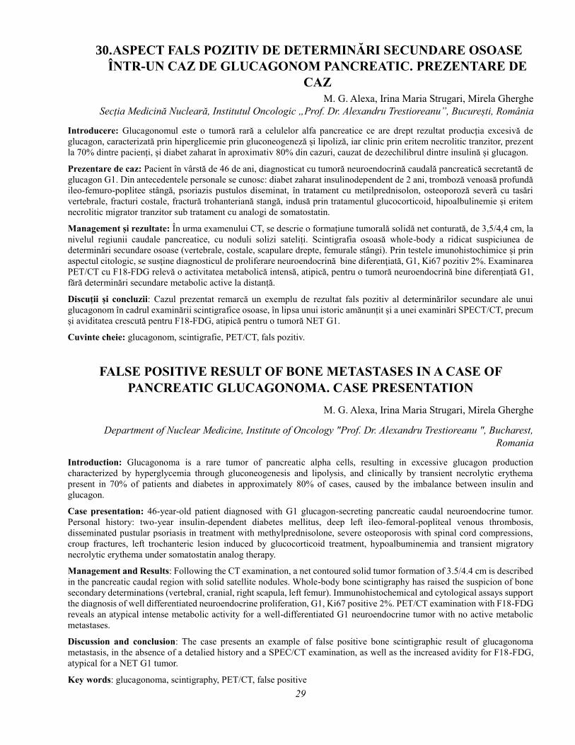
29
30. ASPECT FALS POZITIV DE DETERMINĂRI SECUNDARE OSOASE
ÎNTR-UN CAZ DE GLUCAGONOM PANCREATIC. PREZENTARE DE
CAZ
M. G. Alexa, Irina Maria Strugari, Mirela Gherghe
Secția Medicină Nucleară, Institutul Oncologic „Prof. Dr. Alexandru Trestioreanu”, București, România
Introducere: Glucagonomul este o tumoră rară a celulelor alfa pancreatice ce are drept rezultat producția excesivă de
glucagon, caracterizată prin hiperglicemie prin gluconeogeneză și lipoliză, iar clinic prin eritem necrolitic tranzitor, prezent
la 70% dintre pacienți, și diabet zaharat în aproximativ 80% din cazuri, cauzat de dezechilibrul dintre insulină și glucagon.
Prezentare de caz: Pacient în vârstă de 46 de ani, diagnosticat cu tumoră neuroendocrină caudală pancreatică secretantă de
glucagon G1. Din antecedentele personale se cunosc: diabet zaharat insulinodependent de 2 ani, tromboză venoasă profundă
ileo-femuro-poplitee stângă, psoriazis pustulos diseminat, în tratament cu metilprednisolon, osteoporoză severă cu tasări
vertebrale, fracturi costale, fractură trohanteriană stangă, indusă prin tratamentul glucocorticoid, hipoalbulinemie și eritem
necrolitic migrator tranzitor sub tratament cu analogi de somatostatin.
Management și rezultate: În urma examenului CT, se descrie o formațiune tumorală solidă net conturată, de 3,5/4,4 cm, la
nivelul regiunii caudale pancreatice, cu noduli solizi sateliți. Scintigrafia osoasă whole-body a ridicat suspiciunea de
determinări secundare osoase (vertebrale, costale, scapulare drepte, femurale stângi). Prin testele imunohistochimice şi prin
aspectul citologic, se susţine diagnosticul de proliferare neuroendocrină bine diferenţiată, G1, Ki67 pozitiv 2%. Examinarea
PET/CT cu F18-FDG relevă o activitatea metabolică intensă, atipică, pentru o tumoră neuroendocrină bine diferențiată G1,
fără determinări secundare metabolic active la distanță.
Discuţii și concluzii: Cazul prezentat remarcă un exemplu de rezultat fals pozitiv al determinărilor secundare ale unui
glucagonom în cadrul examinării scintigrafice osoase, în lipsa unui istoric amănunțit și a unei examinări SPECT/CT, precum
și aviditatea crescută pentru F18-FDG, atipică pentru o tumoră NET G1.
Cuvinte cheie: glucagonom, scintigrafie, PET/CT, fals pozitiv.
FALSE POSITIVE RESULT OF BONE METASTASES IN A CASE OF
PANCREATIC GLUCAGONOMA. CASE PRESENTATION
M. G. Alexa, Irina Maria Strugari, Mirela Gherghe
Department of Nuclear Medicine, Institute of Oncology "Prof. Dr. Alexandru Trestioreanu ", Bucharest,
Romania
Introduction: Glucagonoma is a rare tumor of pancreatic alpha cells, resulting in excessive glucagon production
characterized by hyperglycemia through gluconeogenesis and lipolysis, and clinically by transient necrolytic erythema
present in 70% of patients and diabetes in approximately 80% of cases, caused by the imbalance between insulin and
glucagon.
Case presentation: 46-year-old patient diagnosed with G1 glucagon-secreting pancreatic caudal neuroendocrine tumor.
Personal history: two-year insulin-dependent diabetes mellitus, deep left ileo-femoral-popliteal venous thrombosis,
disseminated pustular psoriasis in treatment with methylprednisolone, severe osteoporosis with spinal cord compressions,
croup fractures, left trochanteric lesion induced by glucocorticoid treatment, hypoalbuminemia and transient migratory
necrolytic erythema under somatostatin analog therapy.
Management and Results: Following the CT examination, a net contoured solid tumor formation of 3.5/4.4 cm is described
in the pancreatic caudal region with solid satellite nodules. Whole-body bone scintigraphy has raised the suspicion of bone
secondary determinations (vertebral, cranial, right scapula, left femur). Immunohistochemical and cytological assays support
the diagnosis of well differentiated neuroendocrine proliferation, G1, Ki67 positive 2%. PET/CT examination with F18-FDG
reveals an atypical intense metabolic activity for a well-differentiated G1 neuroendocrine tumor with no active metabolic
metastases.
Discussion and conclusion: The case presents an example of false positive bone scintigraphic result of glucagonoma
metastasis, in the absence of a detalied history and a SPEC/CT examination, as well as the increased avidity for F18-FDG,
atypical for a NET G1 tumor.
Key words: glucagonoma, scintigraphy, PET/CT, false positive

30
31. DIAGNOSTICUL DIFERENȚIAL ȘI STABILIREA CONDUITEI
TERAPEUTICE PENTRU O TUMORĂ OSOASĂ SCAPULARĂ
SUPRASPINOASĂ PRIN SCINTIGRAFIA OSOASĂ ÎN TREI FAZE
VERSUS ALTE TEHNICI IMAGISTICE
M. Dragoteanu1, Cecilia Pîgleșan1, Ioana Duca2
1Institutul Regional de Gastroenterologie-Hepatologie” Prof. Dr. Octavian Fodor”, Cluj-Napoca 2Clinica Medicală II, Cluj-Napoca
Pacient 41 ani, diagnostic clinic, Rx, CT, ecografie de părți moi: tumoră malignă supra-spinoasă a omoplatului stâng. Fără
fenomene locale celsiene evidente.
RADIOGRAFIE: Tumoră scapulară supraspinoasă, zone de distrucție osoasă, deformări ale corticalei; aspect de sarcom osos,
puțin probabil metastaza unică, exclude osteomielita.
ECOGRAFIA de părți moi: Distrucție extinsă a corticalei osoase, întreruperi periostale, aspect tumoral (sarcom?). RMN:
Colecție intraarticulară scapulo-humerală importantă (abces?). Aspect inflamator al musculaturii periarticulare. CT: Aspect
de tumoră condromatoasă scapulară primitivă malignă.
REPETARE RADIOGRAFIE: Zonă osteolitică imprecis delimitată, interesând procesul coracoid, posibil sechestru osos. Nu
poate diferenția abces / proces malign, eventual suprainfectat.
SCINTIGRAFIA OSOASĂ ”în TREI FAZE”: 1. Faza dinamică precoce: Hiperperfuzie intensă supraspinoasă stângă. Aspect
tipic de osteomielită. 2. Scintigrafia statică imediată: Fixare intensă supraspinoasă. Susține diagnosticul de osteomielită, nu
exclude tumora malignă suprainfectată. 3. Scintigrafia statică tardivă segmentală și de corp întreg, 3h postinjectare:
Hiperfixarea locală supraspinoasă, fără alte zone focale hipercaptante. Nu tranșează diagnosticul diferențial tumoră malignă
/ osteomielită.
CONDUITA TERAPEUTICĂ: Pacientul refuză propunerea ortopedului de amputare a brațului și centurii scapulo-humerale.
Biopsie osoasă chirurgicală din zona supraspinoasă, examen morfopatologic, însămânțare pe medii de cultură. Intraoperator:
aspect osos tumoral. Externare, tratament ambulatorial două săptămâni cefalosporine + ciprofloxacină.
REZULTATE LABORATOR: Confirmarea infecției stafilocococice. Examen histopatologic: fără caractere de malignitate.
Antibioterapie prelungită 2 luni; dispariție totală a simptomatologiei.
CONCLUZIE: Scintigrafia osoasă in trei faze este foarte performantă în evaluarea diferențiată a imaginilor segmentale și
corp întreg. Diagnosticul de tumoră osoasă primitivă malignă pus exclusiv prin tehnici imagistice morfologice este riscant.
Cuvint cheie: Scintigrafie osoasă, sarcom, metastază osoasă, osteomielită, diagnostic diferențial imagistic
32. TUMORI PRIMARE ASOCIATE CANCERULUI TIROIDIAN
K. Gabora1,2, B. Grădinaru2, E. Spînu2, G. Rusu2, L. Hîțu2, A. Alexiev2, H. Haba2, G. Kermoison2, E. Olariu1,2,
A. Piciu1,2, D. Piciu1,2 1Universitatea de Medicină și Farmacie ”Iuliu Hațieganu” Cluj Napoca
2Institutul Oncologic ”Prof. Dr. Ion Chiricuță” Cluj Napoca
Introducere: Incidența patologiilor neoplazice este în creștere continuă, fiind legată de expunerea individului la factori de
risc predispozanți, existența unei condiționări genetice, vârsta, sexul etc. Cancerul tiroidian este cea mai frecventă patologie
neoplazică endocrină al cărei tratament presupune cel mai frecvent chirurgie și radioiodoterapie. Apariția unui proces malign
asociat cu neoplazia tiroidiană nu este o raritate. Rata apariției unei neoplasm asociat cu cancerul de tiroidă raportat în
literatură este de 13,1% pentru sexul masculin şi de 13,7% pentru sexul feminin.
Metode: Studiul actual s-a desfășurat în Institutului Oncologic “Prof. Dr. Ion Chiricuţă”, Cluj-Napoca. S-au luat în evidenţă
pacienţi cu neoplazii primare asociate cancerului tiroidian care s-au prezentat la control oncologic în perioada noiembrie
2016 - aprilie 2017. S-a efectuat un studiu retrospectiv în care au fost incluși pacienți diagnosticați cu cancer tiroidian, la
care s-a asociat o altă tumoră primară.
Rezultate: Au fost incluși în studiu 50 de pacienți cu cancer tiroidian și alte neoplasme primare asociate. S-au identificat 16
cazuri de cancer mamar (32%), 12 cazuri de melanom (24%), 13 cazuri de cancer colorectal (26%), 9 cazuri de neoplasm
bronho-pulmonar (18%). Majoritatea cazurilor au fost de sex feminin, tipul de asociere cel mai frecvent a fost de tip metacron

31
cu un procentaj de 77% față de cel sincron 23%. O singură pacientă a fost identificată cu trei cancere primare asociate,
repectiv neoplasm tiroidian, colo-rectal și melanom, la care s-a testat mutația BRAF V600E din toate tipurile de tumoră.
Concluzii: Sunt puține studii care abordează tema cancerelor primare asociate, deși ele sunt importante atât din punct de
vedere a evaluării, supravegherii cât și a conduitei terapeutice la acești pacienți.
Cuvinte cheie: glanda tiroidă, cancer tiroidian, neoplasme asociate
Referințe
1.Thyroid cancer statistics [Internet]. Cancer Research UK. 2016 [cited 7 December 2016]. Available from:
http://www.cancerresearchuk.org/health-professional/cancer-statistics/statistics-by-cancer-type/thyroid-cancer
2.Piciu D, Irimie A, Piciu A. Investigation of thyroid carcinoma over 40 years, using the database of the Ion Chiricuta
Institute of Oncology Cluj-Napoca. J BUON. 2014 Apr-Jun; 19(2): 524–529.
3.Rubinstein J, Brown T, Christison-Lagay E, Zhang Y, Kunstman J, Juhlin C et al. Shifting patterns of genomic variation in
the somatic evolution of papillary thyroid carcinoma. BMC Cancer. 2016;16(1).
4.Hsu C, Huang C, Hsu Y, Iqbal U, Nguyen P, Jian W. Co-occurrence of second primary malignancy in patients with thyroid
cancer. QJM. 2014;107(8):643-648.
5.Subramanian S, Goldstein D, Parlea L, Thabane L, Ezzat S, Ibrahim-Zada I et al. Second Primary Malignancy Risk in
Thyroid Cancer Survivors: A Systematic Review and Meta-Analysis. Thyroid. 2007;17(12):1277-1288.
6.Sawka A, Thabane L, Parlea L, Ibrahim-Zada I, Tsang R, Brierley J et al. Second Primary Malignancy Risk After
Radioactive Iodine Treatment for Thyroid Cancer: A Systematic Review and Meta-analysis. Thyroid. 2009;19(5):451-457.
7.Piciu D, Pestean C, Barbus E, Larg M, Piciu A. Second malignancies in patients with differentiated thyroid carcinoma
treated with low and medium activities of radioactive I-131. Clujul Medical. 2016;89(3):384.
8.Atta I, Al Qahtani F. Thyroid, Renal, and Breast Carcinomas, Chondrosarcoma, Colon Adenomas, and Ganglioneuroma:
A New Cancer Syndrome, FAP, or Just Coincidence. Case Reports in Medicine. 2016;2016:1-5.
9.Jelsig A, Qvist N, Brusgaard K, Nielsen C, Hansen T, Ousager L. Hamartomatous polyposis syndromes: A review. Orphanet
Journal of Rare Diseases. 2014;9(1):101.
10.Nosé V. Genodermatosis Affecting the Skin and Mucosa of the Head and Neck: Clinicopathologic, Genetic, and Molecular
Aspect—PTEN-Hamartoma Tumor Syndrome/Cowden Syndrome. Head and Neck Pathol. 2016;10(2):131-138.
11.Bailey E, Tantravahi S, Poole A, Agarwal A, Straubhar A, Batten J et al. Correlation of Degree of Hypothyroidism With
Survival Outcomes in Patients With Metastatic Renal Cell Carcinoma Receiving Vascular Endothelial Growth Factor
Receptor Tyrosine Kinase Inhibitors. Clinical Genitourinary Cancer. 2015;13(3):e131-e137.
12.Mondello P, Sindoni A, Pitini V, Scisca C, Altavilla G, Benvenga S. Thyroid function, autoimmunity and nodules in
hematological malignancies. Arch Endocrinol Metab. 2015;59(3):236-244.
13.de Filette J, Jansen Y, Schreuer M, Everaert H, Velkeniers B, Neyns B et al. Incidence of Thyroid-Related Adverse Events
in Melanoma Patients Treated with Pembrolizumab. The Journal of Clinical Endocrinology & Metabolism. 2016;jc.2016-
2300.
14.Tanaka R, Fujisawa Y, Maruyama H, Nakamura Y, Yoshino K, Ohtsuka M et al. Nivolumab-induced thyroid dysfunction.
Japanese Journal of Clinical Oncology. 2016;46(6):575-579.
15.Fujiwara Y. Hypothyroidism in patients with colorectal carcinoma treated with fluoropyrimidines. Oncology Reports.
2013
16. Piciu D. Nuclear endocrinology. Berlin: Springer; 2012.
17.Cui G, Liu D, Li W, Fu X, Liang Y, Li Y et al. A meta-analysis of the association between BRAF mutation and nonsmall
cell lung cancer. Medicine. 2017;96(14):e6552.
18.Sanz-Garcia E, Argiles G, Elez E, Tabernero J. BRAF mutant colorectal cancer: prognosis, treatment, and new
perspectives. Annals of Oncology. 2017;28(11):2648-2657.
19.Kang X, Zeng Y, Liang J, Li J, Ren D, Chai L et al. Aberrations and clinical significance of BRAF in malignant melanoma.
Medicine. 2018;97(1):e9509.

32
PRIMARY TUMORS ASSOCIATED WITH THYROID CANCER
K. Gabora1.2, B. Grădinaru2, E. Spînu2, G. Rusu2, L. Hîțu2, A. Alexiev2, H. Haba2, G. Kermoison2, E. Olariu1,2,
A. Piciu1,2, D. Piciu1,2
1University of Medicine and Pharmacy ”Iuliu Hațieganu” Cluj Napoca 2Institute of Oncology ”Prof. Dr. Ion Chiricuță” Cluj Napoca
Introduction: The incidence of cancer is steadily increasing worldwide, being related to the exposure of the individual to
predisposing risk factors, the existence of genetic conditioning, age, gender, etc. Thyroid cancer is the most common
endocrine cancer whose treatment involves usually surgery and radioiodine therapy. The occurrence of a malignant process
associated with thyroid cancer is not a rarity. The rate of occurrence of a primary neoplasm associated with thyroid cancer
reported in the literature is 13.1% for males and 13.7% for females.
Methods: The present study was conducted at the Institute of Oncology "Prof. Dr. Ion Chiricuţă", Cluj-Napoca. We
conducted a retrospective study to identify patients with thyroid cancer and an associated primary malignancy. We included
in our study patients who underwent oncological control during November 2016 - April 2017 in our department for thyroid
cancer, who in association presented another primary malignant tumor.
Results: 50 patients with thyroid cancer and other associated primary neoplasms were enrolled in this study. We identified
16 cases of breast cancer (32%), 12 cases of melanoma (24%), 13 cases of colorectal cancer (26%), 9 cases of
bronchopulmonary neoplasm (18%). Most cases were female, the most common type of association was metachronous with
77% of the cases, while 23% were synchronous. One patient was identified with three associated primary cancers, in
particular ,thyroid cancer, colorectal cancer and melanoma, the BRAF V600E mutation was tested in all tumor types.
Conclusions: There are few studies addressing the issue of associated primary malignancies, although they are important
both in terms of evaluation, supervision and treatment in these patients.
Key words: thyroid gland, thyroid cancer, associated primary tumors
References
1.Thyroid cancer statistics [Internet]. Cancer Research UK. 2016 [cited 7 December 2016]. Available from:
http://www.cancerresearchuk.org/health-professional/cancer-statistics/statistics-by-cancer-type/thyroid-cancer
2.Piciu D, Irimie A, Piciu A. Investigation of thyroid carcinoma over 40 years, using the database of the Ion Chiricuta
Institute of Oncology Cluj-Napoca. J BUON. 2014 Apr-Jun; 19(2): 524–529.
3.Rubinstein J, Brown T, Christison-Lagay E, Zhang Y, Kunstman J, Juhlin C et al. Shifting patterns of genomic variation in
the somatic evolution of papillary thyroid carcinoma. BMC Cancer. 2016;16(1).
4.Hsu C, Huang C, Hsu Y, Iqbal U, Nguyen P, Jian W. Co-occurrence of second primary malignancy in patients with thyroid
cancer. QJM. 2014;107(8):643-648.
5.Subramanian S, Goldstein D, Parlea L, Thabane L, Ezzat S, Ibrahim-Zada I et al. Second Primary Malignancy Risk in
Thyroid Cancer Survivors: A Systematic Review and Meta-Analysis. Thyroid. 2007;17(12):1277-1288.
6.Sawka A, Thabane L, Parlea L, Ibrahim-Zada I, Tsang R, Brierley J et al. Second Primary Malignancy Risk After
Radioactive Iodine Treatment for Thyroid Cancer: A Systematic Review and Meta-analysis. Thyroid. 2009;19(5):451-457.
7.Piciu D, Pestean C, Barbus E, Larg M, Piciu A. Second malignancies in patients with differentiated thyroid carcinoma
treated with low and medium activities of radioactive I-131. Clujul Medical. 2016;89(3):384.
8.Atta I, Al Qahtani F. Thyroid, Renal, and Breast Carcinomas, Chondrosarcoma, Colon Adenomas, and Ganglioneuroma:
A New Cancer Syndrome, FAP, or Just Coincidence. Case Reports in Medicine. 2016;2016:1-5.
9.Jelsig A, Qvist N, Brusgaard K, Nielsen C, Hansen T, Ousager L. Hamartomatous polyposis syndromes: A review. Orphanet
Journal of Rare Diseases. 2014;9(1):101.
10.Nosé V. Genodermatosis Affecting the Skin and Mucosa of the Head and Neck: Clinicopathologic, Genetic, and Molecular
Aspect—PTEN-Hamartoma Tumor Syndrome/Cowden Syndrome. Head and Neck Pathol. 2016;10(2):131-138.
11.Bailey E, Tantravahi S, Poole A, Agarwal A, Straubhar A, Batten J et al. Correlation of Degree of Hypothyroidism With
Survival Outcomes in Patients With Metastatic Renal Cell Carcinoma Receiving Vascular Endothelial Growth Factor
Receptor Tyrosine Kinase Inhibitors. Clinical Genitourinary Cancer. 2015;13(3):e131-e137.
12.Mondello P, Sindoni A, Pitini V, Scisca C, Altavilla G, Benvenga S. Thyroid function, autoimmunity and nodules in
hematological malignancies. Arch Endocrinol Metab. 2015;59(3):236-244.

33
13.de Filette J, Jansen Y, Schreuer M, Everaert H, Velkeniers B, Neyns B et al. Incidence of Thyroid-Related Adverse Events
in Melanoma Patients Treated with Pembrolizumab. The Journal of Clinical Endocrinology & Metabolism. 2016;jc.2016-
2300.
14.Tanaka R, Fujisawa Y, Maruyama H, Nakamura Y, Yoshino K, Ohtsuka M et al. Nivolumab-induced thyroid dysfunction.
Japanese Journal of Clinical Oncology. 2016;46(6):575-579.
15.Fujiwara Y. Hypothyroidism in patients with colorectal carcinoma treated with fluoropyrimidines. Oncology Reports.
2013
16. Piciu D. Nuclear endocrinology. Berlin: Springer; 2012.
17.Cui G, Liu D, Li W, Fu X, Liang Y, Li Y et al. A meta-analysis of the association between BRAF mutation and nonsmall
cell lung cancer. Medicine. 2017;96(14):e6552.
18.Sanz-Garcia E, Argiles G, Elez E, Tabernero J. BRAF mutant colorectal cancer: prognosis, treatment, and new
perspectives. Annals of Oncology. 2017;28(11):2648-2657.
19.Kang X, Zeng Y, Liang J, Li J, Ren D, Chai L et al. Aberrations and clinical significance of BRAF in malignant melanoma.
Medicine. 2018;97(1):e9509.
33. STRUCTURAL AND FUNCTIONAL IMAGING ASPECTS OF
NEUROENDOCRINE CARCINOMA OF THE CERVIX : A CASE REPORT
Wael Jalloul1, Ruxandra Tibu1, Ana-Maria Statescu1, Cipriana Ștefănescu1, 2 1 Nuclear Medicine Laboratory, County Emergency Hospital „Sf. Spiridon” Iasi, Romania
2 University of Medicine and Pharmacy U.M.F „Grigore T. Popa” Iasi, Romania
Background and Aim: Neuroendocrine carcinoma of the cervix (NECC) is an aggressive histological variant accounting
for about 1–1.5% of all cervical cancers. Small cell carcinomas (NEC) is the most common type of NECC. It is more likely
to invade the lymphovascular space and to spread to the regional lymph node basin at the time of diagnosis.
The purpose of our study was to describe the imaging aspects in a case of NECC and to look for a theranostic approach to
treat it.
Materials and Methods: We present the case of 70-year-old woman diagnosed with NECC (small cell NEC). After uterine
curettage and electrical conization, followed by chemotherapy, she was sent for a 99mTc-HDP bone scan in order to obtain
further informations about the nature of a lesion described on a follow up MRI on the left ischium and to look for other
possible bone metastasis.
Results and Discussions:
-Anatomopathological exam: a neuroendocrine cervical small cell carcinoma (poorly differentiated).
-Immunohistochemical exam: positive Chromogranin A, Synaptophysin and Ki67.
-Abdominal and pelvic MRI: the exact topography and extension of the neoplasia. Four metastatic sites were discovered in
the liver and one nodular lesion at the base of the right lung.
-Thoracic CT scan: a pulmonary nodule on the right lung.
-A follow up MRI (6 months after the first MRI): a possible metastatic lesion on the left ischium. No other aspects of active
disease were discovered.
-99mTc-HDP bone scan: A high uptake on the left ischium (corresponding to the MRI description) and an other one on the
mid shaft of the tibia, which could have a metastatic etiology.
The theranostic approach with the use of somatostatin analogues was not possible in this case because of the high value of
Ki67 (40-50%).
Conclusion: MRI, CT and 99mTc-HDP bone scan are essential tools in the pre and post-therapeutic assessment of NECC for
local staging and to determine the extent of nodal and metastatic spread.
In our case, the patient has simultaneously liver, lung and bone metastasis which is rarely described.
Key words: Neuroendocrine carcinoma of the cervix, 99mTc-HDP bone scan, metastasis.
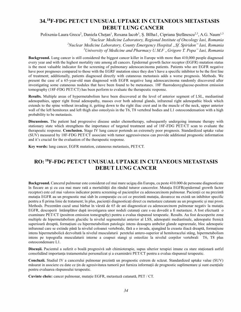
34
34. 18F-FDG PET/CT UNUSUAL UPTAKE IN CUTANEOUS METASTASIS
DEBUT LUNG CANCER
Polixenia-Laura Grecu2, Daniela Chețan1, Roxana Iacob3, Ș. Bîlha1, Cipriana Ștefănescu2,3, A.G. Naum1,3 1Nuclear Medicine Laboratory, Regional Institute of Oncology Iasi, Romania
2Nuclear Medicine Laboratory, County Emergency Hospital „Sf. Spiridon” Iasi, Romania 3University of Medicine and Pharmacy U.M.F „Grigore T. Popa” Iasi, Romania
Background. Lung cancer is still considered the biggest cancer killer in Europe with more than 410,000 people diagnosed
every year and with the highest mortality rate among all cancers. Epidermal growth factor receptor (EGFR) mutation status
is the most valuable indicator for the screening of pulmonary adenocarcinoma patients. Patients who are EGFR negative
have poor prognoses compared to those with the EGRF mutation since they don’t have a specific inhibitor to be the first line
of treatment; additionally, patients diagnosed directly with cutaneous metastasis adds a worse prognosis. Methods. We
present the case of a 65-year-old man diagnosed with EGFR negative lung adenocarcinoma randomly discovered after
investigating some cutaneous nodules that have been found to be metastases. 18F fluorodeoxyglucose-positron emission
tomography (18F-FDG PET/CT) has been perform to evaluate the therapeutic response.
Results. Multiple areas of hypermetabolism have been discovered at the level of anterior segment of LSL, mediastinal
adenopathies, upper right frenal adenopathy, masses over both adrenal glands, infrarenal right adenopathic block which
extends to the spine without invading it, getting down to the right iliac crest and in the muscle of the neck, upper anterior
wall of the left hemitorax and left thigh also osteolysis in the T6, T8 vertebral bodies and L1 osteocondensation with a high
probability to be metastasis.
Discussions. The patient had progressive disease under chemotherapy, subsequently undergoing immune therapy with
stationary state which strengthens the importance of targeted treatment and of 18F-FDG PET/CT scan to evaluate the
therapeutic response. Conclusion. Stage IV lung cancer portends an extremely poor prognosis. Standardized uptake value
(SUV) measured by 18F-FDG PET/CT associate with tumor aggressiveness can provide additional prognostic information
and it’s crucial for the evaluation of the therapeutic response.
Key words: lung cancer, EGFR mutation, cutaneous metastasis, PET/CT.
RO: 18F-FDG PET/CT UNUSUAL UPTAKE IN CUTANEOUS METASTASIS
DEBUT LUNG CANCER
Background. Cancerul pulmonar este considerat cel mai mare ucigaș din Europa, cu peste 410.000 de persoane diagnosticate
în fiecare an și cu cea mai mare rată a mortalității din rândul tuturor cancerelor. Mutația EGFR(epidermal growth factor
receptor) este cel mai valoros indicator pentru screening-ul pacienților cu adenocarcinom pulmonar. Pacienții ce nu prezintă
mutația EGFR au un prognostic mai slab în comparație cu cei ce prezintă mutația, deoarece nu există un inhibitor specific
pentru a fi prima linie de tratament; în plus, pacienții diagnosticați direct cu metastaze cutanate au un prognostic și mai prost.
Methods. Prezentăm cazul unui bărbat în vârstă de 65 de ani diagnosticat cu adenocarcinom pulmonar negativ la mutația
EGFR, descoperit întâmplător după investigarea unor noduli cutanați care s-au dovedit a fi metastaze. A fost efectuată o
examinare PET/CT (positron emission tomography) pentru a evalua răspunsul terapeutic. Results. Au fost descoperite zone
multiple de hipermetabolism glucidic la nivelul segmentului anterior al LSS, adenopatii mediastinale, adenopatie frenică
superioară dreaptă, formațiuni cu hipermetabolism patologic intens deasupra ambelor glande suprarenale, bloc adenopatic
infrarenal care se extinde până la nivelul coloanei vertebrale, fără a o invada, ajungând la creasta iliacă dreaptă, formațiune
intens hipermetabolică dezvoltată la nivelul musculaturii peretelui antero-superior al hemitoracelui stâng, hipermetabolism
intens pe topografia musculaturii interne a coapsei stangi și osteolize la nivelul corpilor vertebrali T6, T8 plus
osteocondensare L1.
Discușii. Pacientul a suferit o boală progresivă sub chimioterapie, supus ulterior terapiei imune cu stare staționară astfel
consolidând importanța tratamentului personalizat și a examinării PET/CT pentru a evalua răspunsul terapeutic.
Concluzii. Stadiul IV a cancerului pulmonar prezintă un prognostic extrem de scăzut. Standardized uptake value (SUV)
măsurat in asociere cu date ce țin de agresivitatea tumorii pot furniza informații de prognostic suplimentare și sunt esențiale
pentru evaluarea răspunsului terapeutic.
Cuvinte cheie: cancer pulmonar, mutație EGFR, metastază cutanată, PET / CT.

35
35. DILATAREA ISCHEMICĂ TRANZITORIE DE EFORT A
VENTRICULULUI STÂNG: PREZENTARE DE CAZ
I.Muntea³, A. Chirilă1,2, R. Hagiu1, G. Andries2,3 1Institutul Inimii „Niculae Stăncioiu” Cluj-Napoca
2UMF „Iuliu Haţieganu” Cluj-Napoca 3Spitalul Clinic Județean de Urgența Cluj-Napoca
Dilatarea ischemică tranzitorie de efort este un fenomen indus de prezenţa ischemiei subendocardice, care generează creşterea
în volum a cavităţii ventriculului stâng pe imaginile post-efort, comparativ cu imaginile de repaus. Aspectul scintigrafic este
specific afectării multivasculare sau stenozelor de trunchi principal semnificative hemodinamic.Vă prezentăm cazul unei
paciente de 66 ani, diabetică, fără angor, cu antecedente de dublu by-pass aorto-coronarian. Imaginile de gated SPECT relevă
dilatare ichemică tranzitorie de efort, cu un indice de dilatare de 1.31, valori normale ale volumelor end-sistolice la efort și
repaus, și anomalii semnificative de performanță ventriculară la efort. Imaginile de perfuzie au arătat hipoperfuzie cronică
de repaus și ischemie extinsă post-efort. Rezultatele obtinute au fost validate prin explorare invazivă: patologie
multivasculară, implicarea trunchiului principal al arterei coronare stângi și ocluzie de graft venos. S-a efectuat angioplastie
coronariana percutană cu rezultat final bun. Examinarea scintigrafică s-a dovedit utilă în stabilirea eligibilităţii pentru
explorare invazivă şi ghidarea terapiei de revascularizare.
Cuvinte cheie: dilatare ischemică tranzitorie de efort, ischemie miocardica, gated SPECT
Referințe:
1.Verberne, H., Acampa, W. and Anagnostopoulos, C. (2015). EANM procedural guidelines for radionuclide myocardial
perfusion imaging with SPECT and SPECT/CT: 2015 revision. European Journal of Nuclear Medicine and Molecular
Imaging, 42(12), pp.1929-1940.
2.Halligan, W., Morris, P. and Schoepf, U. (2014). Transient Ischemic Dilation of the Left Ventricle on SPECT: Correlation
with Findings at Coronary CT Angiography. Journal of Nuclear Medicine, 55(6), pp.917-922.
3.Xu, Y., Arsanjani, R. and Clond, M. (2012). Transient ischemic dilation for coronary artery disease in quantitative analysis
of same-day sestamibi myocardial perfusion SPECT. Journal of Nuclear Cardiology, 19(3), pp.465-473.
4.Slomka, P., Berman, D. and Germano, G. (2016). Normal limits for transient ischemic dilation with 99mTc myocardial
perfusion SPECT protocols. Journal of Nuclear Cardiology, 24(5), pp.1709-1711.
5.Abidov, A., Germano, G. and Berman, D. (2007). Transient ischemic dilation ratio: A universal high-risk diagnostic marker
in myocardial perfusion imaging. Journal of Nuclear Cardiology, 14(4), pp.497-500.
6.Robinson, V., Corley, J. and Marks, D. (2000). Causes of Transient Dilatation of the Left Ventricle During Myocardial
Perfusion Imaging. American Journal of Roentgenology, 174(5), pp.1349-1352.
7.Sharir, T. (2017). Transient ischemic dilation: An old but not obsolete marker of extensive coronary artery disease. Journal
of Nuclear Cardiology, 25(3), pp.738-741.
LEFT VENTRICULAR TRANSIENT ISCHAEMIC DILATION: A CASE
REPORT
I.M. Muntea³, A. Chirilă1,2, R. Hagiu1, G. Andries2,3
1The Heart Institute „Niculae Stăncioiu” Cluj-Napoca, ROMANIA 2University of Medicine and Pharmacy UMF „Iuliu Haţieganu” Cluj-Napoca, ROMANIA
3Department of Nuclear Medicine- Clinical Emergency County Hospital Cluj-Napoca, ROMANIA
Transient ischaemic dilation (TID) is a phenomenon induced by the presence of extensive subendocardial ischaemia,
generating an increase in the left ventricular cavity on the post-exercise images, compared to rest images. The scintigraphic
aspect is specific to multivessel disease or main trunk stenosis with hemodynamic significance. We present the case of a 66-
year-old female diabetic, no typical chest pain, with a history of double aortic coronary bypass. Gated SPECT revealed
transient exercise-induced dilation, with a TID index of 1.31, normal values of end-systolic volumes at stress and rest and

36
significant abnormalities of ventricular performance at stress. Perfusion images showed rest abnormalities and extensive
stress ischaemia. The scintigraphic findings were validated by invasive exploration: multivessel pathology, involvement of
left main coronary and venous graft occlusion. Percutaneous coronary intervention (PCI) was performed, with good clinical
outcome. Scintigraphic examination is useful in establishing the eligibility for invasive exploration and in guiding the
revascularization therapy.
Key words: transient ischaemic dilation, miocardial ischemia, gated SPECT
References:
1.Verberne, H., Acampa, W. and Anagnostopoulos, C. (2015). EANM procedural guidelines for radionuclide myocardial
perfusion imaging with SPECT and SPECT/CT: 2015 revision. European Journal of Nuclear Medicine and Molecular
Imaging, 42(12), pp.1929-1940.
2.Halligan, W., Morris, P. and Schoepf, U. (2014). Transient Ischemic Dilation of the Left Ventricle on SPECT: Correlation
with Findings at Coronary CT Angiography. Journal of Nuclear Medicine, 55(6), pp.917-922.
3.Xu, Y., Arsanjani, R. and Clond, M. (2012). Transient ischemic dilation for coronary artery disease in quantitative analysis
of same-day sestamibi myocardial perfusion SPECT. Journal of Nuclear Cardiology, 19(3), pp.465-473.
4.Slomka, P., Berman, D. and Germano, G. (2016). Normal limits for transient ischemic dilation with 99mTc myocardial
perfusion SPECT protocols. Journal of Nuclear Cardiology, 24(5), pp.1709-1711.
5.Abidov, A., Germano, G. and Berman, D. (2007). Transient ischemic dilation ratio: A universal high-risk diagnostic marker
in myocardial perfusion imaging. Journal of Nuclear Cardiology, 14(4), pp.497-500.
6.Robinson, V., Corley, J. and Marks, D. (2000). Causes of Transient Dilatation of the Left Ventricle During Myocardial
Perfusion Imaging. American Journal of Roentgenology, 174(5), pp.1349-1352.
7.Sharir, T. (2017). Transient ischemic dilation: An old but not obsolete marker of extensive coronary artery disease. Journal
of Nuclear Cardiology, 25(3), pp.738-741.
36. EXAMINAREA PET/CT CU F18-FDG VERSUS SCINTIGRAFIA
RECEPTORILOR DE SOMATOSTATINA IN CAZUL UNEI TUMORI
NEUROENDOCRINE ILEALE G2
M. Mutuleanu, Romina Costache, Mirela Gherghe
Institutul Oncologic “Prof. Dr. Al. Trestioreanu”, București, România, Secția Medicină Nucleară
Introducere: Tumorile neuroendocrine sunt tumori dezvoltate din celule stem multipotente care migrează din creasta
neurală. Aceste tumori se dezvoltă din celulele enterocromafine localizate în țesutul neuroendocrin răspândit în întreg
organismul. Clasificarea morfologică a acestora propusă de OMS se face după indicele de proliferare Ki67 în: tumori G1 (
≤2%-bine diferențiate), tumori G2 (3-20%-moderat diferențiate) și tumori G3 (≥20%-slab diferentiate). Metoda gold
standard de identificare a unei tumori neuroendocrine cu punct de plecare neprecizat este scintigrafia neuroendocrină sau
examenul PET/CT cu radiotrasori specifici ce se cuplează de receptorii de somatostatină (Ga68- DOTATATE și 111In-
Pentetreotide sau 99mTC- Tektrotyd).
Diagnostic și rezultate: Prezentăm cazul unui pacient cu simptomatologie gastrointestinală nespecifică diagnosticat în urma
unei ecografii de rutină cu formațiuni înlocuitoare de spațiu hepatic; examenul bioptic efectuat a evidențiat determinări
secundare hepatice de carcinom neuroendocrin G2.
Pentru depistarea tumorii primare, nefiind disponibili analogii de somatostatină, se indică examen PET/CT cu F18-FDG care
a evidențiat determinarea secundară hepatică, inactivă metabolic la F18-FDG și determinări secundare ganglionare
retroperitoneale moderat active metabolic. Nu s-a reușit determinarea exactă a tumorii primare. Examinarea scintigrafică a
evidențiat: tumora primară ileală și determinare secundară ganglionară iliacă comună stângă intens hipercaptante pentru
radiotrasorul specific.
Concluzii: Cazul prezentat remarcă un exemplu de rezultat fals negativ al unei tumori neuroendocrine G2 cu punct de plecare
neprecizat în cadrul examinării PET/CT F18-FDG, dar care a fost identificată în urma unui examen scintigrafic cu analogi
de somatostatină radiomarcați (99mTC- Tektrotyd) punând în evidență limitele ficărei metode.
Cuvinte cheie: tumoră neuroendocrină, PET/CT, scintigrafie neuroendocrină, fals negativ.

37
PET / CT WITH F18-FDG EXAMINATION VERSUS SOMATOSTATIN
RECEPTOR SCINTIGRAPHY IN A PATIENT WITH NEUROENDOCRINE
ILEAL G2 TUMOR
M. Mutuleanu, Romina Costache, Mirela Gherghe
Oncology Institute "Prof. Dr. Al. Trestioreanu", Bucharest, Romania, Nuclear Medicine Department
Introduction: Neuroendocrine tumors are tumors developed from multipotent stem cells migrating from the neural crest.
These tumors develop from enterocromaphin cells located in neuroendocrine tissue spread throughout the body. Their
morphological classification proposed by WHO is based on the Ki67 proliferation index in G1 tumors (≤2% - highly
differentiated), G2 tumors (3-20% - moderate differentiated) and G3 tumors (≥20% - low differentiated). The gold standard
method for identifying an ocult primary neuroendocrine is neuroendocrine scintigraphy or specific PET/CT examinations
with somatostatin analogs radiotracers that attach themselves to somatostatin receptors (Ga68-DOTATATE and 111In-
Pentetreotide or 99mTC-Tektrotyd).
Diagnosis and Results: We present the case of a patient with non-specific gastrointestinal symptoms diagnosed following a
routine ultrasound with liver tumor formations; the bioptic examination performed revealed hepatic metastasis of
neuroendocrine carcinoma G2.
For the identification of primary tumor, because somatostatin analogs are not available, a F18-FDG PET / CT test is indicated
which revealed metabolic inactive to F18-FDG metastasis of the liver, and moderately metabolic active retroperitoneal
ganglionary metastasis. The exact location of the primary tumor was not identified. The scintigraphic examination revealed:
an intens accumulation of the specific radiotracer in the ileal primary tumor and secondary giant lymph node of the left
common iliac.
Conclusions: The case represents an example of a false negative result of an ocult primary G2 neuroendocrine tumor on the
F18-FDG PET/CT examination but was identified following a scintigraphic examination with somatostatin analogs
radiotracers (99mTC- Tektrotyd) highlighting the limits of both methods.
Key words: neuroendocrine tumor, PET / CT, neuroendocrine scintigraphy, false negative.
37. DUPLICAȚIA RENALĂ: 99mTc –DTPA ȘI 99mTc-DMSA ÎȘI UNESC
FORȚELE PENTRU A DETERMINA FUNCȚIA ȘI VIABILITATEA
Ruxandra Țibu1, T.M. Ionescu1, Irena Cristina Grierosu1,3, A.C. Țârcă1, M.M. Guțu1,3, Magdalena Stârcea2,
Cipriana Ștefănescu1,3 1Laboratorul de Medicină Nucleară, Spitalul Județean de Urgență „Sf. Spiridon” Iași, România
2Departamentul de Nefrologie Pediatrică, Spitalul ”Sfânta Maria”, Iași, România 3Universitatea de Medicină și Farmacie „Grigore T. Popa” Iași, România
Introducere: Anomaliile renale congenitale reprezintă cea mai comună malformație, însumând aproximativ o treime din
totalul anomaliilor fetale. Incidența duplicației renale este de aproximativ 1%. Scintigrafia renală este frecvent utilizată pentru
evaluarea funcției fiecărui rinichi, iar în completarea sa poate fi efectuată scintigrama renală statică pentru a identifica
viabilitatea și cicatricile renale. Particularitatea cazului este determinarea funcției si viabilității folosind două proceduri
scintigrafice distincte, în cazul unei duplicații renală cu istoric de infecții renale repetitive.
Metodă: Pacienta în vârstă de 2 ani a fost diagnosticată postnatal consecutiv infecțiilor urinare repetate, prin URO-CT, cu
duplicație reno-pielo-ureterală bilaterală, hidronefroză a unității superioare a rinichiului stâng și ureterocel drept. În prima zi
a fost realizată nefrograma cu 99mTc-DTPA imediat după injectarea i.v. a radiofarmaceuticului, urmată de administrarea
Furosemidului în minutul 1, iar în ziua următoare a fost efectuată o renogramă cu 99mTc-DMSA.
Rezultate: Nefrograma a decelat o funcție aparent normală pentru rinichiul stang după administrarea diureticului, cu
mențiunea unei funcții mai reduse a polului renal superior comparativ cu cel inferior. În cazul rinichiului drept s-a evidențiat
o obstrucție mecanică incompletă, care ulterior a fost cuantificată. Scintigrama renală relevat dimensiuni scintigrafice diferite
ale rinichiului drept comparativ cu cel stâng.
Discuții: Renograma și nefrograma sunt investigații complementare folosite pentru a evalua o malformație renală complexă
din perspective diferite. Nefrograma cu 99mTc-DTPA determină funcția fiecărei unități a rinichiului afectat în cadrul

38
duplicației renale bileterale, datorită mecanismului său de captare specific (filtrare glomerulară). Scintigrama renală statică
cu 99mTc-DMSA evaluează viabilitatea și zonele cicatriceale post-infecție datorită captării sale corticale.
Concluzii: În relație cu cazul prezentat, concluzionăm că scintigrafia renală statică și dinamică poate determina viabilitatea
și funcția fiecărui rinichi în parte, în vederea elaborării unei strategii terapeutice personalizate.
Cuvinte cheie: duplicație renală bilaterală, test la Furosemid, nefrogramă, renogramă
DUPLICATE KIDNEY: 99mTc-DTPA AND 99mTc-DMSA JOIN FORCES FOR
FUNCTION AND VIABILITY
Ruxandra Țibu1, T.M. Ionescu1, Irena Cristina Grierosu1,3, A. C. Țârcă1, M.M. Guțu1,3, Magdalena Stârcea2,
Cipriana Ștefănescu1,3
1Nuclear Medicine Laboratory, County Emergency Hospital „Sf. Spiridon” Iasi, Romania 2Pediatric Nephrology Department, Saint Maria Hospital, Iasi, Romania
3University of Medicine and Pharmacy U.M.F „Grigore T. Popa” Iasi, Romania
Background: Congenital renal anomalies are the most common malformation types, representing almost a third of fetal
anomalies. Duplex kidneys have an incidence of approximately 1%. Dynamic renal scintigraphy is used to determine the
functionality of each kidney. In addition to the nephrogram, static renal scintigraphy can be done to assess viability and
scarring. The particularity of our case is determining the functionality and viability using two distinctive scintigraphic
procedures in a case of bilateral duplicated kidney and repetitive urinary tract infections.
Method: Our 2 years old patient was diagnosed postnatally by URO-CT, subsequent to repetitive urinary tract infections,
with bilateral renopieloureteral duplication, left superior unit hydronephrosis and right ureterocele. In the first day it was
performed a nephrogram with 99mTc-DTPA immediately after the iv injection of the radiopharmaceutical and Furosemide
administration in the first minute. In the next day a renal scan with 99mTc-DMSA was added.
Results: The nephrogram revealed an apparent normal function for the left kidney after the diuretic with the upper pole
functioning less then the inferior pole. The right kidney revealed an incomplete mechanical obstruction that was later
quantified. The renogram unveiled modified scintigraphic dimensions of the right kidney compared to the left.
Discussions: The renogram and nephrogram are complementary investigations used to approach a complex renal
malformation from different points of view. 99mTc-DTPA dynamic renal scan is able to determine the functionality of each
unit of the affected kidney in the context of bilateral renal duplication, owing to it’s specific uptake mechanism (glomerular
filtration). 99mTc-DMSA can evaluate the viability and post-infection scarring, due to its cortical uptake.
Conclusion: In our particular case, dynamic and static renal scintigraphy can determine the functionality and viability of
each kidney for a personalized management and treatment approach.
Key words: bilateral duplicated kidney, furosemide test, nefrogram, renogram
38. THE ROLE OF F18-FDG PET/CT BRAIN SCANNING IN PATIENTS
WITH ONCOLOGICAL PATHOLOGY FOR ASSESSMENT OF
PITUITARY METABOLISM: PRIMARY TUMORS, METASTASIS OR
PHYSIOLOGICAL UPTAKE AND SUBSEQUENT MANAGEMENT
E.Spinu*; B. Gradinaru*; D.Piciu; E.Olariu; L.Hitu; G.Rusu; H. Haba; G. Kermoison. A.Piciu; K. Gabora;
I.Cecan.
Department of Nuclear Medicine, the Institut of Oncology ''Prof. Dr. Ion Chiricuta'', Cluj-Napoca, Romania
*E Spinu si B Gradinaru have equally contributed to this paper
Introduction: The F18-FDG PET/CT scanning protocol for most oncological pathologies includes assessment from the base
of the skull up to mid-femoral bones. This article highlights the importance of including brain scan in oncological patients
for the evaluation of pituitary metabolism and possible incidentalomas.
39
Materials and Method: The article includes 4 cases with different oncological pathologies: thyroid folliculo-papillary
carcinoma, typical pulmonary neuroendocrine tumor, small cell lung carcinoma and parotid adenocarcinoma. Included in the
multidisciplinary therapeutic management for their pathologies, patients were evaluated with F18-FDG PET/CT scans, which
included the cerebral region. Although the assessment of cerebral metabolism is difficult to assess properly due to increased
glucose consumption, the pituitary gland can have varying degrees of radiopharmaceutical uptake.
Results: The study includes 4 patients with different cancers, known or not with pituitary abnormalities. Following the F18-
FDG PET/CT evaluation, the pituitary metabolic status is particular for each patient.
We would also like to mention that the incidence of pituitary findings identified on F18-FDG PET/CT scans performed in
our medical department is 0.04% out of over 8000 scans.
Conclusions: Given the particularities of hybrid imaging in pituitary metabolism assessment, we consider that brain scans
should be performed in all F18-FDG PET/CT scanning protocols, while taking into account the possible findings: primary
tumors, metastases, functioning/non-functioning adenoma, normal functioning pituitary gland with increased metabolic
activity - artifact. In order to identify the causes of abnormal findings we recommend further tests including contrast enhanced
MRI for pituitary gland and endocrine and metabolic evaluation.