VOLATOLOMICS ANALYSIS OF LUNG AND COLON CANCER...
37
UNIVERSITI TEKNOLOGI MALAYSIA VOLATOLOMICS ANALYSIS OF LUNG AND COLON CANCER USING TERAHERTZ AND INFRARED SPECTROSCOPY AHMAD ZULHILMI BIN ARSHAD
Transcript of VOLATOLOMICS ANALYSIS OF LUNG AND COLON CANCER...

UNIVERSITI TEKNOLOGI MALAYSIA
VOLATOLOMICS ANALYSIS OF LUNG AND COLON CANCER USING
TERAHERTZ AND INFRARED SPECTROSCOPY
AHMAD ZULHILMI BIN ARSHAD

.
VOLATOLOMICS ANALYSIS OF LUNG AND COLON CANCER USING
TERAHERTZ AND INFRARED SPECTROSCOPY
SEPTEMBER 2016
Faculty of Bioscience and Medical Engineering
Universiti Teknologi Malaysia
A thesis submitted in fulfilment of the
requirements for the award of the degree of
Doctor of Philosophy (Biomedical Engineering)
AHMAD ZULHILMI BIN ARSHAD

iii
A special dedication to my parents,
Arshad Mustapa & Rohana Hassan
To my beloved siblings,
Herman, Anis, Faiz, Hisyam & Tasya
To my beloved best friends,
Sarhan, Sya, Krik, Arep, Azani,
Razak, Fauzi, Syamil
Thank you for everything.
~ipsa scientia potestas est~
“KNOWLEDGE ITSELF IS POWER”
…with love and care
a.zulhilmi
DEDICATION

iv
Bismillahirrahmanirrahim… In the name of Allah S.W.T., the most Gracious,
Merciful and Knowledgeable. All this work is belonging to Him, the owner of all
knowledge and creatures.
I would like to express my sincere gratitude to my supervisor Associate
Professor Dr. Yusof Munajat for his mentoring and guidance. He is untiring
enthusiasm, support, advice and guidance throughout my study. He has encouraged
me to reach my full potential as a research scientist and to be an independent and
critical thinker. He provided me with many academic opportunities on campus to
widen my horizons. Without him, this study would not have been possible to finish.
I also would like to acknowledge all my co-supervisors, Dr. Raja
Kamarulzaman Raja Ibrahim, Dr. Salehhuddin Hamdan and Dr. Nasrul Humaimi
Mahmood, for their advice, time, critical feedback and input. I would like to thanks
Dr. Noradzimah Johdi and Mr. Hanif Zulkhairi from UKM Medical Molecular
Biology Institute (UMBI) for facilitating collaborative work with us. Not to forget the
Photonics Research Centre (PRC), Universiti Malaya for providing Terahertz time
domain spectroscopy instrument to analyse the samples.
I would also like to thanks everyone who helped with experiments, talking
through ideas and being resourceful and to my colleagues in the Physics Departments,
Faculty of Sciences, and Department of Biotechnology & Medical Engineering,
Faculty of Biosciences and Medical Engineering, for making the campus enjoyable
ACKNOWLEDGEMENT

v
and intellectuality stimulating. Do not forget the member of the Applied Optics
Research lab, Kak Sara, Hamdi, Ayu, Farha, Wani and Elham.
Special thanks to Razak, Fauzi, Azani, Rizal, Hafizi, Sham and Aidil for their
valuable discussions and suggestions in finishing my study. I extend many thanks to
my best friend group since secondary school for their support, care and motivation
through my difficulties and sorrows over a decade. I love them all.
Finally, yet importantly, I would like to say a heartfelt thank you to my family.
Thanks to my parents for their endless support, love and faith in me over the years.
Also, for Malaysia Ministry of Higher Education (MOHE) for providing MyBrain15
(MyPhD) scholarship throughout my three years and half of studies. This study is
funded by two Fundamental Research Grant Scheme (FRGS - grant no: 4F178 and
4F648) and one UTM Research University Grant (RUG Tier 1: 06H63). Thank you.

vi
Terahertz and infrared spectroscopy are effective analytical spectroscopic
techniques to identify and study the conformation and molecular interaction of the
biomolecules. It has a huge potential in cancer diagnosis because these spectroscopies
are non-invasive technique and do not require labelling for tissues and cells.
Volatolomics analysis is a technique to analyse the volatile organic compounds
(VOCs) emitted and released by human metabolites, which are not limited to breathe
analysis. VOCs that are released by cancerous cells can be one of the bio-diagnostics
techniques to diagnose cancer. Although studies on breath analysis have been widely
carried out, the study of the volatolomics analysis by using Fourier transform infrared
spectroscopy (FTIR) and Terahertz time-domain spectroscopy (THz-TDS) is still new.
Both FTIR and THz-TDS instruments are installed with a gas cell sampling tools by
absorption technique to analyse and detect the key species released from the VOCs.
Lung cancer (NCL-H1299) and colon cancer (COLO320DM) cell lines are uas
samples to identify the key species of each of the cancerous cells. The experiment has
been verified and validated by comparing with control samples such as normal lung
(MRC-5) cell lines, normal colon (CCD112CoN) cell lines, empty flask, air from the
culture media and normal lab air. All the samples have been cultured into different
sealed flasks for 24 to 120 hours, before the VOCs are collected and transferred into
the gas cells to analyse using FTIR and THz-TDS. Hydrogen chloride and benzamide
have been identified as key species for lung and colon cancer, respectively. These
findings have been verified and validated by using residual gas analyser (RGA), gas
chromatography – mass selective detector (GC-MSD), and confirmed by earlier
literatures. A chemometric statistical analysis also has been applied to this study to
extract the important information of the biochemical data from the VOCs with the
greatest discriminative power and highest precision. These findings demonstrate the
potential use of FTIR and THz-TDS as clinical tools through the volatolomics analysis.
In addition, more work is needed if it is to be applied in clinical practice.
ABSTRACT

vii
Spektroskopi terahertz dan inframerah merupakan teknik spektroskopik
analitikal yang efektif dalam mengenalpasti dan mengkaji struktur interaksi molekul
bagi sesuatu biomolekul. Ia merupakan potensi yang besar dalam proses diagnosis
kanser kerana teknik spektroskopik ini adalah tidak invasif dan tidak memerlukan
pelabelan untuk tisu dan sel. Analisis volatolomik pula merupakan satu teknik untuk
menganalisis sebatian organik yang mudah meruap (VOCs) yang terhasil daripada
proses metabolisme manusia, yang mana tidak terhad kepada analisis pernafasan
sahaja. VOCs yang dihasilkan oleh sel kanser boleh menjadi salah satu teknik bio-
diagnostik sel kanser. Walaupun kajian mengenai analisis pernafasan telah banyak
dijalankan, tetapi kajian analisis volatolomik dengan mengunakan spektroskopi infra
merah transformasi Fourier (FTIR) dan spektroskopi Terahertz domain masa (THz-
TDS) masih baru. Kedua-dua instrument FTIR dan THz-TDS telah dipasangkan pada
satu alat persampelan sel gas melalui teknik penyerapan untuk menganalisa dan
mengesan spesies petunjuk daripada VOCs yang dilepaskan. Titisan sel-sel bagi
kanser paru-paru (NCL-H1299) dan kanser kolon (COLO320DM) digunakan di dalam
kajian ini untuk mengesan spesies petunjuk bagi setiap kanser. Ujikaji yang dijalankan
telah diverifikasi dan divalidasi dengan membandingkan sampel terkawal seperti
titisan sel paru-paru normal (MRC-5), sel kolon normal (CCD112CoN), udara
kelalang kosong, udara daripada medium kultur dan udara persekitaran makmal.
Semua sampel titisan sel telah dikultur melalui kelalang-kelalang yang kedap yang
berbeza selama 24 jam hingga 120 jam, sebelum VOCs dikumpul dan dipindahkan ke
sel-sel gas untuk dianalisis menggunakan FTIR dan THz-TDS. Hidrogen klorida dan
benzamida telah dikenalpasti sebagai spesies petunjuk bagi kanser paru-paru dan
kanser kolon. Penemuan ini telah diverifikasi dan divalidasi dengan menggunakan
penganalisis gas sisa (RGA), kromatografi gas – pengesan jisim terpilih (GC-MSD)
dan disahkan oleh literature terdahulu. Satu statistikal analisis kemometri juga
diterapkan untuk kajian ini bagi mengekstrak maklumat penting data biokimia
daripada VOCs dengan kuasa diskrimitif terbesar dan kepersisan tertinggi. Hasil kajian
ini menunjukkan potensi penggunaan FTIR dan THz-TDS sebagai peralatan klinikal
menerusi analisis volatolomik. Di samping itu, kajian lanjut masih diperlukan jika ia
ingin diaplikasikan di dalam amalan klinikal.
ABSTRAK

viii
TABLE OF CONTENTS
CHAPTER
TITLE PAGE
DECLARATION ii
DEDICATION iii
ACKNOWLEDGEMENT iv
ABSTRACT vi
ABSTRAK vii
TABLE OF CONTENTS viii
LIST OF TABLES xii
LIST OF FIGURES xiv
LIST OF ABBREVATIONS xix
LIST OF APPENDICES xx
1 INTRODUCTION 1
1.1 Overview 1
1.2 Background of the Study 1
1.3 Motivation of the Work 3
1.3.1 Why is volatolomics analysis used in cancer diagnosis? 3
1.3.2 Why should a volatolomics analysis system be
developed? 4
1.4 Problem Statement 4
1.5 Objectives 5
1.6 Scope of Study 5

ix
1.7 Significance and Original Contributions of This Study 6
1.8 Thesis Structure and Organization 7
1.9 Summary 7
2 LITERATURE REVIEW 8
2.1 Overview 8
2.2 Volatolomics 8
2.2.1 Cancer 9
2.2.2 Breath Analysis 18
2.2.3 Analytical Instruments 20
2.3 Fourier Transform Infrared Spectroscopy 21
2.3.1 Functional Group 23
2.4 Terahertz Time-domain Spectroscopy (THz-TDS) 24
2.4.1 THz-TDS system 26
2.4.2 THz-TDS Gas Recognition Technique 27
2.4.3 Terahertz Radiation Functional Group 27
2.5 Data Analysis using Chemometric 29
2.6 The Approach of this Study 30
2.7 Summary 31
3 METHODOLOGY 33
3.1 Overview 33
3.2 Samples and Apparatus 33
3.2.1 Apparatus 34
3.2.2 Types of Samples 34
3.2.3 Samples Preparation 35
3.3 Instrument Configurations and Characterization 37

x
3.3.1 Fourier transform infrared spectroscopy (FTIR) 37
3.3.2 Terahertz time-domain spectroscopy (THz-TDS) 40
3.4 Flowchart 43
3.4.1 Phase I: Developing A Technique 46
3.4.2 Phase II: Measuring & Identifying of Key Species 47
3.4.3 Phase III: Verification and Validation 47
3.5 Data Analysis 48
3.5.1 Spectral Analysis 48
3.5.2 Quantitative Analysis 49
3.5.3 Chemometric Analysis 50
3.6 Summary 51
4 RESULTS AND DISCUSSIONS 52
4.1 Overview 52
4.2 Developing A Technique 52
4.2.1 Viability Cells 53
4.3 Identification of Key Species 62
4.3.1 Infrared 62
4.3.2 Terahertz 83
4.4 Verification, Validation and Statistics 100
4.4.1 Verification 101
4.4.2 Validation 105
4.4.3 Statistical Analysis 106
4.5 Summary 112

xi
5 SCONCLUSION 113
5.1 Overview 113
5.2 Overall Conclusion 113
5.3 Future Works and Recommendations 114
REFERENCES 117
Appendices A – B 128-129

xii
LIST OF TABLES
TABLE NO.
TITLE PAGE
2.1 Type of cancer [19] 13
2.2 Possible compounds found from lung cancer from breath
analysis 15
2.3 Possible compounds found from colon cancer from
breath analysis 17
2.4 Current studies on biomedical through VOCs detection 19
2.5 Comparison of Electromagnetic waves characteristics
[83] 25
2.6 Volatile organic compounds based on terahertz spectrum 28
2.7 Current research in biomedical application which had
been using chemometric analysis in their study 30
3.1 Sample types used in this study 35
3.2 Normal samples used in this study 35
3.3 Control samples used in this study 35
3.4 General information on the cell lines 36
3.5 Growth medium and culture condition of the cell lines 36
3.6 The water and carbon dioxide regions in infrared
spectrum 38
3.7 Region 1, Region 2 and Region 3 of the infrared
spectrum 40
3.8 Selected specifications for the Spectra-Physics short
pulse Tsunami laser 40
4.1 Viability assay for lung cancer cell lines 54

xiii
4.2 Viability assay for lung normal cell lines 55
4.3 Viability assay for colon cancer cell lines 58
4.4 Viability assay for colon normal cell lines 59
4.5 Selected types of samples based on their most live cells
in flasks 61
4.6 Result of lung cancer in terahertz regions 90
4.7 The area below curve ratio of lung cancer, lung normal,
control 1, control 2 and control 3 92
4.8 Result of colon cancer in terahertz regions 98
4.9 The area below curve ratio of lung cancer, lung normal,
control 1, control 2 and control 3 100
4.10 All compounds listed from the GC-MSD 104

xiv
LIST OF FIGURES
FIGURE NO.
TITLE PAGE
2.1 The cancer spread in cells [16] 10
2.2 The incidence and mortality of cancer reported in 2012
by GLOBOCAN [1] 12
2.3 Colon cancer in the human colorectal system 16
2.4 Functional groups in the infrared fingerprint region [79] 22
2.5 The functional group of chemical compounds 24
2.6 Position of terahertz radiation in electromagnetic
spectrum 25
3.1 Fourier transform infrared spectroscopy (FTIR)
experimental set-up with gas cell 38
3.2 The infrared regions of (a) water, (b) carbon dioxide, and
(c) mixture spectra of water and carbon dioxide 39
3.3 Optical layout of Ekspla Terahertz time-domain
spectroscopy kit in transmission configuration [115] 41
3.4 Terahertz time-domain spectroscopy (THz-TDS) set-up.
(a) the position of gas cell in the set-up, and (b) the
computer and amplifier set-up to the THz-TDS system 42
3.5 Overall flowchart of this study 44
3.6 Work flow of the study 45
3.7 The experimental flowchart 46
4.1 Viability cells of lung cancer. The cells are observed in
(a) day 1, (b) day 2, (c) day 3, (d) day 4, and (e) day 5 54

xv
4.2 Viability cells for normal lung. The cells are observed in
(a) day 1, (b) day 2, (c) day 3, (d) day 4, and (e) day 5 56
4.3 Viability cells for colon cancer. The cells are observed in
(a) day 1, (b) day 2, (c) day 3, (d) day 4, and (e) day 5 57
4.4 Viability cells for normal colon. The cells is observed in
(a) day 1, (b) day 2, (c) day 3, (d) day 4, and (e) day 5 58
4.5 Control experiment of lung cell lines. (a) empty flask, (b)
media I (RPMI-1640 medium), and (c) media II (EMEM) 60
4.6 Control experiment of colon cell lines. (a) empty flask,
(b) media I (RPMI-1640 medium), and (c) media II
(EMEM) 61
4.7 The preliminary study of breast cancer cell lines which is
matching with methanol spectrum from the FTIR. 63
4.8 The VOCs of lung cancer cell lines is observed by using
FTIR for (a) day 1, (b) day 2, (c) day 3, (d) day 4, and (e)
day 5. 66
4.9 The VOCs of normal lung cell lines is observed by using
FTIR for (a) day 1, (b) day 2, (c) day 3, (d) day 4, and (e)
day 5. 67
4.10 The VOCs of lung control samples are observed by using
FTIR for (a) empty flask, (b) media I (RPMI), and (c)
media II (EMEM). 68
4.11 Colour mapping for the full infrared spectrum for lung
samples. (D5-upper) cancer day 5, (D5) normal day 5,
(C1) empty flask, (C2) media I (RPMI), and (C3) media
II (EMEM) 69
4.12 Region 1 of infrared region for lung samples. (a) lung
cancer day 5, (b) lung normal day 5, (c) control 1 – empty
flask, (d) control 2 – RPMI media, and (e) control 3 –
EMEM media 71
4.13 Region 2 of infrared region for lung samples. (a) lung
cancer day 5, (b) lung normal day 5, (c) control 1 – empty
flask, (d) control 2 – RPMI media, and (e) control 3 –
EMEM media 72

xvi
4.14 Region 3 of infrared region for lung samples. (a) lung
cancer day 5, (b) lung normal day 5, (c) control 1 – empty
flask, (d) control 2 – RPMI media, and (e) control 3 –
EMEM media 73
4.15 The VOCs of colon cancer cell lines are observed by
using FTIR for (a) day 1, (b) day 2, (c) day 3, (d) day 4,
and (e) day 5. 75
4.16 The VOCs of normal colon cell lines are observed by
using FTIR for (a) day 1, (b) day 2, (c) day 3, (d) day 4,
and (e) day 5. 76
4.17 The VOCs of colon control samples are observed by
using FTIR for (a) empty flask, (b) media I (RPMI), and
(c) media II (EMEM). 77
4.18 Colour mapping for the full infrared spectrum for lung
samples. (D5) cancer day 5, (D4) normal day 4, (C1)
empty flask, (C2) media I (RPMI), and (C3) media II
(EMEM) 78
4.19 Region 1 of infrared spectrum for colon samples (a)
cancer day 5, (b) normal day 4, (c) empty flask, (d) media
I (RPMI), and (e) media II (EMEM) 80
4.20 Region 2 of infrared spectrum for colon samples (a)
cancer day 5, (b) normal day 4, (c) empty flask, (d) media
I (RPMI), and (e) media II (EMEM) 81
4.21 Region 3 of infrared spectrum for colon samples (a)
cancer day 5, (b) normal day 4, (c) empty flask, (d) media
I (RPMI), and (e) media II (EMEM) 82
4.22 Terahertz spectra for lung cancer cell for five days
observation, (a) day 1, (b) day 2, (c) day 3, (d) day 4 and
(e) day 5. 85
4.23 The VOCs of lung normal cell lines from THz spectrum
from 0.175 to 2 THz. (a) Day 1, (b) day 2, (c) day 3 (d)
day 4, and (e) day 5 of collected VOCs. 86
4.24 Control samples of VOCs collected. (a) empty flask, (b)
media I (RPMI), and (c) media II (EMEM) 87

xvii
4.25 Colour mapping for the full terahertz spectrum for lung
samples. (LCD5) cancer day 5, (LND5) normal day 5,
(LCO1) empty flask, (LCO2) media I (RPMI), and
(LCO3) media II (EMEM) 88
4.26 Specific region of terahertz spectrum for lung samples.
(a) lung cancer day 5, (b) lung normal day 5, (c) control 1
– empty flask, (d) control 2 – RPMI media, and (e)
control 3 – EMEM media 89
4.27 Area below curves between 1.76 – 1.98 THz for lung
samples. (a) lung cancer day 5, (b) lung normal day 5, (c)
control 1 – empty flask, (d) control 2 – RPMI media, and
(e) control 3 – EMEM media 91
4.28 Terahertz spectra for colon cancer cell for five days
observation, (a) day 1, (b) day 2, (c) day 3, (d) day 4 and
(e) day 5. 93
4.29 VOCs of colon normal cell lines from THz spectrum
from 0.175 to 4 THz. (a) Day 1, (b) day 2, (c) day 3 (d)
day 4, and (e) day 5 of collected VOCs. 94
4.30 Terahertz spectra for colon control samples 95
4.31 Colour mapping for the full terahertz spectrum for colon
samples. (LCD5) cancer day 5, (LND4) normal day 4,
(LCO1) empty flask, (LCO2) media I (RPMI), and
(LCO3) media II (EMEM) 96
4.32 Specific region of terahertz spectrum for colon samples.
(a) lung cancer day 5, (b) lung normal day 4, (c) control 1
– empty flask, (d) control 2 – RPMI media, and (e)
control 3 – EMEM media 97
4.33 Area below curves between 1.64 – 1.79 THz for colon
samples. (a) colon cancer day 5, (b) colon normal day 4,
(c) control 1 – empty flask, (d) control 2 – RPMI media,
and (e) control 3 – EMEM media 99
4.34 (a) Hydrogen chloride and others compounds have been
detected by using RGA of the lung cancer samples (b) the
enlarged view of the result. 102

xviii
4.35 Bar chart of the lung cancer compounds using RGA 103
4.36 Compounds analyzed of the colon cancer using GC-MSD 104
4.37 PCA of lung samples 107
4.38 PLS of lung samples 108
4.39 PCA of colon samples 109
4.40 PLS of colon samples 110
4.41 PCA score for lung cancer and normal lung 111
4.42 PCA score for colon cancer and normal colon 111
xix
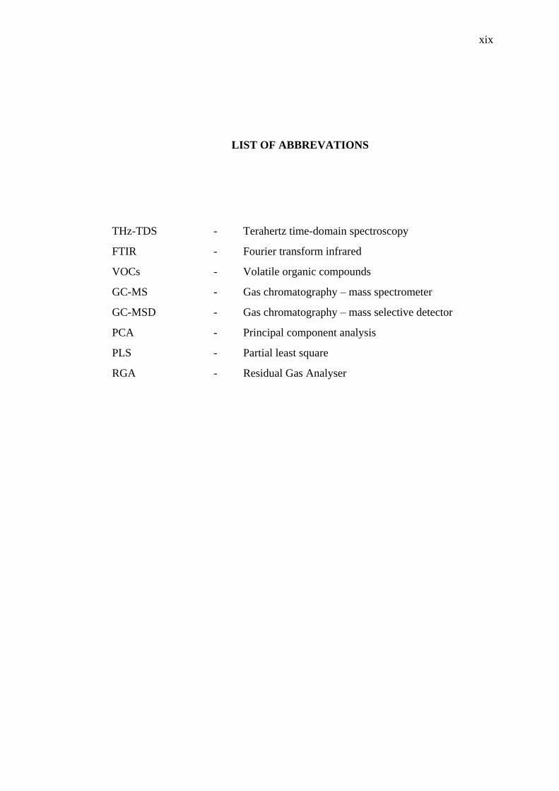
LIST OF ABBREVATIONS
THz-TDS - Terahertz time-domain spectroscopy
FTIR - Fourier transform infrared
VOCs - Volatile organic compounds
GC-MS - Gas chromatography – mass spectrometer
GC-MSD - Gas chromatography – mass selective detector
PCA - Principal component analysis
PLS - Partial least square
RGA - Residual Gas Analyser

xx
LIST OF APPENDICES
APPENDIX
TITLE PAGE
A Publications 128
B Articles Will Be Published 129

INTRODUCTION
1.1 Overview
This introductory chapter illustrates the core of this study, including the
background which stimulates this research work, the motivation for the focus of the
work which highlighted the importance of the volatolomics analysis used in disease
and cancer diagnosis, the problem statement, aim and objectives, scope, and
significance of the study. This chapter is structured to introduce the huge potential of
cancer detection through volatolomics analysis using Fourier transform infrared
spectroscopic and Terahertz time-domain spectroscopy technique.
1.2 Background of the Study
Cancer is a major cause of mortality in this world with more than half a million
deaths by the year 2013 in the United States alone, and the number of cancer cases are
increasing every year, especially in the low and mid-income countries [1]. The
imbalanced socio-economics from these countries led to lack of awareness, expertise
and equipment to diagnose the cancer accurately, in short duration and cost effective
manners.
2
In general, cancer can be diagnosed by invasive and non-invasive techniques.
The invasive techniques, such as biopsy and endoscopy, involve making an incision in
the body to gain access to the target area. The non-invasive techniques, such as
imaging and laboratory testing, do not involve surgical procedures. There also are
alternative techniques to detect and identify the cancer, such as breath analysis [2],
chemometric analysis [3], bio-fluids analysis [4]s and others.
Fourier transform infrared spectroscopy (FTIR) and Terahertz time-domain
spectroscopy (THz-TDS) based technique in cancerous tissue diagnostic are dependent
on terahertz and infrared spectral analysis between healthy and cancerous tissue
samples. However, spectrum broadening is the most challenging to determine [5,6].
The broadening character of the terahertz and infrared spectrum in particular recorded
from liquid or solid samples can mask and also interfere with other cancerous tissue
constituents, including water spectrum thus affecting the measurement resolution.
Detection of volatile organic compounds (VOCs) produced by cancerous cell can be a
better option as the terahertz and infrared spectrum of the gas exhibit less broadening
character.
This approach allows for a better terahertz and infrared spectrum of key
products of the VOCs to be identified [7,8]. This key product then can be used as a
fingerprint for that particular cancer type. Other diagnostic tools have not been able to
yield the biochemical information to identify the key species of the cancer. Even so,
the current techniques to detect the cancer have many disadvantages, such as high cost,
many procedure, contamination or side effects. The proposed techniques using FTIR
and THz-TDS complement the other techniques in assisting the physician to diagnose
the cancer effectively.
In this study, the development of cancer fingerprints uses FTIR and THz-TDS
is to identify the key species of volatile organic compounds (VOCs). This approach
will utilize the gas released from cancerous cell, which contains VOCs, and infrared
light absorbance to monitor specific absorbance patterns which produced the following
changes in chemical compound species: for example, 6-aldehydes, isoprene, n-butyl

3
acetate, and n-propyl propionate released by hepatocellular carcinoma cell using GC-
MS [9].
1.3 Motivation of the Work
This study involves three key aspects: (i) developing a system to capture and
measure the volatile organic compounds released by cancerous cells, (ii) measuring
and identifying the key species, and (iii) verifying and validating the key species of
the cancer. The following sub-section highlight the significance of the study and the
importance of the current work.
1.3.1 Why is volatolomics analysis used in cancer diagnosis?
Volatolomics analysis is the examination of the volatile organic compounds
(VOCs) released by all metabolites from living things for the presence of certain
compounds to determine the presence of cancers or diseases of the human body.
Volatolomics analysis is not limited to breath analysis, but it is covered the VOCs
released from breath, sweat, skin, urine, faeces and vaginal secretions. Volatolomics
analysis has huge potential in detection and identification of diseases and cancer
diagnosis [10,11], especially when it is involved in the end products of cellular
processes of the human as well as a non-destructive technique.
There are many advantages by using Volatolomics analysis to diagnose the
cancerous or diseases samples compared to other conventional methods; for example,
this analysis technique is a non-invasive method which may reduce the risks and be
less harmful to the patient and personnel. Furthermore, the results can also analyze and
appear immediately if we have an established database of the diseases.

4
1.3.2 Why should a volatolomics analysis system be developed?
This volatolomics analysis technique will assist physicians and medical experts
to diagnose the cancer or other diseases effectively. The non-destructive sample
collection technique will help some patients who have problems with conventional
sampling and diagnostics technique.
1.4 Problem Statement
The VOCs released by cancerous cells can be one of the bio-diagnostics
techniques to diagnose the cancer. Previous works on VOCs detection released by
cancerous cells have been performed by using a few analytical instruments, such as
gas chromatography – mass spectrometer (GS-MS), electronic nose, proton transfer
reaction mass spectrometry (PTR-MS), selected ion flow tube – mass spectrometry
(SIFT-MS) and ion mobility spectrometry (IMS). However, each of these analytical
instruments had their limitations, such as the need to change the filters, cannot measure
in real time and simultaneously, need sample preparations, not effective and time
consuming. Fourier transform infrared spectroscopy (FTIR) and Terahertz time-
domain spectroscopy (THz-TDS) with gas absorption sampling techniques can
overcome this limitation. The key species from VOCs released by cancerous cells can
be identified and obtained from the literatures and National Institute of Science and
Technology (NIST) [12] databases. The key species will be verified with other gas
recognition technique and validated with the literature. Then, the samples are analysed
by using chemometric statistical analysis technique for the highest discrimination and
precision of results.

5
1.5 Objectives
The aim of this study was to carry out a detailed study of the application of
Fourier transform infrared spectroscopy (FTIR) and Terahertz time-domain
spectroscopy (THz-TDS) as potential diagnostics tools for clinical use for detection of
cancer through volatolomics analysis. The study shows the application of FTIR and
THz-TDS as a method to characterise biochemical differences that detect and
distinguish the volatile organic compounds released by cancerous cells. This is based
on the FTIR and THz-TDS analytical instruments measurements and statistical
analysis of lung and colon cancer cell lines as well as normal cell lines and control
experiment.
The study aims to address the following objectives:
a) To develop a technique to capture and measure the volatile organic
compounds (VOCs) from lung and colon cancer cell lines
b) To measure and identify the key species of the samples using Fourier
transform infrared spectroscopy (FTIR) and Terahertz time-domain
spectroscopy (THz-TDS)
c) To analyse the samples using chemometric analysis and validate the key
species
1.6 Scope of Study
This study is focused on identification of two types of cancer fingerprint, such
as lung and colon cancer, through release and uptake of volatile organic compounds
by cell lines by using Fourier transform infrared spectroscopy (FTIR) and Terahertz
time-domain spectroscopy (THz-TDS).

6
This study consists of four parts of science and mathematics areas. Firstly, the
concept of physics radiation of infrared and terahertz for detection of the samples.
Second is biomedical samples, such as cancerous cell lines, to be detected and
identified by the analytical instruments. Thirdly, the chemical compounds need to be
identified as key species of the particular cancer from the volatile organic compounds
(VOCs). Fourth is the mathematical statistical analysis of the sample for the highest
discrimination and accuracy of the samples by using chemometric analysis.
1.7 Significance and Original Contributions of This Study
Detection and identification of cancer fingerprints are very important and have
a high impact to the community. In this world of health and medical practice, fast
techniques, cost-effective and accurate detection and identification of diseases and
cancer is very crucial to assist the medical practitioner. The cancer also affects human
health and causes the most human mortality every year, worldwide. In addition,
research on identification of cancer fingerprint through release and uptake of volatile
organic compounds using analytical spectroscopy instruments such as FTIR and THz-
TDS is still new and not established yet.
The spectroscopic analytical instruments, such as FTIR and THz-TDS, with a
combination of gas cell sampling tools and the analytical capabilities of this
combination were demonstrated by simultaneous in vitro gas monitoring and detection
of cancerous cells. Furthermore, assistance of chemometric analysis technique will
provide the highest discriminative power and highest precision of the result.
The THz-TDS with gas analysis technique is a preliminary study to identify
the cancer key species through volatile organic compounds (VOCs). The terahertz
spectroscopy technique can capture the signal directly, simultaneously and effective

7
from the samples. All of the key species will be saved into a database and can be used
as cancer identifier for patient using mobile THz-TDS – gas analyzer in the near future.
1.8 Thesis Structure and Organization
Chapter 2 of this thesis reviews the literature highlighting applications of FTIR
and THz-TDS in biomedicine, cancer identification and volatolomics analysis. This
includes detailed discussion on spectroscopic studies in various diseases in human
tissues, cells and bio-fluids. This is followed by an outline of the methodology and
materials used for analysis and identification of cancerous cells in Chapter 3. This
chapter also covers system optimization and data analysis. The results are presented in
Chapter 4 with discussion as the data was analysed. This chapter is divided into three
sections, presenting and discussing results from a) cell lines analysis, b) detection of
key species, and c) verification and validation. The conclusions drawn from the work
will follow in Chapter 5 with suggestions for future work.
1.9 Summary
This chapter summarizes the foundation of this study to make sure this study
will be achieved within the prescribed scope. Two types of cancerous cells will be
investigated in this study, namely colon and lung cancer. Fourier transform infrared
spectroscopy (FTIR) and Terahertz time-domain spectroscopy (THz-TDS) is used as
analytical instruments. Chemometric analysis is also used to refine and to determine
the highest discrimination and accuracy of the results.

REFERENCES
1. J, F., I, S., M, E., R, D., et al., GLOBOCAN 2012 v1.0: Cancer Incidence and
Mortality Worldwide: IARC CancerBase No. 11, Lyon, France, 2013.
2. Dent, A.G., Sutedja, T.G., Zimmerman, P. V., Exhaled breath analysis for lung
cancer. J. Thorac. Dis. 2013, 5.
3. Madsen, R., Lundstedt, T., Trygg, J., Chemometrics in metabolomics-A review
in human disease diagnosis. Anal. Chim. Acta 2010, 659, 23–33.
4. Ollesch, J., Drees, S.L., Heise, H.M., Behrens, T., et al., FTIR spectroscopy of
biofluids revisited: an automated approach to spectral biomarker identification.
Analyst 2013, 138, 4092–102.
5. Hayat, A., Ginzburg, P., Orenstein, M., Measurement and model of the infrared
two-photon emission spectrum of GaAs. Phys. Rev. Lett. 2009, 103.
6. Balan, E., Delattre, S., Roche, D., Segalen, L., et al., Line-broadening effects
in the powder infrared spectrum of apatite. Phys. Chem. Miner. 2011, 38, 111–
122.
7. Li, Y., Wang, J.D., Spatially resolved investigation of remote sensing FTIR for
the quantitative determination of air toxic VOCs. Spectrosc. Spectr. Anal. 2003,
23, 885–887.
8. Dai, J., Xie, X., Zhang, X.C., Detection of broadband terahertz waves with a
laser-induced plasma in gases. Phys. Rev. Lett. 2006, 97.
9. Mochalski, P., Sponring, A., King, J., Unterkofler, K., et al., Release and
uptake of volatile organic compounds by human hepatocellular carcinoma cells
(HepG2) in vitro. Cancer Cell Int. 2013, 13, 72.
10. Amann, A., Miekisch, W., Schubert, J., Buszewski, B., et al., Analysis of
exhaled breath for disease detection. Annu. Rev. Anal. Chem. (Palo Alto. Calif).
2014, 7, 455–82.
11. Braun, P.X., Gmachl, C.F., Member, S., Dweik, R.A., Bridging the

118
Collaborative Gap : Realizing the Clinical Potential of Breath Analysis for
Disease Diagnosis and Monitoring – Tutorial 2012, 12, 3258–3270.
12. Commerce, U.S.S. of, The NIST WebBook 2016.
13. Broza, Y.Y., Zuri, L., Haick, H., Combined Volatolomics for Monitoring of
Human Body Chemistry. Sci. Rep. 2014, 2–7.
14. Wolkoff, P., Nielsen, G.D., Organic compounds in indoor air—their relevance
for perceived indoor air quality? Atmos. Environ. 2001, 35, 4407–4417.
15. Król, S., Zabiegała, B., Namieśnik, J., Monitoring VOCs in atmospheric air I.
On-line gas analyzers. TrAC Trends Anal. Chem. 2010, 29, 1092–1100.
16. NCI, N.C.I., Understanding Cancer Series: Cancer. Natl. Institue Heal. 2005,
6.
17. Siegel, R., Naishadham, D., Jemal, A., Cancer Statistics , 2013 2013, 63, 11–
30.
18. Ministry of Health Malaysia, National Cancer Registry Report, Ministry of
Health Malaysia, Malaysia, 2011.
19. Hesketh, R., Introduction to Cancer Biology, Cambridge University Press, New
York, 2013.
20. Dunning, M.B., Fischbach, F., Common Laboratoty & Diagnostic Tests,
Lippincott Williams & Wilkins, Philadelphia, 2011.
21. Schnell, Z.B., Van Leeuwen, A.M., Kranpitz, T.R., Davis’s Comprehensive
Handbook of Laboratory and Diagnostic Tests with Nursing Implications, F.A.
Davis Company, Canada, 2003.
22. Wilson, D.D., Manual of Laboratory & Diagnostic Tests, McGraw-Hill, United
States of America, 2008.
23. Ward, E., Ph, D., Ries, L.A.G., Wingo, P.A., et al., Annual Report to the Nation
on the Status of Cancer , 1975 – 2001 , with a Special Feature Regarding 2004,
4251, 675–690.
24. Sridhar, S., Karnani, N., Connell, D.W., Millington, K.A., et al., Increased Risk
of Mycobacterium tuberculosis Infection in Household Child Contacts Exposed
to Passive Tobacco Smoke. Pediatr. Infect. Dis. J. 2014, 33, 1303–1306.
25. Weitberg, A.B., Cancer of the Lung: From Molecular Biology to Treatment
Guidelines, 2002.
26. Risby, T.H., Solga, S.F., Current status of clinical breath analysis. Appl. Phys.
B 2006, 85, 421–426.

119
27. Tittel, F.K., Current status of midinfrared quantum and interband cascade lasers
for clinical breath analysis. Opt. Eng. 2010, 49, 111123.
28. Amann, A., Smith, D., Breath Analysis for Clinical Diagnosis and Therapeutic
Monitoring, World Scientific Publishing, Singapore, 2005.
29. Lourenço, C., Turner, C., Breath Analysis in Disease Diagnosis:
Methodological Considerations and Applications. Metabolites 2014, 4, 465–
498.
30. Giovannucci, E., Physical Activity, Obesity, and Risk for Colon Cancer and
Adenoma in Men. Ann. Intern. Med. 1995, 122, 327.
31. Willett, W., The search for the causes of breast and colon cancer. Nature 1989,
338, 389–394.
32. Giovannucci, E., Insulin and colon cancer. Cancer Causes Control 1995, 6,
164–179.
33. Gerson, C., Healing Colon, Liver and Pancreas Cancer, The Gerson Institute,
California, 2002.
34. Zimmermann, D., Hartmann, M., Moyer, M.P., Nolte, J., Baumbach, J.I.,
Determination of volatile products of human colon cell line metabolism by
GC/MS analysis. Metabolomics 2007, 3, 13–17.
35. Frame, G.M., A collaborative study of 209 PCB congeners and 6 Aroclors on
20 different HRGC columns. Fresenius. J. Anal. Chem. 1997, 357, 714–722.
36. Kim, K.-H., Jahan, S.A., Kabir, E., A review of breath analysis for diagnosis
of human health. TrAC Trends Anal. Chem. 2012, 33, 1–8.
37. Cao, W., Duan, Y., Breath analysis: potential for clinical diagnosis and
exposure assessment. Clin. Chem. 2006, 52, 800–11.
38. Phillips, M., Cataneo, R.N., Ditkoff, B.A., Fisher, P., et al., Volatile markers of
breast cancer in the breath. Breast J. 2003, 9, 184–91.
39. Olopade, C.O., Zakkar, M., Swedler, W.I., Rubinstein, I., Exhaled pentane
levels in acute asthma. Chest 1997, 111, 862–5.
40. Paredi, P., Kharitonov, S. a, Barnes, P.J., Elevation of exhaled ethane
concentration in asthma. Am. J. Respir. Crit. Care Med. 2000, 162, 1450–4.
41. Montuschi, P., Corradi, M., Ciabattoni, G., Nightingale, J., et al., Increased 8-
isoprostane, a marker of oxidative stress, in exhaled condensate of asthma
patients. Am. J. Respir. Crit. Care Med. 1999, 160, 216–20.
42. Corradi, M., Majori, M., Cacciani, G.C., Consigli, G.F., et al., Increased

120
exhaled nitric oxide in patients with stable chronic obstructive pulmonary
disease. Thorax 1999, 54, 572–5.
43. Phillips, M., Cataneo, R.N., Cummin, A.R.C., Gagliardi, A.J., et al., Detection
of Lung Cancer With Volatile Markers in the Breath. Chest J. 2003, 123, 2115–
2123.
44. Pyo, J.S., Ju, H.K., Park, J.H., Kwon, S.W., Determination of volatile
biomarkers for apoptosis and necrosis by solid-phase microextraction-gas
chromatography/mass spectrometry: a pharmacometabolomic approach to
cisplatin’s cytotoxicity to human lung cancer cell lines. J. Chromatogr. B.
Analyt. Technol. Biomed. Life Sci. 2008, 876, 170–4.
45. Kaji, H., Hisamura, M., Saito, N., Murao, M., Evaluation of volatile sulfur
compounds in the expired alveolar gas in patients with liver cirrhosis. Clin.
Chim. Acta 1978, 85, 279–284.
46. Novak, B.J., Blake, D.R., Meinardi, S., Rowland, F.S., et al., Exhaled methyl
nitrate as a noninvasive marker of hyperglycemia in type 1 diabetes. PNAS
2007, 104, 15613–15618.
47. Phillips, M., Sabas, M., Greenberg, J., Increase pentane and carbon disulfide in
breath of patients with schizophrenia. J. Clin. Pathol. 1993, 46, 861–864.
48. Phillips, M., Boehmer, J.P., Cataneo, R.N., Cheema, T., et al., Prediction of
heart transplant rejection with a breath test for markers of oxidative stress. Am.
J. Cardiol. 2004, 94, 1593–4.
49. Studer, S.M., Orens, J.B., Rosas, I., Krishnan, J. a, et al., Patterns and
significance of exhaled-breath biomarkers in lung transplant recipients with
acute allograft rejection. J. Heart Lung Transplant. 2001, 20, 1158–66.
50. Phillips, M., Basa-Dalay, V., Bothamley, G., Cataneo, R.N., et al., Breath
biomarkers of active pulmonary tuberculosis. Tuberculosis (Edinb). 2010, 90,
145–51.
51. Grushka, E., Grinberg, N., Advances in Chromatography, vol. 52, CRC Press,
Florida, 2014.
52. Jennings, W., Shibamoto, T., Qualitative Analysis of Flavor and Fragrance
Volatiles, Academic Press, New York, 1980.
53. Demtröder, W., Laser Spectroscopy: Basic Concepts and Instrumentation, vol.
53, Springer Science+Business Media New York, New York, 1989.
54. Chen, X., Shao, Z., Marinkovic, N.S., Miller, L.M., et al., Conformation

121
transition kinetics of regenerated Bombyx mori silk fibroin membrane
monitored by time-resolved FTIR spectroscopy. Biophys. Chem. 2001, 89, 25–
34.
55. Kno, H., Huber, S., IR spectroscopy of small and weakly interacting molecular
probes for acidic and basic zeolites. J. Chem. Soc. 1998, 94.
56. Wolkers, W.F., Hoekstra, F. a., In situ FTIR assessment of desiccation‒tolerant
tissues. Spectroscopy 2003, 17, 297–313.
57. Ahmad, M.S., Mirza, B., Hussain, M., Hanif, M., et al., ATR-FTIR
spectroscopy detects alterations induced by organotin(IV) carboxylates in
MCF-7 cells at sub-cytotoxic/-genotoxic concentrations. PMC Biophys. 2008,
1, 3.
58. Mourant, J.R., Yamada, Y.R., Carpenter, S., Dominique, L.R., Freyer, J.P.,
FTIR spectroscopy demonstrates biochemical differences in mammalian cell
cultures at different growth stages. Biophys. J. 2003, 85, 1938–47.
59. Llabjani, V., Jones, K.C., Thomas, G.O., Walker, L. a, et al., Polybrominated
diphenyl ether-associated alterations in cell biochemistry as determined by
attenuated total reflection Fourier-transform infrared spectroscopy: a
comparison with DNA-reactive and/or endocrine-disrupting agents. Environ.
Sci. Technol. 2009, 43, 3356–64.
60. Zendehdel, R., Masoudi-Nejad, A., Mohammadzadeh, J., H Shirazi, F.,
Cisplatin Resistant Patterns in Ovarian Cell Line Using FTIR and Principle
Component Analysis. Iran. J. Pharm. Res. IJPR 2012, 11, 235–40.
61. Zendehdel, R., Masoudi-Nejad, A., H Shirazi, F., Patterns Prediction of
Chemotherapy Sensitivity in Cancer Cell lines Using FTIR Spectrum, Neural
Network and Principal Components Analysis. Iran. J. Pharm. Res. IJPR 2012,
11, 401–10.
62. Vitorino, P.G., Alves, J.D., Magalhães, P.C., Magalhães, M.M., et al., Flooding
tolerance and cell wall alterations. Pesq. agropec. bras. 2001, 1027–1035.
63. Romeo, M.J., Quinn, M. a, Burden, F.R., McNaughton, D., Influence of benign
cellular changes in diagnosis of cervical cancer using IR microspectroscopy.
Biopolymers 2002, 67, 362–6.
64. Salman, A., Erukhimovitch, V., Talyshinsky, M., Huleihil, M., Huleihel, M.,
FTIR spectroscopic method for detection of cells infected with herpes viruses.
Biopolymers 2002, 67, 406–12.

122
65. Mostaço-Guidolin, L.B., Bachmann, L., Application of FTIR Spectroscopy for
Identification of Blood and Leukemia Biomarkers: A Review over the Past 15
Years. Appl. Spectrosc. Rev. 2011, 46, 388–404.
66. Mordehai, J., Ramesh, J., Huleihel, M., Cohen, Z., et al., Studies on Acute
Human Infections Using FTIR Microspectroscopy and. Infect. Stud. Using Nov.
Opt. Technol. 2004, 73, 494–502.
67. Shen, Y.C., Davies, a G., Linfield, E.H., Taday, P.F., et al., Determination of
Glucose Concentration in Whole Blood using Fourier-Transform Infrared
Spectroscopy. J. Biol. Phys. 2003, 29, 129–33.
68. Krafft, C., Codrich, D., Pelizzo, G., Sergo, V., Raman and FTIR microscopic
imaging of colon tissue: a comparative study. J. Biophotonics 2008, 1, 154–69.
69. Journal, I., Pharmacology, C., Anti-inflammatory and anti-cancer activities of
essential oils and their biological constituents *. Int. J. Clin. Pharmacol. Ther.
2011, 49, 93–95.
70. Ramesh, J., Salman, A., Mordechai, S., Argov, S., et al., FTIR Microscopic
Studies on Normal , Polyp , and Malignant Human Colonic Tissues. Subsurf.
Sens. Technol. Appl. 2001, 2, 99–117.
71. Peng, G., Hakim, M., Broza, Y.Y., Billan, S., et al., Detection of lung, breast,
colorectal, and prostate cancers from exhaled breath using a single array of
nanosensors. Br. J. Cancer 2010, 103, 542–51.
72. Gao, T., Feng, J., Ci, Y., Human breast carcinomal tissues display distinctive
FTIR spectra: implication for the histological characterization of carcinomas.
Anal. Cell. Pathol. 1999, 18, 87–93.
73. Sokolov, V. V, Frank, A., Minimally invasive and ex vivo diagnostics of breast
cancer tissues by fiber optic evanescent wave Fourier transform IR ( FEW-FTIR
) spectroscopy. SPIE n.d., 3250, 140–147.
74. Szarawara, E., Trybalska, B., Cichocin, M., Stoch, A., et al., FTIR monitoring
of the growth of the carbonate containing apatite layers from simulated and
natural body fluids. J. Mol. Struct. 1999, 512, 287–294.
75. Rettig, R., Virtanen, S., Composition of corrosion layers on a magnesium rare-
earth alloy in simulated body fluids. J. Biomed. Mater. Res. A 2009, 88, 359–
69.
76. Stoch, J., Szaraniec, J., Trybalska, B., Broz, A., FTIR absorption – reflection
study of biomimetic growth of phosphates on titanium implants. J. Mol. Struct.

123
2000, 555, 375–382.
77. Brogden, K. a, Antimicrobial peptides: pore formers or metabolic inhibitors in
bacteria? Nat. Rev. Microbiol. 2005, 3, 238–50.
78. Gasper, R., Dewelle, J., Kiss, R., Mijatovic, T., Goormaghtigh, E., IR
spectroscopy as a new tool for evidencing antitumor drug signatures. Biochim.
Biophys. Acta 2009, 1788, 1263–70.
79. Hynes, A., Scott, D. a, Man, A., Singer, D.L., et al., Molecular mapping of
periodontal tissues using infrared microspectroscopy. BMC Med. Imaging 2005,
5, 2.
80. Kirley, M.P., Booske, J.H., in:, 2015 IEEE Int. Vac. Electron. Conf., IEEE,
Beijing, 2015, pp. 1–2.
81. Alhuwaidi, S., Zubair, K., Song, H., Shellman, Y., et al., in:, 2015 IEEE
Biomed. Circuits Syst. Conf., IEEE, Georgia, USA, 2015, pp. 1–4.
82. Song, H.-J., Nagatsuma, T., Handbook of Terahertz Technologies: Devices and
Applications, CRC Press, Florida, 2015.
83. Zhenwei, Z., Kejia, W., Yong, L., Zhuoyong, Z., et al., Non-destructive
detection of pigments in oil painting by using terahertz tomography. Sci. China
2015, 58, 1–2.
84. Stedwell, C.N., Polfer, N.C., in:, Polfer NC, Dugourd P (Eds.), Laser
Photodissociation Spectrosc. Mass-separated Biomol. Ions, Springer
Science+Business Media New York, Switzerland, 2013, pp. 1–21.
85. Choudhury, B., Menon, A., Jha, R.M., Active Terahertz Metamaterial for
Biomedical Applications, Springer Science+Business Media New York, New
York, 2016.
86. Liu, W., Lu, C., Jiang, Z., Sun, P., in:, Proc. SPIE, vol. 9655, SPIE, Jeju, Korea,
2015, p. 96551G.
87. Jen, C.Y., Richter, C., Sample thickness measurement with THz-TDS:
Resolution and implications. J. Infrared, Millimeter, Terahertz Waves 2014, 35,
840–859.
88. Lin, H., Withayachumnankul, W., Fischer, B.M., Mickan, S.P., Abbott, D., Gas
recognition with terahertz time-domain spectroscopy and spectral catalog: a
preliminary study. Proc. SPIE 2007, 6840, 68400X–68400X–9.
89. Zhang, X.C., Xu, J., Introduction to THz wave photonics, 2010.
90. Wilmink, G.J., Grundt, J.E., in:, J. Infrared, Millimeter, Terahertz Waves, vol.

124
32, 2011, pp. 1074–1122.
91. Mittleman, D.M., Jacobsen, R.H., Neelamani, R., Baraniuk, R.G., Nuss, M.C.,
Gas sensing using terahertz time-domain spectroscopy. Appl. Phys. B-Lasers
Opt. 1998, 67, 379–390.
92. Kawase, K., Iwasaki, A., Shibuya, T., in:, CLEO 2011 Laser Sci. to Photonic
Appl., 2011, pp. 1–3.
93. Lewis, R.A., in:, Proc. IEEE, vol. 95, 2007, pp. 1641–1645.
94. Davies, A.G., Burnett, A.D., Fan, W., Linfield, E.H., Cunningham, J.E.,
Terahertz spectroscopy of explosives and drugs. Mater. Today 2008, 11, 18–26.
95. National Institute of Information and Communications Technolog, THz
Database 2016.
96. Yasuda, H., Hosako, I., Measurement of terahertz refractive index of metal with
terahertz time-domain spectroscopy. Jpn. J. Appl. Phys. 2008, 47, 1632–1634.
97. Laboratory, T., THz database 1998.
98. Laman, N., Harsha, S.S., Grischkowsky, D., Melinger, J.S., High-Resolution
Waveguide THz Spectroscopy of Biological Molecules. Biophys. J. 2008, 94,
1010–1020.
99. Http://webbook.nist.gov/chemistry/thz-ir/, Terahertz Spectral Database 2016.
100. University, P.S., Terahertz Signal Propagation Database 2015.
101. Braakman, R., Gas-Phase Terahertz Spectroscopy and the Study of Complex
Interstellar Chemistry 2010, 2010.
102. Neese, C.F., Medvedev, I.R., Plummer, G.M., Frank, A.J., et al., Compact
submillimeter/terahertz gas sensor with efficient gas collection,
preconcentration, and ppt sensitivity. IEEE Sens. J. 2012, 12, 2565–2574.
103. Danylov, A., THz laboratory measurements of atmospheric absorption between
6% and 52% relative humidity. Submillimeter-Wave Technol. Lab. Univ.
Massachusetts Lowell 2006, 1–7.
104. Andersen, J., Heimdal, J., Mahler, D.W., Nelander, B., Larsen, R.W.,
Communication: THz absorption spectrum of the CO2-H2O complex:
observation and assignment of intermolecular van der Waals vibrations. J.
Chem. Phys. 2014, 140, 091103.
105. Saito, S., Syouji, A., Sakai, K., Fukunaga, K., et al., Broadband terahertz time-
domain spectroscopic system with photoconductive antennas. 2007 Jt. 32nd Int.
Conf. Infrared Millim. Waves 15th Int. Conf. Terahertz Electron. Vols 1 2 2007,

125
475–476.
106. Guo, T., Chemical Analysis of Exhaled Breath by Means of Terahertz
Rotational Spectroscopy. Wright State University, 2014.
107. Gerecht, E., Douglass, K.O., Plusquellic, D.F., Chirped-pulse terahertz
spectroscopy for broadband trace gas sensing. Opt. Express 2011, 19, 8973–84.
108. Jiang, Y., Zhou, F., Wen, X., Yang, L., et al., Terahertz absorption
spectroscopy of benzamide, acrylamide, caprolactam, salicylamide and
sulfanilamide in the solid state. Downloads.Hindawi.Com 2013, 2014, 1–24.
109. Honary, S., Ebrahimi, P., Ghasemitabar, M., Preparation of Gold Nanoparticles
for Biomedical Applications Using Chemometric Technique 2013, 12, 295–
298.
110. Winterhalder, M., Zumbusch, A., Beyond the borders - Biomedical
applications of non-linear Raman microscopy. Adv. Drug Deliv. Rev. 2015, 89,
135–144.
111. Taylor, P., Combined, C.S., Analysis of Olive Fruit Essential Oil : Application
of Gas Analysis of Olive Fruit Essential Oil : Application of Gas
Chromatography-Mass Spectrometry Combined with Chemometrics 2013,
2912, 37–41.
112. Jalali-heravi, M., Arrastia, M., Gomez, F.A., How Can Chemometrics Improve
Micro fl uidic Research? Anal. Chem. 2015, 87, 3544–3555.
113. Sciences, N., Chemometric Classification of Citrus Juices of Moroccan
Cultivars by Infrared Spectroscopy 2015, 2015, 137–142.
114. Wei, S.K., Raja Ibrahim, R.K., Lani, M.N., Abd Razak, S.B., Munajat, Y., in:,
Proc. 4th Int. Sci. Postgrad. Conf. 2016, UTM Press, Johor Bahru, 2016, pp.
314–323.
115. Savanoriu, A., Terahertz spectrometers. Ekspla 2015, 1–14.
116. Gander, W., Hřebíček, J., Solving Problems in Scientific Computing Using
Maple and MATLAB, Springer, New York, 2004.
117. Xiao, G., Dong, D., Liao, T., Zheng, L., A Novel Measurement Method of the
Emission Rules of Greenhouse Gases from Fertilized Soil Based on Fourier
Transform Infrared Spectrometry with Long Optical Path. Spectrosc. Lett. 2015,
48, 572–577.
118. Arshad, A.Z., Munajat, Y., Kamarulzaman, R., Ibrahim, R., et al., Volatolomics
Analysis using FTIR Spectroscopy for Breast Cancer Identification in vitro,

126
IEEE, Kuala Lumpur, Malaysia, 2014.
119. Sellakumar, A.R., Snyder, C.A., Solomon, J.J., Albert, R.E., Carcinogenicity
of formaldehyde and hydrogen chloride in rats. Toxicol. Appl. Pharmacol. 1985,
81, 401–406.
120. Albert, R.E., Sellakumar, A.R., Laskin, S., Snyder, C.A., et al., Gaseous
Formaldehyde and Hydrogen Chloride Induction of Nasal Cancer in the Rat 1,2.
J NCI 1982, 68, 597–603.
121. Chen, C.P., Missett, B., Yom, S.S., Nasal Cavity and Paranasal Sinus Cancer.
Cancer 2010.
122. Shu-Chen, C., Ruey-Hong, W., Li-Jie, S., Ming-Chih, C., Huei, L., Exposure
to Mosquito Coil Smoke May be a Risk Factor for Lung Cancer in Taiwan. J.
Epidemiol. 2008, 18, 19–25.
123. Fagan, P., Moolchan, E.T., Pokhrel, P., Herzog, T., et al., Biomarkers of
tobacco smoke exposure in racial/ethnic groups at high risk for lung cancer. Am.
J. Public Health 2015, 105, 1237–1245.
124. Sethi, M., Fanayan, S., Mass Spectrometry-Based N-Glycomics of Colorectal
Cancer. Int. J. Mol. Sci. 2015, 16, 29278–29304.
125. Protection, D. of E., Bureau of Environmental Cleanup and Brownfields
Division of Storage Tanks REGULATED SUBSTANCES LIST, vol. 2014,
2014.
126. Agency, U.S.E.P., Consolidated List of Chemicals Subject to the Emergency
Planning and Community Right- To-Know Act ( EPCRA ), Comprehensive
Environmental Response , Compensation and Liability Act ( CERCLA ) and
Section 112 ( r ) of the Clean Air Act, vol. 112, 2015.
127. Qi, B.C., Aldrich, C., Biosorption of heavy metals from aqueous solutions with
tobacco dust. Bioresour. Technol. 2008, 99, 5595–5601.
128. Wang, L.K., Chen, J.P., Hung, Y.-T., Shammas, N.K., Heavy Metals in the
Environment: Advances in Industrial and Hazardous Wastes Treatment Series,
Taylor & Francis Group, Florida, 2009.