Vol. II No. 3 July 1985 18, COLLEGE ROAD, MADRAS, INDIA Files/insight0785.pdf · 18, COLLEGE ROAD,...
32
Vol. II No. 3 July 1985 NETHRALAYA INSIGHT HOUSE JOURNAL OF MEDICAL RESEARCH FOUNDAITON & VISION RESEARCH FOUNDATON 18, COLLEGE ROAD, MADRAS, INDIA ISSUE HIGHLIGHTS ! FACETS OF SANKARA NETHRALAYA ! EDITORIAL ! CATARACT EXTRACTION IN A CASE OF CERVICAL ANKYLOSING SPONDYLITIS ! UNUSUAL COMPLICATIONS OF SCLERAL DEPRESSION DURING INDIRECT OPHTHALMOSCOPY ! SILICONE SPONGE EXTRUSION THROUGH THE EYE LID – A CASE REPORT ! PROLIFERATIVE VASCULAR RETINOPATHY IN A CASE OF ANKYLOSING SPONDYLITIS ! VITREOUS HAEMORRHAGE DURING ! ARGON LASER PHOTO- COAGULATION – A RARE MANNER OF OCCURRENCE ! OCULAR METASTATIC TUMOUR SECRONDARY TO MEDULLARY CARCINOMA OF BREAST ! COMBINED OPEN ANGLE AND INVERSE GLAUCOMA ASSOCIATED WITH MICROSPHEROPHAKIA ! ACCOMMODATIVE ESOTROPIA IN TRIPLETS ! TOXOPLASMOSIS SEROPOSITIVITY IN UVEITIS USING THE ELISA EDITOR : MARY ABRAHAM FACTS OF SANKARA NETHRALAYA The correspondence Section, which does tremendous work silently, is headed by Mrs.Ambika Dilip Kumar who joined us recently. She completed her preuniversity in 1968 and has a 5 years experience as stenographer. Her section receives sorts and dispatches the daily mail. Typing out replies to referring doctors, case summaries and certificates are all done here. The department also deals with reimbursement of medical bill. The very demanding task of typing out abstracts, full text of papers and slide material for scientific conferences falls on the shoulders of her five assistants. They have been doing their job exceptionally well, unmindful of the time. They also lend a helping hand to the Administrative office whenever needed. The manuscripts for each
Transcript of Vol. II No. 3 July 1985 18, COLLEGE ROAD, MADRAS, INDIA Files/insight0785.pdf · 18, COLLEGE ROAD,...

Vol. II No. 3 July 1985
NETHRALAYA INSIGHT HOUSE JOURNAL OF
MEDICAL RESEARCH FOUNDAITON & VISION RESEARCH FOUNDATON
18, COLLEGE ROAD, MADRAS, INDIA
ISSUE HIGHLIGHTS ! FACETS OF SANKARA
NETHRALAYA ! EDITORIAL ! CATARACT EXTRACTION IN A
CASE OF CERVICAL ANKYLOSING SPONDYLITIS
! UNUSUAL COMPLICATIONS OF SCLERAL DEPRESSION DURING INDIRECT OPHTHALMOSCOPY
! SILICONE SPONGE EXTRUSION THROUGH THE EYE LID – A CASE REPORT
! PROLIFERATIVE VASCULAR RETINOPATHY IN A CASE OF ANKYLOSING SPONDYLITIS
! VITREOUS HAEMORRHAGE DURING
! ARGON LASER PHOTO-COAGULATION – A RARE MANNER OF OCCURRENCE
! OCULAR METASTATIC TUMOUR SECRONDARY TO MEDULLARY
CARCINOMA OF BREAST ! COMBINED OPEN ANGLE AND
INVERSE GLAUCOMA ASSOCIATED WITH MICROSPHEROPHAKIA ! ACCOMMODATIVE ESOTROPIA IN
TRIPLETS ! TOXOPLASMOSIS
SEROPOSITIVITY IN UVEITIS USING THE ELISA
EDITOR : MARY ABRAHAM FACTS OF SANKARA NETHRALAYA
The correspondence Section, which does tremendous work silently, is headed by Mrs.Ambika Dilip Kumar who joined us recently. She completed her preuniversity in 1968 and has a 5 years experience as stenographer. Her section receives sorts and dispatches the daily mail. Typing out replies to referring doctors, case summaries and certificates are all done here. The department also deals with reimbursement of medical bill. The very demanding task of typing out abstracts, full text of papers and slide material for scientific conferences falls on the shoulders of her five
assistants. They have been doing their job exceptionally well, unmindful of the time. They also lend a helping hand to the Administrative office whenever needed. The manuscripts for each

‘Insight’ is also typed out by them. Mrs . Dilip Kumar is ably assisted by them in the task of co-oridination of the section and in its upkeep.
Miss P.Pushpa plays a leading role in the functioning of the out-patient department. Besides being a graduate in Economics, she has obtained diplomas in diverse subjects – Journalism and Computer Programming (B) and is fluent in Four Indian Languages. Apart from supervising the overall functions of the out-patient departments, she co-ordinates work among the various departments and is actively engaged in Public Relatons. She also schedules the duty roster for Fellows and Post Graduates under training and for the Reception Staff. She schedules
surgeries depending on the nature of the case, availability of the operating room, patients’ preference for dates and the surgeon’s request. All this she does with remarkable efficiency despite the increasing number of out-patients and those requiring surgery.
In patients need excellent nursing care apart from medical care and room facilities. Miss P. K. Lakshmi has been providing this since 1980 Trained at Tata main Hospital. Jamshedpur, she has been an excellent and versatile theatre nurse in the past Helped by 8 staff nurses and 6 auxiliary nurses she provides pre and post operative care for all the in patients apart from maintaining cleanliness and hygiene in the wards. She is considerate and sympathetic and yet firm in dealing with patients. Her care for patients extends beyond the wards – One can often see her stop and enquire
about patient when they come for post perative follow up visits at the out patient. Apart from the ward routine she extends patient care to those at the Dharmashala. She distributes food and medication to “service patients” and devotes time to the running of the mess. She is a fully dedicated nurse who bridges any ago between doctors and patients. Sankara Nethralaya is fortunate to have such a person.
Mr. S.P.Govindarajan has been with us since 1980. He got interested in photography when he was 19 and started his career as Assistant Still camera in a Movie Studio. He later worked as an Industrial and commercial photographer. Backed by 23 years of experience in various types of photography, he has now moulded into a medical photographer. He takes Fluorescein angiograms, colour photographs of the fundus and external photographs of the eye. All black and white films are processed by him. He willingly copes with the increased work load during conference and provides
us with all the necessary slides some times at very short notice. He is very much at ease when he covers any function or a social get together at the hospital.
To keep a hospital clean at ALL TIMES is not all that easy. Mrs. Shantha Seshadri, trained in House – Keeping, is to be commended for her proficiency in the general up keep of the entire hospital and its compound. She supervises the daily cleaning, periodical washing and periodical pesticide spraying at the out patient block wards and Dharmashala. The plants within the premises are also looked after by her. She has shown by her good work that it is possible to keep any place clean no matter what the odds against it are.

CLINICAL BASIC SCIENCE COURSE IN OPHTHALMOLOGY DECEMBER 1985 – JANUARY 1986
C.U.SHAH OPHTHALMIC POST GRADUATE TRAINING CENTRE, SANKARA NETHRALAYA proposes to conduct a CLINICAL BASIC SCIENCE COURSE IN OPHTHALMOLOGY comprising of lectures by eminent teachers in respective fields. It will be a full time 6 week course commencing on 2nd December 1985. The topics will include Ocular Anatomy, Physiology, Biochemistry, Pathology, Bacteriology, Pharmacology, Optics, Refraction, Ocular Motility, Contact Lens, Fluorescein Angiography and Laser Photocogulation. The course is open to all Post graduates and Residents in Ophthalmology Only limited Registration is available. The registration fee is Rs. 250/- THOSE DESIROUS OF REGISTERING please contact Dr. B.SRIDHAR RAO, Secretary Academic Council, Medical Research Foundation, 18 College Road, Madras 600 006. ONE YEAR FELLOWSHIP IN VITREO RETINAL SURGERY AVAILABLE FROM FEBRUARY 1986 (Comprising Fundus Fuorescein Angiography, Photocoagulation, Electro-physiology, Ultrasonography, Retinal Detachment and Vitreous Surgery). Apply with curriculum vitae to:- The Medical Director, Medical Research Foundation Sankara Nethralaya 18, College Road, Madras 600 006.
We acknoledge the gift of two donor eyes sent to us for Keratoplasty by Dr.S.Balasubramaniam and Dr.R.V.Ramani of the Sri Kanchi Kamakoti Medical Centre, Coimbatore and thank them profoundly for their gesture of good will.

EYE OPENER EDITORIAL
This year which has been a busy one is notable for installation of the Yag Laser on 18th February. This has been used to perform 75 posterior capsulotomies and 60 iridectomies so far. The YAG should prove increasingly useful when we start Extra-capsular cataract Surgery with Intraocular Lens Implantation. The Medical Director and Consultants of Sankara Nethralaya took part in a television programme – “Ophthalmology Today” on 25th February when they discussed the role of Laser
Photocoagulation in eye diseases. Mrs. Sandhya Gandhi an active member of Sankara
His Holiness Jagadguru Sri Jayendra Saraswathi Saraswathi Swamigal when he blessed the building site at St. Thomas Mount.
Nethralaya Women’s Auxillary (SWAN) interviewed the speakers.
We have always received the blessings of His Holiness Sri Sankaracharya. His Holiness Jagadguru Sri Jayendra Saraswathi Swamigal visited the institution on 3rd April and blessed the Dharmashala and the staff of the hospital. On 17th April he blessed the new building site acquired at St. Thomas Mount. This piece of land will be utilized to set up a School of
Optometry. Prof. JM Enoch, Dean, School of OthanOSin
Minexinfo
Prof. F.O. Mueller at an informal
ptometry, Berkeley. USA has been playing a vital role in its planning. Prof. F.O.Mueller, from e University of Sarbruken, West Germany, visited us on 5th April when an informal question d answer session on topics of current interest in ophthalmology was held. Prof Eric Attesan, Chairman WHO steering committee for Filariasis and Head Clinical Parasitology ection, National Institute of Health Bethesda, Maryland was yet another distinguished visitor May.
Shri V.R..Ramanatha Iyer Dharmasala
The Dharmashala was inaugurated by the Honourable Vice President Shri R. Venkataraman, at a colourful function on13th April. He presented the V.T.Doshi Gold Medal for the best outgoing fellow 1984 to Drs.Madhivanan Natarajan and Lingam Gopal who were jointly chosen for the same in a stimulating key note address, Prof Ramalingaswami, Director General, Indian Council of
edical Research , New Delhi enumerated 3 strategies to cope with the problem of cataract India – immediate, where more number of surgeries under optimal conditions to clear the isting back log can be performed research to delay the onset of cataract and
terdisciplinary studies of the human lens to understand the basic mechanisms of cataract rmation.

The first two floors of the Dharmashala offer accommodation for patients and their attendants and provide facilities for cooking. The remaining floors provide accommodation for members of staff.SWAN has been instrumental in the opening of a mess at the Dharmashala and a Utility service and gift shop in the out-patient block. This was inaugurated by Smt. M.S.Subbulakshmi on 5th June. Mr.
Santhosh Kumar, Our Bio-Medical Engineer
The Hon.Vice President, Mr. R. Venkataraman, at the inauguration of Shri V.R. Ramanatha Iyer Dharmasala has just returned from a short training programme on YAG and Argon Laser Photocoagulators at Coherent Radiation, U.K. and Carl Zeiss, West Germany. Dr. Prema Padmanabhan, one of our Consultants will soon undergo training with Dr. Akira Momose, Director, Institute of Clinical Pathology, Umeda Kryu Gunma, Japan on Extra-capsular cataract surgery and intraocular lens implant. Dr. Noshir Shroff was with us between 18th June and 25th June, He demonstrated the implantation of anterior chamber lenses and gave a lecture on the same subject. We hope that extra-capsular cataract extraction will reduce the incidence of Aphakic Retinal Detachment and Intraocular Lens Implantation provide better visual rehabilitation after cataract surgery.Dr.M.Thangavelu Former Principal Medical College, Trivandrum : Regional Adviser for Non-communicable Diseases, South East Asia Regional Office,World Health Organisation, New Delhi, has been elected as Member Board of Management. Vision Research Foundation. We welcome him in our midst. Dr.(Mrs)Gyanam Guruprasad Murthy, deputed by the Dean, T.V.Medical College and B.Y.N. Nair Ch. Hospial, Bombay spent two weeks with us observing Fluorescein Angiography, Argon and YAG Laser Photocoagulation in May. Dr. D.N. Gangwar Associate Professor. Post Graduate Institute of Medical Education & Research. Chandigarh is at present observing Vitreo-retinal Surgery for 6 weeks under the Ethicon Fellowship. This issue of ‘Nethralaya Insight’ has articles covering a wide range of subjects which we hope will be of interest to you. We
thank our readers for their letters and look forward to their comments and criticism. Drs. Madhaivanan Natarajan and Lingam Gopal
receiving the V.T. Doshi Gold Medal for the bestout going fellows 1984.
Smt.M.S. Subbulakshmi inaugurating the utility service and gift shop
EDITOR.

CATARACT EXTRACTION IN A CASE OF CERVICAL ANKYLOSING SPONDYLITIS Dr. Natarajan S Dr. Mythili Sriram Dr. Balasundaram S* Dr. Ian Sundaraj* Dr. Yegappan M.ct ** Ankylosing Spondylitis is a progressive disease which cause ankylosis of the interveterbral and costoverterbral joints with ossification of the spinal ligaments and the margins of the interver tebral discs1. When this involves the cervical spine it makes examination and performance of surgical procedures in the supine position difficult for the ophthalmologist. When general anaesthesia is required for these patients. We describe the extraction of a cataract in a patient with ankylosing spondylitis involving the cervical spine where underseen xylocaine toxicity after retrobulbar and facial blocks necessitated general anaesthesia by intubation. CASE REPORT
X A 67 year old malin the left eye. Hiclose to face in teach eye and theclinical findings inloss of usual contdescribed earlier.the head “ fallingdeformity. Kyphoperipheral vision3
calcification of spisystem were norm An Intra capsular was pre medicateThe patient was achieved by simutable as shown inpillows placed ver
-Ray – 1 Cervical Spine – Lateral View X-Ray Cervical Spine A P View
e presented with dense nuclear sclerosis in his right eye and a total cataract s best corrected visual acuity was 6/60 and perception of hand movements he right and left eyes respectively. His intraocular pressure was normal in right fundus normal. His pre operative medical work up showed striking cluding forward flexion of the hear, forward displacement of the head with our in the occipital region, rotation and lateral tilting of the head as has been 2 The patient with this flexion deformity experienced a peculiar sensation of forward” as reported in the literature2. He also had a 650 fixed flexion
sis Compensatory flexion at the hips and knees in order to maintain his . X-Ray cervical spine anterior – posterior and lateral views showed nal ligaments (fig. 3&4). The central nervous cardio vascular and respiratory al.
cataract extraction under local anaesthesia was planned for his left eye. He d with 10mg of diazepam orally, 45 minutes prior to the time of surgery4. postioned supine on the operating table at 300 Trendelenberg.This was ltaneously lowering the head end and raising the foot end of the operating Fig. 1. The gap between his head and operating table, was bridged by two tically. A cushioned head rest was kept over them as shown in Fig. 2.

A canvas belt was applied mid way between hip and knees to prevent the patient from sliding down. The patient was comfortable in this position. Despite lowering the head end of the operating table we had to maximally raise the microscope to get a proper focus. The surgeon’s chair had also to be raised. Regional anaesthesia in the form of retro bulbar block and modified Van Lint’s facial block was given, during which the vital signs were being monitored. A total dose of 12.5 ml of 2% xylocaine (250 mg) and 2.5 ml of 0.5%
Marcaine (12.5 mg) with 1500 I.U. of hylase was administered. Soon after completing the injection, the patient developed twitching of the facial muscles which gradually increase to generalized muscular tremors associated with an increase in pulse rate and blood pressure. This was rapidly followed by deep sedation and respiratory depression. These features were attributed to toxicity of xylocaine and 5 mg of Diazepam was given intravenously to prevent convulsions. The patient was oxygenated with a mask. It was decided to intubate the patient and proced with the planned surgery. Nasal endo tracheal intubation was performed by visualizing only the tip of the epigiottis with the direct laryngoscope and passing the tube below the tip of the epiglottis with Magil’s intubatting forceps. The vocal cords could not be visualized. The patient was ventilated with oxygen and nitrous oxide (50-50). Halothane 1 % was included in the anaesthetic management as the patient showed signs of recovery on surgical stimulation. A classical limbal based flap, peripheral iridectomy, cataract extraction with cryo anterior chamber reformation with air and closure of sclerocorneal wound with interrupted 80 silk was performed without any complications under the operating microscope. Recovery from anaesthesia was uneventful and good. As the patient could only lie with his head raised post operatively, part of the air from the anterior chamber went behind the pupil and resulted in temporary shallowing of the anterior chamber in the upper quadrant at 12’ 0 clock meridian. Following absorption of air, and atropinisation, the pupil remained round with a well formed deep anterior chamber. On final examination two months later, his vision was 6/5:N/5 in the operated left eye Slit Lamp biomicroscopy and ophthalmoscopy revealed no abnormality.
Fig.1 300 Trendelenberg Position
DISCUSSION:
The problems due to fixed flexion deformity were overcome by tilting the operating table and raising the operating microscope. The positioning almost led to the usual situation of head and foot being at the same level, and the operation microscope at a higher level than usual. The patient and surgeon were quite comfortable and there was no feeling of performing an operation under unusual circumstances. The need for intubation was unforeseen. Through surgery could have proceeded following deep sedation due to xylocaine toxicity and with IV diazepam5,
recovery and inco-orinated movements during
Fig.2 Bridging gap between Patients Head and Table with Pillows surgery might have led to unwanted surgicalcomplications.All these were safely avoided by endotracheal intubation and light general anaesthesia. Endotracheal intubation necessitates sedation, muscular, relaxation and laryngobronchial reflex suppression. As the patient was not responding to stimul following IV. Diazepam. (administred for xylocaine toxicity) the situation provided ideal requisites for intubation and endotracheal intubation was performed without other routine medication. Difficulties in endotracheal indubation in patients with ankylosing spondylitis results from a rigid neck, inability of the head to be extended on the cervical verterbrae or rotated laterally and trismus

of varying severity6. Nasal intubation was required in this patient as in cases where complete dental extraction or bilateral resection of mandibular condyles is intended. Our patient is somewhat similar to the case described by Munson and Cullen8. They have described a most unusal case of severe cervical spondylosis in a man unable to lift his head off his chest and unable to open his mouth precluding tracheostomy. Awake intubation was skillifully performed without moving the patient by using a nasal endotracheal catheter and a oral wire hook to direct the tip of the catheter into the larynx. All these above descriptions reveal that nasal endotracheal intubation is done without visualization in cases of cervical ankylosing spondylitis. We could however visualize the epiglottis with direct laryngoscope because there was no trismus Visualisation of vocal cords was not possible because of the rigid neck. Toxicity to Xylocaine can occur due to over-dosage inadvertent intra-vascular injection and rapid absorption of ordinary doses. Xylocaine toxicity could be promptly managed because qualified anaesthestists monitor every case done under local anaesthesia. Reformation of anterior chamber could have been done with balanced salt solution instead of air if the post operative position of the paitnet would have been anticipated. SUMMARY: The positioning of a patient with Ankylosing Cervical Spondylitis for cataract extraction and the induction of general Anaesthesia by Nasal intubation following Xylocaine Toxicity is described. REFERENCE:
1. Wright. V. & Moll. J.M.H.Ankylosing Spondylitis Br. J Hosp Med1973. 9. 331 – 341 2. Sharp J and Purser, D.W. Spontaneous atlanto axial dislocation in ankylosing
spondylitis and Rheumatoid Arthritis. Ann Rheum, Dis 1961.20 47 – 77 3. Nathan Smuker “Arthritis of Spine” from the book “ The Spine” second edition W.B.
Saunders Company, New York 1982, 906-938 4. Ronald A Miller, Anaesthesia Churchill Livingstone New York, 1981. Vol.1 581 – 582 5. Chandran Abraham S. S.Badrinath, et al intravenous and intra muscular diazepam as
pre medication in ocular surgery, Silver Jubilee Conference of the Madras State ophthalmological Association 1977.
6. T Cecil Georj et al General Anaesthesia. Fourth Edition 13 Butter Worth, London, 1980 Vol 2, 1278 – 1288
7. Walters D.J. Guided blind Endotracheal intubation Anaesthesia 1963, 18 158 – 162. 8. Munson E.S. & Cullen S.R. Endotracheal intubation in a patient with Ankylosing
Spondylitis of the cervical spine. Anaesthesiology 1965 26 365.

UNUSAL COMPLICATIONS OF SCLERAL DEPRESSION DURING INDIRECT OPHTHALMOSCOPY Dr. NATARAJAN S. Dr. BADRINATH S S Scleral depression has been considered as a safe examination technique in all eyes except those with suspected rupture globe, recent or weak surgical wounds as in cataract surgery and in vascular retinopathies. We report two unusal complications – macular faemorrhage and hyphaema resulting from scleral depression. CASE I: A 29 year old male presented with episodes of repeated periodic haemorrhages inside the eyes associated with visual loss followed by spontaneous recovery for the past thirteen years. It occurred following a dive from a a diving board at the swimming pool. He had giddiness on walking and flatulent dyspepsia. His mother and sister had a similar ocular history. His best corrected visual acuity was 6/5 N/5 in each eye. Refraction was R.E.+ 2.50 Dsph with + 3.50 D cyl X 90 and L.E.: + 3.50 D Sph with + 3.50 D Cyl X 90. Anterior segments were normal in both the eyes. Fundus examination with the Binocular Indirect Ophthalmoscope showed tortuous retinal vessels in the posterior pole and around the fovea of the right eye. The peripheral retinal vasculature was normal, there were no new vessels or other vascular anomalies 3600 scleral depression revealed no abnormality. When the left fundus was being examined and as the inferior quadrant was being depressed, the patient complained of “bleeding inside the right eye”. Re-examination of the right eye showed multiple fresh haemorrhages at the macula. The blood was located at the fovea and in two small areas superior to the fovea in relation to arterioles (Fig. 1). The haemorhages were retinal and subretinal. Visual acuity dropped from 6/5 to counting finger at 1 metre distance. Fundus Fluorescein Angiography showed tortuous retinal vessels around the fovea (Fig. 2 & 2A). There was no evidence of any choroidal or retinal vascular changes either at the macula or elsewhere. Amsler charting and visual field recorded with Goldman perimeter revealed a central scotoma with 50 range.
count, 7100/mm3 NeutrophAtypical lymphocytes 2%; 1,91,800 mm3;Reticulocytestests were also normal with bilirubin 0.16 mgs% total pratio 1.50 : 1.00; serum alkaand Thymol Turbidity 2.5 M(Bleeding time, 3 minutesProthrombin time: Test 17
Fig 1 – Showing Multiple Macular Haemorrhages in right eye
Systemic examination revealed an angiomatous spot in the right Lower Lid extending to the left check. His blood pressure was normal (130/80 mm Hg). Gastroscopy sigmoidoscopy and ultrasonography of the abdomen and electrocardiography performed elsewhere were found to be normal. There was no neurological deficit according to his Neurologist, and a C T Scan was reported to be normal. His haemotologic data were normal. (Haemogolobin 14.4 gm% haematocrit 44% white blood cell ils, 47%;Eosinophils 5%:Lymphocytes 44%: Monocytes 2%; erythrocyte sedimentation rate 2mm fall in 1 hour; platelets 0.8%; MCV 120/mm3;MCH 39 pg:MCHC 33%). His liver function
total bilirubin 0.52 mgs%;conjugated bilirubin 0.36%;unconjugated rotein 7.0 gms%; Albumin 4.2 gms %; Globulin 2.8 gms %;A/G line phosphastase 5.2 KA units, SGOT 20 units; SGPT 10 units; aclagan units. The coagulation work up showd no abnormality. ;clotting time, 14 minutes; II tube clotted T 11½ minutes; seconds; control, 17 seconds; Ratio 1:1 Partial Thromboplastin
time, Test 49 seconds and Euglobulin lysis time was 3 hours (normal: 2 to 4 hours): Factor VIII was normal).
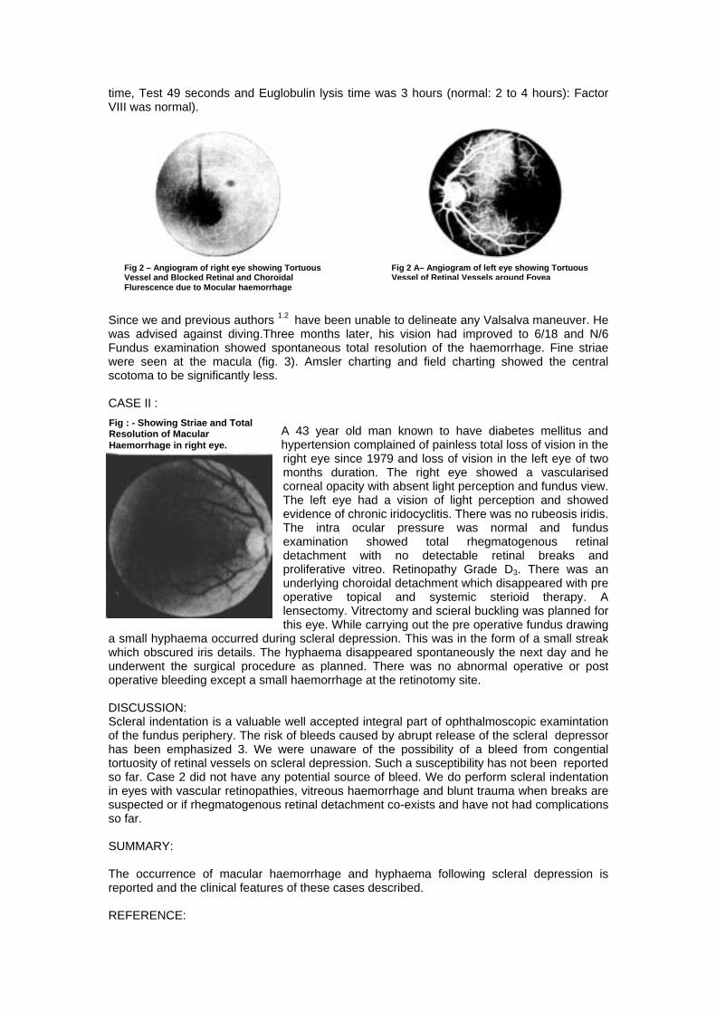
Fig 2 – Angiogram of right eye showing Tortuous
Vessel and Blocked Retinal and Choroidal Flurescence due to Mocular haemorrhage
Since we and previous authors 1.2 have been unablwas advised against diving.Three months later, hFundus examination showed spontaneous total rewere seen at the macula (fig. 3). Amsler chartinscotoma to be significantly less. CASE II : A 43 year old ma hypertension compl
right eye since 197months duration. corneal opacity withThe left eye had evidence of chronicThe intra ocularexamination shodetachment with proliferative vitreo.underlying choroidaoperative topical lensectomy. Vitrectthis eye. While carr
a small hyphaema occurred during scleral depressiwhich obscured iris details. The hyphaema disappeunderwent the surgical procedure as planned. Thoperative bleeding except a small haemorrhage at th
Fig : - Showing Striae and Total Resolution of Macular Haemorrhage in right eye.
DISCUSSION: Scleral indentation is a valuable well accepted integof the fundus periphery. The risk of bleeds caused bhas been emphasized 3. We were unaware of thtortuosity of retinal vessels on scleral depression. Suso far. Case 2 did not have any potential source ofin eyes with vascular retinopathies, vitreous haemosuspected or if rhegmatogenous retinal detachmentso far. SUMMARY: The occurrence of macular haemorrhage and hyreported and the clinical features of these cases des REFERENCE:
Fig 2 A– Angiogram of left eye showing Tortuous Vessel of Retinal Vessels around Fovea
e to delineate any Valsalva maneuver. He is vision had improved to 6/18 and N/6 solution of the haemorrhage. Fine striae g and field charting showed the central
n known to have diabetes mellitus and ained of painless total loss of vision in the 9 and loss of vision in the left eye of two The right eye showed a vascularised absent light perception and fundus view.
a vision of light perception and showed iridocyclitis. There was no rubeosis iridis. pressure was normal and fundus wed total rhegmatogenous retinal
no detectable retinal breaks and Retinopathy Grade D3. There was an l detachment which disappeared with pre
and systemic sterioid therapy. A omy and scieral buckling was planned for ying out the pre operative fundus drawing on. This was in the form of a small streak ared spontaneously the next day and he ere was no abnormal operative or post e retinotomy site.
ral part of ophthalmoscopic examintation y abrupt release of the scleral depressor e possibility of a bleed from congential ch a susceptibility has not been reported
bleed. We do perform scleral indentation rrhage and blunt trauma when breaks are co-exists and have not had complications
phaema following scleral depression is cribed.

1.Golding M.F. Pollack, I.P. Green R. Familial retinal arteriolar tortuosity with retinal haemorrhage Am.J.Ophth 73:183:191. 1972 2.Sir stewart Duke Elder System of Ophthalmology, Herry Kimpton, London vol.III – Part 2 PP 784 785 1964. 3. Freeman, H.M.General discussion of preoperative examination. In Schepens C.L. and Regan, C.D.J. (Eds) Controversial Aspects of the Management of Retinal Detachment Boston, Little Brown P 54-56 1965. SILICONE SPONGE EXTRUSION THROUGH THE EYE LID A CASE REPORT. DR. ARUN EL HENCE DR. BADRINATH S S Extrusion of silicone implants and sponge is a well known complication pf Retinal Detachment surgery. Silicone sponge are believed to have higher rates of infection and extrusion than solid silicone tires 1.2.3 . Their bulk and their lack of conformity to the globe may me responsible for this3. However this is contradicted by explant surgeons4.5 . In this article we report a cse where the silicone sponge extruded not only throught the conjunctiva but also through the eyelid. There is no mention of such a complication in the literature. CASE REPORT: A 29 year old Sikh patient came to us in November 1980 with history of recurrent rhegmatogenous retinal detachment despite four repeated surgeries in his right eye. He had earlier suffered a blunt injury in this eye. On examination he was found to have visual acuities of perception of hand movements close to face, and 6/4 in the right and left eyes respectively. Binocular indirect ophthalmoscopy revealed a total rhegmatogenous retinal detachment with multiple breaks in the upper temporal quadrant and a single large posteriorly placed break., four disc diameters from the optic nerve head in the 12o clock meridian. The left eye was normal. Revision scleral buckling was performed in November 1980. A solid silicone the (No.280) was placed under three mattress sutures after cryo application and dissection of scleral flaps in the upper temporal quadrant. A silicone sponge was placed radially. In the upper nasal quadrant, under scleral flaps, to take care of the posterior break. Sub-retinal fluid drainage was performed in the usual way. Upto a month postoperatively the retina was attached except for the presence of two dry retinal folds. When the patient returned at two months after surgery, he was found to have a recurrent retinal detachment with proliferative vitreo-retinopathy Grade D3. In 1982 the patient had developed episodes of nagging pain and purulent discharge which
subsided with use of topical antibiotic drops prescribed by his local ophthalmologist. He had also noticed a red fleshy growth under the upper Iid about the same time. There was no history of recurrent conjunctival bleeds. In 1983 he became aware of a painless swelling in the upper lid which gradually increased in size. About two weeks prior to being seen here, it ulcerated and began discharging pus. A small piece of rubbery material could be seen within the fistula. This material slowly began
protruding out and finally extruded spontaneously, while the patient was cleaning his eye on the day of his examination here (Fig. 1)
Fig 1 – Extuded Silicon sponge
Examination showed a fistulous opening in the upper lid about the medial canthus of the right eye (Fig 2). Both tarsal and bulbar conjunctivae were congested and there was a pedunculated granulomatous swelling in the upper fornicial conjunctiva, in the upper nasal quadrant (Fig 3). There was no apparent conjunctival deficiency or exposed suture. The eye was blind and had complicated cataract and hypotony.

TREATMENT: Fig. 2 Fistulous Opening in Right Upper Ltd Fig 3. Penduculated Granulomatous Swwelling in Upper Formix and Complicated Cataract.
Conjunctival scraping revealed a growth of coagulase positive staphylococcus pyogenes. The patient was put on an hourly regimen of Garamycin eye drops pre-operatively. The remaining silicone tire with its retention sutures was removed under general anaesthesia. There was no evidence of the sponge or its retention sutures in the upper nasal quadrant. There was no infection in the region of the silicone implant, or any area of scleral necrosis. DISCUSSION: Probably, chronic buckle infection in the region of the silicone sponge was responsible for the pain and discharge that the patient had in the late post operative period. Through a fistulous opening located in the fore shortend upper fomix a pedunculated granuloma grew, again due to chronic subcutaneous tissue of the upper lid., overlying the foreshortened upper fornix, through an opening in the orbital septum damaged by repeated surgery and / or chronic infection. Bursting of the subcutaneous abcess resulted in discharge of purulent material and a fistulous tract through which the probably predisposed to infection because of repeated reoperations, long duration of surgery and the use of silicone sponge. CONCLUSION: This article mentions a hitherto unreported complication of scleral buckling in which the silicone sponge extruded through the eyelid tissues. Reoperations exposure of sclera due to long duration of surgery and use of silicone sponge may be cited as the reasons for occurrence of such a complication. REFERENCE: 1.Schepens C.L.Rationale of surgical procedures, Chap 23, p 313 Retinal Congress Appleton Century Crafts. New York. 2.Okamura I.D.: Implants in Retinal Surgery Chap 24, p 319 Retina Congress Appleton Century Crafts, New York. 3.Russo C.E. Ruis R.S.: Silicone sponge rejection Archives of Ophthal Vol 85: p 647, 1971 4.Lincoff H Nadel A & O Connor P. The changing character of the infected scleral implant Archives of Ophthal Vol. 84. p647, 1971. 5.Langsten R. Lincoff H.Mc Leen J.M :II Experimental production in Animals Archives of Ophthal Vol 74: p 665 – 668 1965.

PROLIFERATIVE VASCULAR RETINOPATHY IN A CASE OF ANKYLOSING SPONDYLITIS
Dr. Natarajan S Ankylosing spondylitis also known as rheumatoid spondylitis and Marie Strumpell diseases is a chronic and usually progressive inflammatory disease involging the articulation of the spine and adjacent soft tissues. The sacro iliac joints, hip and shoulder joints are commonly affected. Peripheral joints are affected less frequently2. This disease predominantly affects young men and begins most often in the third decade. A high association has been found between the disorder and the histo compatability antigen HLA-B-27. The clinical features of this disease entity are different from those of rheumatoid arthritis. The etiology is unknown2
. Several disorders like Reiter’s syndrome and psioriatic spondyloarthritis possess similar clinical features as ankylosing spondylitis. The role of HLA B 27 antigen in the pathogenesis of these disorders is not known. The currently favoured hypothesis is that the HLA-B-27 antigen because of a close association with the immune response genes, is only a marker distinguishing a group, is only a marker distinguishing a group of individuals whose immuni response to an as yet underfined infectious agent leads to one of the HLA B-27 associated forms of arthritis. We report a case of proliferative vascular retinopathy. Eales’ disease in a case of ankylosing spondylitis. CASE REPORT. A 38 year old make with Ankylosing spondylitis of nine years duration came with the complaints of floaters and diminution of vision in the left eye of one month’s duration, Ocular examination revealed a visual acuity of 6/6 and 1/60 in the right and left eye respectively. He was orthophoric. The anterior segments were normal in both the eyes except for a small subconjunctival haemorrhage in the left eye. The intra ocular pressure was 12 mm of mercury each eye. The right fundus showed a small sub hyaloid haemorrhage in the lower nasal quadrant and vascular imprints on the posterior hyaloid in the upper nasal quadrant. Fundus fluorescein Angiography showed dilated capillaries on the optic disc which leaked minimally. There was leakage of dye from some of the major vessels. No new vessels were evident and the capillaries were intact. The left eye showed sub hyaloid haemorrhage in the lower nasal quadrant and haemorrhage into the vitreous gel, caused by disc neovascularisation. In addition a few superficial retinal haemorrhages were seen in the upper temporal quadrant. While waiting for vitrectomy, the vitreous haemorrhage in his left eye cleared remarkably and vision improved to 6/9, N/6. Some of the blood settled to the bottom of the vitreous cavity. He needed pan retinal photo coagulation therapy. He could not sit because the spondylitis involved the cervical and lumbar vertebrae, the sacro iliac and hip joints. As the knee joint was normal, he was made to kneel on two pillows for laser photocoagulation (Fig.1).
Fig. 1 – Posture adopted for Laser Photocoagulation Therapy. The laser machine and examiner’s stool had to be lowered in order to position the patient on the slit lamp. He was photocoagulated in four divided sittings. A total of 1,521 applications time of 0.1 second with power adjusted to create an optimal reaction were made. All areas of the fundus except the lower quadrant which was obscured by vitreous haemorhage was ablated. The treatment was performed without any discomfort to the patient. Systemic examination revealed a staff neck, and loss of movement of the
lumber and cervical vertebrae. Movement at the hip and shoulder joints were restricted. He also had a right hip fixed flexion deformity. X-ray pelvis and dorsal spine (Fig 2 and Fig 3) indicated that ankylosis was complete in the sacro iliac and hip joints. Ligamentous calcification of the Vertebrae gave the appearance of a bamboo spine.

DISCUSSION: Advanced ankylosing spondylitis with such extensive involvement of the joints is reported to be rare2 Whether Eales’ disease and ankylosing spondylitis were two independent disorders co-existing in the same individual or whether they were inter connected in same way is diffcult to interpret. While HLA antigen studies are specific for ankylosing spondylitis, it has been shown that they do not play any significant role in Eales’ disease4. This inter link is probably no more significant than the association of Eales’ disease to Tuberculosis, Neurological, Haematological3 and auditory disorders6 in other publications.
Fig 2 – X-Ray Dorsal Spine showing Ligamentous Fig 3 – X-Ray Pelvis showing Ankylosis of Sacro lilac and Calcification giving Bamboo Spine Appearance Hip Joints. SUMMARY: A case of severe ankylosing spondylitis involving cervical and lumbar vertebrae, sacro iliac and hip joints associated with Eales’ disease is reported and discussed. The association between these two conditions is probably reported for the first time. REFERENCE:
1. Nathan Smukler, Anrhritis of spine from the book “ The Spine”, Second edition, W.B. Saunders Company, Philadelphia pp 906-938. 1982.
2. Bruce E Giltiland, Mart Monnik Ankylosing spondyilitis from the book “Harrisson’s principles of Internal Medicine, Ninth Edition Mc Graw Hill, pp 1880 – 82 1980.
3. Blumberg, B and Ragan, C The natural history of rheumatoid spondylitis. Medicine 35: 1 – 31, 1956.
4. Jorg beriram, Manfred Spitznas and Maxmillan Rommelfanger, missing evidence for HLA antigen association with Eales disease, choriorethinales., Central serous retinopathy and malignant choroidal melonoma Invest Ophthalmol Visual Sci Pr 918 – 920 – 1978.
5. Isaac C Michaelson, Text book of the Fundus of the Eye, Churchill Livingstone, Edinburg. Pp 382 1980.
6. Robert P. Murhphy Amall Patz et al. “A survey of patients with Eales’ Disease” from the book in Management of Retinal Vascular and Macular disorder. Williams and Wilkins Baltimore pp 28-31. 1983.

VITREOUS HAEMORRHAGE DURING ARGON LASER PHOTO-COAGULATION – A RARE MANNER OF OCCURRENCE Dr. Chandran Abraham Mr. Santhosh Kumar Prof. Govindarajan S.R Vitreous haemorrhage during photocoagulation can occur when abnormal vascular lesions are being directly coagulated or if excessive energy is coagulated or if excessive energy is inadvertently applied to apparently normal target tissue2. This article describes the occurrence of vitreous haemorhage in a patient with proliferative Diabetic Retinopathy and Cataract at a site remote from where the blue-green argon laser was aimed at and fired. CASE REPORT; A 60 year old male who has been regularly followed up since 1980 for non-proliferative diabetic retinopathy underwent focal argon laser photocoagulation without complications for new vessels without vitreous haemorhage in the upper temporal quadrant of the right eye in March 1982 and for another tuft of new vessel formation at the lower nasal quadrant in April 1985. His visual acuity had always been around 6/18 in the right eye and 6/6 in the left. Nuclear Sclerosis posterior subcapsular lenticular changes and peripheral spokes have been in evidence since his first visit and showed no significant progression. His fluorescein angiograms have always been of mediocre quality because of cataracts and there was always difficulty in visualizing the dye during fluorescein ophthalmoscopy. However, reasonably useful information was obtained from these during his follow up visits. In May 1985, he developed disc neovascularisation in both the eyes as determined by ophthalmoscopy and fluorescein angiography. The right eye had vitreous haemorrhage which occupied the lower half of the vitreous cavity. His visual acuity remained at 6/18 in the right eye and 6/6 in the left eye. There was no clinical worsening of his cataracts and there was no rubeosis indis in either eye. A pan retinal laser photocoagulation was completed in the right eye, with a setting of 300 mircon/0.7w/0.1 sec on the Argon Laser Photocoagulator utilizing bluegreen light. The cataract and the haze in vitreous caused by haemorrhage hindered treatment but there was no complication, and whatever area could be visualized was treated. Four sessions of laser pan retinal photocoagulation with a setting of 300 micron/0.5 to 0.6w/01. sec on the same machine went on uneventfully in the left eye except for a little difficulty in treating around the lens opacities. At the 5th and last planned session a trickle of blood in the vitreous was observed to come from above the field of observation on the patient’s fundus. At this moment the central lens of the Goldmann was being used to view and treat the region above the optic disc and a burn had just been fired with a setting of 300 micron/0.5W/0.1sec2. The patient’s gaze was directed slightly upward and the patient had not moved his eye. The aiming beam which was slightly vertically oval was faintly visible at the site aimed at just prior to firing the laser and a faint (less than desired) burn was evident on the area aimed at1. Optimal pressure was applied to the eye with the Goldmann Contact Lens to prevent further bleedin. A rapid and thorough examination of the Iris, papillary border and the area of fundus under observation did not show the site of origin of the bleed. A re-check was made on the power reading, spot size and exposure time. They remained at the pre-set level. Judging from the flow of blood (which was from above and to the right of the observer), examination of the superior temporal quadrant with the equatorial mirror and the patient looking straight ahead showed the site of bleed. This was just posterior to the equator. A layer of fresh blood about ¼ disc diameter obscured the underlying retina. This area did not contain any normal retinal venules or new vessels. There was no significant difference in the tissue characteristics between the site aimed at and the site in which the bleed occurred, except for minimal oedema in the region above the disc. The area surrounding the bleed was coagulated with 3 rows of confluent laser burns. The power was first raised to 0.7W and spot size changed to 500u. When this produced a fairly heavy burn, the power was brought down to 0.55W. Further haemorrhage from the site was prevented by maintaining pressure on the eye with the contact lens. Coagulation of the entire upper temporal and temporal quadrants was then

completed with total of 505 applications as planned earlier. The pressure of the contact lens of the eye was then gradually decreased and the contact lens removed when no further oozing was observed for about a minute when only a pressure sufficient enough to maintain the lens in contact with the eye was applied. Indirect Ophthalmoscopy after a couple of minutes did not show any further bleed. The patient was advised to avoid any activity the would raise the venous pressure in his eye. Examination the next day showed 6/6 – 3 visual acuity signs of clearing of the vitreous haemorrhage and no further bleed. He has been advised to return after a month. DISSUSSION: The manner in which the vitreous haemorrhage occurred in this case is unique for two reasons:
1. The site of bleed was remote from the area being visualized and from where the aiming beam was visibly focused.
2. The bleed occurred from an area in the retina which was free from a potential source like new vessels or a normal retinal venule. Inadvertant machine settings, and inadvertent firing of the laser were ruled out prior to attempting an explanation of this phenomenon. Since the aiming beam was visible at point E, it must be assumed that what was visible to the observer was only a part of it was directed elsewhere (in this instance point S where it could not be seen by the observer). The spokes in the cataract must have acted as a prism producing deviation of the beam45 (Fig 1&2). The deviation in a small angled prism is governed by the formula δ = (µ - 1) ∝ Where δ is the angled deviation and µ is the ratio of µ-2 (the refractive index of the material refracting the incident ray) to µ-1 (the refractive index for the surrounding material) and ∝ is the angle of the prism. In this particular case we assume that the refractive index of the lens spoke was greater than the refractive index of the surrounding lens resulting in a deviation of the incident laser beam to point S. Had the refractive index of the lens spoke been less than that of the lens, the deviation of the beam would have been to a point below E. Moreover, the value of µ should have been quite considerable to justify the magnitude of the deviation observed. The beam may have been incident such that the spoke deviated most of the laser beam to be focused at point E. The deviation was presumably caused a stronger focus at point S (Fig 1 & 2).
For such a beam to produce a bleed in apparently normal retina means that the energy level at that site has shown an increase. It now becomes increasingly difficult to explain this phenomenon* because well known phenomenon of laser interaction with ocular tissue like scatter, absorption and thermal blooming – particularly in relation to a Cataractous lens will always result in a lesser amount of energy delivered to the retina13. Though we are unable to prove mathematically, we speculate that the laser beam traveled a greater length from the lens to point S with resultant decrease in the pre-set 300 µ spot size at the area of impact. Alternatively we do not completely rule out the remote chance of Resonant Amplification of the Argon Laser at the site of lens spoke in a manner similar to what can occur in a Ruby Crystal under Excitation. The possible damage to the impact site was the formation of a retinal hole which was covered by a layer of blood.It was for this reason that it was surrounded by 3 rows of confluent laser burns. It is likely that the slight difference in tissue characteristics at point E and S might have slightly contributed to the altered reaction at point S (Fig 1 & 2). For such a beam to produce a bleed in apparently normal retina means that the energy level at that site has shown an increase. It now becomes increasingly difficult to explain this phenomenon* because well known phenomenon of laser interaction with ocular tissue like scatter, absorption and thermal blooming – particularly in relation to a Cataractous lens will always result in a lesser amount of energy delivered to the retina13. Though we are unable to prove mathematically, we speculate that the laser beam traveled a greater length from the lens to point S with resultant decrease in the pre-set 300 µ spot size at the area of impact.
Alternatively we do not completely rule out the remote chance of Resonant amplification of the Argon Laser at the site of the lens spoke in a Ruby Crystal under Excitation. The possible damage to the impact site was the formation of a retinal hole which was covered by a layer of blood. It was for this reason that it was surrounded by 3 rows of confluent laser burns. It is likely that the slight difference in tissue charactgeristics at point E and S might have slightly contributed to the altered reaction at point S (Figs 1 & 2). The settings used in this situation were also not unusual and coagulation of the fellow eye which also had similar changes was completed with a higher energy level without complications. The field of view with the Goldmann 3 mirror lens had not permitted a simultaneous viewing of points E and S. May be one could have observed two aiming beams prior to firing the laser if a pan fundoscope had been used. SUMMARY: A rare instance in which the Argon laser beam was defiected away from the site aimed at and produced a vitreous haemorrhage is reported and a possible explanation offered. REFERENCES;
1. L’esperance F A Jr Oculur Photocoagulation – A Stereoscopic Atlas Page 33 – 34, 292. The CUMosly Co.St Louis 1975
2. Zweng H.C.little H.L.A W D vassiliadis Argon Laser Photocoagulation Page 34 – 37. The C.U.Mosbu Co. St.Louis 1977
3. Pomerantzff O Simple Physics of Photocoagulation Page III 1 – 23- Annual Course on Photocoagulation Department of Retina Research Eye Research Institute of Retina Foundation Boston 1979.
4. Ramamoorthy S. Krishnamurthy K.S and Rajagopalan P.R. A Text book of light including geometrical and physical optics Page 52 – 53, 56 – 57, the National Publishing Co. Madras – 1 1964
5. Yavousky B.M. and Detalf A.A. A Modern Handbook of Physics Page 481 – 487. MIR Publishers Moscow 1982.
Bio-Medical Engineer, Sankara Nethralaya ** Consultant School of Optometry, Sankara Nethralaya Former Prof of Physics Loyola College and Principal, Vaishnava College.
Fig 1 – 2.
Medical Research Foundation
18, College Road, MADRAS 600 006.

OCULAR METASTATIC TUMOUR SECONDARY TO MEDULLARY CARCINOMA OF BREAST Dr. Jyotimary Biswas Cancer that metastastizes to the eye has been considered a rare disease. By 1950 Grear1 estimated the total number reported to be 300. In 1967 Albert Rubenstein & Scheie2 reviwed the literature and reported that total number of recorded intraocular metastatic tumours was 4632 . Bloch & Garner3 in 1971 reported 23 ocular metastases in 230 patients with autopsy proven carcinomas (10%). Of all the tumours that metastasize to the eye carcinoma breast in women and bronchogenic carcinoma in men are most common. Carcinoma breast accounts for 65% of uveal metastases4. Though pulmonary metastasis appears to be a prerequisite for subsequent embolism of tumour cells to the eye, no other metastatic lesions were found in about 15% of autopsies in persons have ocular metastasis. 75% of ocular metastatic carcinomas are associated with serous detachment of retina. As women in the age group 40 to 70 years are predisposed to breast carcinoma, metastatic tumours are prevalent in this age group. We report a case of metastatic tumour in the choroids with secondary retinal detachment in a Patient with histologically proved carcinoma of breast.
Ultrasonography showing Large Convex Mass filling Sub-Retinal Space Temporally and Inferiorly.
CASE REPORT: A 36 yeat old female was seen at Sankara Nethralaya on 17th April 1985 complaining of pain and redness in the right eye for one week and was being treated with topical sulphacetamide drops. She had undergone radical mastectomy for carcinoma of the right breast. Histopathologically the tumour was found to be a medullary carcinoma. She was being treated with methotre and trifuloro uracil initially and later on with Tamoxyfen Oral Citrate two tablet thrice daily and tablet Predinisolone one tablet twice daily. She subsequently developed breathlessness, puffiness of face and pain over the legs. Chest X-ray P.A. and penetrated PA views showed nodular opacities in the right hemithorax with plural effusion. Two calcified specks were seen one was clear. Skeletal survey did not reveal any evidence of metastasis. Her best corrected visual acuity was 6/18, N/6 in right eye and 6/4, N/6 in left eye. There was a dilated tortuous vessel on the nasal conjunctiva. A bullous fold of detached retina was seen posterior to the lens on slit lamp examination. Intraocular pressure in the right eye was unrecordably low. Indirect Ophthalmoscopy showed a highly bullous, smooth surfaced detached retina extending from 1 O’ clock to 7 O’clock meridian clockwise. The macula was on. There was shifting of sub-retinal fluid, and no tumour mass could be detected. Ultrasonogrraphy showed a large convex. Mass Lesion filling the Sub Retinal Space temporally and inferiorly. This mass lesion was full of densely packed echoes with corresponding moderate amplitude echoes on A Scan. Evidence of retinal detachment was seen in addition. No choroidal excavation or absorption effects were seen. The fellow eye was normal. No specific treatment was given for the eye problem. The patient was advised to come for review after one month but her general

condition deteriorated further and she died two and a half months after our examination and 2 years after Radical Masterctomy. DISCUSSION The Diagnosis of 20 Deposits in the eye was obvious in this case because of the patients history.When a tumour metastatizing to intraocular structures is suspected on the basis of ocular examination alone, a detailed systemic evaluation is mandatory to find out the primary site. However many patients seen by the Ophthalmologist have no history of cancer. The Ophthalmologist plays a major role it. The diagnosis and follow-up of these patients. Seerous retinal detachment associated with a metastatic tumour can be differentiated from rhegmatogenous retinal detachment by the characteristic smooth bullous elevation and shifting of subretinal fluid. Harada’s disease, which can also produce serous detachment of retina, tends to be dilateral and to have signs to anterior uveitis and features of Vogt-Koyanagi syndrome. Uveal Effusion Syndrome can produce non rhegmatogenous retinal detachment with dramatically shifting sub-retinal fluid. But it is found characteristically associated with choroidal detachment in the periphery a finding that is rather uncommon with metastatic tumour. A number of ancillary ophthalmic procedures may aid in the diagnosis of metastatic tumours. These include Fluorescein Angiography, Radio Active phosphorus uptake P22 test and in some cases a fine needle aspiration biopsy. None of these was needed in this case. Complete systemic evaluation and laboratory test for cancer is essential. Plasma carcinoembryonic antigen is elevated in patients with tumours that have metastatized to the eye particularly in those with entodermally derived primary neoplasm5. This test can be helpful in differentiating this tumour from a primary amelanotic melanoma of choroids. The preferred treatment for a tumour that has metastetized to the eye depends on the patient’s symptoms, the degree of control obtainable with Chemotherapy, the location and extent of the tumour and the site of primary lesions. It may involve simple observation, irradiation and surgical resection. In general, if the patient is asymptomatic and the secondary deposits in the eye appear to be controlled with chemotherapy, no specific ocular treatment is indicated as in our case. Though metastatic tumours from the breast to uvea usually show a dramatic response to irradiation with diminution of tumour size, resolution of retinal detachment and improvement of visual acuity the prognosis for life is poor for such patients. Patients with metastasis from breast cancer have an average survival time of about 13 months after ocular diagnosis although some patients survive for more than five years. In some instance enucleation or local surgical excision of a metastatic tumour is justified to relieve intractable pain caused by secondry glaucoma. SUMMARY: A case of metastic tumour of eye with secondary retinal detachment in a patient with medullary carcinoma of breast following radical masterctomy is reported. The clinical features, differential diagnosis, and prognosis are discussed. REFERENCES:
1. Grear, J.N. Jr Metastatic Carcinoma of the eye, Am.J.Ophthalmol 33: 1015 1025,1950
2. Albert D.M.Rubenstein R A and Scheie, H.G. Tumour metastasis to the eye, Incidence in 213 adult patients with generalized malignancy, Am.J.Ophthalmol,63:723.726,1967.
3. Block R.S. and Gartner, The Incidence of Ocular Metastatic carcompma, Arch-Ophthalmol,85:673-675,1971
4. Stephens R.F. Shields J.A. Diagnosis and management of Cancer metastatic to the uvea, A study of 70 cases, Ophthalmology, 86:1336.1349, 1979

5. Michelson, J.B.Felbert, N.T, Snidds J.A & Fostor L.Carcino embryonic antigen positive metastatic adeno carcinoma of choroids Arch-Ophthalmol, 93:794.794, 1975
LIBRARY & INFORMATION NETWORK FOR OPHTHALMOLOGICAL RESEARCH & TRAINING The broad objectives of the network will be :
1. Creation of a Central Reference Library for Ophthalmology.
4. Acceding to requests for current- awareness Service and SDI (selective Dissemination of Information) Service in the field of Ophthalmology.
2. Provision of at least one copy each of all worthwhile materials to be made available for the PG Students, researchers and specialists in Ophthalmology through the Central Reference Library.
5. Compilation of a directory of Ophthalmic teaching and training and its distribution. This will allow for sharing of data about Ophthalmic personnel, training facilities, research projects undertaken etc.
3. Effective and quick dissernination of information pertaining to Ophthalmology through mail, telephone, telex and computer facilities.
Under this network scheme, the members of the network will both be recipients of information on and contributors to new development in the field of Ophthalmology. This network will pave the way for co-operation among its members in identifying and establishing better ways of improving the flow of information related to Ophthalmology. Please fill in the attached questionnaire and mail it to us immediately. Cut along the dotted line
LIBRARY & INFORMATION NETWORK FOR OPHTHALMOLOGICAL RESEARCH AND TRAINING – A PROPOSAL
Questionnaire 1.Do you think this project will be of service to you? YES/NO 2.If yes, how can you help? 3.If no,please state reason 4.Name: Address: Tel.No.

COMBINED OPEN ANGLE AND INVERSE GLAUCOMA ASSOCIATED WITH MICROSPHEROPHAKIA. Dr. Vinay Nangia Dr. Sridhar Rao B Inverse Glaucoma is a condition characterized by relief of or prevention of a state of raised IOP by the use of mydriatics. It is seen in ectopia lentis os varied aetiology. Microspherophakia with ectopia lentis and glaucoma may occur as an isolated familial anomaly either as an autosomal dominant or autosomal recussive pattern2 and may also be seen in association with Marfan’s Syndrome homecystinuria2 Alport’s syndrome and Klinefelters syndrome. These patients have a short stature, stubby hands and feet, brachycephaly, microspherophakia and severe limitation of mobility of hands and feet. This is a discussion of a patient with certain features of Weil Marchesani Syndrome, with microspherophakia who presented with open angle glaucoma involving both eyes. With Medical treatment of open angle glaucoma, the patient developed inverse glaucoma in both eyes. The discussion highlights certain practical aspects in the magement of such a case. CASE REPORT: A 16 year old Mohammedan was referred to Sankara Nethralaya as a case of Weil Marchesani syndrome with raised IOP in both the eyes. (R.E.42mm/Hg and L.E. 35 mm/Hg) uncontrolled on Timoptic 0.5% b.i.d. for both eyes. He complained of occasional pain and seeing coloured haloes in both eyes. He was happy with his existing distant vision in each eye with glasses (R.E – 14.00 = 2.00 X 90 : L.E. – 14.25 = 2.00 X 95) and was more comfortable without glasses while reading. His parents were third cousins and there were no ocular problems in the family. On examination, his best corrected visual acuity was 6/12 R.E. and 6/18 L.E. His near vision was N/6 each eye without glasses. Anterior segment examination of both eyes showed similar features. The anterior chamber persistent pigmented papillary membrane, iridodonesis and phakodoness. The lens was in normal position. The lens was small and spherical. The equator was visible on dilatation all round the circumference. The zonules were elongated, but otherwise appeared normal, IOP recording with applanation tonometer was 36mm/Hg.R.E. and 30mm/Hg L.E. Goinoscopy of both eyes showed the angle to be open up to the scleral spur Dense iris processes obscured the trabecular meshwork. Examination of the optic disc showed glaucomatous cupping in both eyes, being more severe in the left. There was a peripheral constriction of fields of both eyes on Goldmann Perimetry. Though the patient was short with characteristically stubby hands and feet there was no brachycephaly or obvious limitation of mobility of his joints. On the basis of the above findings he was diagnosed as a case of juvenile open angle glaucoma both eyes. He was put on pilocarpine 2% b.i.d. epinephrine 2% b.i.d. and timoptic 0.5% b.i.d. for use in each eye. Two days later he presented with severe pain in both eyes. The cornea was hazy and the anterior chamber shallow in both eyes. There was papillary block. Both eyes had inverse glaucoma with an IOP recording of 45 mm/Hg each eye. (Fig. 1). Pilocarpine was discontinued and he was given I.V. mannitol 20% w/v 350 c.c. stat. He was administered diamox 250 mg.g.1.d. homatropine 2% q.i.d. and drosyn 10% q.i.d. in both eyes.
Combined Open Angle & Inverse Glaucoma associated with Microsperophakia Fig 1.

Two hours later IOP in the R.E. was 26mm/Hg and in the L.E 14mm/Hg Corneal transparency improved considerably
particularly in the left eye. The left pupil was widely dilated and the equator of the lens was visible. (Fig. 2). The pupil in the R.E. was mid dilated and there was a persistent papillary block which remained for another 6 hours. This was relieved by a laser peripheral iridectomy. The Argon Laser was first used to stretch the iris by applying 20 burns in the form of a ring. With a 100 micron spot size, at 100 m.w. in the extreme periphery of the 1.0 laser was then used to explode the central portion of this ring. 144
applications were given at an energy level of 6.9. m.j. However the appearance of a small bleed at the target area obscured the site preventing completion of the iridectomy.
Combined Open Angle & Inverse Glaucoma associated with T Microsperophakia Fig. 2
Combined Open Angle & Inverse Glaucoma associated with T Microsperophakia. Fig. 3
Next morning IOP R.E was 34mm/Hg and L.E. 8mm/Hg. The blood had cleared from the target site. YAG iridectomy was completed at the same site using an energy range of 5.4 to 7.0 m.j. 174 applications were needed. The resulted in deepening of the anterior chamber IOP measured in the evening was 10 mm/Hg in the R.E. and 4mm/Hg in the L.E. Diamox was then discontinued. Reviewed the next day peripheral
irridectomy was patent. Both pupils were widely dilated. (Fig. 3). IOP in each eye was 6 mm/Hg. He was asked to use timoptic 0.50% b.i.d. atropine (eye drops) 1% o.d. and drosyn 10% b.i.d. for both eyes. On follow up for a week he maintained an IOP less than 10 mm/Hg B.E. with the above regime. He was asked to continue with the same combination. DISCUSSION:
Combined Open Angle & Inverse Glaucoma associated with Microperophakia Fig. 4
Combined Open Angle & Inverse Glaucoma associated with Microperophakia Fig. 5

This patient had most of the features of Weil-Marchesani syndrome except brachycephaly and restricted mobility of joints. Therefore he can be said to have a “Weil Marchesani Like” condition. Consanguinity is known to be present in a large proportion of cases in which microspherophakia and Brachymorphia are present2 A positive history of consanguinity in this case typifies the statement. The isolated affection of this patient in his family suggests an autosomal recessive inheritance. It is interesting to note that the lens was normal in position and the glaucoma was manifest with a normally positioned lens. As judged by his disc changes and visual fields it is likely, that he must have been suffering from glaucoma for a longer period of time though it was detected only 5 months age (Fig. 4&5). The presence of dense iris processes over the trabecular meshwork has been reported in Weil marchesani syndrome4. This could be a likely cause of open angle glaucoma in him as noted on gonioscopy although a primary alteration in the structure of the trabecular meshwork itself cannot be ruled out. Pilocarpine was given on the principle of its use in open angle glaucoma, but its use demonstrated emphatically the potential of inverse glaucoma in both eyes, to mydriatic therapy. The right eye did not respond. This could be due to a greater anteroposterior diameter of the lens in the R.E. (Fig. 6 & 7). On this basis it is difficult to explain the almost identical acceptance of spectacle prescription of both eyes, unless there was a slight difference in the antero posterior length of the eye balls the left being longer than the right.
Combined Open Angle & Inverse Glaucoma associated with Microsperophakia The zonules also play an important role in precipitating inverse glaucoma. The zonules were elongated but intact in both eyes of this patient and appeared to have a normal insertion. No further comment could be made on biomicroscopy, Inverse glaucoma can be precipitated only if zonular attachments are still present and functional Personal experience has shown that because of the increased thickness and greater degree of pigmentation of iris on the Indian subcontinent, there is great difficulty in achieving an iridectomy tends used alone. Further the iridectomy tends to shrink and even close, probably because of the intense inflammation induced at the site, due to the greater number of applications. YAG used alone tends to cause bleeding at the site, as it is a cold laser and does not coagulate the blood vessels. Even otherwise. It may not suffice to create an opening in such irides. Argon was used to make a ring in order to stretch the iris, and to coagulate the iris blood vessels, following which an opening was created with the YAG. The use of Argon and YAG lasers in relieving papillary block in the R.E. saved the patient from surgical intervention. The greater energy required and the greater number of YAG applications needed was due to the bogginess of the iris due to raised IOP, and could also be due to an increase in thickness of the iris due to an increased component of mesodermal tissue, anomalies of which are known to occur in such cases. Even with the increased difficulty in obtaining an indectomy with laser, it would still be indicated as surgical complications are common in such cases and vitreous loss more frequent because of loss of protection of vitreous face by the lens. A peripheral iridectomy would have been ideal left eye.However since the pupils dilated at once, it would be extremely difficult to obtain, a peripherally situated laser iridectomy through a relaxed and peripherally bunched up iris. Surgical iridectomy because of it s attendant complications was not attempted. Further, good control of IOP was achieved by medication.

Atropine was presecribed for cycloplegia and mydriasis drosyn for mydriasis drosyn for mydriasis and timolol for control of IOP. Prolonged use of mydriatics can increase chances of dislocation of lens in the anterior chamber. However the presence of functionally normal zonules, the normal position of the lens and the tendency for inverse glaucoma justifies their use. The continue use of anti-glaucoma medication in the right eye despite laser iridectomy is to control the associated open angle glaucoma component. A fresh evaluation would be made when the patient comes for follows up 3 months hence. SUMMARY: This paper highlights the difficulties one may have in the management of open angle Glaucoma in micro sperophakia and creates an awarness of the potential dangers of parasympathomimetic therapy in such situations. BIBLIOGRAPHY:
1. Johnson, V.P. Groyson, M and Christaian, J.G. Dominant Microspherophakia, Arch Ophthalmol 85:534, 1971.
2. Cross, H.E. and Jensen, A.D. Ocular manifestations in the Marfans syndrome and homecystinuria, Am.J.Ophthalmol 75:405,1973.
3. Robert Ritch Glaucoma secondary to lens intumescence and dislocation Chapter X pp 131:149. In “The secondary glaucomas” Ed. Robert Rich and Shields M.B. The C.V.Mosby Company 1982.
4. Feiler Ofrey, V Stein R and Godel V Marchesani Syndrome and chamber angle anomalies. Am.J.Ophthalmol 65:862:1968
5. Chandier P.L. and Grant M.W.”Glaucoma associated with congenital and spontaneous dislocation of lens. Chapter XIX pp.213:215 In Glaucoma Chandier P.L. and Grant M.W. 2nd edition Lea and Febiger 1979.

ACCOMMODATIVE ESOTROPLA IN TRIPLETS Dr. Surendran T.S. Esodeviation in all three children who are triplets is a rare phenomenon. A case of accommodative esotropia in triplets is reported. CASE REPORT: The triplets, who were four years old had intermittent convergent squint since they were six months of age. They were prescribed glasses for accommodative esotropia and advised biweekly Atropin ointment for both eyes at Bangalore. They were also advised alternate occlusion. All three of them wore spectacle correction of +5.00 Sph. In each eye and had Atropine Flash Value no family history of squint. The other elder sister was normal.
Triplets with Accomodative Esotropia without glasses. CASE I Patient TLR had a visual acuity of 6/18+ and 6/12 for distance and N/8 for near in the right and left eyes respectively which had not improved with glasses. Visual acuity with Allen Picture Card was 4/9 in both eyes. Orthoptic evaluation revealed 35 base out prisms in upgaze. 10 base out prisms in primary gaze and 8 base out prisms in down gaze for distance and 40 base out prisms for near revealing a high AC/A ratio with glasses. The squient was more marked without glasses thus revealing the partially accommodative nature of the squint. The deviation was 90 base out 70 base out and 30 base out prisms in up primary and down gazes respectively for distance and 90 base out for near without glasses revealing ‘A’ phenomenon. Extraocular movements showed minimal overaction of superior obliques in both eyes. CASE II Patient T.L.S. had a visual acuity of 6/12 in each eye with correction and 4/9 with Allen Cards and had similar complaints. Orthoptic evaluation revealed orthophoria for primary gaze,20 base out prisms in up gaze and ortho in down gaze for distance, 15 base out prisms for near with glasses. Without glasses, she had 50 base out for primary. 70 base out in up and 40 base out in down gaze and 60 base out for near revealing “V” phenomenon. Extraocular movements revealed 1+ overaction of inferior obliqaues in both eyes.

One of the triplets with Total Accomodative Esotropia corrected with glasses.
CASE III Patient T.L.K. had a visual acuity of 6/18 + in each eye and 4/9 with Allen Cards with correction. Orthoptic evaluation revealed 70 base out prisms in upgaze, 75 base out in primary and 80 base out in down gaze for distance and 90 base out for near with glasses. Without glasses she had 100 base out in down upgaze, 105 base out in primary and 105 base out in downgaze for distance and 110 base out for near. Extraocular movements revealed minimal overaction of inferior obliques in both the eyes. DISCUSSION: Accommodative esodeviation is observed in (i) refractive and a normally linked accommodative convergence and (ii) non –refractive and abnormal convergence mechanisms. In the above cases all the three had pure accommodative esodeviation to start with and subsequently two developed a non-accommodative component. Waardenburg’s studies of twins have proved the genetic origin of primary concomitant convergent strabismus. The genetic origin of Primary concomitant convergent strabismus has been established by Waardenburg3. 69 such case (inclusive of 11 reported by Waardenburg) in Monozygotic twins have been reported so far. All three cases had good BSV and were able to control their deviation. In the first and third case we plan to do surgical correction for the residual ET and correct the accommodative element with high AC/A ratio with appropriate executive bifocals. ACKNOWLEDGEMENT We are thankful to Prof K. Subramaniam, Bangalore who referred these patients to us. REFERENCE: 1. Brenin G.M. Chin N.B. Ripps H.A. Rationale for therapy of Accommodative Esotropia
A.J.Ophthalmol 61:1030 – 1037, 1966. 2. Vonnoorden G.K.Morris, J Eddmom. Effcacy of Bifocals in the treatment of
Accommodative Esotropia Am.J.Ophthalmol 85:830-834 1978 3. Waardenburg P.J. In Forsby. A (editor): Modern trends in Ophthalmology. London
1948 Butterworth & Co. Ltd.

TOXOPLASMOSIS SEROPOSITIVITY IN UVEITIS USING THE ELISA Dr. Vivekanandan S Dr. Rajinikantha Toxoplasma gondii, the protozoan parasite is one of the cause of Uveitis and allied ocular complications (1) . the diagnosis of human Tyoxoplasmosis depends mainly on serological methods for detecting antibodies to Toxoplasma gondill, but ELISA has been found in be more sensitive than any other serological assay (Naot and Remington: 1981: Dahl and Johnson, 1984: Wagle et al 1984). This report communicates the Toxoplasmosis seropositivity (lgG) in patients with various types of Uveitis. MATERIAL & METHODS: Antigen Preparation : Antigens were obtained by sonification of trophozoites of Toxoplasma gondii, which had been cultured in peritoneal cavities of mice. Mice inoculated with the parasite suspension were killed after 72 hours and about 1 ml of peritoneal fluid was aspirated. Parasites were washed by centifugation twice (2000xg for 10 min). and resuspended in an equal volume of distilled water. The parasites were distrupted by Bronson Sonifier at 60 cycles/sec for 30 seconds twice. The suspension was stored at – 200 C The antigen obtained from the peritoneal cavities of 3 mice after injecting sterile normal saline was used as contral. Patient: The antibody titre reported here were obtained from blood samples taken from 80 uveits patients (52 males and 28 females) of six major diagnostic categories as well as from eighteen normal laboratory workers. (Tuber Culosis Research Centre, Madras) Thirty four patients (32, males and 2 females) with Eales’ disease (before therapy) and one patient (Male) with periphlebitic Uveitis were included as abnormal control for this study. Except 18 normal controls all other specimens were obtained from patients at our Uveitis and Eales clinic. All the Uveitis patients were examined by one of us. (RK). Preparation of Serum Samples: Serum was drawn off from 10 ml clotted blood, clarified by low spped centrifugation, heat inactivated (560 for 30 min ) and stored at – 200C. IgG ELISA: The conventional IgG ELISA procedure was carried out as described by Naot et al (1983) with the following modifications. 16% normal goat serum (NGS) was used instead of 4% bovine serum albumin with PBS – Tween 20 washing buffer 16% NGS was used in anti human IgG Peroxidase conjugate also. After the assay was over absorbance at 492 nm was read using Titertek multiskan vertical photometer. IgG titre was determined as the highest serum dilution that exhibited an absorbance of at least twice that of the same dilution of the negative control determinations for binding of antigen, patient antibody (Serum) 2nd antibody and enzyme substrate to antigen uncoated wells in the absence of patient serum were included. These were always negligible (A492 < 0.100). The human positive control sera for Toxoplasmosis were obtained from the parasitology section AIIMS, New Delhi and from the Dept. of Parasitology. PGI, Chandigarh, India. For those samples laboratory diagnosis was established by IHAT in both centers (AIIMS & PGI). These sera (with high and low titres) were used as the positive control sera in this assay. Coefficient of variation between days was found to be 6.6%.

RESULT: Table 1 reveals the spefificity of the ELISA. Among sera that were negative in the reference test. None was found to be positive in the ELISA thus showing no additional cross reactions and no interference from other parasitic diseases and antinuclear antibodies. Table II displays the results of Toxoplasmosis serology from patients with various types of Uveitis in ELISA. Among the six entities of Uveitis studied only choroiditis and retino choroiditis showed seropositivity (13.63%) for Toxoplasmosis. One male (1:1600) and one female (1:6400) with choroiditis showed titres more than 1:100 (Significant). In retino choroiditis only one patient (male) showed sero positivity (1:1600).Since these patients did not come for regular follow up repeat tests to demonstrate rise in the titre to establish acute acquired toxoplasmosis could not be done. The patient with 1:6400 titre of anti Toxoplasma IgG antibody, had a typical healed pigmented patch of Toxoplasmic choroiditis. The other two patients with 1:1600 titre did not show the typical ophthalmoscopic picture of Toxoplasmosis . Table II also demonstrates the none of the normal (healthy) and abnormal (diseased) control groups showed toxoplasmosis seropositivity in ELISA. These results show the overall seropositivity to Toxoplasmosis in Uveitis population in our hospital records is only 3.75%. DISCUSSION; Our results clearly show that the assay is sensitive and specific. Infection with Toxoplasma results in the production of IgG and IgM antibodies which are detectable by ELISA. The presence of antibody, however, is insufficient to protect the host from infection. Even in the presence of high levels of antibody, the parasite can persist (Friedlaender 1979). Since high titres may persist for many years after acute infection and are present in the general population, the diagnosis of established by rising serologic test titres (Remington and Desmonts 1977). The percentage of seropositivity of toxoplasmosis, determined by this assay according to our hospital records is only 3.75% Recently using pencillinase – ELISA Wagle et al (1985) have reported 29.4% of Toxoplasmosis seropositivity in chorioretinitis. Maculo choroiditis (14.2%) and in uveitis (50%) However the number of patients with macular choroiditis(14) and uveitis(2) is small Moreover there are no studies establishing the specificity of the assay in their report. Cross reactions have previously been reported between Toxoplasma gondii and sera containing antibodies to sarcocysts and Trypanosoma cruzl in the Sabin – Feldman dye test (Awad and Lanison 1954. Van Theil 1958) and between Toxoplasma gondii and antibodies to Besnoitia Jellisoni in the complement fixation test and IHAT test (Suggs et al 1968). In our personal observation we found cross reaction between Toxoplasma gondii and serum containing antibodies to Cysticercus cellulosae (upto 1:1600) repeatedly in one patient. Being a tropical country possibilities for cross reactions are more. The difference between our results and those of Wagle et al (1985) can probably be explained by the above mentioned criteria. Despite the cross reaction in a single ocular cysticercus patient these results led us to conclude that the specific ELISA should become a reliable routine test for the measurment of IgG antibodies to T.gondii. SUMMARY Toxoplasmosis seropositivity (lgG) was determined in eighty patients with choroiditis. Panuveitis, acute anterior Uveitis, Vitritis secondary to Uveitis, Parsplanitis and sympathetic ophthalmitis. Eighteen healthy laboratory volunteers and thirty seven diseased (eye) subjects served as normal and abnormal control groups. Seropositivity (13.63%) was recorded only in choroiditis. Since ELISA is more specific, it has been suggested as a reliable routine test for Toxoplasmosis is serology. (IgG). ACKNOWLEDGEMENT We wish to express our thanks to Dr. Samanth Ray. MD Parasitology section. All India Institute of Medical Sciences, New Delhi, Prof. M.D. Mahajan, M.D. M.A.M.S. Dept of Parasitology PGI, Chandigarh, for their control serum samples, angigens and encouragement. We are greatful to Dr. P.R. Narayanan, PhD, Asst. Director and Head,

Division of Immunology, Tuberculosis Research Centre, Madras for his kind permission to use the ELISA reader and for his unfailing guidance and suggestions. REFERENCE: 1. Schoegel, Jr.T.F.: Toxoplasmosis In Fraunfelder, F.T. and Hampton Roy, F(Eds):
Current Ocular Therapy 2, Philadelphia, W.B. Saunders PP80-82 1985. 2. Noot Y and Remington J.S. Use of ELISA for detection of monoclonal antibodies:
Experience with antigens of Toxoplasma gondii. J. Immunol Meth 43 : 333 – 341 (1981).
3. Wagle N.M.Deshpande O.K. and Bhave G.G. Serodiagnosis of Toxoplasmosis by Indirect Haemagglutination test J. Postgrad Med 30 : 163 (1984)
4. Dahl R J and Johnson A M Specificity of the ELISA for Toxoplasma IgG antibody. Trans Royal Soc. Trop. Med. Hyg 78: 661 – 662 (1984).
5. Naot Y, Guptill, D.R. Murlenax J and Remington J.S. Characterization of Toxoplasma gondii antigens that react with human immunoglobulin M and Immunoglubulin g antibodies inject Immun, 41: 331 – 338 (1983)
6. Wagle, N.M. Deshpande, C. K. and Geeta G Bhave. ELISA using penicillinase in serodiagnosis of Toxoplasmosis Ind. J. Med. Res. 81: 275 – 280 (1985)
7. Awad, F I and Lainson R A note on the serology of sarcosporidiosis and Toxoplasmosis J. Clin Pathol7: 152 – 156 (19540)
8. Van Thiel, P.H. the problem of the specificity of the Methylene blue dye test, Antonie Van Leeuwenhoek 24: 113 – 133 (1958).
9. Suggs. M. Walls, KW and Kagan, IG Comparative antigenic study of Besnoitia jellisoni, B. panaments and five Toxoplasma gondii isolates, J Immunol 101 : 166 – 175 (1968)
10. Remington, JS and Desmonts G Toxoplasmosis p 191 – 332. In J.S.Remington and J.O.Klein (eds. Infections diseases of the fetus and new born infant – W.B. Saunders Company, Philadelphia. U.S.A (1977).
11. Friedlaender, MH The eye and systemic infections, In Allergy and Immunology of the eye, Harper and Row Publishers, Maryland, U.S.A. p 175 (1979).
TABLE I Results of testing various sera for Toxoplasmosis IgG
antibody by reference tests and ELISA
Sl. No.
Type of Sera
No. of Sera
tested
No. of Sera Negative by
Reference test
No. of Sera Positive in
ELISA 1. Echinococcus
granulosus 3
2
0
2. Ascariasis 8 4 0 3. Entamoeba
histolytica 2
2
0
4. Antinuclear antibody
5
4
0
5. Rheumatoid factor positive
1
1
0

TABLE 2 RESULTS OF TOXOPLASMOSIS SEROLOGY FROM PATIENTS WITH VARIOUS EYE
DISEASES IN ELISA
Sl.No Disease Negative (<1:100) Positive (>1:100) % of positive I. WITH UVEITIS 1. Choroiditis 19 3(1:600X2)
(1:6400X1) 13.63
2. Pan Uveitis 20 0 - 3. Acute Anterior Uveitis 16 - - 4. Vitreitis secondary to uveitis 15 - - 5. Parsplanitis 9 - - 6. Sympathetic Ophthalmitis 1 - - Total 80 3 3.75 II. WITHOUT UVEITIS 1. Eales Disease 34 - - 2. Others (Scleritis (1)
Periphlebitic Uveitis (2))
3
- -
Total 37 0 0 III Normal Volunteers 18 0 0
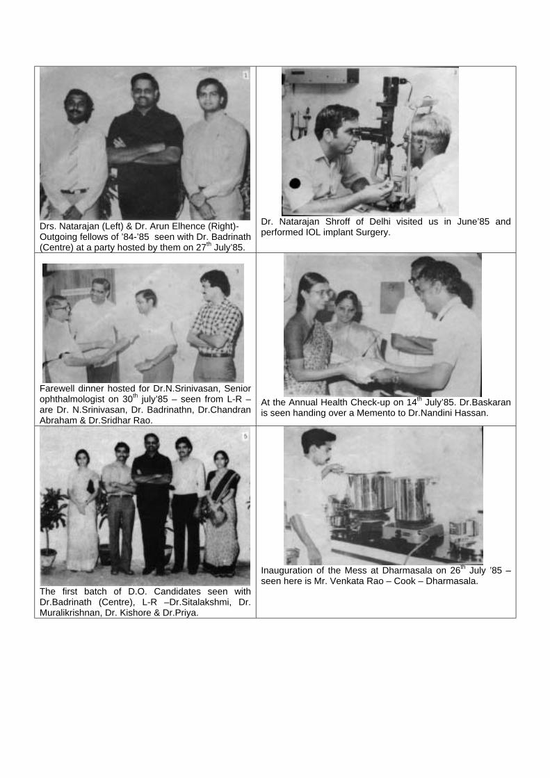
Drs. Natarajan (Left) & Dr. Arun Elhence (Right)- Outgoing fellows of ’84-’85 seen with Dr. Badrinath (Centre) at a party hosted by them on 27th July’85.
Dr. Natarajan Shroff of Delhi visited us in June’85 and performed IOL implant Surgery.
Farewell dinner hosted for Dr.N.Srinivasan, Senior ophthalmologist on 30th july’85 – seen from L-R – are Dr. N.Srinivasan, Dr. Badrinathn, Dr.Chandran Abraham & Dr.Sridhar Rao.
At the Annual Health Check-up on 14th July’85. Dr.Baskaran is seen handing over a Memento to Dr.Nandini Hassan.
The first batch of D.O. Candidates seen with Dr.Badrinath (Centre), L-R –Dr.Sitalakshmi, Dr. Muralikrishnan, Dr. Kishore & Dr.Priya.
Inauguration of the Mess at Dharmasala on 26th July ’85 – seen here is Mr. Venkata Rao – Cook – Dharmasala.

EDITOR: Dr. MARY ABRAHAM – FOR PRIVATE CIRCULATION ONLY – DESIGNED & PRINTED BY: ADIMAGE-MADRAS


![THE [JAGADGURU RAMANANDACHARYA] RAJASTHAN …](https://static.fdocuments.net/doc/165x107/627e18521798244daf6f76ae/the-jagadguru-ramanandacharya-rajasthan-.jpg)