Visualization of transfer of a fluorescently-labeled...
8
Gene Therapy and Molecular Biology Vol 9, page 135 135 Gene Ther Mol Biol Vol 9, 135-142, 2005 Visualization of transfer of a fluorescently-labeled membrane raft protein to T cells using lentivirus Research Article Jennifer Byrum 1 and William Rodgers 1,2, * 1 Molecular Immunogenetics Program, Oklahoma Medical Research Foundation 2 Department of Microbiology and Immunology, Department of Pathology, Oklahoma University Health Sciences Center __________________________________________________________________________________ *Correspondence: Dr. William Rodgers, Molecular Immunogenetics Program, Oklahoma Medical Research Foundation, 825 NE 13th St MS 17, Oklahoma City, Oklahoma 73104; Phone: (405) 271-7393; Fax: (405) 271-8237; E-mail: [email protected] Key words: Rafts, lentivirus, T cells Abbreviations: T cell receptor, (TCR); yellow fluorescent protein, (YFP) Received: 21 June 2005; Accepted: 01 July 2005; electronically published: July 2005 Summary Lentivirus vector systems have been developed for the safe delivery of foreign genes to target tissues. However, the use of these systems for delivering specific proteins to target cells has been largely unexplored. To test this concept, the lentivirus expression plasmid pLenti was utilized to overexpress in producer cells a YFP-fusion protein that is specifically targeted to glycolipid-enriched membrane rafts, which is the site of virus assembly. Our data show that virus generated in producer cells that expressed the YFP fusion protein were able to effectively label target cells by a 2-3 hr incubation with the virus. Labeling of the target cells was specific to the lentivirus, as it was blocked by pre- incubating the virus with antibody to the surface protein, and it was not affected by pre-treating the target cells with cyclohexamide. T cells that were labeled using the lentivirus underwent a robust stimulation following crosslinking the T cell receptor, thus showing that T cells labeled using lentivirus remained responsive to extracellular cues. Altogether, these results show that overexpression of foreign proteins in lentivirus producer cells can yield protein-loaded viruses, which can then function to deliver the protein to target cells. Thus, our findings suggest an avenue for targeting specific proteins to cells where foreign gene expression is not feasible. I. Introduction HIV-based lentiviral vectors represent an emerging tool in gene therapy, as they offer important advantages over retroviral vectors in gene delivery to target cells (Lever et al, 2004; Wiznerowicz and Trono, 2005). Towards this end, HIV-based vectors with improved biosafety have been developed for generation of lentivirus using transfected producer cells (Poznansky et al, 1991; Shimada et al, 1991; Reiser et al, 1996; Naldini et al, 1996; Dull et al, 1998). Production of lentivirus in these systems is dependent upon efficient packaging of the virus by the producer cells. In the case of HIV, packaging of virus is dependent upon the glycolipid-enriched membrane rafts that occur in cell membranes (Nguyen and Rildreth, 2000; Ono and Freed, 2001; Manes et al, 2003). Lipid rafts are a distinct class of membrane domains that are composed of cholesterol, sphingolipids, and specific proteins (Simons and Toomre, 2000; Brown and London, 1998). The precise mechanism by which rafts function in viral production is not known, but it may be related to rafts providing an optimal site for assembly of virus particles and their budding (Manes et al, 2003). The observed incorporation of rafts and raft- associated proteins in the envelope of lentivirus (Nguyen and Hildreth, 2000; Manes et al, 2003) suggests that the viruses may represent an effective tool for delivering proteins to target cells from producer cells. For example, just as HIV may deliver endogenous proteins from the host cells in order to improve their infectivity (Manes et al, 2003), foreign gene expression in producer cells may lead to loading of the gene product in the viral envelope for its delivery to target cells. Such direct delivery of foreign proteins to target cells by lentivirus could have important therapeutic applications. For example, cells that are deficient in a key enzyme could have the protein delivered directly by the virus, rather than relying on gene expression in the target cell. To test the notion of delivery of foreign protein to target cells using lentivirus, we utilized a recently developed HIV-based vector system (Dull et al, 1998) to express and load virus with a membrane raft-associated yellow fluorescent protein (YFP) fusion peptide (Rodgers, 2002). This expression system consists of an HIV-based vector (pLenti) that contains a modified hybrid 5' LTR that
-
Upload
trinhthuan -
Category
Documents
-
view
217 -
download
0
Transcript of Visualization of transfer of a fluorescently-labeled...
Gene Therapy and Molecular Biology Vol 9, page 135
135
Gene Ther Mol Biol Vol 9, 135-142, 2005
Visualization of transfer of a fluorescently-labeledmembrane raft protein to T cells using lentivirusResearch Article
Jennifer Byrum1 and William Rodgers1,2,*1Molecular Immunogenetics Program, Oklahoma Medical Research Foundation2Department of Microbiology and Immunology, Department of Pathology, Oklahoma University Health Sciences Center__________________________________________________________________________________*Correspondence: Dr. William Rodgers, Molecular Immunogenetics Program, Oklahoma Medical Research Foundation, 825 NE 13thSt MS 17, Oklahoma City, Oklahoma 73104; Phone: (405) 271-7393; Fax: (405) 271-8237; E-mail: [email protected]
Key words: Rafts, lentivirus, T cells
Abbreviations: T cell receptor, (TCR); yellow fluorescent protein, (YFP)
Received: 21 June 2005; Accepted: 01 July 2005; electronically published: July 2005
SummaryLentivirus vector systems have been developed for the safe delivery of foreign genes to target tissues. However, theuse of these systems for delivering specific proteins to target cells has been largely unexplored. To test this concept,the lentivirus expression plasmid pLenti was utilized to overexpress in producer cells a YFP-fusion protein that isspecifically targeted to glycolipid-enriched membrane rafts, which is the site of virus assembly. Our data show thatvirus generated in producer cells that expressed the YFP fusion protein were able to effectively label target cells bya 2-3 hr incubation with the virus. Labeling of the target cells was specific to the lentivirus, as it was blocked by pre-incubating the virus with antibody to the surface protein, and it was not affected by pre-treating the target cellswith cyclohexamide. T cells that were labeled using the lentivirus underwent a robust stimulation followingcrosslinking the T cell receptor, thus showing that T cells labeled using lentivirus remained responsive toextracellular cues. Altogether, these results show that overexpression of foreign proteins in lentivirus producer cellscan yield protein-loaded viruses, which can then function to deliver the protein to target cells. Thus, our findingssuggest an avenue for targeting specific proteins to cells where foreign gene expression is not feasible.
I. IntroductionHIV-based lentiviral vectors represent an emerging
tool in gene therapy, as they offer important advantagesover retroviral vectors in gene delivery to target cells(Lever et al, 2004; Wiznerowicz and Trono, 2005).Towards this end, HIV-based vectors with improvedbiosafety have been developed for generation of lentivirususing transfected producer cells (Poznansky et al, 1991;Shimada et al, 1991; Reiser et al, 1996; Naldini et al,1996; Dull et al, 1998). Production of lentivirus in thesesystems is dependent upon efficient packaging of the virusby the producer cells. In the case of HIV, packaging ofvirus is dependent upon the glycolipid-enriched membranerafts that occur in cell membranes (Nguyen and Rildreth,2000; Ono and Freed, 2001; Manes et al, 2003). Lipid raftsare a distinct class of membrane domains that arecomposed of cholesterol, sphingolipids, and specificproteins (Simons and Toomre, 2000; Brown and London,1998). The precise mechanism by which rafts function inviral production is not known, but it may be related to raftsproviding an optimal site for assembly of virus particlesand their budding (Manes et al, 2003).
The observed incorporation of rafts and raft-associated proteins in the envelope of lentivirus (Nguyenand Hildreth, 2000; Manes et al, 2003) suggests that theviruses may represent an effective tool for deliveringproteins to target cells from producer cells. For example,just as HIV may deliver endogenous proteins from the hostcells in order to improve their infectivity (Manes et al,2003), foreign gene expression in producer cells may leadto loading of the gene product in the viral envelope for itsdelivery to target cells. Such direct delivery of foreignproteins to target cells by lentivirus could have importanttherapeutic applications. For example, cells that aredeficient in a key enzyme could have the protein delivereddirectly by the virus, rather than relying on geneexpression in the target cell.
To test the notion of delivery of foreign protein totarget cells using lentivirus, we utilized a recentlydeveloped HIV-based vector system (Dull et al, 1998) toexpress and load virus with a membrane raft-associatedyellow fluorescent protein (YFP) fusion peptide (Rodgers,2002). This expression system consists of an HIV-basedvector (pLenti) that contains a modified hybrid 5' LTR that

Byrum and Rodgers: Protein transfer by lentivirus
136
provides a constitutive transcription of downstreamsequences (Dull et al, 1998). We show that pLentigenerated abundant expression of the YFP protein inproducer cells, and produced virus that effectively labeledtarget cells during a 2-3 hr incubation. Expression of theYFP gene in producer cells trans to pLenti by co-transfection with a separate plasmid also generated virusthat labeled target cells. Altogether, our results suggest anovel pathway for directly delivering foreign protein totarget cells, and that may facilitate therapeutic approachesin systems that are not adaptable to foreign geneexpression.
II. Materials and MethodsA. Generation of lentivirusAll reagents for production of lentivirus were purchased
from Invitrogen (VirapowerTM Lentiviral Gateway® Expressionkit, Invitrogen, Carlsbad, CA)). L10-YFP in the lentivirusexpression plasmid pLenti6 (L10-YFP/pLenti) was generated bysubcloning L10-YFP from pWay20 into an EcoRI site that wasinserted into pLenti using LR clonase (Invitrogen) recombinationreaction from pENTR4 (Invitrogen). Correct orientation of L10-YFP in pLenti for expression by the RSV/5' LTR and CMVpromoters was screened by transient expression in HeLa cells asdescribed, and confirmed by DNA sequencing.
293FT producer cells (Invitrogen) were maintained inDMEM containing 10% FCS and antibiotics. Lentivirus wasproduced by co-transfecting 293FT cells with L10-YFP/pLentiand VirapowerTM packaging mix per the manufacturersinstructions, using 9 µg of the packaging mix, 3 µg of L10-YFP/pLenti, and 36 µl of LipofectamineTM 2000. The DNA-lipofectamine mix was added to a 10 cm dish containing 6x10⁶
293FT cells in 5 ml of Opti-MEM +10% FCS. Fresh media wasadded on day 2, and the culture was harvested on day 4. Forharvesting, the media was collected and media centrifuged at3000 rpm (GS-6R, Beckman) for 15 min at 4 oC, after which thesupernatant was passaged through a 0.45 µm filter (Millex-HV,Millipore, Bellerica, MA). The filtrate was then centrifuged at13,500 rpm (SW 28, Beckman, Fullerton, CA) for 90 min, 4 oC.Virus stock was prepared by suspending the resulting pellet inapproximately 0.5 ml of media, which was then aliquoted andstored at -70 oC.
B. Infection106 Jurkat cells were washed and suspended in 100 µl of
RPMI containing 1% FCS plus antibiotics. The cell suspensionwas transferred to a 96 well plate, and to this was added theindicated volume of virus stock, RPMI containing 1% FCS, andpolybrene (Sigma-Aldrich, St. Louis, MO) to a final volume of200 µl and polybrene concentration of 5 µg/ml. The sample wasthen incubated at 37oC for the indicated time, washed with PBS,and then either seeded onto poly-L-lysine (Sigma-Aldrich)coated coverslips for fluorescence microscopy, or resuspended ina minimal volume of media for flow cytometry. For experimentsusing cell imaging, the virus stock was briefly sonicated (550Sonic Dismembrator, Fisher Scientific, Pittsburgh, PA) prior toadding to the cells.
C. Fluorescence measurementMicroscopy was performed using a Nikon Eclipse E400
fluorescence microscope equipped with an Olympus DWIOcamera, and 40X (NA 0.75) and 100X (NA 1.3) objectives. Forimaging YFP fluorescence, the samples were excited withwavelengths 465 to 495 nm, and the fluorescence was detectedusing wavelengths 505 to 555 nm. Flow cytometry was
performed using a Becton Dickinson FACScan, with excitationof the samples at 488 nm, and collection of wavelengths between515 and 545 nm for the detection of emission.
D. Cell stimulationJurkat cells were washed with RPMI containing 50 mM
HEPES (pH 7.4), and suspended in the same at a finalconcentration of 107 cells/ml. The samples were then pre-incubated at 37 oC for 5 min, after which 3 µg of OKT3 wasadded. The samples were incubated for an additional minute,then sedimented, wash with chilled PBS, and then suspended in50 µl of SDS-PAGE sample buffer. The samples were separatedby SDS-PAGE, and protein tyrosine phosphorylation wasmeasured by immunoblotting with 4G10 (Cell SignalingSolutions, Charlottesville, VA), and detected using enhancedchemiluminescence (Pierce, Rockford, IL).
III. ResultsA. Generation of lentivirus in producer
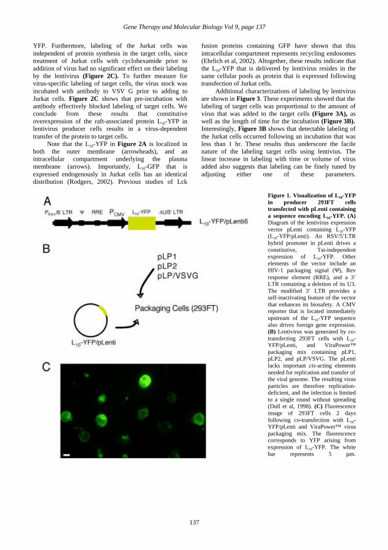
cells expressing L10-YFPThe vector used for expression of L10-YFP and
generation of lentivirus is illustrated in Figure 1A. Thegene encoding L10-YFP was subcloned into pLenti, whereexpression is driven by a hybrid 5’ LTR composed of theRSV enhancer/promoter in place of the U3 region of theHIV 5’ LTR. The hybrid LTR provides a constitutive, Tat-independent transcription of downstream sequences (Dullet al, 1998). An internal CMV promoter that isimmediately upstream of the L10-YFP also drivesconstitutive transcription (Dull et al, 1998).
Infectious particles were generated by co-transfecting293FT cells with L10-YFP/pLenti and three other separateand nonoverlapping expression plasmids required forencapsulation and targeting of the virus (Figure 1B): LP1and LP2, which contain gag and pol, and rev, respectively,and pLP/VSV G, which encodes the G protein of vesicularstomatitis virus (VSV). pLP1, pLP2, and pLP/VSV Gwere introduced using the Virapower™ packaging mix(Materials and Methods). Imaging of transfected 293FTcells showed a robust expression of L10-YFP in theproducer cells (Figure 1C).
B. Facile and effective labeling of targetcells using L10-YFP/lentivirus
Culture supernatants of transfected 293FT cells wereharvested 48 hr following transfection, after which thevirus was concentrated by centrifugation. Accordingly, a10 cm plate of 293FT cells was used to generateapproximately 0.5 ml of concentrated virus stock. Forlabeling, a 100 µl aliquot of virus stock was brieflysonicated, and then added to cell cultures for a 3 hrincubation at 37 oC.
A fluorescence image demonstrating labeling ofJurkat T cells by incubation with lentivirus from producercells that expressed L10-YFP is shown in Figure 2A . In aseparate experiment, Jurkat cells were incubated withlentivirus from 293FT cells that were transfected with anempty pLenti plasmid that did not contain L10-YFP, inwhich case no labeling of the target cells was detected(Figure 2B). Thus, labeling of the Jurkat cells was specificto virus derived from producer cells that expressed L10-
Gene Therapy and Molecular Biology Vol 9, page 137
137
YFP. Furthermore, labeling of the Jurkat cells wasindependent of protein synthesis in the target cells, sincetreatment of Jurkat cells with cyclohexamide prior toaddition of virus had no significant effect on their labelingby the lentivirus (Figure 2C). To further measure forvirus-specific labeling of target cells, the virus stock wasincubated with antibody to VSV G prior to adding toJurkat cells. Figure 2C shows that pre-incubation withantibody effectively blocked labeling of target cells. Weconclude from these results that constitutiveoverexpression of the raft-associated protein L10-YFP inlentivirus producer cells results in a virus-dependenttransfer of the protein to target cells.
Note that the L10-YFP in Figure 2A is localized inboth the outer membrane (arrowheads), and anintracellular compartment underlying the plasmamembrane (arrows). Importantly, L10-GFP that isexpressed endogenously in Jurkat cells has an identicaldistribution (Rodgers, 2002). Previous studies of Lck
fusion proteins containing GFP have shown that thisintracellular compartment represents recycling endosomes(Ehrlich et al, 2002). Altogether, these results indicate thatthe L10-YFP that is delivered by lentivirus resides in thesame cellular pools as protein that is expressed followingtransfection of Jurkat cells.
Additional characterizations of labeling by lentivirusare shown in Figure 3. These experiments showed that thelabeling of target cells was proportional to the amount ofvirus that was added to the target cells (Figure 3A), aswell as the length of time for the incubation (Figure 3B).Interestingly, Figure 3B shows that detectable labeling ofthe Jurkat cells occurred following an incubation that wasless than 1 hr. These results thus underscore the facilenature of the labeling target cells using lentivius. Thelinear increase in labeling with time or volume of virusadded also suggests that labeling can be finely tuned byadjusting either one of these parameters.
Figure 1. Visualization of L10-YFPin producer 293FT cellstransfected with pLenti containinga sequence encoding L10-YFP. (A)Diagram of the lentivirus expressionvector pLenti containing L10-YFP(L10-YFP/pLenti). An RSV/5’LTRhybrid promoter in pLenti drives aconstitutive, Tat-independentexpression of L10-YFP. Otherelements of the vector include anHIV-1 packaging signal (Ψ), Revresponse element (RRE), and a 3’LTR containing a deletion of its U3.The modified 3’ LTR provides aself-inactivating feature of the vectorthat enhances its biosafety. A CMVreporter that is located immediatelyupstream of the L10-YFP sequencealso drives foreign gene expression.(B) Lentivirus was generated by co-transfecting 293FT cells with L10-YFP/pLenti, and ViraPower™packaging mix containing pLP1,pLP2, and pLP/VSVG. The pLentilacks important cis-acting elementsneeded for replication and transfer ofthe viral genome. The resulting virusparticles are therefore replication-deficient, and the infection is limitedto a single round without spreading(Dull et al, 1998). (C) Fluorescenceimage of 293FT cells 2 daysfollowing co-transfection with L10-YFP/pLenti and ViraPower™ viruspackaging mix. The fluorescencecorresponds to YFP arising fromexpression of L10-YFP. The whitebar represents 5 µm.

Byrum and Rodgers: Protein transfer by lentivirus
138
Figure 2. Visualization of L10-YFP delivery to target cells using lenitivirus. (A) Fluorescence image of Jurkat cells labeled byincubation with lentivirus that was collected from producer cells that were transfected as described in Figure 1. For labeling, 106 cellswere incubated with 100 µl of sonicated virus stock for 3 hr at 37°C. (B) Fluorescence (top) and bright field (bottom) images of Jurkatcells that were incubated with virus harvested from 293FT cells that were co-transfected with an empty pLenti that did not contain theL10-YFP sequence, and Virapower™ virus packaging mix. The white bars in (A) and (B) represent 5 µm. (C) L10-YFP labeling bylentivirus is independent of protein synthesis in target cells. The separate plots represent Jurkat cells that were either treated with 50µg/ml cyxlohexamide for 2 hr prior to addition to virus (red), or did not receive cyclohexamide (blue). In the treated sample, thecyclohexamide was maintained in the culture during incubation with the virus. Both samples were labeled by incubating with 100 µl oflentivirus for 3 hr at 37°C. The blue plot represents control Jurkat cells that did not receive cyclohexamide and were not incubated withvirus. (D) Antibody to VSV G effectively blocks labeling of Jurkat cells by lentivirus. 106 Jurkat cells were incubated with virus stockthat was either untreated (+ Virus), or that was pre-incubated with a 1:100 dilution of sera to VSV (Rose and Bergmann, 1983) for 30min at 37°C (+ αVSV G, Virus). One sample received no virus, and served as a control for nonspecific fluorescence in the Jurkat cells.The mean relative fluorescence intensity (Mean Rel. Fl. Int.) of each sample was measured by flow cytometry.
Figure 3. Titrationmeasurements of labeling ofJurkat cells by lentivirus. (A) 106
cells were incubated with 0, 10,20, 50, and 100µl of lentivirusstock for 2 hr at 37°C, each in afinal volume of 200 µl. (B) 106
cells were incubated with 60 µl ofvirus in a final volume of 200 µlfor 5 min, 30 min, 1 hr, 2 hr, and 4hr. The fluorescence labeling ofeach sample in (A) and (B) wasmeasured by flow cytometry.

Gene Therapy and Molecular Biology Vol 9, page 139
139
C. Labeling by virus from producer cellsexpressing L10-YFP trans to pLenti
Figures 2 and 3 show that the plasmid pLenticontaining L10-YFP functions to generate infectiousparticles that deliver the L10-YFP from producer cells totarget cells. Next, to determine if virus from producer cellsexpressing L10-YFP trans to pLenti also labeled targetcells, L10-YFP was expressed in 293FT cells using thevector pWay20 rather than pLenti. The pWay20 plasmidcontains the L10-YFP gene under the transcriptionalcontrol of a CMV promoter (Rodgers, 2002).
The manner by which L10-YFP-loaded virus wasproduced by trans expression of L10-YFP is illustrated inFigure 4A. First, in order to ensure that expression of L10-YFP was sufficient to provide effective loading of thevirions, 293FT cells were transfected with the pWay20plasmid 24 hr prior to co-transfection with pLenti and theViraPower™ packaging mix. Virus was then harvested 48hr following the second transfection. As shown in Figure4B, lenitvirus generated by trans expression of L10-YFP inproducer cells was also able to effectively label target
cells.
D. Lentivirus-dependent labeling of cellsdoes not disrupt cell stimulation
One concern was that the viral load that was requiredfor detectable labeling of the target cells would bedisruptive to the cells. For example, delivery of foreignproteins by lentivirus could be accompanied by cellcytotoxicity that is associated with incubating cells withrelatively high titers of virus for several hours. To test forthis possibility, we compared the response of labeled andunlabeled Jurkat cells to stimulation by crosslinking the Tcell receptor (TCR). The response to stimulation wasassayed by measuring protein phosphotyrosine signals inwhole cell lysates. As shown in Figure 5, a control samplethat did not receive virus demonstrated a robust increase inthe tyrosine phosphorylation of multiple proteinsfollowing stimulation, as did the sample that was labeledusing lentivirus prior to stimulation. Thus, labeling Jurkatcells using lentivirus was not deleterious to the cells asmeasured by their response to TCR-dependent signaling.
Figure 4. Visualization of labeling of Jurkat cells by lentivirus from producer cells expressing L10-YFP trans to pLenti. (A) Forgeneration of lentivirus by trans expression of L10-YFP. 293 FT cells were transfected with L10-YFP in the expression vector pWay20.On day 2, the cells were washed and co-transfected with empty pLenti and ViraPower™ virus packaging mix. Virus was harvested fromculture supernatants on day 4. (B) Fluorescence image of Jurkat cells labeled using lentivirus from producer cells expressing L10-YFPtrans to pLenti. For labeling, 106 cells were incubated with 100µl of virus stock for 3 hr at 37°C. The white bar represents 5 µm.

Byrum and Rodgers: Protein transfer by lentivirus
140
Figure 5. Jurkat cells labeled usinglentivirus remain responsive toTCR crosslinking. Phosphotyrosineimmunoblot of control unstimulatedand OKT3-stimulated Jurkat cells. Inone set of samples, the cells werepre-labeled with L10-YFP usinglentivirus that was harvested fromL10-YFP/pLenti-transfected producercells. Molecular weights (inthousands) are indicated on the left.
IV. DiscussionLentivirus systems using HIV-based expression
vectors are a developing tool in gene therapy. Thesesystems use an HIV-derived machinery to assemble andbud viruses, but are genetically altered from the wild typeHIV so as to increase their biosafety (Poznansky et al,1991; Shimada et al, 1991; Naldini et al, 1996; Reiser etal, 1996; Dull et al, 1998). One feature of HIV is that itsassembly and budding occurs in glycolipid-enrichedmembrane rafts within cell membranes (Nguyen andRildreth, 2000; Ono and Freed, 2001; Manes et al, 2003).Membrane rafts are a ubiquitous class of membranedomains which function in cell signaling, proteintrafficking, and pathogen entry and exit from cells (Brownand London, 1998; Simons and Toomre, 2000; Manes etal, 2003) (Helms and Zurzolo, 2004; Rodgers et al, 2005;Rodgers and Smith, 2005). In the case of lentiviruses, raftsmay function as a scaffold for assembly of the virusparticles. Rafts may also represent an optimal point forbudding of nascent virions from the plasma membrane.For example, studies have shown that separate phaseregions of membranes, including those enriched with rafts,have a larger curvature than the remaining bilayer(Baumgart et al, 2003; Bacia et al, 2005), and this mayfunction to augment membrane budding in cellmembranes.
Using one example of an HIV-derived lentivirusexpression vector, we have shown here that constitutiveoverexpression of a raft-associated protein in producercells resulted in its efficient delivery to target cells.Labeling of target cells by virus was specific to virus-dependent fusion to the target cell, as it was effectively
blocked by pre-incubating the virus stock with antibody tothe surface glycoprotein of the virus that was produced forthis study. Also, protein that was delivered to the targetcells appeared to become incorporated in the same cellularpools as that produced by gene expression. This latterobservation underscores the potential utility of celllabeling by viral delivery, such that a foreign protein canbe delivered to discrete pools within cells through theappropriate targeting signals.
One possible mechanism for transfer of L10-YFP totarget cells through lentivirus is by loading of the L10-YFPinto the viral envelope during assembly and budding of thevirus. The loaded protein could then be released into thecell membrane pools following fusion of the virus to thetarget cell. One explanation for the efficient labelingachieved with L10-YFP is that, as a raft-associated protein,it is preferentially incorporated into the viral envelope. Itfollows that proteins that are excluded from rafts will beincorporated into the virions less efficiently than proteinssuch as L10-YFP. Consistent with this hypothesis, wefound that labeling of target cells with lentivirus producedfrom cells expressing the construct S15-YFP, which isexcluded from rafts, was less efficient than the labeling byan equivalent titer of the L10-YFP/lentivirus (data notshown).
Another factor besides raft-association that is likelyto be a determinant regarding the efficiency by which thetargeted protein is loaded into the virus is the size of theforeign protein. We are currently exploring the molecularweight cut off for virus loading and delivery to target cells,and whether this system is amendable for fusion proteinsother than GFP. One outcome of these studies may

Gene Therapy and Molecular Biology Vol 9, page 141
141
therefore be evidence that lentivirus can be used toefficiently transfer enzymatic activity to target cells fortherapeutic purposes.
One concern with using the lentivirus as a proteindelivery device is the biohazard associated with this classof viruses. The system employed in this study is a thirdgeneration lentivirus vector that contains a number ofsafeguards that eliminate the possibility of production ofreplication-competent virions (Dull et al, 1998). Theseinclude usage of a limited set of the HIV genes, which areexpressed trans on non-overlapping plasmids, therebyguarding against generation of productive recombinants.
In summary, we have described a facile method fordelivering foreign proteins to target cells using a lentivirusplasmid that was originally developed for gene delivery.Our findings suggest that protein delivery occurs bygeneration of loaded virus through constitutive expressionof the foreign protein in producer cells. The currentfindings therefore demonstrate a novel strategy forlabeling rafts in cells, and future studies may demonstrateapplication of this technology for delivery of enzymes tooffset certain pathologies.
AcknowledgementsThis work was supported by grants from the National
Institutes of Health P50 RR015577, ROl 6M070001, andthe Oklahoma Center for the Advancement of Science andTechnology grant HRO2-009.
ReferencesBacia K, Schwille P and Kurzcbalia T (2005) Sterol structure
determines the separation of phases and the curvature of theliquid-ordered phase in model membranes. Proc Natl AcadSci. U S A 102, 3272-3277.
Baumgart T, Hess ST and Webb WW (2003) Imaging coexistingfluid domains in biomembrane models coupling curvatureand line tension. Nature 425, 821-824.
Brown DA and London E (1998) Functions of lipid rafts inbiological membranes. Annu Rev Cell Dev Biol 14, 111-136.
Dull T, Zufferey R, Kelly M, Mandel RJ, Nguyen M, Trono Dand Naldini L (1998) A third-generation lentivirus vectorwith a conditional packaging system. J Virol 72, 8463-8471.
Ehrlich LI, Ebert PJ, Krummel MF, Weiss A and Davis MM(2002) Dynamics of pS6lck translocation to the T cellimmunological synapse following agonist and antagoniststimulation. Immunity 17, 809-822.
Helms JB and Zurzolo C (2004) Lipids as targeting signals: lipidrafts and intracellular trafficking. Traffic 5, 247-254.
Lever AM, Strappe PM and Zhao J (2004) Lentiviral vectors. JBiomed Sci 11, 439-449.
Manes S, del Real 0 and Martinez AC (2003) Pathogens: rafthijackers. Nat Rev Immunol 3, 557-568.
Naldini L, Blomer U, Gallay P, Ory D, Mulligan R, Gage FH,Verma IM and Trono D (1996) In vivo gene delivery andstable transduction of nondividing cells by a lentiviral vector.
Science 272, 263-267.Nguyen DH and Hudreth JE (2000) Evidence for budding of
human immunodeficiency virus type 1 selectively fromglycolipid-enriched membrane lipid rafts. J Virol 74, 3264-3272.
Ono A and Freed EG (2001) Plasma membrane rafts play acritical role in HIV-1 assembly and release. Proc Natl AcadSci USA 98, 13925-13930.
Poznansky M, Lever A, Bergeron L, Haseltine W and Sodroski, J(1991) Gene transfer into human lymphocytes by a defectivehuman immunodeficiency virus type 1 vector. J Virol 65,532-536.
Reiser J, Harmison G, Kluepfel-Stahl S, Brady RO, Karlsson Sand Schubert M (1996) Transduction of nondividing cellsusing pseudotyped defective high-titer HIV type 1 particles.Proc Natl Acad Sci USA 93, 15266-15271.
Rodgers W (2002) Making membranes green: construction andcharacterization of GFP-fusion proteins targeted to discreteplasma membrane domains. Biotechniques 32, 1044-1051.
Rodgers W and Smith K (2005) Properties of glycolipid-enrichedmembrane rafts in antigen presentation. Crit Rev Immunol25, 19-30.
Rodgers W, Farris D and Mishra S (2005) Merging complexes:properties of membrane raft assembly during lymphocytesignaling. Trends Immunol 26, 97-103.
Rose JK and Bergmann JE (1983) Altered cytoplasmic domainsaffect intracellular transport of the vesicular stomatitis virusglycoprotein. Cell 34, 513-524.
Shimada T, Fuj ii, H, Mitsuya H and Nienhuis AW (1991)Targeted and highly efficient gene transfer into CD4+ cellsby a recombinant human immunodeficiency virus retroviralvector. J Clin Invest 88, 1043-1047.
Simons K and Toomre D (2000) Lipid rafis and signaltransduction. Nature Mol Cell Biol Rev 1,31-39.
Wiznerowicz M. and Trono D (2005) Harnessing HIV fortherapy, basic research and biotechnology. TrendsBiotechnol 23, 42-47.
Jennifer Byrum and William Rodgers

Byrum and Rodgers: Protein transfer by lentivirus
142