VIII-Ⅱ-1. Project Research Project 5frogs and Japanese green sea urchins, with mammalian DAEP....
9
VIII-Ⅱ-1. Project Research Project 5
Transcript of VIII-Ⅱ-1. Project Research Project 5frogs and Japanese green sea urchins, with mammalian DAEP....

VIII-Ⅱ-1. Project Research
Project 5

採択課題番号 24P5 放射線や紫外線照射によるタンパク質の異常凝集と プロジェクト
その防御及び修復機構に関する研究 (京大・原子炉)藤井紀子
PR5 Project Research on the Abnormal Aggregation of Proteins by
Post-Translational Modifications, and Study of Repair Mechanism
N. Fujii
Research Reactor Institute, Kyoto University
Objectives and Allotted Research Subjects:
The aim of this project research is to elucidate the cor-
relation between the change of the protein structure in-
duced by various post-translational modifications with
UV irradiation, gamma-irradiation, aging and protein
function. We also investigate the repair mechanism for
the damaged protein by irradiation. This research pro-
gram has started in 2011. In this year, the 7 research sub-
jects were carried out. The allotted research subjects
(ARS) are as follows;
ARS-1: Protein deuteration for small-angle neutron
Scattering (M. Sugiyama, N. Fujii and N. Fujii) ARS-2: Comparison of properties of mammalian and
aquatic animal D-aspartyl endopeptidases.
(T. Kinouchi and N. Fujii)
ARS-3: Damage to biological molecules induced by
ionizing radiation and biological defense mechanisms
provided by radical scavengers II.
(T. Saito and N. Fujii)
ARS-4: Effect of higher order structure of recombinant
human A-crystallin on the rate of -linkage
isomerization of specifc aspartyl residue.
(Y. Sadakane and N. Fujii)
ARS-5: Analysis of environmental stress-related
imbalance in mice. (N. Ohgami and N. Fujii)
ARS-6: Factors permitting the combination of D-serine
and pyridoxal 5′-phosphate at the active site of
tryptophanase. (A. Shimada, N. Fujii and T. Saito)
ARS-7: A rapid survey of Asp isomers in lens proteins
from the cataract. (N. Fujii, N. Fujii, H. Sakaue and H.
Sasaki)
Main Results and Contents of This Project
ARS-1: Sugiyama et.al. investigated a structure of
α-crsytallin complex by subunit deuteration and SANS.
As a result, it was revealed that there exists subunit ex-
change between αA-crsytallin and αB-crystallin and also
between αB-crystallins.
ARS-2: Kinouchi et al. searched for and attempted to
purify D-aspartyl-endopeptidase (DAEP) in aquatic ani-
mals. As a result, high DAEP activity was detected in
gonads of African clawed frogs (Xenopus laevis) and
Japanese green sea urchins (Hemicentrotus pulcherrimus).
Purified DAEP from Xenopus oocytes indicated common
specific features with mouse DAEP: mitochondrial lo-
calization, high molecular weight, and sensitivity to a
synthesized DAEP inhibitor. We are characterizing its
fundamental functions of frog DAEP.
ARS-3: Saito et al. showed that astaxanthin inhibits or
promots gamma radiation induced oxidative degradation
of -linolenic acid depending on the conditions, sug-
gesting that carotenoids are involved in the protection
against damage to lipid structures induced by gamma
irradiation in vivo.
ARS-4: Sadakane et al. determined the rate of β-linkage
isomerization of a specific Asp residue both in the re-
combinant human αA-crystallin and fragmentary peptide
at 50°C and 90°C, and revealed that higher order struc-
ture of protein suppressed the β-linkage isomerization of
Asp58 residue of αA-crystallin.
ARS-5: Ohgami et al. showed that chronic exposure to
low frequency noise (below 0.5 kHz) at moderate levels
causes impaired balance involving morphological im-
pairments of the vestibule with enhanced levels of oxida-
tive stress. We are investigating whether imbalance in-
volves aggregation of a specific protein in the vestibule in
inner ears.
ARS-6: Shimada et al. analyzed the kinetics of trypto-
phanase against various tryptophan-analogous inhibitors
in terms of double reciprocal plots. The results indi-
cated that small stereo-structural change was necessary to
form an aldimine bond between D-serine and pyridoxal
5′-phosphate.
ARS-7: Fujii et al. showed that a convenient and ro-
bust biochemical method for identifying the isomeric Asp
sites in crystallins using LC-MS systems. There are many
advantages to this new method: 1) No requirement for
large amounts of sample proteins, 2) No requirement for
the purification of proteins 3) No requirement for com-
plicated analytical steps. This new method is able to
search comprehensively for the Asp isomers in damaged
or aged proteins from all living tissues and cells.
Protein Deuteration for Small-Angle Neutron Scattering
M. Sugiyama, N. Fujii and N. Fujii
Research Reactor Institute, Kyoto University
INTRODUCTION: Small-Angle Scattering (SAS) is one of promising methods to analyze protein structure in an aqueous solution. The probes of SAS are X-ray and neutron: they are called Small-Angle X-ray and Neutron Scatterings (SAXS) and (SANS). A merit of use of X-ray as probe is strong intensity of source by using a synchro-tron. On the other hand, typical neutron sources are weaker than that of X-ray but neutron is very sensitive for isotope effect. An interesting point of neutron probe for biological samples is the large difference in scattering length between proton and deuteron: the scattering length of proton is 3.74 fm but that of deuteron is 6.64 fm. Be-cause hydrogen is one of main components of protein, we can make the scattering length density of the protein and/or a subunit changed by replacing the proton by deuteron. In other word, deuteration can be for used a labeling method with less modification of the protein itself.
Crystallin複合体の会合様態の研究 プロジェクト
Recently, Sugiyama et al. applied the combination technique of subunit deuteraion and SANS for measure-ment of large scale kinetics in protein, such as subunit exchange. This technique has possibility to analyze the similar large scale kinetics in the other proteins. So, we begin to apply this technique to analyze the formation and time evolution of α-Crystalin. It is well-known that α-Crystalin consists of two kinds of subunits, named as αA-Crystalin and αB-Crystalin. Moreover, the ratio of two subunits in α-Crystalin could be change by aging. However, the mechanism has been unknown but the subunit exchange could be one of candidates of the mechanism. Therefore, our first goal is to prove the ex-istence of subunit exchange in α-Crystalin system. To perform the combination technique of subunit deuteraion and SANS, the key technique is protein deuteration. Here, we report the results of our preparation of deuterated protein (α-Crystalin) and SANS measurements with them.
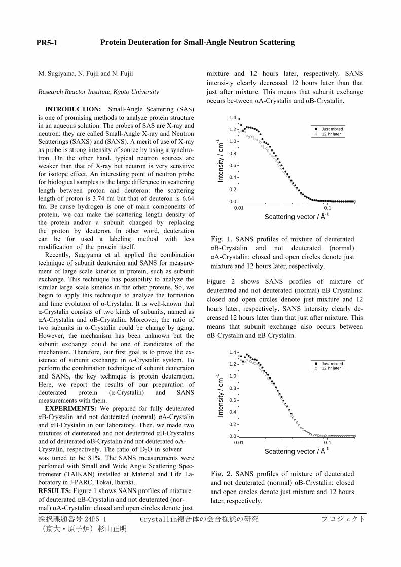
EXPERIMENTS: We prepared for fully deuterated αB-Crystalin and not deuterated (normal) αA-Crystalin and αB-Crystalin in our laboratory. Then, we made two mixtures of deuterated and not deuterated αB-Crystalins and of deuterated αB-Crystalin and not deuterated αA-Crystalin, respectively. The ratio of D2O in solvent was tuned to be 81%. The SANS measurements were perfomed with Small and Wide Angle Scattering Spec-trometer (TAIKAN) installed at Material and Life La-boratory in J-PARC, Tokai, Ibaraki. RESULTS: Figure 1 shows SANS profiles of mixture of deuterated αB-Crystalin and not deuterated (nor-mal) αA-Crystalin: closed and open circles denote just
mixture and 12 hours later, respectively. SANS intensi-ty clearly decreased 12 hours later than that just after mixture. This means that subunit exchange occurs be-tween αA-Crystalin and αB-Crystalin.
0.01 0.10.0
0.2
0.4
0.6
0.8
1.0
1.2
1.4
Inte
nsity
/ cm
-1Scattering vector / Å-1
Just mixted 12 hr later
Fig. 1. SANS profiles of mixture of deuterated αB-Crystalin and not deuterated (normal) αA-Crystalin: closed and open circles denote just mixture and 12 hours later, respectively.
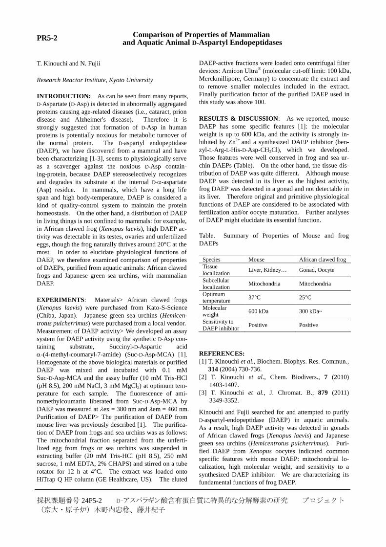
Figure 2 shows SANS profiles of mixture of deuterated and not deuterated (normal) αB-Crystalins: closed and open circles denote just mixture and 12 hours later, respectively. SANS intensity clearly de-creased 12 hours later than that just after mixture. This means that subunit exchange also occurs between αB-Crystalin and αB-Crystalin.
0.01 0.10.0
0.2
0.4
0.6
0.8
1.0
1.2
1.4
Inte
nsity
/ cm
-1
Scattering vector / Å-1
Just mixted 12 hr later
Fig. 2. SANS profiles of mixture of deuterated and not deuterated (normal) αB-Crystalin: closed and open circles denote just mixture and 12 hours later, respectively.
採択課題番号 24P5-1
(京大・原子炉)杉山正明
PR5-1
採択課題番号 24P5-2 D-アスパラギン酸含有蛋白質に特異的な分解酵素の研究 プロジェクト
(京大・原子炉)木野内忠稔、藤井紀子
PR5-2 Comparison of Properties of Mammalian and Aquatic Animal D-Aspartyl Endopeptidases
T. Kinouchi and N. Fujii
Research Reactor Institute, Kyoto University
INTRODUCTION: As can be seen from many reports,
D-Aspartate (D-Asp) is detected in abnormally aggregated
proteins causing age-related diseases (i.e., cataract, prion
disease and Alzheimer's disease). Therefore it is
strongly suggested that formation of D-Asp in human
proteins is potentially noxious for metabolic turnover of
the normal protein. The D-aspartyl endopeptidase
(DAEP), we have discovered from a mammal and have
been characterizing [1-3], seems to physiologically serve
as a scavenger against the noxious D-Asp contain-
ing-protein, because DAEP stereoselectively recognizes
and degrades its substrate at the internal D--aspartate
(Asp) residue. In mammals, which have a long life
span and high body-temperature, DAEP is considered a
kind of quality-control system to maintain the protein
homeostasis. On the other hand, a distribution of DAEP
in living things is not confined to mammals: for example,
in African clawed frog (Xenopus laevis), high DAEP ac-
tivity was detectable in its testes, ovaries and unfertilized
eggs, though the frog naturally thrives around 20°C at the
most. In order to elucidate physiological functions of
DAEP, we therefore examined comparison of properties
of DAEPs, purified from aquatic animals: African clawed
frogs and Japanese green sea urchins, with mammalian
DAEP.
EXPERIMENTS: Materials> African clawed frogs
(Xenopus laevis) were purchased from Kato-S-Science
(Chiba, Japan). Japanese green sea urchins (Hemicen-
trotus pulcherrimus) were purchased from a local vendor.
Measurement of DAEP activity> We developed an assay
system for DAEP activity using the synthetic D-Asp con-
taining substrate, Succinyl-D-Aspartic acid
-(4-methyl-coumaryl-7-amide) (Suc-D-Asp-MCA) [1].
Homogenate of the above biological materials or purified
DAEP was mixed and incubated with 0.1 mM
Suc-D-Asp-MCA and the assay buffer (10 mM Tris-HCl
(pH 8.5), 200 mM NaCl, 3 mM MgCl2) at optimum tem-
perature for each sample. The fluorescence of ami-
nomethylcoumarin liberated from Suc-D-Asp-MCA by
DAEP was measured at ex = 380 nm and em = 460 nm.
Purification of DAEP> The purification of DAEP from
mouse liver was previously described [1]. The purifica-
tion of DAEP from frogs and sea urchins was as follows:
The mitochondrial fraction separated from the unferti-
lized egg from frogs or sea urchins was suspended in
extracting buffer (20 mM Tris-HCl (pH 8.5), 250 mM
sucrose, 1 mM EDTA, 2% CHAPS) and stirred on a tube
rotator for 12 h at 4°C. The extract was loaded onto
HiTrap Q HP column (GE Healthcare, US). The eluted
DAEP-active fractions were loaded onto centrifugal filter
devices: Amicon Ultra® (molecular cut-off limit: 100 kDa,
Merckmillipore, Germany) to concentrate the extract and
to remove smaller molecules included in the extract.
Finally purification factor of the purified DAEP used in
this study was above 100.
RESULTS & DISCUSSION: As we reported, mouse
DAEP has some specific features [1]: the molecular
weight is up to 600 kDa, and the activity is strongly in-
hibited by Zn2+
and a synthesized DAEP inhibitor (ben-
zyl-L-Arg-L-His-D-Asp-CH2Cl), which we developed.
Those features were well conserved in frog and sea ur-
chin DAEPs (Table). On the other hand, the tissue dis-
tribution of DAEP was quite different. Although mouse
DAEP was detected in its liver as the highest activity,
frog DAEP was detected in a gonad and not detectable in
its liver. Therefore original and primitive physiological
functions of DAEP are considered to be associated with
fertilization and/or oocyte maturation. Further analyses
of DAEP might elucidate its essential function.
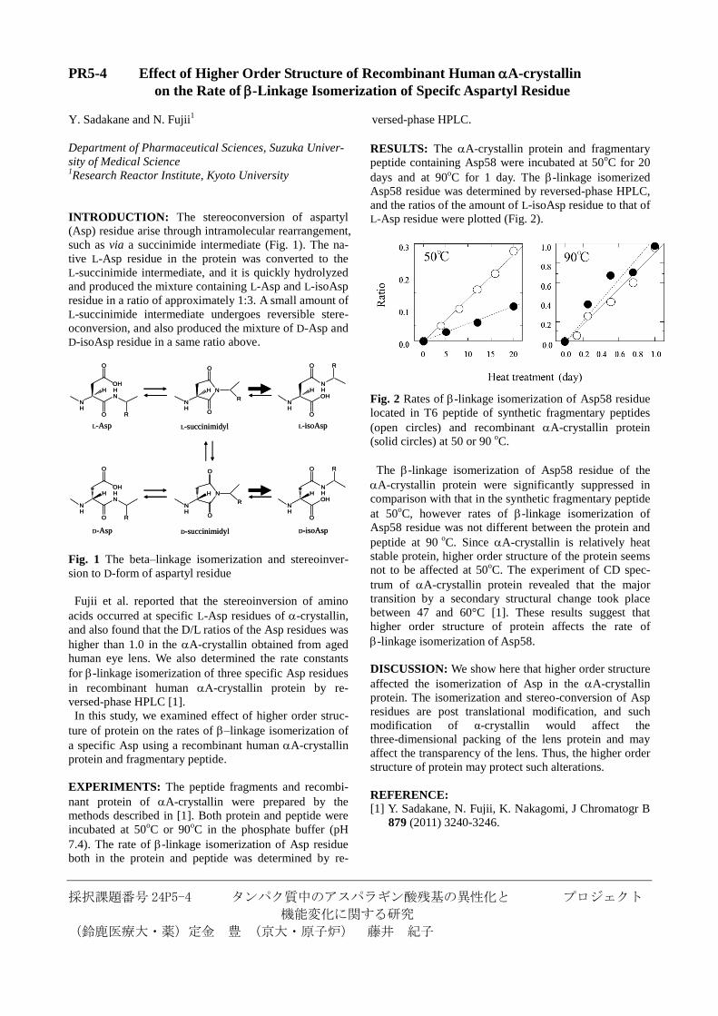
Table. Summary of Properties of Mouse and frog
DAEPs
Species Mouse African clawed frog
Tissue
localization Liver, Kidney… Gonad, Oocyte
Subcellular
localization Mitochondria Mitochondria
Optimum
temperature 37°C 25°C
Molecular
weight 600 kDa 300 kDa~
Sensitivity to
DAEP inhibitor Positive Positive
REFERENCES:
[1] T. Kinouchi et al., Biochem. Biophys. Res. Commun.,
314 (2004) 730-736.
[2] T. Kinouchi et al., Chem. Biodivers., 7 (2010)
1403-1407.
[3] T. Kinouchi et al., J. Chromat. B., 879 (2011)
3349-3352.
Kinouchi and Fujii searched for and attempted to purify
D-aspartyl-endopeptidase (DAEP) in aquatic animals.
As a result, high DAEP activity was detected in gonads
of African clawed frogs (Xenopus laevis) and Japanese
green sea urchins (Hemicentrotus pulcherrimus). Puri-
fied DAEP from Xenopus oocytes indicated common
specific features with mouse DAEP: mitochondrial lo-
calization, high molecular weight, and sensitivity to a
synthesized DAEP inhibitor. We are characterizing its
fundamental functions of frog DAEP.

PR5-3 Damage to Biological Molecules Induced by Ionizing Radiation
and Biological Defense Mechanisms Provided by Radical Scavengers II
T. Saito and N. Fujii
Research Reactor Institute, Kyoto University
INTRODUCTION: Some bacteria exhibit extreme
resistance to ionizing radiation [1]. A common feature of
these bacteria is that they contain red carotenoid pig-
ments [1, 2, 3]. Colorless mutants of these radioresistant
bacteria are more sensitive to gamma irradiation than
wild types [1]. Therefore, carotenoids are thought to be
involved in the bacterial defense mechanisms against
ionizing radiation [1]. Biological effects induced by
low-linear energy transfer ionizing radiation are mainly
attributed to radicals generated by radiolysis. Carotenoids
have high radical scavenging activity, and they are local-
ized in cell surface lipids in prokaryotes. These facts in-
dicate that carotenoids are likely to defend the cell sur-
face lipids of radioresistant bacteria against ionizing ra-
diation.
When considering the biological defense mechanism
of these radioresistant bacteria against ionizing radiation,
it is important to elucidate the effects of carotenoids on
damage to biological molecules, especially biological
lipids. In this study, we analyzed the effect of astaxanthin,
a typical carotenoid, on gamma radiation induced oxida-
tive degradation of -linolenic acid, a type of fatty acid.
EXPERIMENTS: Sample Preparation: Linolenic acid
was dissolved in benzene at a final concentration of 5.0
10-1
M, and astaxanthin was added at a final concentra-
tion of 5.0 10-8
to 5.0 10-4
M. Gamma Irradiation:
The prepared solutions were irradiated with 60
Co gamma
rays at a dose of 30 kGy and a dose rate of 400 Gy/min.
Analysis of Oxidative Degradation of -Linolenic Acid:
The method described by Buege and Aust was used with
some modifications [4]. TCA-TBA-BHT-HCl regent
(15% trichloroacetic acid, 0.375% thiobarbituric acid,
0.04% butylated hydroxytoluene, and 0.25N hydrochlo-
ric acid) was prepared. The gamma irradiated sample was
diluted 50-fold with benzene. The diluted solution (3.0
mL) was evaporated in vacuo. The residue was dispersed
in 9.0 mL of PBS(-) with a sonicator. The dispersed solu-
tion (1.0 mL) was combined with 2.0 mL of
TCA-TBA-BHT-HCl regent and thoroughly mixed. The
mixed solution was heated in a boiling water bath for 15
min, and absorption at 535 nm was measured. The
amount of malondialdehyde (MDA) formed was calcu-
lated using the molar absorption coefficient () of the
color substance formed by the reaction (i.e., 1.56 105 at
535 nm). Thus, oxidative degradation of -linolenic acid
was evaluated by measuring the amount of MDA.
RESULTS: Under the experimental conditions used,
5.0 10-4
M astaxanthin inhibited the oxidative degrada-
tion of -linolenic acid induced by gamma irradiation,
although it was not statistically significant (Fig. 1). In
contrast, treatment with 5.0 10-7
and 5.0 10-8
M
astaxanthin significantly promoted the oxidative degra-
dation of -linolenic acid (Fig. 1). We have previously
reported that-carotene, a type of carotenoid, shows
effects similar to those of astaxanthin [5]. We found that
8.5 10-3
M -carotene significantly inhibited oxidative
degradation of -linolenic acid induced by gamma irra-
diation. These results suggest that carotenoids play a role
in the protection against damage to the lipid structure
induced by gamma irradiation in radioresistant bacteria
and that the intracellular concentrations of carotenoids
are strictly regulated.
REFERENCES: [1] T. Saito, Viva Origino, 30 (2007) 85-92.
[2] T. Saito et al., Arch. Microbiol., 162 (1994) 414-421.
[3] T. Saito et al., Microbios, 95 (1998) 79-90.
[4] J. A. Buege and S. D. Aust, Meth. Enzymol., 52
(1978) 302-310.
[5] T. Saito and N. Fujii, KURRI Progress Report 2009,
(2010) 137.
採択課題番号 24P5-3 放射線照射による生体分子の損傷と プロジェクト ラジカルスカベンジャーによる生体防御機構
(京大・原子炉)齊藤 毅、藤井紀子
Fig. 1. Effects of astaxanthin on gamma radiation-induced oxidative degradation of -linolenic acid. The horizontal axis shows the concentration of astaxanthin and the vertical axis shows the amount of MDA formed. Each data set is presented as the mean ± SD of six samples from three independent experiments. An asterisk (*) indicates a sample with a significant difference from the control sample with no astaxanthin added (P < 0.05).
採択課題番号 24P5-4 タンパク質中のアスパラギン酸残基の異性化と プロジェクト
機能変化に関する研究
(鈴鹿医療大・薬)定金 豊 (京大・原子炉) 藤井 紀子
PR5-4 Effect of Higher Order Structure of Recombinant Human A-crystallin
Department of Pharmaceutical Sciences, Suzuka Univer-
sity of Medical Science 1Research Reactor Institute, Kyoto University
INTRODUCTION: The stereoconversion of aspartyl
(Asp) residue arise through intramolecular rearrangement,
such as via a succinimide intermediate (Fig. 1). The na-
tive L-Asp residue in the protein was converted to the
L-succinimide intermediate, and it is quickly hydrolyzed
and produced the mixture containing L-Asp and L-isoAsp
residue in a ratio of approximately 1:3. A small amount of
L-succinimide intermediate undergoes reversible stere-
oconversion, and also produced the mixture of D-Asp and
D-isoAsp residue in a same ratio above.
N
NH
O
O
R
H
N
NH
O
O
R
H
NH
HN
OH
O
O
R
H
NH
OH
NH
O
O R
H
NH
OH
NH
O
O R
H
NH
HN
OH
O
O
R
H
L-Asp L-succinimidyl
D-succinimidyl
L-isoAsp
D-Asp D-isoAsp
N
NH
O
O
R
H
N
NH
O
O
R
H
NH
HN
OH
O
O
R
H
NH
OH
NH
O
O R
H
NH
OH
NH
O
O R
H
NH
HN
OH
O
O
R
H
L-Asp L-succinimidyl
D-succinimidyl
L-isoAsp
D-Asp D-isoAsp
Fig. 1 The beta–linkage isomerization and stereoinver-
sion to D-form of aspartyl residue
Fujii et al. reported that the stereoinversion of amino
acids occurred at specific L-Asp residues of -crystallin,
and also found that the D/L ratios of the Asp residues was
higher than 1.0 in the A-crystallin obtained from aged
human eye lens. We also determined the rate constants
for -linkage isomerization of three specific Asp residues
in recombinant human -crystallin protein by re-
versed-phase HPLC [1].
In this study, we examined effect of higher order struc-
ture of protein on the rates of –linkage isomerization of
a specific Asp using a recombinant human A-crystallin
protein and fragmentary peptide.
EXPERIMENTS: The peptide fragments and recombi-
nant protein of A-crystallin were prepared by the
methods described in [1]. Both protein and peptide were
incubated at 50oC or 90
oC in the phosphate buffer (pH
7.4). The rate of -linkage isomerization of Asp residue
both in the protein and peptide was determined by re-
on the Rate of -Linkage Isomerization of Specifc Aspartyl Residue
Y. Sadakane and N. Fujii1 versed-phase HPLC.
RESULTS: The A-crystallin protein and fragmentary
peptide containing Asp58 were incubated at 50oC for 20
days and at 90oC for 1 day. The -linkage isomerized
Asp58 residue was determined by reversed-phase HPLC,
and the ratios of the amount of L-isoAsp residue to that of
L-Asp residue were plotted (Fig. 2).
Fig. 2 Rates of -linkage isomerization of Asp58 residue
located in T6 peptide of synthetic fragmentary peptides
(open circles) and recombinant A-crystallin protein
(solid circles) at 50 or 90 oC.
The -linkage isomerization of Asp58 residue of the
A-crystallin protein were significantly suppressed in
comparison with that in the synthetic fragmentary peptide
at 50oC, however rates of -linkage isomerization of
Asp58 residue was not different between the protein and
peptide at 90 o
C. Since A-crystallin is relatively heat
stable protein, higher order structure of the protein seems
not to be affected at 50oC. The experiment of CD spec-
trum of A-crystallin protein revealed that the major
transition by a secondary structural change took place
between 47 and 60°C [1]. These results suggest that
higher order structure of protein affects the rate of
-linkage isomerization of Asp58.
DISCUSSION: We show here that higher order structure
affected the isomerization of Asp in the A-crystallin
protein. The isomerization and stereo-conversion of Asp
residues are post translational modification, and such
modification of α-crystallin would affect the
three-dimensional packing of the lens protein and may
affect the transparency of the lens. Thus, the higher order
structure of protein may protect such alterations.
REFERENCE: [1] Y. Sadakane, N. Fujii, K. Nakagomi, J Chromatogr B
879 (2011) 3240-3246.

採択課題番号 24P5-5 騒音ストレスによる内耳タンパク質中の プロジェクト
アスパラギン酸残基のプロジェクト異性化の解析
(中部大学)大神信孝(京大・原子炉)藤井紀子
PR5-5 Analysis of Environmental Stress-Related Imbalance in Mice
N. Ohgami and N. Fujii1
Units of Environmental Health Sciences, Department
of Biomedical Sciences, College of Life and Health
Sciences, Chubu University.
1Research Reactor Institute, Kyoto University
INTRODUCTION: Exposure to noise generated in
occupational and daily environments is one of the
community hazards [1,2]. Noise contains sound with
broad frequencies, but there is very limited infor-
mation regarding the frequency-dependent influence of
noise on health. Low frequency noise (LFN) is gener-
ated from natural and artificial sources at all times.
The frequency range of LFN is defined as being below
100 Hz [3]. LFN is detected in various situations in
our modern society and is generated from many occu-
pational and daily sources including transportation
systems, industrial devices, air movement devices and
household appliances. Thus, we are constantly exposed
to LFN generated from various devices in the daily
environment. Inner ears have the vestibule in the vi-
cinity of the organ of Corti. Vestibular hair cells cov-
ered with otoconia play an important role in mecha-
notransduction, by which gravity stimulus are con-
verted into neural impulses. Dysfunctions of vestibular
hair cells have been shown to cause abnormal behav-
iors including balance [4]. Thus, the vestibule plays an
important role for balance. On the other hand, expo-
sure to audible noise (at 1-20 kHz) has been shown to
induce damage of hair cells with increased oxidative
stress in the organ of Corti in the inner ear, leading to
noise-induced hearing loss in mice and humans. Thus,
exposure to noise can cause damage of hair cells with
increased oxidative stress in inner ears, although most
of the previous studies used broadband noise with no
consideration of specific frequencies. However, there
is no information regarding whether exposure to LFN
induces oxidative stress in vestibular hair cells, which
play a critical role in regulation of balance. In this
study, therefore, we used LFN (0.1 kHz) at a moderate
level of 70 dB SPL for exposure of mice to noise in
order to analyze the pathogenesis of imbalance caused
by LFN stress.
EXPERIMENTS: Randomly bred wild-type female
mice (ICR) at 6 weeks of age were used for exposure
experiments. All experiments were authorized by the
Institutional Animal Care and Use Committee in
Chubu University (approval number: 2410030) and
followed the Japanese Government Regulations for
Animal Experiments. Mice were continuously exposed
for 1 month to LFN at 70 dB SPL from a speaker as
previously reported [6]. Measurement of balance and
morphological analyses were performed according to
previous studies [5, 6].
RESULTS: After exposure for one month to LFN at
70 dB SPL, behavior analyses including rotarod,
beam-crossing and footprint analysis showed impaired
balance in LFN-exposed mice but not in non-exposed
mice. Morphological analyses including immuno-
histochemistry showed a decreased number of vestib-
ular hair cells and an increased number of
D-beta-Asp-positive cells in LFN-exposed mice com-
pared to those in non-exposed mice. Our results sug-
gest that chronic exposure to LFN at moderate levels
causes imbalance involving morphological impair-
ments of the vestibule with enhanced levels of im-
paired proteins caused by oxidative stress. Thus, the
results of this study suggest the importance of consid-
ering the risk of chronic exposure to LFN at a moder-
ate level for imbalance. Further studies are needed to
identify the impaired proteins in vestibule caused by
LFN stress.
REFERENCES:
[1] J.D. Dougherty, O.L. Welsh N Engl J Med 275
(1966) 759-765.
[2] M. Wallenius J Environ Psych 24 (2004) 167–177.
[3] G. Leventhall (2003) A Review of Published Re-
search on Low Frequency Noise and its Effects,
Department of Environment, Food, and Rural Af-
fairs (DEFRA), United Kingdom.
[4] X. Zhao et al., Neuroscience 153 (2008) 289-299.
[5] N. Ohgami et al., Proc. Natl. Acad. Sci. USA., 107
(2010) 13051-13056.
[6] H. Tamura, N. Ohgami et al., PLoS ONE, 7(6) (2012)
e39807

採択課題番号 24P5-6 トリプトファナーゼ活性部位内において プロジェクト
D-セリンとピリドキサール5′-リン酸との結合を可能にする条件の検討
(筑波大・生命環境系)島田秋彦,(京大・原子炉) 藤井紀子,齋藤 毅
Factors Permitting the Combination of D-serine and Pyridoxal 5′-phosphate Pyridoxal 5′-phosphate at the Active Site of Tryptophanase
A. Shimada, N. Fujii1 and T. Saito
1
Sustainable Environmental Studies, Graduate School of
Life and Environment Sciences, University of Tsukuba 1Research Reactor Institute, Kyoto University
INTRODUCTION: A stable supply of homochiral
molecules, which is necessary for synthesizing biological
macromolecules with highly organized stereostructures
such as proteins or polysaccharides, is achieved by en-
zyme enantioselectivity that functions to select the right
enantiomer; and therefore the stability of this enantiose-
lectivity is essential for sustaining vital activity. We have
so far studied the enantioselectivity of tryptophanase,
with the aim of acquiring a better understanding of the
enantioselectivity. Tryptophanase is usually known as an
enzyme that has a very wide substrate specificity for
L-tryptophan derivatives and various β-substituted
L-amino acids, but has an extremely tight enantioselec-
tivity. Because of this absolute enantioselectivity, tryp-
tophanase has no activity on D-tryptophan and D-serine at
all. However, previous studies have shown that tryp-
tophanase becomes active towards the D-enantiomers in
highly concentrated diammoniumhydrogen phosphate
((NH4) 2HPO4), DAP for short) solution. Tryptophanase
enantioselectivity is very flexible in the presence of DAP,
contrary to conventional knowledge about enantioselec-
tivity. D-tryptophan or D-serine, respectively, was de-
graded or synthesized through β-elimination or β-
replacement reactions after D-tryptophan or D-serine
formed an aldimine bond with pyridoxal 5'-phosphate.
The formation of the external aldimine bond between the
D-enantiomers and pyridoxal 5'-phosphate was the first
step for D-enantiomers to function as active substrates.
However, it is not enough for the flexible tryptophanase
enantioselectivity to convert D-enantiomers into active
substrates. Perhaps other factors such as conformational
change are required as well. Previous reports showed
that tryptophanase underwent a small reversible confor-
mational change in the presence of DAP. We think this
small conformational change possibly modifies the enan-
tioselectivity of tryptophanase so that D-enantiomers can
be activated via the external aldimine bond formation.
Inhibitors, which structurally resemble their enzymes’
substrates but either do not react or react only very
slowly compared to the substrate, are commonly used to
probe the conformational nature of a substrate-binding
site to elucidate the enzymes’ catalytic mechanisms.
Therefore, a tryptophan-analogous inhibitor was used for
kinetic analysis in this study because it was powerful
enough to understand what happens at the active site of
the tryptophanase with a conformational change.
EXPERIMENTAL: When tryptophanase degraded
L-tryptophan, all reaction mixtures included 0.2 mM of
pyridoxal 5′-phosphate and 0.23 μM of tryptophanase in
100 mM potassium phosphate buffer of pH 8.3. DAP
concentrations were prepared to the required concentra-
tions of 0, 0.6, 1.2, 1.9 or 3.1 M LL-tryptophan. The re-
action mixture was prepared to a concentration of 98,
108, 128, 137, 147, 167, 196, 225, 245, 294, 343, 392,
490, 680 or 980 μM for kinetic analyses. Reactions were
performed at 37 °C for 30 min in a Dry Thermo Unit
DTU-1B (Taitec, Tokyo, Japan). On the other hand, the
reaction condition of D-tryptophan degradation differed
in several ways from that of L-tryptophan degradation.
Each concentration of pyridoxal 5′-phosphate and tryp-
tophanase was 1.2 mM and 0.92μM, and additionally
reaction temperature and time was 55 °C and 2 h, respec-
tively. All the other reaction conditions were the same as
in the L-tryptophan degradation. Tryptophanase activity
was calculated as described elsewhere.
RESULTS AND DISCUSSION: Tryptophanase is one
of the enzymes with absolute enantioselectivity, but it
reversibly changes in concentrated DAP solution. This
flexible enantioselectivity is caused by a small confor-
mational change in the presence of DAP. When
D-tryptophan, pyruvate, indole pyruvate and D-histidine
were used as inhibitors, their inhibition patterns were
examined in terms of kinetics to see how the confor-
mation affected the activity of tryptophanase on
L-tryptophan degradation. The inhibition of
D-tryptophan shifted from competitive to noncompeti-
tive via mixed type inhibition with increasing DAP con-
centrations. This inhibition pattern was also obtained
from experiments with indole pyruvate. We indicated
that a heterocyclic benzene ring of D-tryptophan was
responsible for the inhibition pattern on the basis of ex-
perimental results using D-histidine. Additionally, we
suggested that tryptophanase had two different confor-
mations (L-conformation and D-conformation) between
which it switched depending on the saline environment
in aqueous solution. Our results shed light on the origin
of homochirality; that is to say they provide the possibil-
ity that today’s exclusive use of L-amino acids in the
biological world might be associated with the saline en-
vironment in vivo. This study provides an attractive
mechanism to explain the origin of homochirality.
PR5-6

採択課題番号 24P5-7 タンパク質中のアスパラギン酸残基の異性化と異常凝集 プロジェクト
(京大・原子炉)藤井 紀子、藤井 智彦、坂上 弘明、(金沢医大)佐々木 洋
PR5-7 A Rapid Survey of Asp Isomers in Lens Proteins from the Cataract
N. Fujii, N. Fujii, H. Sakaue and H. Sasaki1
Research Reactor Institute, Kyoto University 1Kanazawa Medical University
INTRODUCTION: A cataract, which is the most
common age-related disease, is caused by clouding of the
eye lens that may lead to a partial or total loss of vision.
The mechanism of cataract development is not well un-
derstood. However, it is thought that eye lens proteins of
a cataract are abnormally aggregated, resulting in
clumping that scatters the light and interferes with fo-
cusing on the retina. Human lens proteins are mainly
composed from the -, -, and -crystallin superfamily of
proteins. The overall structure, stability and short-range
interactions of these proteins are thought to contribute to
the transparent properties of the lens [1]. Because the
lens crystallins are long-lived proteins, they undergo
various posttranslational modifications including isom-
erization, inversion, deamidation, oxidation, glycation
and truncation. These posttranslational modifications
may lead to age-related cataract. Therefore, analysis of
posttranslational modifications of individual amino acid
residues in proteins is important. However, detection of
the optical isomers of amino acids formed in these pro-
teins is difficult because optical resolution is only
achieved using complex methodology. In this study, we
describe a new method for the analysis of isomerization
of individual Asp residues in proteins using LC-MS and
the corresponding synthetic peptides containing the Asp
isomers. This makes it possible to analyze isomers of Asp
residues in proteins precisely and quickly.
EXPERIMENTS: Lens samples from elderly individ-
uals were homogenized in 20 mM Tris/HCl, pH 7.8, 150
mM NaCl, 1 mM phenylmethylsulfonyl fluoride (PMSF),
1 mM ethylenediaminetetraacetic acid (EDTA) by ultra-
sonication and fractionated into water-insoluble (WI) and
water-soluble (WS) fractions by centrifugation at 16,000
g for 20 min at 4°C. The WI proteins were dissolved in 8
M urea, 50 mM Tris/HCl, pH 7.8, 1 mM CaCl2, and then
the urea concentration diluted to less than 1 M in 50 mM
Tris/HCl pH 7.8, 1 mM CaCl2 buffer before enzymatic
digestion. The WI and WS proteins were digested with
trypsin and the resulting peptides were analyzed by
LC-MS using a nano flow HPLC system (Paradigm MS4,
Michrom BioResources, USA). Mass spectrometry (MS)
was performed on an Ion Trap (IT) system (LCQ Fleet,
Thermo, USA).
RESULTS: The detection of the isomeric Asp residues
in the protein was achieved by the combination of find-
ing peptides with the same mass which are separated into
multiple peaks in the LC-MS followed by their MS/MS
analysis. Fig. 1 shows the 4 different isomers Asp 58 of
αA-crystallin. We found that Asp 58, 76, 84 and 151 of
αA-crystallin, and Asp 62 and 96 of αB-crystallin are
highly converted to Lβ-, Dβ- and Dα-isomers. The
amount of isomerization of Asp is greater in the insoluble
fraction at all Asp sites in lens proteins [2].
DISCUSSION: Here, we describe a convenient and
robust biochemical method for identifying the isomeric
Asp sites in crystallins using LC-MS systems. There are
many advantages to this new method: 1) No requirement
for large amounts of sample proteins, 2) No requirement
for the purification of lens proteins from WI and WS
fractions, 3) No requirement for complicated analytical
steps which usually include the hydrolysis of the peptides
followed by derivatization to the diastereoisomers of
amino acids. This new method is able to search compre-
hensively for the Asp isomers in damaged or aged pro-
teins from all living tissues and cells.
REFERENCES:
[1] H. Bloemendal, et al., Prog. Biophys. Mol. Biol. 86,
(2004) 407-485.
[2] N. Fujii, H. Sakaue, H. Sasaki H. and N. Fujii.
J. Biol. Chem. 287, (2012) 39992-40002.
. .