· Web viewLacrimal Apparatus Lacrimal Gland The lacrimal gland consists of a large orbital part...
20
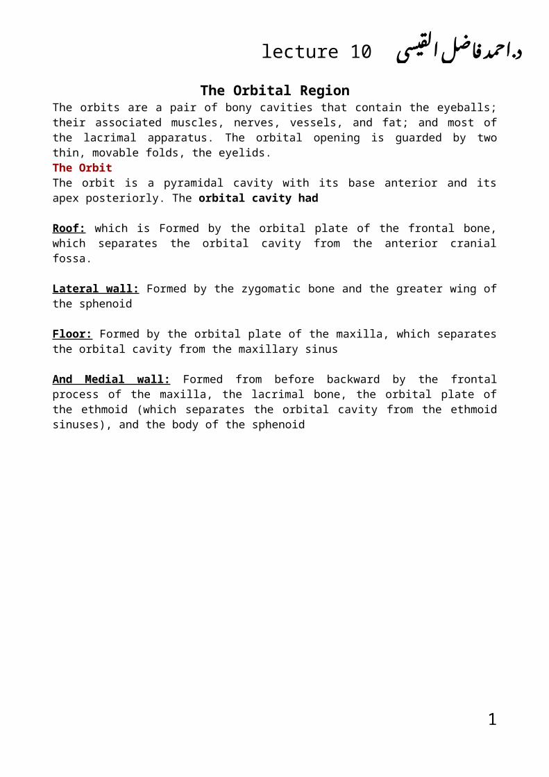
The Orbital Region The orbits are a pair of bony cavities that contain the eyeballs; their associated muscles, nerves, vessels, and fat; and most of the lacrimal apparatus. The orbital opening is guarded by two thin, movable folds, the eyelids. The Orbit The orbit is a pyramidal cavity with its base anterior and its apex posteriorly. The orbital cavity had Roof: which is Formed by the orbital plate of the frontal bone, which separates the orbital cavity from the anterior cranial fossa. Lateral wall: Formed by the zygomatic bone and the greater wing of the sphenoid Floor: Formed by the orbital plate of the maxilla, which separates the orbital cavity from the maxillary sinus And Medial wall: Formed from before backward by the frontal process of the maxilla, the lacrimal bone, the orbital plate of the ethmoid (which separates the orbital cavity from the ethmoid sinuses), and the body of the sphenoid 1 ي س ي ق ل ل ا ض ا مد ف ح د.اlecture 10
Transcript of · Web viewLacrimal Apparatus Lacrimal Gland The lacrimal gland consists of a large orbital part...
The Orbital RegionThe orbits are a pair of bony cavities that contain the eyeballs; their associated muscles, nerves, vessels, and fat; and most of the lacrimal apparatus. The orbital opening is guarded by two thin, movable folds, the eyelids.The OrbitThe orbit is a pyramidal cavity with its base anterior and its apex posteriorly. The orbital cavity had
Roof: which is Formed by the orbital plate of the frontal bone, which separates the orbital cavity from the anterior cranial fossa.
Lateral wall: Formed by the zygomatic bone and the greater wing of the sphenoid
Floor: Formed by the orbital plate of the maxilla, which separates the orbital cavity from the maxillary sinus
And Medial wall: Formed from before backward by the frontal process of the maxilla, the lacrimal bone, the orbital plate of the ethmoid (which separates the orbital cavity from the ethmoid sinuses), and the body of the sphenoid
1
القيسي. فاضل احمد lecture 10 دAnatomy
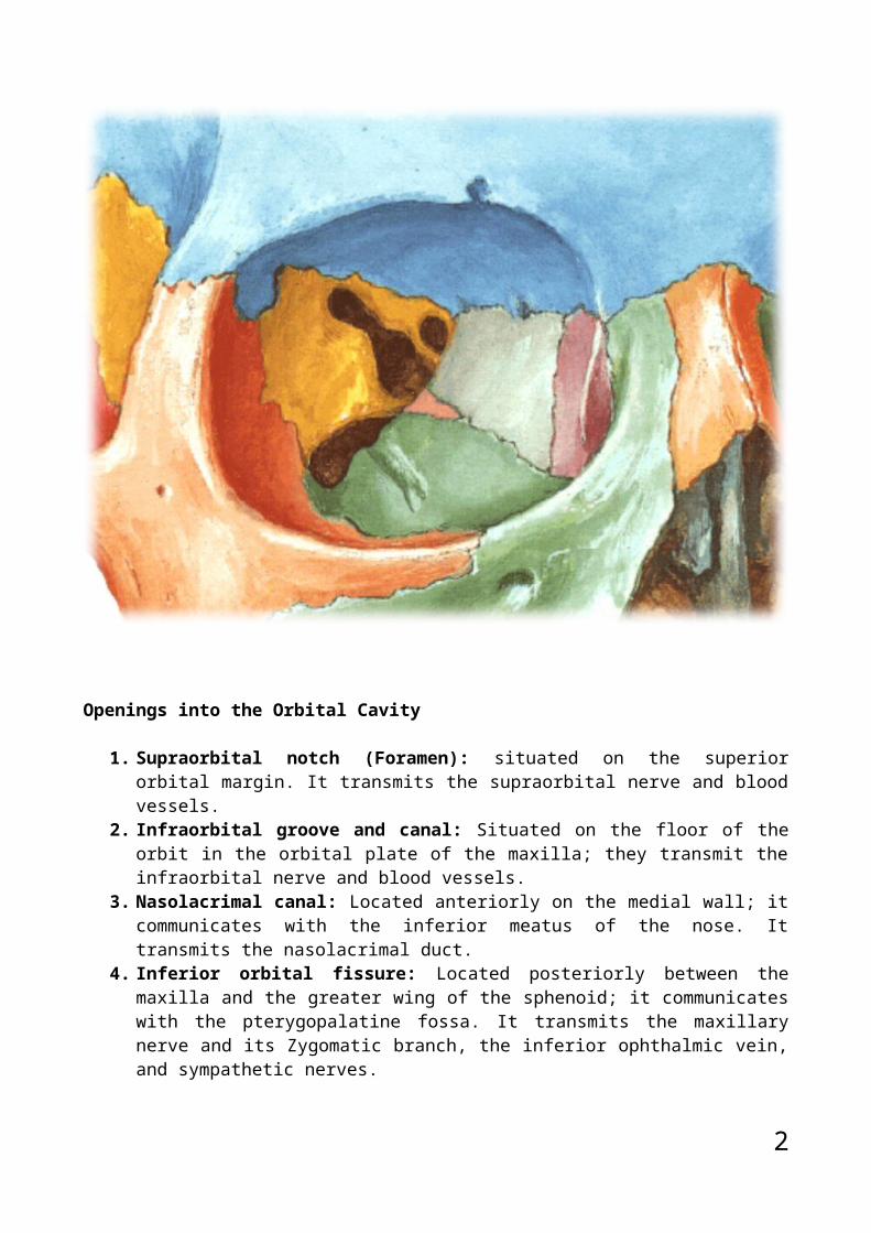
Openings into the Orbital Cavity
1. Supraorbital notch (Foramen): situated on the superior orbital margin. It transmits the supraorbital nerve and blood vessels.
2. Infraorbital groove and canal: Situated on the floor of the orbit in the orbital plate of the maxilla; they transmit the infraorbital nerve and blood vessels.
3. Nasolacrimal canal: Located anteriorly on the medial wall; it communicates with the inferior meatus of the nose. It transmits the nasolacrimal duct.
4. Inferior orbital fissure: Located posteriorly between the maxilla and the greater wing of the sphenoid; it communicates with the pterygopalatine fossa. It transmits the maxillary nerve and its Zygomatic branch, the inferior ophthalmic vein, and sympathetic nerves.
5. Superior orbital fissure: Located posteriorly between the greater and lesser wings of the sphenoid; it communicates with the middle cranial fossa. It transmits the lacrimal nerve, the frontal nerve, the trochlear nerve, the oculomotor nerve (upper and lower divisions), the abducent nerve, the nasociliary nerve, and the superior ophthalmic vein.
6. Optic canal: Located posteriorly in the lesser wing of the sphenoid; it communicates with the middle cranial fossa. It transmits the optic nerve and the ophthalmic artery.
Orbital FasciaThe orbital fascia is the periosteum of the bones that form the walls of the orbit. It is loosely attached to the bones and is continuous through the foramina and fissures with the periosteum covering the outer surfaces of the bones. The muscle of Müller, or orbitalis muscle, is a thin layer of smooth muscle that bridges the inferior orbital fissure. It is supplied by sympathetic nerves, and its function is unknown.
EyelidsThe eyelids protect the eye from injury and excessive light by their closure. The upper eyelid is larger and more mobile than the lower, and they meet each other at the medial and lateral angles. The palpebral fissure is the elliptical opening between the eyelids and is the entrance into the conjunctival sac.
2
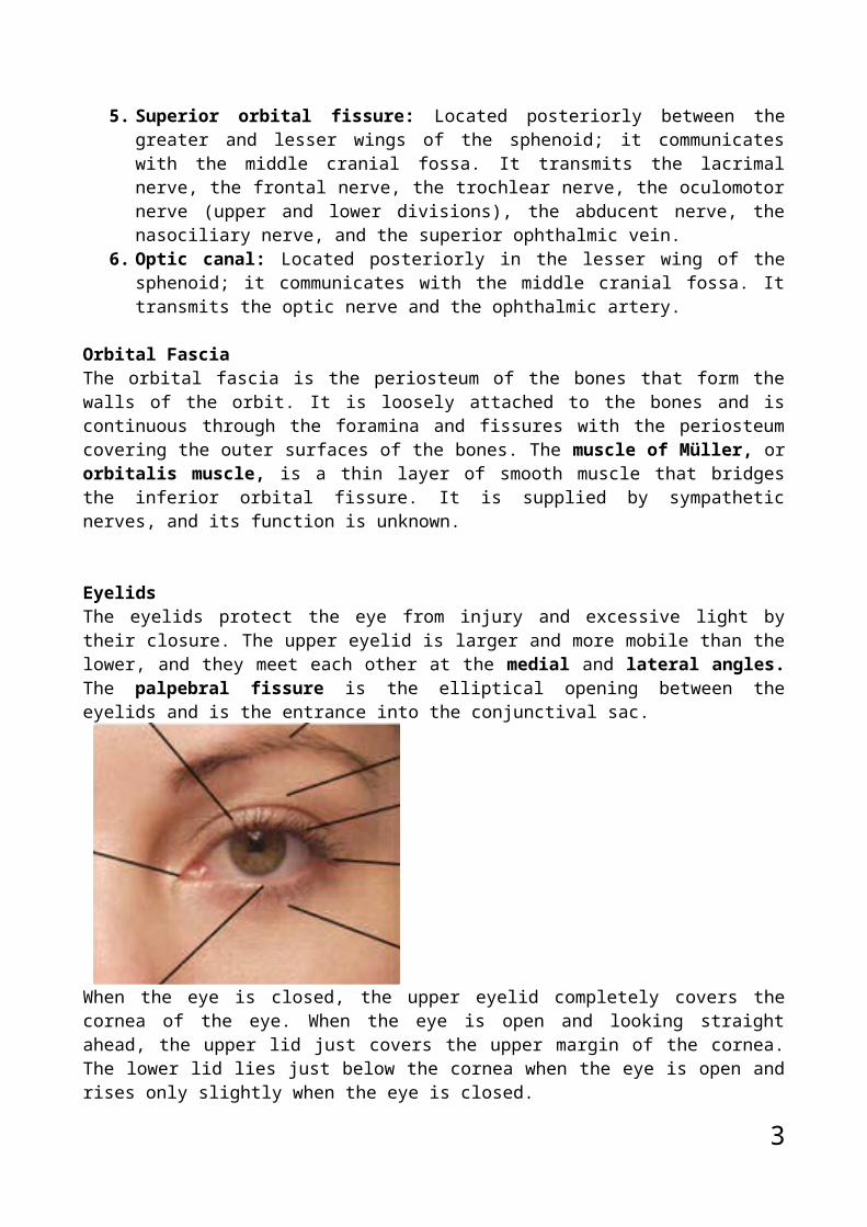
When the eye is closed, the upper eyelid completely covers the cornea of the eye. When the eye is open and looking straight ahead, the upper lid just covers the upper margin of the cornea. The lower lid lies just below the cornea when the eye is open and rises only slightly when the eye is closed.
The superficial surface of the eyelids is covered by skin, and the deep surface is covered by a mucous membrane called the conjunctiva. The eyelashes are short, curved hairs on the free edges of the eyelids. They are arranged in double or triple rows at the mucocutaneous junction. The sebaceous glands (glands of Zeis) open directly into the eyelash follicles. The ciliary glands (glands of Moll) are modified sweat glands that open separately between adjacent lashes. The tarsal glands are long, modified sebaceous glands that pour their oily secretion onto the margin of the lid; their openings lie behind the eyelashes. This oily material prevents the overflow of tears and helps make the closed eyelids airtight.
The conjunctiva is a thin mucous membrane that lines the eyelids and is reflected at the superior and inferior fornices onto the anterior surface of the eyeball. The upper lateral part of the superior fornix is pierced by the ducts of the lacrimal gland. The framework of the eyelids is formed by a fibrous sheet, the orbital septum. This is attached to the periosteum at the orbital margins. The orbital septum is thickened at the margins of the lids to form the superior and inferior tarsal plates. The lateral ends of the plates are attached by a band, the lateral palpebral ligament, to a bony tubercle just within the
3
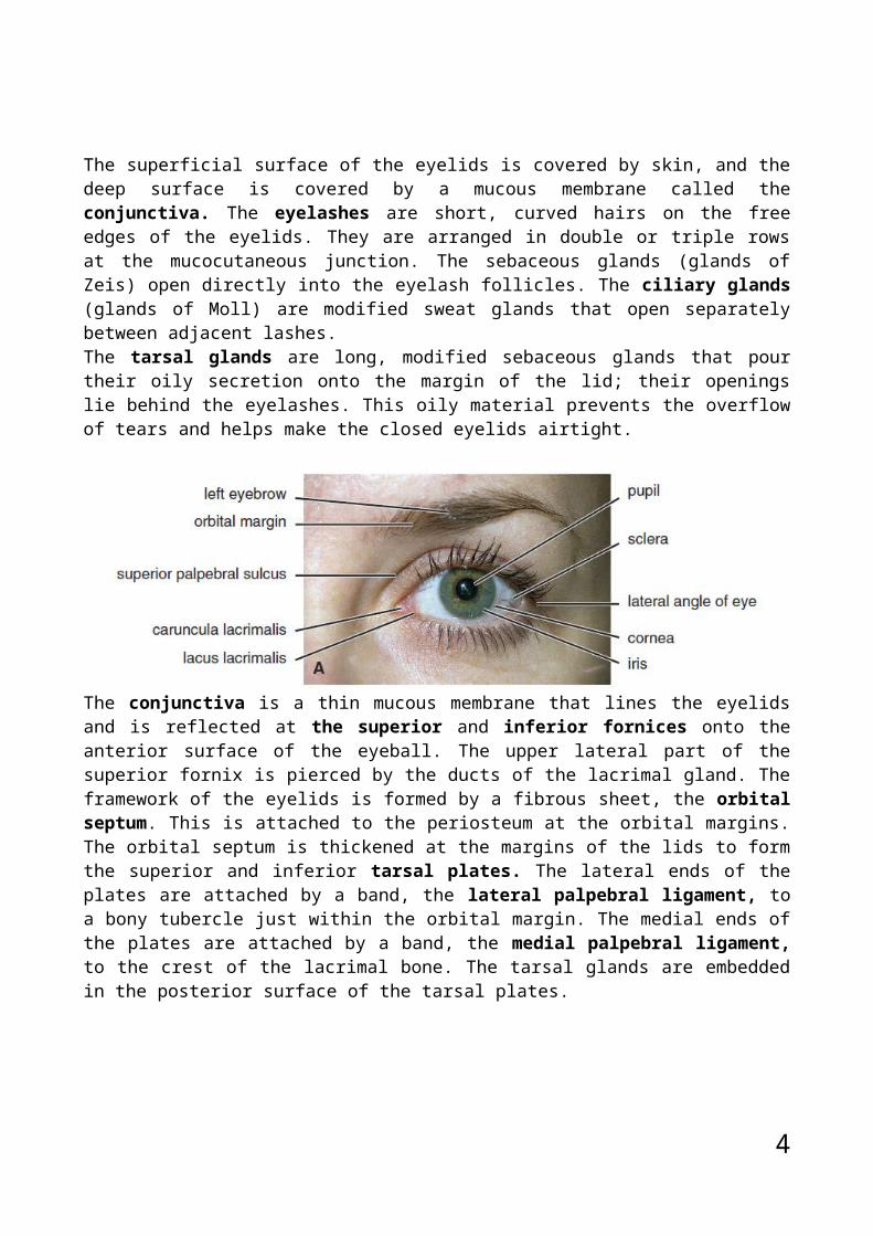
orbital margin. The medial ends of the plates are attached by a band, the medial palpebral ligament, to the crest of the lacrimal bone. The tarsal glands are embedded in the posterior surface of the tarsal plates.
4
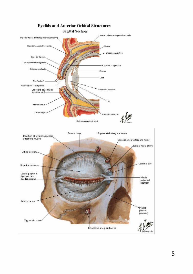
Movements of the EyelidsThe position of the eyelids at rest depends on the tone of the orbicularis oculi and the levator palpebrae superioris muscles and the position of the eyeball. The eyelids are closed by the contraction of the orbicularis oculi and the relaxation of the levator palpebrae superioris muscles. The eye is opened by the levator palpebrae superioris raising the upper lid. On looking upward, the levator palpebrae superioris contracts, and the upper lid moves with the eyeball.On looking downward, both lids move, the upper lid continues to cover the upper part of the cornea, and the lower lid is pulled downward slightly by the conjunctiva, which is attached to the sclera and the lower lid.
Lacrimal ApparatusLacrimal GlandThe lacrimal gland consists of a large orbital part and a small palpebral part, which are continuous with each other around the lateral edge of the aponeurosis of the levator palpebrae superioris. It is situated above the eyeball in the anterior and upper part of the orbit posterior to the orbital septum. The gland opens into the lateral part of the superior fornix of the conjunctiva by 12 ducts. The parasympathetic secretomotor nerve supply is derived from the lacrimal nucleus of the facial nerve. The preganglionic fibers reach the pterygopalatine ganglion (sphenopalatine ganglion) via the nervus intermedius and its great petrosal branch and via the nerve of the pterygoid
5
canal. The postganglionic fibers leave the ganglion and join the maxillary nerve. They then pass into its Zygomatic branch and the zygomaticotemporal nerve. They reach the lacrimal gland within the lacrimal nerve. The sympathetic postganglionic nerve supply is from the internal carotid plexus and travels in the deep petrosal nerve, the nerve of the pterygoid canal, the maxillary nerve,the zygomatic nerve, the zygomaticotemporal nerve, and finally the lacrimal nerve.
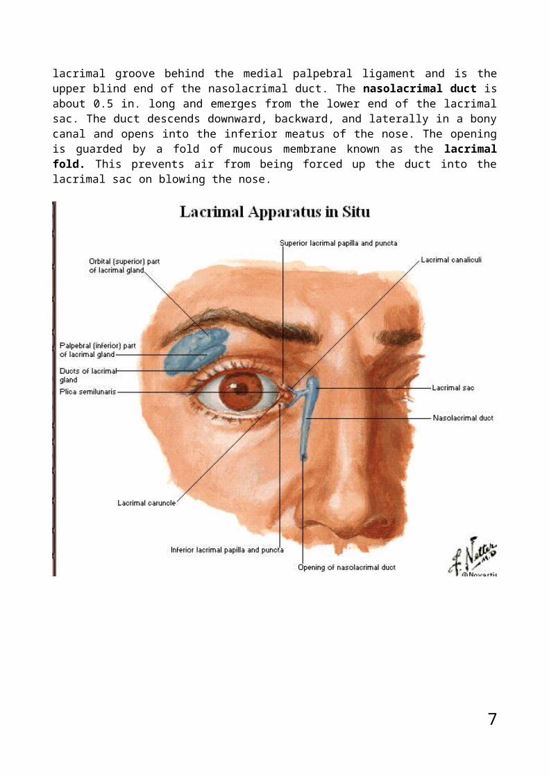
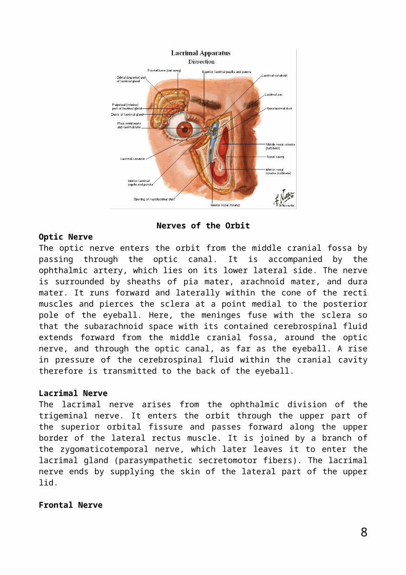
Lacrimal DuctsThe tears circulate across the cornea and accumulate in the lacus lacrimalis. From here, the tears enter the canaliculi lacrimales through the puncta lacrimalis. The canaliculi lacrimales pass medially and open into the lacrimal sac, which lies in the lacrimal groove behind the medial palpebral ligament and is the upper blind end of the nasolacrimal duct. The nasolacrimal duct is about 0.5 in. long and emerges from the lower end of the lacrimal sac. The duct descends downward, backward, and laterally in a bony canal and opens into the inferior meatus of the nose. The opening is guarded by a fold of mucous membrane known as the lacrimal fold. This prevents air from being forced up the duct into the lacrimal sac on blowing the nose.
6
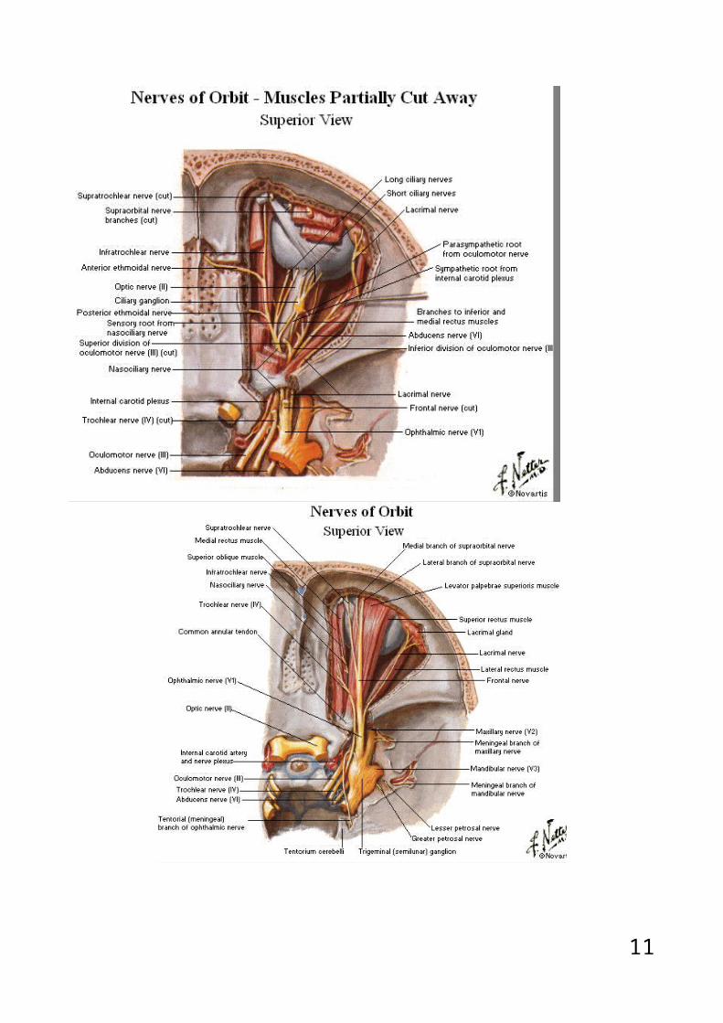
Nerves of the Orbit7
Optic NerveThe optic nerve enters the orbit from the middle cranial fossa by passing through the optic canal. It is accompanied by the ophthalmic artery, which lies on its lower lateral side. The nerve is surrounded by sheaths of pia mater, arachnoid mater, and dura mater. It runs forward and laterally within the cone of the recti muscles and pierces the sclera at a point medial to the posterior pole of the eyeball. Here, the meninges fuse with the sclera so that the subarachnoid space with its contained cerebrospinal fluid extends forward from the middle cranial fossa, around the optic nerve, and through the optic canal, as far as the eyeball. A rise in pressure of the cerebrospinal fluid within the cranial cavity therefore is transmitted to the back of the eyeball.
Lacrimal NerveThe lacrimal nerve arises from the ophthalmic division of the trigeminal nerve. It enters the orbit through the upper part of the superior orbital fissure and passes forward along the upper border of the lateral rectus muscle. It is joined by a branch of the zygomaticotemporal nerve, which later leaves it to enter the lacrimal gland (parasympathetic secretomotor fibers). The lacrimal nerve ends by supplying the skin of the lateral part of the upper lid.
Frontal NerveThe frontal nerve arises from the ophthalmic division of the trigeminal nerve. It enters the orbit through the upper part of the superior orbital fissure and passes forward on the upper surface of the levator palpebrae superioris beneath the roof of the orbit. It divides into the supratrochlear and supraorbital nerves that wind around the upper margin of the orbital cavity to supply the skin of the forehead; the supraorbital nerve also supplies the mucous membrane of the frontal air sinus.
Trochlear NerveThe trochlear nerve enters the orbit through the upper part of the superior orbital fissure. It runs forward and supplies the superior oblique muscle.
Oculomotor NerveThe superior ramus of the oculomotor nerve enters the orbit through the lower part of the superior orbital fissure. It supplies the superior rectus muscle, then pierces it, and supplies the levator palpebrae superioris muscle. The inferior ramus of the oculomotor nerve enters the orbit in a similar manner and supplies the inferior rectus, the medial rectus, and the inferior oblique muscles. The nerve to the inferior oblique gives off a branch that passes to the ciliary ganglion and carries parasympathetic fibers to the sphincter pupillae and the ciliary muscle.
Nasociliary NerveThe nasociliary nerve arises from the ophthalmic division of the trigeminal nerve. It enters the orbit through the lower part of the superior orbital fissure. It crosses above the optic nerve, runs forward along the upper margin of the
8
medial rectus muscle, and ends by dividing into the anterior ethmoidal and infratrochlear nerves.
Branches of the Nasociliary Nerve:
1. The communicating branch to the ciliary ganglion is a sensory nerve. The sensory fibers from the eyeball pass to the ciliary ganglion via the short ciliary nerves, pass through the ganglion without interruption, and then join the nasociliary nerve by means of the communicating branch.
2. The long ciliary nerves, two or three in number, arise from the nasociliary nerve as it crosses the optic nerve. They contain sympathetic fibers for the dilator pupillae muscle. The nerves pass forward with the short ciliary nerves and pierce the sclera of the eyeball. They continue forward between the sclera and the choroid to reach the iris.
3. The posterior ethmoidal nerve supplies the ethmoidal and sphenoidal air sinuses.
4. The infratrochlear nerve passes forward below the pulley of the superior oblique muscle and supplies the skin of the medial part of the upper eyelid and the adjacent part of the nose.
5. The anterior ethmoidal nerve passes through the anterior ethmoidal foramen and enters the anterior cranial fossa on the upper surface of the cribriform plate of the ethmoid. It enters the nasal cavity through a slitlike opening alongside the crista galli. After supplying an area of mucous membrane, it appears on the face as the external nasal branch at the lower border of the nasal bone, and supplies the skin of the nose down as far as the tip.
Abducent NerveThe abducent nerve enters the orbit through the lower part of the superior orbital fissure. It supplies the lateral rectus muscle.
Ciliary GanglionThe ciliary ganglion is a parasympathetic ganglion about the size of a pinhead and situated in the posterior part of the orbit. It receives its preganglionic parasympathetic fibers from the oculomotor nerve via the nerve to the inferior oblique. The postganglionic fibers leave the ganglion in the short ciliary nerves, which enter the back of the eyeball and supply the sphincter pupillae and the ciliary muscle. A number of sympathetic fibers pass from the internal carotid plexus into the orbit and run through the ganglion without interruption.
9
10
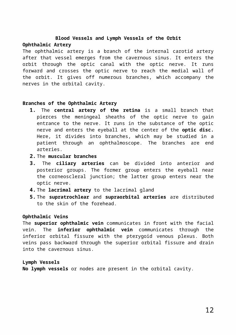
Blood Vessels and Lymph Vessels of the OrbitOphthalmic ArteryThe ophthalmic artery is a branch of the internal carotid artery after that vessel emerges from the cavernous sinus. It enters the orbit through the optic canal with the optic nerve. It runs forward and crosses the optic nerve to reach the medial wall of the orbit. It gives off numerous branches, which accompany the nerves in the orbital cavity.
Branches of the Ophthalmic Artery1. The central artery of the retina is a small branch that pierces the
meningeal sheaths of the optic nerve to gain entrance to the nerve. It runs in the substance of the optic nerve and enters the eyeball at the center of the optic disc. Here, it divides into branches, which may be studied in a patient through an ophthalmoscope. The branches are end arteries.
2. The muscular branches3. The ciliary arteries can be divided into anterior and posterior groups.
The former group enters the eyeball near the corneoscleral junction; the latter group enters near the optic nerve.
4. The lacrimal artery to the lacrimal gland 5. The supratrochlear and supraorbital arteries are distributed to the
skin of the forehead.
Ophthalmic VeinsThe superior ophthalmic vein communicates in front with the facial vein. The inferior ophthalmic vein communicates through the inferior orbital fissure with the pterygoid venous plexus. Both veins pass backward through the superior orbital fissure and drain into the cavernous sinus.
Lymph VesselsNo lymph vessels or nodes are present in the orbital cavity.
11
Fascial Sheath of the EyeballThe fascial sheath surrounds the eyeball from the optic nerve to the corneoscleral junction. It separates the eyeball from the orbital fat and provides it with a socket for free movement. It is perforated by the tendons of the orbital muscles and is reflected onto each of them as a tubular sheath. The sheaths for the tendons of the medial and lateral recti are attached to the
12
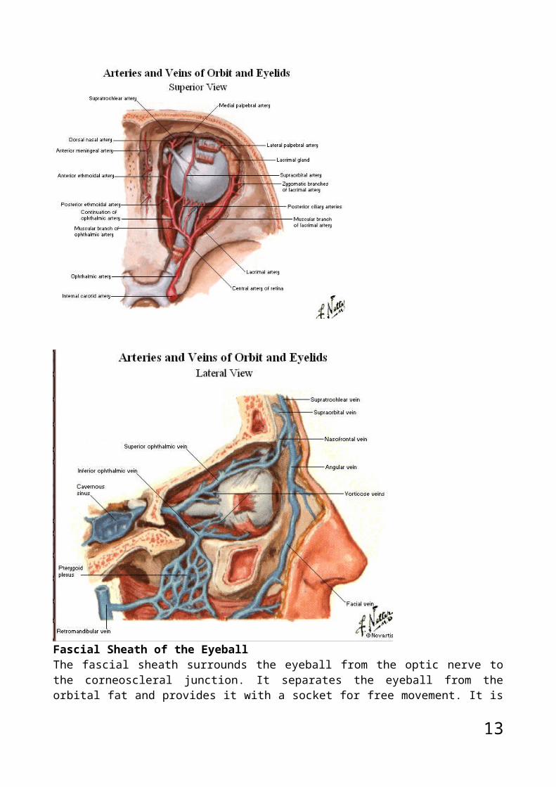
medial and lateral walls of the orbit by triangular ligaments called the medial and lateral check ligaments. The lower part of the fascial sheath, which passes beneath the eyeball and connects the check ligaments, is thickened and serves to suspend the eyeball; it is called the suspensory ligament of the eye . By this means, the eye is suspended from the medial and lateral walls of the orbit, as if in a hammock.
Structure of the EyeThe eyeball is embedded in orbital fat but is separated from it by the fascial sheath of the eyeball. The eyeball consists of three coats, which, from outside to inside, are the fibrous coat, the vascular pigmented coat, and the nervous coat.
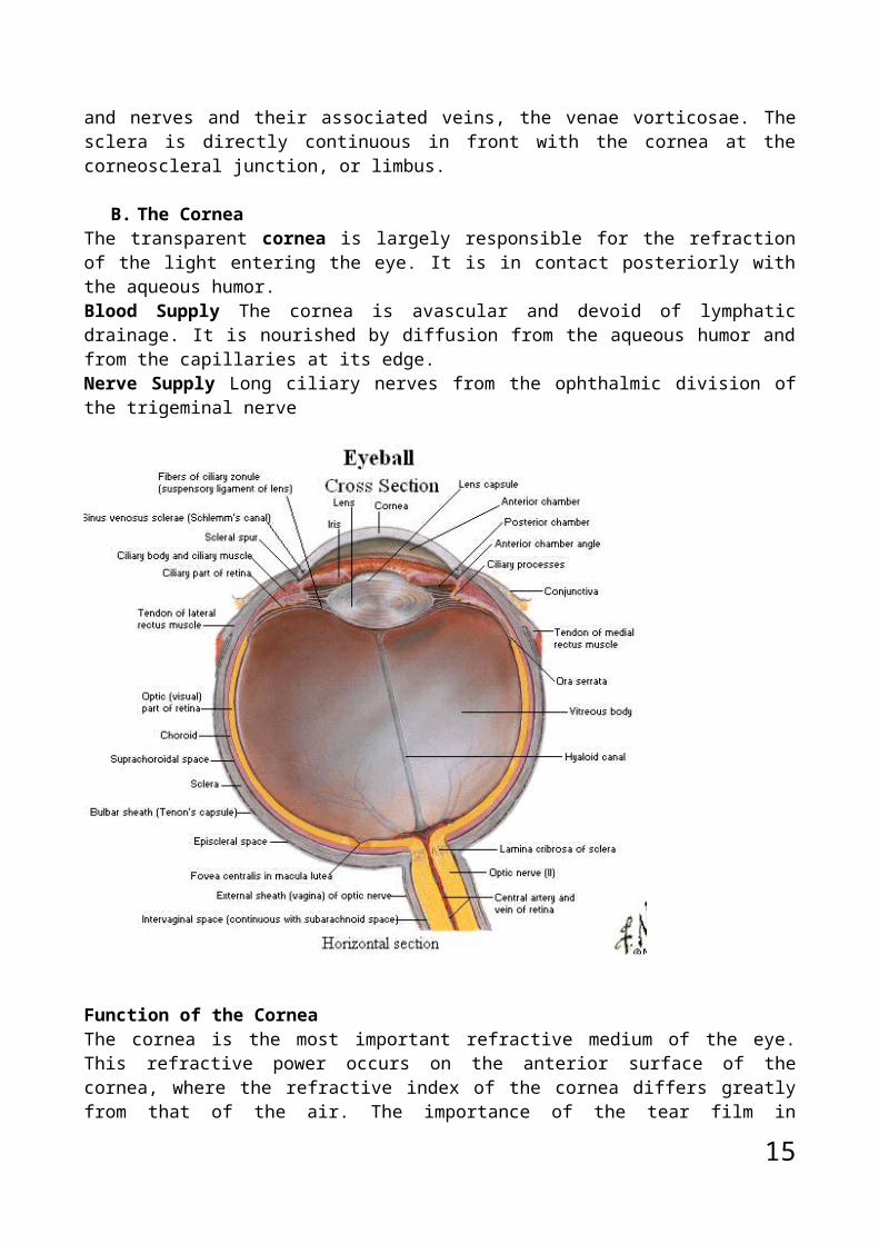
Coats of the EyeballFibrous CoatThe fibrous coat is made up of a posterior opaque part, the sclera, and an anterior transparent part, the cornea.
A. The ScleraThe opaque sclera is composed of dense fibrous tissue and is white. Posteriorly, it is pierced by the optic nerve and is fused with the dural sheath of that nerve. The lamina cribrosa is the area of the sclera that is pierced by the nerve fibers of the optic nerve. The sclera is also pierced by the ciliary arteries and nerves and their associated veins, the venae vorticosae. The sclera is directly continuous in front with the cornea at the corneoscleral junction, or limbus.
B. The CorneaThe transparent cornea is largely responsible for the refraction of the light entering the eye. It is in contact posteriorly with the aqueous humor.Blood Supply The cornea is avascular and devoid of lymphatic drainage. It is nourished by diffusion from the aqueous humor and from the capillaries at its edge.
13
Nerve Supply Long ciliary nerves from the ophthalmic division of the trigeminal nerve
Function of the CorneaThe cornea is the most important refractive medium of the eye. This refractive power occurs on the anterior surface of the cornea, where the refractive index of the cornea differs greatly from that of the air. The importance of the tear film in maintaining the normal environment for the corneal epithelial cells should be stressed.
Vascular Pigmented CoatThe vascular pigmented coat consists, from behind forward, of the choroid, the ciliary body, and the iris.
A. The ChoroidThe choroid is composed of an outer pigmented layer and an inner, highly vascular layer.
B. The Ciliary BodyThe ciliary body is continuous posteriorly with the choroid, and anteriorly it lies behind the peripheral margin of the iris. It is composed of the ciliary ring, the ciliary processes, and the ciliary muscle.The ciliary ring is the posterior part of the body, and its surface has shallow grooves, the ciliary striae. The
14
ciliary processes are radially arranged folds, or ridges, to the posterior surfaces of which are connected the suspensory ligaments of the lens.The ciliary muscle is composed of meridianal and circular fibers of smooth muscle. The meridianal fibers run backward from the region of the corneoscleral junction to the ciliary processes. The circular fibers are fewer in number and lie internal to the meridianal fibers.
Nerve supply: The ciliary muscle is supplied by the parasympathetic fibers from the oculomotor nerve. After synapsing in the ciliary ganglion, the postganglionic fibers pass forward to the eyeball in the short ciliary nerves.Action: Contraction of the ciliary muscle pulls the ciliary body forward. This relieves the tension in the suspensory ligament, and the elastic lens becomes more convex. This increases the refractive power of the lens.
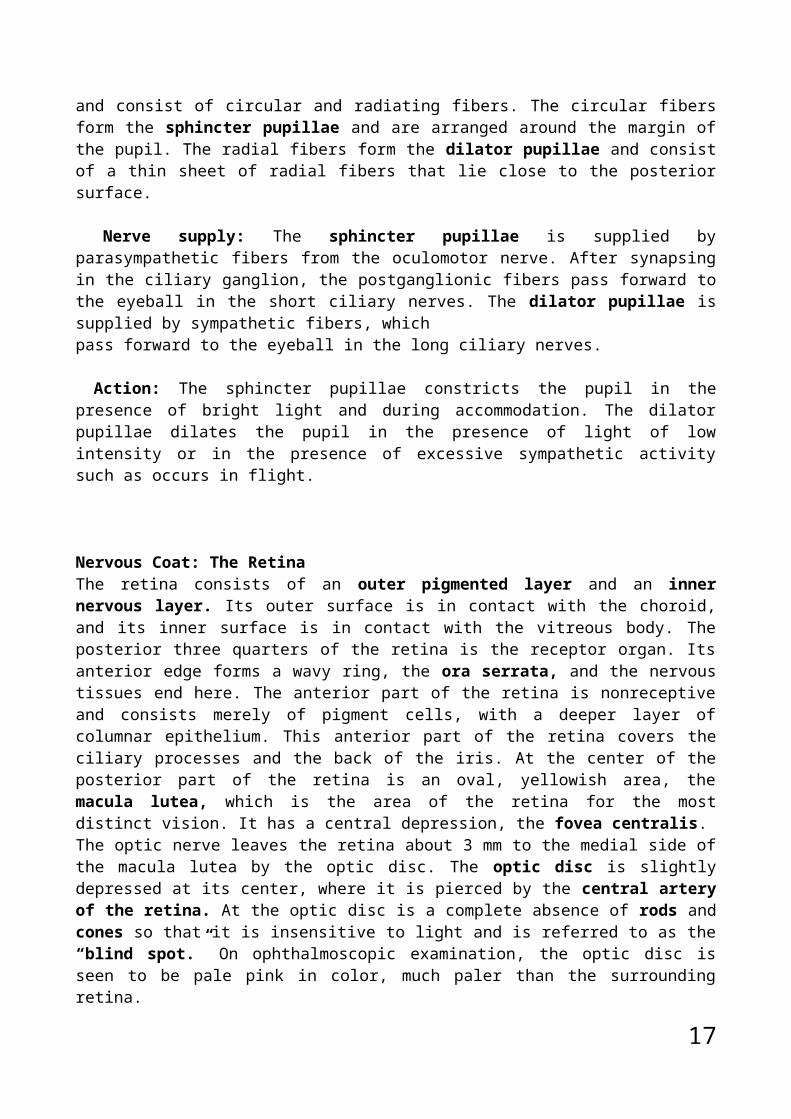
C. The Iris and PupilThe iris is a thin, contractile, pigmented diaphragm with a central aperture, the pupil. It is suspended in the aqueous humor between the cornea and the lens. The periphery of the iris is attached to the anterior surface of the ciliary body. It divides the space between the lens and the cornea into an anterior and a posterior chamber. The muscle fibers of the iris are involuntary and consist of circular and radiating fibers. The circular fibers form the sphincter pupillae and are arranged around the margin of the pupil. The radial fibers form the dilator pupillae and consist of a thin sheet of radial fibers that lie close to the posterior surface.
Nerve supply: The sphincter pupillae is supplied by parasympathetic fibers from the oculomotor nerve. After synapsing in the ciliary ganglion, the postganglionic fibers pass forward to the eyeball in the short ciliary nerves. The dilator pupillae is supplied by sympathetic fibers, whichpass forward to the eyeball in the long ciliary nerves.
Action: The sphincter pupillae constricts the pupil in the presence of bright light and during accommodation. The dilator pupillae dilates the pupil in the presence of light of low intensity or in the presence of excessive sympathetic activity such as occurs in flight.
Nervous Coat: The RetinaThe retina consists of an outer pigmented layer and an inner nervous layer. Its outer surface is in contact with the choroid, and its inner surface is in contact with the vitreous body. The posterior three quarters of the retina is the receptor organ. Its anterior edge forms a wavy ring, the ora serrata, and the nervous tissues end here. The anterior part of the retina is nonreceptive and consists merely of pigment cells, with a deeper layer of columnar epithelium. This anterior part of the retina covers the ciliary processes and the back of the iris. At the center of the posterior part of the retina is an oval, yellowish area, the macula lutea, which is the area of the retina for the most distinct vision. It has a central depression, the fovea centralis.
15

The optic nerve leaves the retina about 3 mm to the medial side of the macula lutea by the optic disc. The optic disc is slightly depressed at its center, where it is pierced by the central artery of the retina. At the optic disc is a complete absence of rods and cones so that it is insensitive to light and is referred to as the “blind spot.” On ophthalmoscopic examination, the optic disc is seen to be pale pink in color, much paler than the surrounding retina.
Contents of the EyeballThe contents of the eyeball consist of the refractive media, the aqueous humor, the vitreous body, and the lens.
Aqueous HumorThe aqueous humor is a clear fluid that fills the anterior and posterior chambers of the eyeball. It is believed to be a secretion from the ciliary processes, from which it enters the posterior chamber. It then flows into the anterior chamber through the pupil and is drained away through the spaces at the iridocorneal angle into the canal of Schlemm. Obstruction to the draining of the aqueous humor results in a rise in intraocular pressure called glaucoma. This can produce degenerative changes in the retina, with consequent blindness. The function of the aqueous humor is to support the wall of the eyeball by exerting internal pressure and thus maintaining its optical shape. It also nourishes the cornea and the lens and removes the products of
16
metabolism; these functions are important because the cornea and the lens do not possess a blood supply.
Vitreous BodyThe vitreous body fills the eyeball behind the lens and is a transparent gel. The hyaloid canal is a narrow channel that runs through the vitreous body from the optic disc to the posterior surface of the lens. The function of the vitreous body is to contribute slightly to the magnifying power of the eye. It supports the posterior surface of the lens and assists in holding the neural part of the retina against the pigmented part of the retina.
The LensThe lens is a transparent, biconvex structure enclosed in a transparent capsule. It is situated behind the iris and in front of the vitreous body and is encircled by the ciliary processes. The lens consists of an elastic capsule, and lens fibers. The lens fibers make up the bulk of the lens. The elastic lens capsule is under tension, causing the lens constantly to endeavor to assume a globular rather than a disc shape. The circumference of the lens is attached to the ciliary processes of the ciliary body by the suspensory ligament. The pull of the radiating fibers of the suspensory ligament tends to keep the elastic lens flattened so that the eye can be focused on distant objects.
17

18


![[PPT]PowerPoint Presentation - DeannaRussler - Home · Web viewLacrimal apparatus Consists of lacrimal gland and several ducts Ducts drain lacrimal secretions into nasal cavity Gland](https://static.fdocuments.net/doc/165x107/5ae7f9f47f8b9acc268f6a96/pptpowerpoint-presentation-deannarussler-home-viewlacrimal-apparatus-consists.jpg)