mrlscience.weebly.commrlscience.weebly.com/.../16631190/bio...16_14-15.docx · Web view-Finish...
26
Name: ___________________________Period: _______ Biology Week #16 Week of: December 1 st – 5 th Day Root Words In-Class Homework 12/1: Monday Word: Definition: As in: - Picture: -Goals -Cell Pink Notes -Grade Graph -Notes on Microscopes 12/2: Tuesday Word: Definition: As in: - Picture: - Microscope Lab 12/3: Wednesda y Word: Definition: As in: - Picture: - Cell Notes/Analogy - Finish Microscope Lab 12/4: Thursday Word: Definition: As in: - Picture: -Cell Notes/Analogy -Plant vs. Animal Microscope Lab 12/5: Friday Word: Definition: As in: - Picture: - Quiz over Microscopes/Cells -Finish Plant vs. Animal Microscope Lab Need Help? Talk to me in class. I’m available during periods 4, 5 and 8. You can call me at 708-434-3616 or email [email protected] Also, Mr. Hill is in the Tutoring Center before school on Tuesdays and Thursdays. Be sure to use the website mrlscience.weebly.com 1
Transcript of mrlscience.weebly.commrlscience.weebly.com/.../16631190/bio...16_14-15.docx · Web view-Finish...

Name: ___________________________Period: _______
Biology Week #16Week of: December 1st – 5th
Day Root Words In-Class Homework
12/1: Monday
Word:Definition:
As in:-
Picture: -Goals-Cell Pink Notes-Grade Graph-Notes on Microscopes
12/2: Tuesday
Word:Definition:
As in:-
Picture:
- Microscope Lab
12/3: Wednesday
Word:Definition:
As in:-
Picture:
- Cell Notes/Analogy- Finish Microscope Lab
12/4: Thursday
Word:Definition:
As in:-
Picture:-Cell Notes/Analogy-Plant vs. Animal Microscope Lab
12/5: Friday
Word:Definition:
As in:-
Picture:- Quiz over Microscopes/Cells-Finish Plant vs. Animal Microscope Lab
Need Help? Talk to me in class. I’m available during periods 4, 5 and 8. You can call me at 708-434-3616 or email [email protected] Also, Mr. Hill is in the Tutoring Center before school on Tuesdays and Thursdays. Be sure to use the website
mrlscience.weebly.com
Microscope NotesProblem: What is a microscope? Why are microscopes important?
1

Name: ___________________________Period: ________________________________________________________________________________________
_________________________________________________________________________________
_________________________________
What is a compound light microscope?
What is a stereoscope?
What is a scanning electron and transmission electron microscope?
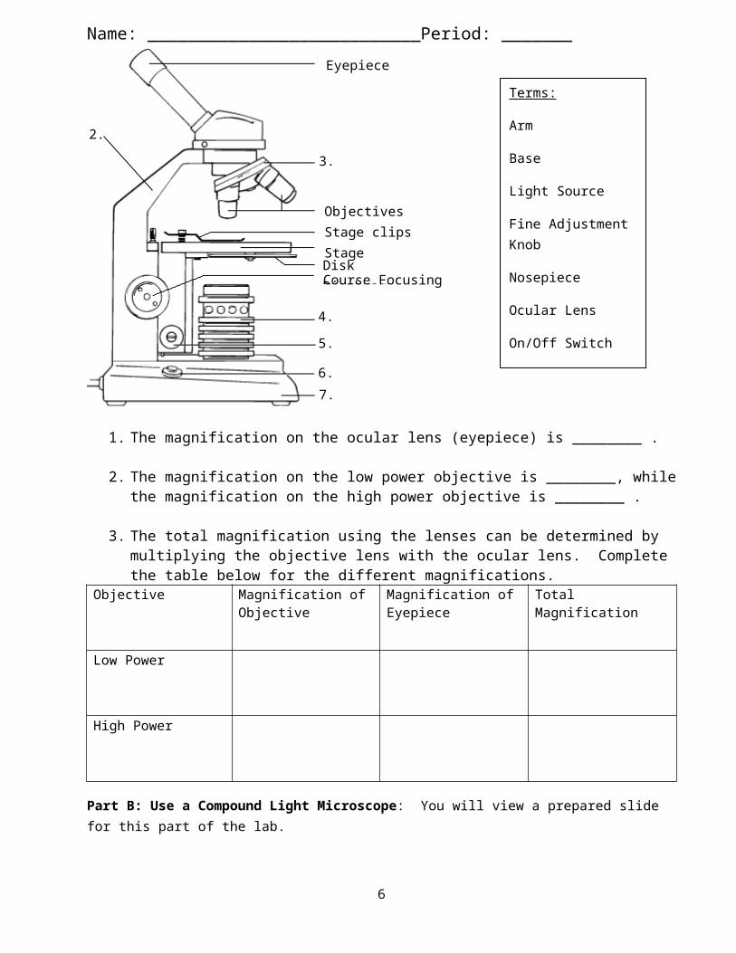
Parts of the Light Microscope
PART FUNCTIONArm Carry the scope with arm in one handBase Carry the scope with base in other handEyepiece (Ocular lens) (10X) To look at specimen (10X magnification)Revolving nosepiece Turn to get desired objective lensScanning Power Objective (4X)
Starting position, magnifies object 40X
2
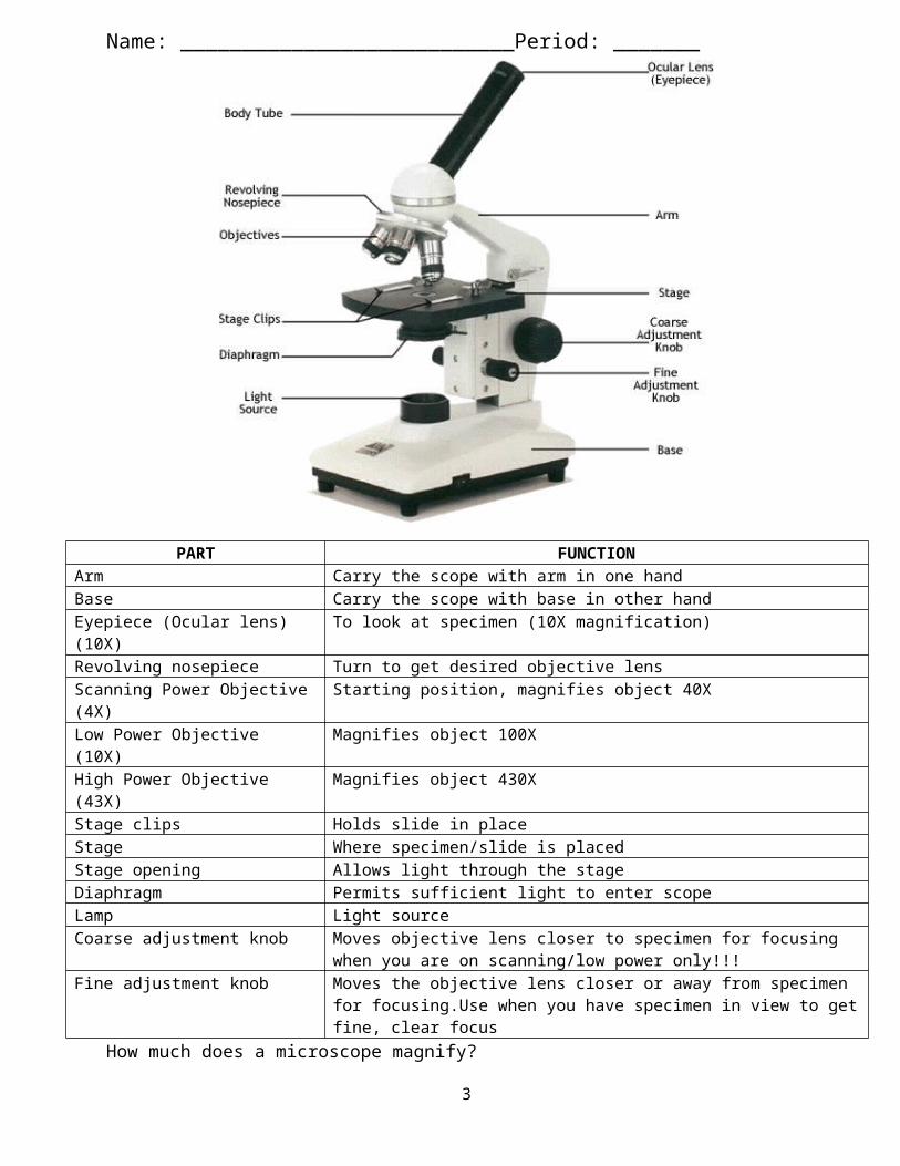
Name: ___________________________Period: _______Low Power Objective (10X) Magnifies object 100XHigh Power Objective (43X) Magnifies object 430XStage clips Holds slide in placeStage Where specimen/slide is placedStage opening Allows light through the stageDiaphragm Permits sufficient light to enter scopeLamp Light sourceCoarse adjustment knob Moves objective lens closer to specimen for focusing when you
are on scanning/low power only!!!Fine adjustment knob Moves the objective lens closer or away from specimen for
focusing.Use when you have specimen in view to get fine, clear focus
How much does a microscope magnify?
How do I prepare a microscope?
How do I focus a microscope?
What are the two types of slides?
How do I clean up a microscope?
Problem: What is a microscope? Why are microscopes important? ___________________________________________________________________________________________________________________________________________________________________________________________________
Introduction to the Light MicroscopeIntroduction:
Many objects are too small to be seen by the eye alone. They can be seen, however, with the use of an instrument that magnifies, or visually enlarges, the object. One such instrument, which is of great importance to biologists and other scientists, is the compound light microscope.
A compound light microscope consists of a light source or mirror that illuminates the object to be observed, an objective lens that magnifies the image of the object, and
3

Name: ___________________________Period: _______an eyepiece (ocular lens) that further magnifies the image of the object and projects it into the viewer’s eye.
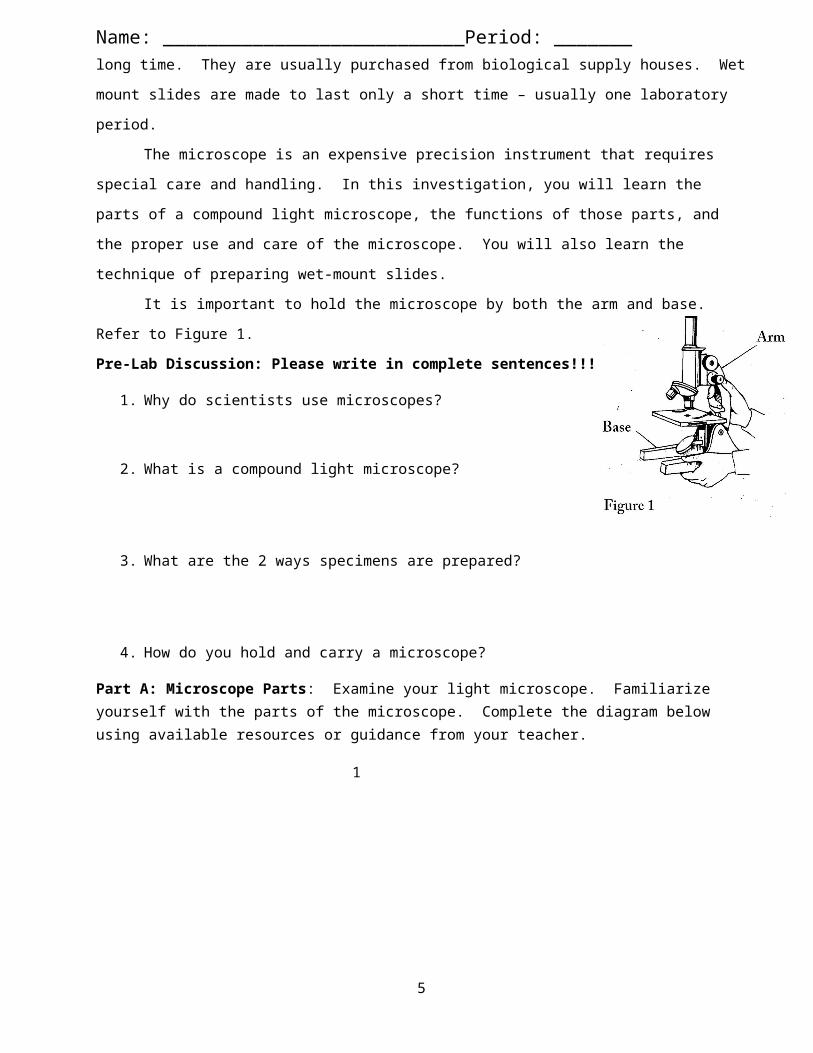
Objects, or specimens, to be observed under a microscope are generally prepared in one of two ways. Prepared slides are made to last a long time. They are usually purchased from biological supply houses. Wet mount slides are made to last only a short time – usually one laboratory period.
The microscope is an expensive precision instrument that requires special care and handling. In this investigation, you will learn the parts of a compound light microscope, the functions of those parts, and the proper use and care of the microscope. You will also learn the technique of preparing wet-mount slides.
It is important to hold the microscope by both the arm and base. Refer to Figure 1.Pre-Lab Discussion: Please write in complete sentences!!!
1. Why do scientists use microscopes?
2. What is a compound light microscope?
3. What are the 2 ways specimens are prepared?
4. How do you hold and carry a microscope?
Part A: Microscope Parts: Examine your light microscope. Familiarize yourself with the parts of the microscope. Complete the diagram below using available resources or guidance from your teacher.
4
1.
Name: ___________________________Period: _______
1. The magnification on the ocular lens (eyepiece) is ________ .
2. The magnification on the low power objective is ________, while the magnification on the high power objective is ________ .
3. The total magnification using the lenses can be determined by multiplying the objective lens with the ocular lens. Complete the table below for the different magnifications.
Objective Magnification of Objective
Magnification of Eyepiece
Total Magnification
Low Power
High Power
Part B: Use a Compound Light Microscope: You will view a prepared slide for this part of the lab.
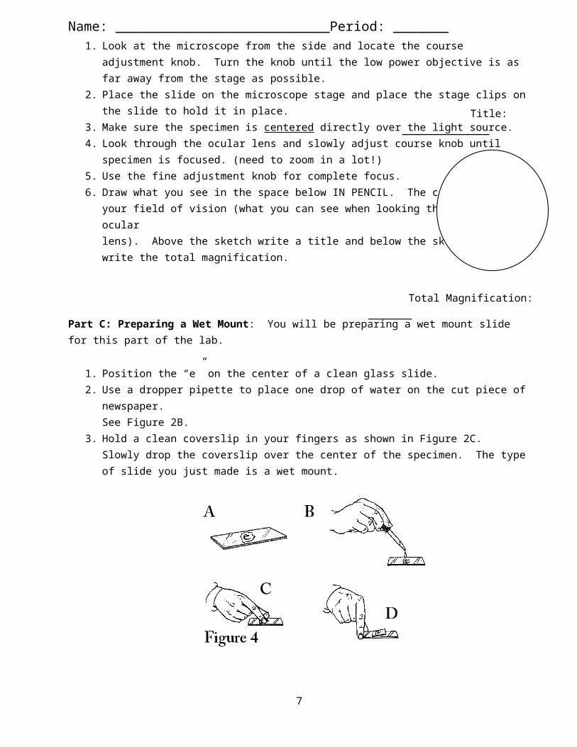
1. Look at the microscope from the side and locate the course adjustment knob. Turn the knob until the low power objective is as far away from the stage as possible.
5
Eyepiece
ObjectivesStage clipsStageDisk DiaphragmCourse Focusing Knob
2.
3.
4.
5.
6.
7.
Terms:
Arm
Base
Light Source
Fine Adjustment Knob
Nosepiece
Ocular Lens
On/Off Switch
Name: ___________________________Period: _______2. Place the slide on the microscope stage and place the stage clips on the slide to
hold it in place.3. Make sure the specimen is centered directly over the light source.4. Look through the ocular lens and slowly adjust course knob until
specimen is focused. (need to zoom in a lot!)5. Use the fine adjustment knob for complete focus.6. Draw what you see in the space below IN PENCIL. The circle is
your field of vision (what you can see when looking through the ocular lens). Above the sketch write a title and below the sketch write the total magnification.
Part C: Preparing a Wet Mount: You will be preparing a wet mount slide for this part of the lab.
1. Position the “e” on the center of a clean glass slide.2. Use a dropper pipette to place one drop of water on the cut piece of newspaper.
See Figure 2B.3. Hold a clean coverslip in your fingers as shown in Figure 2C. Slowly drop the
coverslip over the center of the specimen. The type of slide you just made is a wet mount.
4. Center the “e” on the stage over the light and place the stage clips over the sides of the slide.
5. Make sure the “e” is in its upright normal position (so you can read it normal).6. Start with the low power objective and focus the slide. Draw what you see under
low-power magnification IN PENCIL. Now focus the letter under the high power objective. Draw what you see under high-power magnification IN PENCIL. Again, title the sketch and calculate total magnification.
Title: ______________ Title: ______________
6
Title: ______________
Total Magnification: _______
Name: ___________________________Period: _______
Total Magnification: _________ Total Magnification: _________
7. While looking through the ocular lens, move the slide to the left. Notice the way the letter seems to move. Now move the slide to the right. Again, notice the way the letter seems to move. Move the slide up and down and observe the direction the letter moves. Describe what is happening.
Take apart the wet mount. Rinse the slide and coverslip with tap water. Carefully dry the slide and coverslip with a paper towel and place them back into their box.
Part D: Viewing Depth
1. View the prepared slide of the threads under the microscope. Remember to use proper procedure (slide on stage held by stage clips, start with lowest power using course focus, finally use fine adjustment).
2. What do you see? Can you focus all 3 threads at the same time? Why or why not? (Discuss with your teacher if you do not know the answer to this question) Draw a picture of what you see.
Part E: Stereoscope
7
A. What happens when you move the slide to the left? ____________________________________________________________
B. What happens when you move the slide up? ____________________________________________________________
C. Look at the slide with the naked eye (from the side of the microscope). Then look at it under the microscope. What happens to the letter? ____________________________________________________________
Title: ______________
Total Magnification: _______
________________________________________________________
________________________________________________________
________________________________________________________

Name: ___________________________Period: _______At the last station you will find a stereoscope or a dissecting microscope. Notice that while using the stereoscope you will be looking at the object through two ocular lenses instead of just one. In addition the magnification of the stereoscope is much lower than a compound light microscope. Look at the object under the stereoscope and draw your observations below.
Part F: Analysis and Conclusions: Answer the questions below in COMPLETE SENTENCES.
1. How is the image of an object seen through the high-power magnification different from the image seen through the low-power magnification?
2. How does the letter “e” as seen through the microscope differ from the way an “e” normally appears?
3. What is the difference between a prepared slide and a wet mount slide?
4. What level objective (high or low) do you start your viewing? Why?
8
Title: ______________
Total Magnification: _______

Name: ___________________________Period: _______5. Why might it be a good idea to keep your microscope at least 10 cm from the
edge of the table?
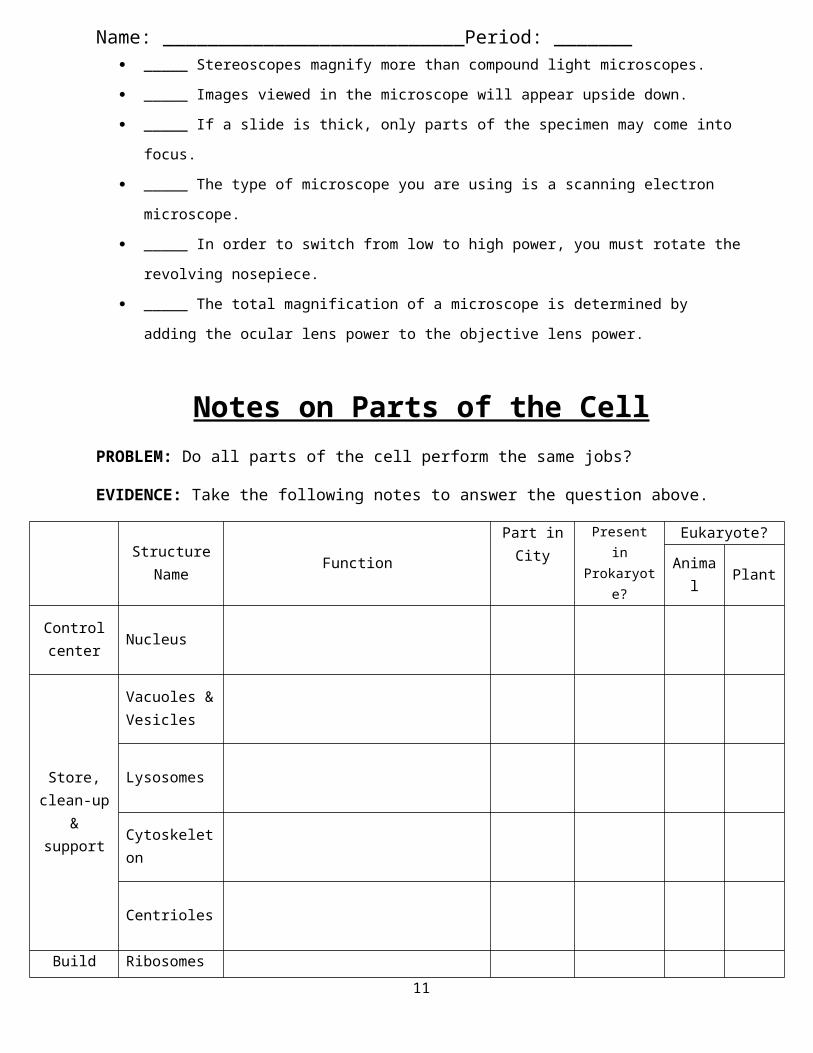
Part H: True or False - Answer true or false to each of the following statements.
_____ On high power, you should use the coarse adjustment knob. _____ The diaphragm determines how much light shines on the specimen. _____ Stereoscopes magnify more than compound light microscopes. _____ Images viewed in the microscope will appear upside down. _____ If a slide is thick, only parts of the specimen may come into focus. _____ The type of microscope you are using is a scanning electron microscope. _____ In order to switch from low to high power, you must rotate the revolving
nosepiece. _____ The total magnification of a microscope is determined by adding the ocular
lens power to the objective lens power.
Notes on Parts of the CellPROBLEM: Do all parts of the cell perform the same jobs?
EVIDENCE: Take the following notes to answer the question above.
Structure Name Function
Part in City
Present in Prokaryote
?
Eukaryote?Anim
al Plant
Control center Nucleus
Store, clean-up & support
Vacuoles & Vesicles
Lysosomes
Cytoskeleton
Centrioles
Build proteins
Ribosomes
9
Name: ___________________________Period: _______
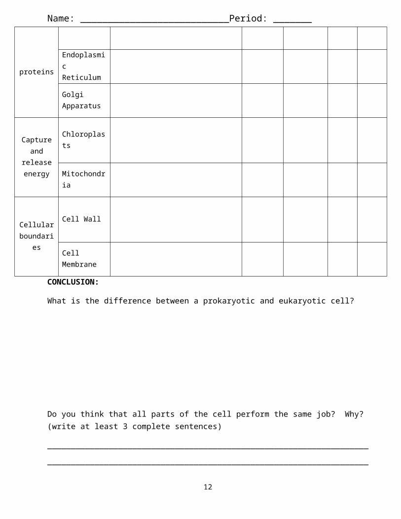
Endoplasmic Reticulum
Golgi Apparatus
Capture and
release energy
Chloroplasts
Mitochondria
Cellular boundarie
s
Cell Wall
Cell Membrane
CONCLUSION:
What is the difference between a prokaryotic and eukaryotic cell?
Do you think that all parts of the cell perform the same job? Why? (write at least 3 complete sentences)
____________________________________________________________________________________________________________________________________________________________________________________________________________________________________________________________________
What are some parts in the Doughnuts factory and their similar parts in the cell (name at least three parts)?
10
Robert Hooke’s sketch of cork
Name: ___________________________Period: ____________________________________________________________________________________________________________________________________________________________________________________________________________________________________________________________________________________________________________________________________________
Various Types of CellsPROBLEM: Are all cells the exact same?
EVIDENCE: Read, annotate, and answer the following questions.
The Discovery of Cells“Seeing is believing,” an old saying goes. It would be hard to find a better example of this than the discovery of the cell. Without the instruments to make them visible, cells remained out of sight and, therefore, out of mind for most of human history. All of this changed with a dramatic advance in technology—the microscope.
Early Microscopes In the late 1500s, eyeglass makers in Europe discovered that using several glass lenses in combination could magnify even the smallest objects to make them easy to see. Before long, they had built the first true microscopes from these lenses, opening the door to the study of biology as we know it today.
1. Microscopes use (one/several – circle one) lenses to magnify objects.
In 1665, Englishman Robert Hooke used an early microscope to look at a nonliving thin slice of cork, a plant material. Under the microscope, cork seemed to be made of thousands of tiny empty chambers. Hooke called these chambers “cells” because they reminded him of a monastery's tiny rooms, which were called cells. The term cell is used in biology to this day. Today we know that living cells are not empty chambers, that in fact they contain a huge array of working parts, each with its own function.
11
Name: ___________________________Period: _______2. Why did Robert Hooke choose the term “cell”?
________________________________________________________________________________________________________________________
In Holland around the same time, Anton van Leeuwenhoek used a single-lens microscope to observe pond water and other things. To his amazement, the microscope revealed a fantastic world of tiny living organisms that seemed to be everywhere, in the water he and his neighbors drank, and even in his own mouth. Leeuwenhoek's illustrations of the organisms he found in the human mouth—which today we call bacteria—are shown to the right.
3. What did van Leeuwenhoek see? ___________________________________
The Cell TheorySoon after van Leeuwenhoek, observations by scientists made it clear that cells are the basic units of life. Many discoveries, confirmed by biologists, are summarized in the cell theory, a fundamental concept of biology.
The cell theory states: • All living things are made up of cells. • Cells are structure & function in living things. • New cells are produced from existing cells.
4. List & sketch examples of the 3 parts of the cell theory.
Cell Theory Sketch
12
Name: ___________________________Period: _______Prokaryotic Vs. Eukaryotic Cells
Cells come in an amazing variety of shapes and sizes. Despite their differences, all cells, at some point in their lives, contain DNA, the molecule that carries biological information. In addition, all cells are surrounded by a thin flexible barrier called a cell membrane. There are other similarities as well, as you will learn in the next lesson.
1. What are two items ALL cells have in common? _____________________________________________________________________________________
Cells fall into two broad categories, depending on whether they contain a nucleus. The nucleus (plural: nuclei) is a large structure that contains genetic material in the form of DNA and controls many of the cell's activities. Eukaryotes (yoo KAR ee ohts) are cells that enclose their DNA in nuclei. Prokaryotes (pro KAR ee ohts) are cells that do not enclose DNA in nuclei.
2. Complete the table below regarding Eukaryotes and Prokaryotes by placing a check mark in the columns if the cells contain that structure.
DNA Membrane NucleusEukaryote
Prokaryote
Prokaryotes As seen in the figure below, prokaryotic cells are generally smaller and simpler than eukaryotic cells, although there are many exceptions to this rule. Prokaryotic cells do not separate their genetic material within a nucleus. Despite their simplicity, prokaryotes carry out every activity associated with living things. They grow, reproduce, respond to the environment, and, in some cases, glide along surfaces or swim through liquids. The organisms we call bacteria are prokaryotes.
13

Name: ___________________________Period: _______
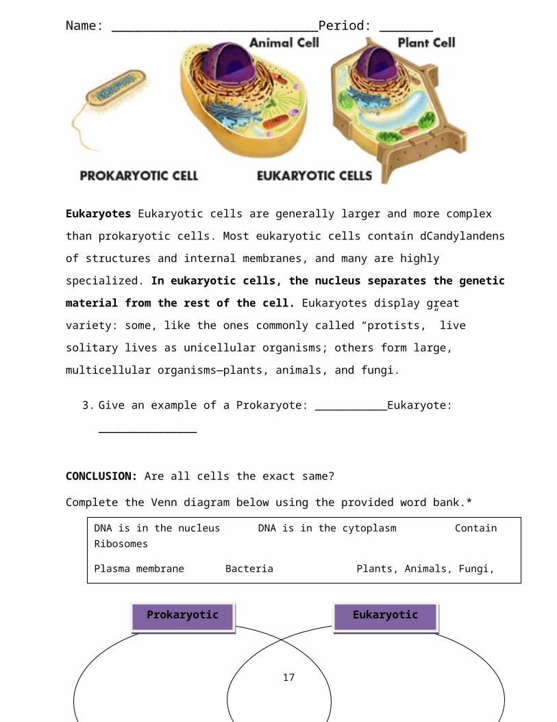
Eukaryotes Eukaryotic cells are generally larger and more complex than prokaryotic cells. Most eukaryotic cells contain dCandylandens of structures and internal membranes, and many are highly specialized. In eukaryotic cells, the nucleus separates the genetic material from the rest of the cell. Eukaryotes display great variety: some, like the ones commonly called “protists,” live solitary lives as unicellular organisms; others form large, multicellular organisms—plants, animals, and fungi.
3. Give an example of a Prokaryote: ___________Eukaryote: _______________
CONCLUSION: Are all cells the exact same?
Complete the Venn diagram below using the provided word bank.*
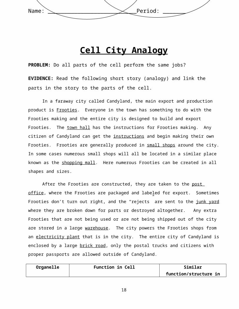
Cell City AnalogyPROBLEM: Do all parts of the cell perform the same jobs?
14
DNA is in the nucleus DNA is in the cytoplasm Contain Ribosomes
Plasma membrane Bacteria Plants, Animals, Fungi, Protists
Membrane bound organelles Living Genetic Material
Prokaryotic Eukaryotic

Name: ___________________________Period: _______EVIDENCE: Read the following short story (analogy) and link the parts in the story to the parts of the cell.
In a faraway city called Candyland, the main export and production product is Frooties. Everyone in the town has something to do with the Frooties making and the entire city is designed to build and export Frooties. The town hall has the instructions for Frooties making. Any citizen of Candyland can get the instructions and begin making their own Frooties. Frooties are generally produced in small shops around the city. In some cases numerous small shops will all be located in a similar place known as the shopping mall. Here numerous Frooties can be created in all shapes and sizes.
After the Frooties are constructed, they are taken to the post office, where the Frooties are packaged and labeled for export. Sometimes Frooties don’t turn out right, and the “rejects” are sent to the junk yard where they are broken down for parts or destroyed altogether. Any extra Frooties that are not being used or are not being shipped out of the city are stored in a large warehouse. The city powers the Frooties shops from an electricity plant that is in the city. The entire city of Candyland is enclosed by a large brick road, only the postal trucks and citizens with proper passports are allowed outside of Candyland.
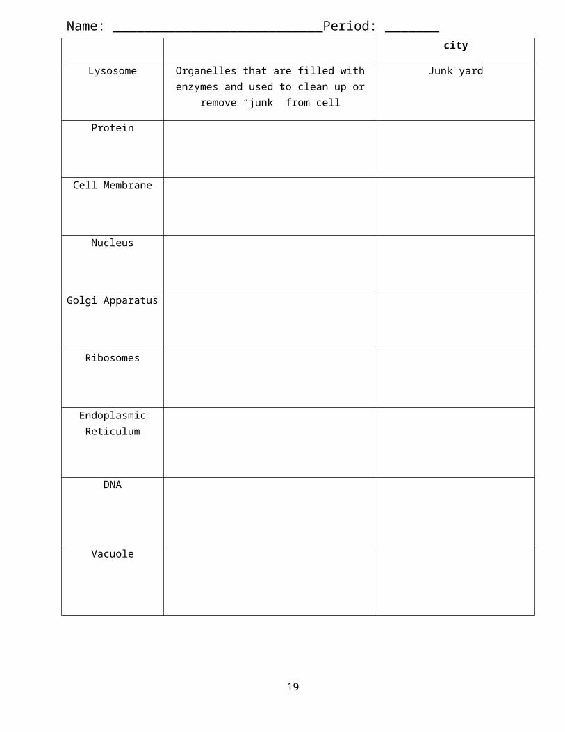
Organelle Function in Cell Similar function/structure in city
Lysosome Organelles that are filled with enzymes and used to clean up or remove “junk”
from cell
Junk yard
Protein
Cell Membrane
Nucleus
15

Name: ___________________________Period: _______Golgi Apparatus
Ribosomes
Endoplasmic Reticulum
DNA
Vacuole
Mitochondria / Chloroplast
CONCLUSION: Do you think that all parts of the cell perform the same job? Why? (write at least 3 complete sentences)
____________________________________________________________________________________________________________________________________________________________________________________________________________________________________________________________________
Plant Vs Animal Cell LabIntroduction: When different types of cells are viewed under a microscope, different cell parts can be seen. Some parts may be present in a cell but not observable if the cell is alive. Often, stain has to be added to a specimen to allow an organelle to be seen, but this will probably destroy the cell.Purpose: In this investigation, you will:
1. Observe a variety of living and once living materials under the microscope.
16
Name: ___________________________Period: _______2. Study and locate under the microscope five specific cell parts – cell wall, cell
membrane, cytoplasm, nucleus, and chloroplasts.3. Compare the cell parts found in plant and animal cells. Use your text to
determine if there are other differences between plants and animals not observed in lab.
Procedure:Part A: Onion Cells
Onion cells may be used to show a cell’s nucleus. This structure appears within most living cells. The nucleus will appear as a round structure inside each cell.
1. Prepare a wet mount slide.a. Peel some of the epidermis skin and place it flat on a clean microscope
slide. The epidermis is the thin layer of translucent cells in between the layers of the onion.
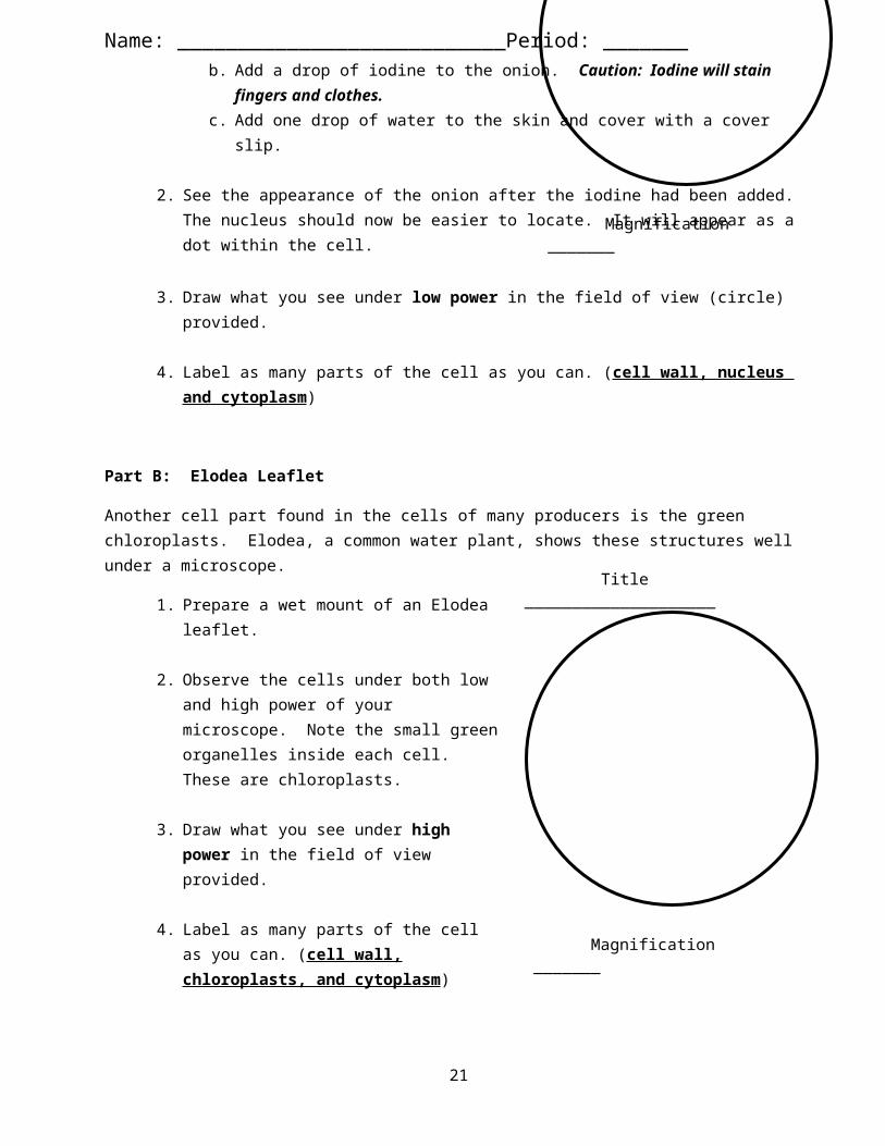
b. Add a drop of iodine to the onion. Caution: Iodine will stain fingers and clothes.
c. Add one drop of water to the skin and cover with a cover slip.
2. See the appearance of the onion after the iodine had been added. The nucleus should now be easier to locate. It will appear as a dot within the cell.
3. Draw what you see under low power in the field of view (circle) provided.
4. Label as many parts of the cell as you can. (cell wall, nucleus and cytoplasm)
Part B: Elodea Leaflet
Another cell part found in the cells of many producers is the green chloroplasts. Elodea, a common water plant, shows these structures well under a microscope.
1. Prepare a wet mount of an Elodea leaflet.
2. Observe the cells under both low and high power of your microscope. Note the small green organelles inside each cell. These are chloroplasts.
17
Magnification _______
Title ____________________
Title ____________________
Name: ___________________________Period: _______3. Draw what you see under high power in the field of view provided.
4. Label as many parts of the cell as you can. (cell wall, chloroplasts, and cytoplasm)
Part C: Human Skin Cells
Human skin cells may be used for viewing the cell membrane and cytoplasm.
1. Using a small (2cm by 2cm) strip of sticky tape place this on the underside of a well washed wrist.
2. Gently remove the sticky tape and place it sticky side up on the slide; stain with 2 or 3 drops methylene blue (this can STAIN your clothes, so be careful).
3. Place a cover slip over the sticky tape.4. Examine the slide under the microscope.5. Draw what you see under high power in the
field of view provided.6. Label as many parts of the cell as you can.
Remember this is an animal. (cell membrane and cytoplasm)
Part D: Blood
1. Observe and draw the prepared slides of blood under high power.
2. Label as many parts of the cell as you can. Remember this is an animal. (cell membrane, nucleus, and cytoplasm)
Analysis Questions: Use your notes and sketches/info from this lab to complete the following questions in complete sentences!
1. What was the difference between the onion epidermis cells with and without the iodine?
18
Magnification _______
Title ____________________
Magnification _______
Title ____________________
Name: ___________________________Period: _______
2. Describe the chloroplasts as you viewed them in the Elodea plants. Why didn’t you see chloroplasts in the skin cells?
3. What was one major difference you viewed between a plant and your skin cell?
4. Compare the size of the blood cells to your skin cell.
5. What is the purpose of the nucleus?
CONCLUSION: Are all cells the exact same? (Please answer the following questions in 5 sentences using 5 pieces of evidence that you SAW in the lab today)
__________________________________________________________________________________________________________________________________________________________________________________________________________________________________________________________________________________________________________________________________________________________________________________________________________________________________________________________________________________________________________________________________________________________________________________________________________________________________________________________________________
CONCLUSION: Are all cells the exact same?
Complete the Venn diagram below using the provided word bank.*
19
Nucleus Chloroplasts Cell membrane
Cytoplasm Cell Wall Living DNA
Plant Animal
Name: ___________________________Period: _______
20
Name: ___________________________Period: _______Cell Structures – Complete the Review Table Below based on your notes and what you just read… this is all on the quiz tomorrow!!!
Structure Name Function
Present in Prokaryote
?
Eukaryote?Anim
al Plant
Control center Nucleus
Store, clean-up & support
Vacuoles & Vesicles
Lysosomes
Cytoskeleton
Centrioles
Build proteins
Ribosomes
Endoplasmic Reticulum
Golgi Apparatus
Capture and
release energy
Chloroplasts
Mitochondria
Cellular boundari
es
Cell Wall
Cell Membrane
21