Vestibular function in superficial siderosis - BioMed Central
9
RESEARCH ARTICLE Open Access Vestibular function in superficial siderosis Toru Miwa 1 , Ryosei Minoda 1* and Hidetake Matsuyoshi 2 Abstract Background: Superficial siderosis (SS) is caused by repeated or continuous bleeding into the subarachnoid space that results in iron from hemoglobin (hemosiderin) being deposited on the surface of the brain. Clinically, the condition is characterized by sensorineural deafness, ataxia, and pyramidal signs. However the mechanism of peripheral vestibular disturbance was not revealed. We show the vestibular function of SS patients, and shed light on saccule-inferior vestibular nerve. Methods: Over the past 9 years, 5 patients were definitively diagnosed with SS by MRI in our department. These patients were subjected to balance testing. Results: Vestibular evoked myogenic potential (VEMP) was observed in patients who had suffered from SS for a short period but tended to be diminished or absent in patients who had suffered from the condition for a longer period. Conclusions: These findings in SS patients suggest that saccule-inferior vestibular function is maintained at early stages of the disorder. Our study may help to clarify the mechanism of SS. Keywords: Hemosiderin, Superficial siderosis, Vertigo, Vestibular function, Clinical neurology examination Background Superficial siderosis (SS) of the central nervous system (CNS) is caused by repeated or continued bleeding into the subarachnoid space resulting in the deposition of iron from hemosiderin onto the surface of the brain [1,2]. SS is classified into two groups based on its cause: idiopathic, for which the source of the bleeding is not identified, and symptomatic, for which a source of bleed- ing is identified. A review of 270 SS patients identified idiopathic SS in 35% of cases, while the causes of symptomatic SS included current or previous CNS tu- mors (21%), head or back trauma (13%), arteriovenous malformations/aneurysms (9%), post-surgical changes related to neurosurgeries (7%), brachial plexus injury (6%), amyloid angiopathy (AA) (3%), and other chronic subdural hematomas (6%) [3]. Clinically, SS is character- ized by sensorineural deafness, ataxia, pyramidal signs, and dementia. Vestibular deficits due to SS have rarely been reported in the otolaryngological literature because early reports noted the selective deposition of hemosiderin around the CNS and/or the 8th nerve in contact with the cerebrospinal fluid, most notably the cerebellum, brain- stem, lining of the ventricles, and spinal cord [1,4]. These deposits around CNS structures and/or the 8th nerve were considered to be the changes most respon- sible for the disequilibrium of SS. However, Fukiyama et al. reported that the cause of impaired balance lies in the damage to the inner hair cells. The deposition of he- mosiderin in the inner ear and the subsequent fibrosis thicken the dura mater, decreasing peripheral blood flow in the inner ear [5]. Over the past 9 years, we have performed balance test- ing on 483 patients who suffer from impaired balance or hearing loss. Among them, 5 patients were definitively diagnosed with SS by magnetic resonance imaging (MRI). This study provides a retrospective report on these five patients. We examined the root cause of the vestibular deficits due to SS through otolaryngological vestibular balance testing. Vestibular evoked myogenic potential (VEMP) testing is frequently utilized in the assessment of a variety of vestibular etiologies. The VEMP response is obtained by measuring the release of the sternocleidomastoid (SCM) muscle from a contracted state provoked by delivering auditory stimuli to the ipsilateral ear [6]. VEMP responses are considered to be a reflection of vestibulospinal projec- tions to the neck, which offer information regarding the * Correspondence: [email protected] 1 Department of Otolaryngology and Head and Neck Surgery Kumamoto University, 1-1-1 Honjo, Kumamoto, Japan Full list of author information is available at the end of the article © 2013 Miwa et al.; licensee BioMed Central Ltd. This is an Open Access article distributed under the terms of the Creative Commons Attribution License (http://creativecommons.org/licenses/by/2.0), which permits unrestricted use, distribution, and reproduction in any medium, provided the original work is properly cited. Miwa et al. BMC Ear, Nose and Throat Disorders 2013, 13:5 http://www.biomedcentral.com/1472-6815/13/5
Transcript of Vestibular function in superficial siderosis - BioMed Central

Miwa et al. BMC Ear, Nose and Throat Disorders 2013, 13:5http://www.biomedcentral.com/1472-6815/13/5
RESEARCH ARTICLE Open Access
Vestibular function in superficial siderosisToru Miwa1, Ryosei Minoda1* and Hidetake Matsuyoshi2
Abstract
Background: Superficial siderosis (SS) is caused by repeated or continuous bleeding into the subarachnoid spacethat results in iron from hemoglobin (hemosiderin) being deposited on the surface of the brain. Clinically, thecondition is characterized by sensorineural deafness, ataxia, and pyramidal signs. However the mechanism ofperipheral vestibular disturbance was not revealed. We show the vestibular function of SS patients, and shed lighton saccule-inferior vestibular nerve.
Methods: Over the past 9 years, 5 patients were definitively diagnosed with SS by MRI in our department. Thesepatients were subjected to balance testing.
Results: Vestibular evoked myogenic potential (VEMP) was observed in patients who had suffered from SS for a shortperiod but tended to be diminished or absent in patients who had suffered from the condition for a longer period.
Conclusions: These findings in SS patients suggest that saccule-inferior vestibular function is maintained at early stagesof the disorder. Our study may help to clarify the mechanism of SS.
Keywords: Hemosiderin, Superficial siderosis, Vertigo, Vestibular function, Clinical neurology examination
BackgroundSuperficial siderosis (SS) of the central nervous system(CNS) is caused by repeated or continued bleeding intothe subarachnoid space resulting in the deposition ofiron from hemosiderin onto the surface of the brain[1,2]. SS is classified into two groups based on its cause:idiopathic, for which the source of the bleeding is notidentified, and symptomatic, for which a source of bleed-ing is identified. A review of 270 SS patients identifiedidiopathic SS in 35% of cases, while the causes ofsymptomatic SS included current or previous CNS tu-mors (21%), head or back trauma (13%), arteriovenousmalformations/aneurysms (9%), post-surgical changesrelated to neurosurgeries (7%), brachial plexus injury(6%), amyloid angiopathy (AA) (3%), and other chronicsubdural hematomas (6%) [3]. Clinically, SS is character-ized by sensorineural deafness, ataxia, pyramidal signs,and dementia.Vestibular deficits due to SS have rarely been reported
in the otolaryngological literature because early reportsnoted the selective deposition of hemosiderin aroundthe CNS and/or the 8th nerve in contact with the
* Correspondence: [email protected] of Otolaryngology and Head and Neck SurgeryKumamoto University, 1-1-1 Honjo, Kumamoto, JapanFull list of author information is available at the end of the article
© 2013 Miwa et al.; licensee BioMed Central LCommons Attribution License (http://creativecreproduction in any medium, provided the or
cerebrospinal fluid, most notably the cerebellum, brain-stem, lining of the ventricles, and spinal cord [1,4].These deposits around CNS structures and/or the 8thnerve were considered to be the changes most respon-sible for the disequilibrium of SS. However, Fukiyamaet al. reported that the cause of impaired balance lies inthe damage to the inner hair cells. The deposition of he-mosiderin in the inner ear and the subsequent fibrosisthicken the dura mater, decreasing peripheral blood flowin the inner ear [5].Over the past 9 years, we have performed balance test-
ing on 483 patients who suffer from impaired balance orhearing loss. Among them, 5 patients were definitivelydiagnosed with SS by magnetic resonance imaging(MRI). This study provides a retrospective report onthese five patients. We examined the root cause of thevestibular deficits due to SS through otolaryngologicalvestibular balance testing.Vestibular evoked myogenic potential (VEMP) testing
is frequently utilized in the assessment of a variety ofvestibular etiologies. The VEMP response is obtained bymeasuring the release of the sternocleidomastoid (SCM)muscle from a contracted state provoked by deliveringauditory stimuli to the ipsilateral ear [6]. VEMP responsesare considered to be a reflection of vestibulospinal projec-tions to the neck, which offer information regarding the
td. This is an Open Access article distributed under the terms of the Creativeommons.org/licenses/by/2.0), which permits unrestricted use, distribution, andiginal work is properly cited.

Miwa et al. BMC Ear, Nose and Throat Disorders 2013, 13:5 Page 2 of 9http://www.biomedcentral.com/1472-6815/13/5
saccule and inferior vestibular nerve integrity [7,8]. Thecaloric test, on the other hand, is considered to be a reflec-tion of the otolith and lateral semicircular canal functions.This test provides information regarding the utricle andsuperior vestibular nerve [9]. Few reports have discussedVEMP in SS patients because VEMP is a relatively newtest and SS is a rare disease. We performed VEMP testingon all patients to assess their saccule-inferior vestibularfunction.
MethodsOver the past 9 years, we have performed balance testingon 483 patients suffering from impaired balance or hear-ing loss. Among them, five patients were diagnosed withSS by MRI. The patient ages ranged from 53 to 79 years(mean: 64.5 ± 12.6 years), and the patients includedthree males and two females. SS is classified into twogroups based on its causes, idiopathic and symptomatic.Balance testing consisted of dynamic balance testing via
walking and stepping tests and static balance testing viaMann’s test and stabilometry (eyes open and eyes closed).Testing for gaze nystagmus, spontaneous nystagmus, andpositional and positioning nystagmus was performed usingan infrared camera. Electronystagmography (ENG) was
ba
c d
Figure 1 Patient 1. a. Pure-tone audiometry: bilateral moderate sensorinenystagmus to the right during the supine roll test. c. VEMP: bilateral normabrainstem and the cerebellum, partially the lateral Sylvian fissure and longinerve (arrow).
utilized for an eye-tracking test (ETT), an optokinetic nys-tagmus (OKN) test, and a caloric test. During the calorictest, stimulation was provided by irrigation with 5 ml coldwater (20 degrees Celsius) for 20 sec. The maximum slow-phase velocity was measured based on ENG recordings. Inaddition, a VEMP test was performed. The VEMP test fea-tured 105-dB nHL clicks, 0.1 ms in duration, with a stimu-lation frequency of 5 Hz and an analysis time of 50 ms;the responses to 200 stimuli were averaged, and a band-pass filter of 20–2000 Hz was used. The patient’s neck wasrotated during the testing. The hearing tests consisted ofpure-tone audiometry, speech audiometry, and a distor-tion product otoacoustic emission (DPOAE) test.The MRI of the brain was performed on 3.0 T clinical
units equipped with head coils. T2-weighted and T2*-weighted imaging were performed in the axial planes.
CasesI. Idiopathic SSCase 1: A 53-age-year old woman, without medical his-tory, presented with progressive bilateral hearing loss withtinnitus, headaches, and dizziness for several months. Theotoneurologic examination showed bilateral moderatesensorineural hearing loss (Figure 1a). Body swaying with
ural hearing loss. b. Nystagmus test: mixed horizontal and rotatoryl. d. MRI: T2*-weighted images revealed hemosiderosis around thetudinal cerebral fissure and the base of the brain, and the 8th

Miwa et al. BMC Ear, Nose and Throat Disorders 2013, 13:5 Page 3 of 9http://www.biomedcentral.com/1472-6815/13/5
her eyes open was revealed by both the dynamic and staticpostural tests. The ENG showed that OKN was poor onthe left, with mixed horizontal and rotatory nystagmus tothe right during the supine roll test (Figure 1b), and righthyporeflexia was observed in the caloric test. No findingswere revealed by the VEMP responses (Figure 1c). Mag-netic resonance imaging (MRI) of the brain was performedin the axial planes. The MRI axial T2*-weighted imagesshowed hemosiderosis around the brainstem and the cere-bellum, partially around the surface of the lateral Sylvianfissure and the longitudinal cerebral fissure and the baseof the brain, and the 8th nerve (Figure 1d).Cause of impaired balance: CNS damage + utricle-
superior vestibular dysfunction.Case 2: A 71-age-year old woman, without medical
history, presented with progressive bilateral hearing lossand dizziness for a few years. The otoneurologic examin-ation showed bilateral moderate sensorineural hearingloss (Figure 2a). Speech perception was worse on theleft. Body swaying with her eyes open was revealed byboth dynamic and static postural tests (Figure 2b). TheENG revealed abnormal eye movements with saccadic
Figure 2 Patient 2. a. Pure-tone audiometry: moderate sensorineural hearhearing loss on the left. b. Stabilometry: body swaying with her eyes opensupine roll test. d. VEMP: bilateral absent. e. MRI: T2*-weighted images revethe lateral Sylvian fissure and longitudinal cerebral fissure (arrow).
ocular pursuit, pathological OKN, and horizontal nystag-mus to the right during the supine roll test (Figure 2c),and bilateral areflexia was observed in the caloric test.Bilateral VEMP responses were absent (Figure 2d). MRIT2*-weighted images showed hemosiderosis around thebrainstem and the cerebellum, as well as partially the lat-eral Sylvian fissure and the longitudinal cerebral fissure(Figure 2e).Cause of impaired balance: CNS damage + utricle-
superior and saccule-inferior vestibular dysfunction.
II. Symptomatic SSCase 3: A 55-age-year old man underwent brain surgeryto remove a cavernous hemangioma of the right ven-tricle due to bleeding. Several days later, he complainedof progressive bilateral hearing loss and dizziness. Theotoneurologic examination showed bilateral moderate tosevere sensorineural hearing loss (Figure 3a). Speechperception was worse on the left. Bilateral DPOAE re-sponses were diminished. Body swaying with his eyesclosed was revealed on the dynamic postural test. TheENG revealed abnormal eye movements with saccadic
ing loss on the right and moderate mixed conductive-sensorineural. c. Nystagmus test: horizontal nystagmus to the right during thealed hemosiderosis around the brainstem and the cerebellum, partially

Figure 3 Patient 3. a. Pure-tone audiometry: mild sloping sensorineural hearing loss on the right. Reverse-cookie-bite sensorineural hearing lossat 1000 Hz on the left. b. Nystagmus test: pendular nystagmus during the supine roll test. c. VEMP: bilateral normal. d. MRI: T2-weighted imagesrevealed hemosiderosis around the cerebellum, the medulla oblongata, and the right temporal lobe (arrow).
Miwa et al. BMC Ear, Nose and Throat Disorders 2013, 13:5 Page 4 of 9http://www.biomedcentral.com/1472-6815/13/5
ocular pursuit, pathological OKN, pendular nystagmusduring the supine roll test (Figure 3b), right hyporeflexiain the caloric test and the disappearance of visual sup-pression. No findings were revealed by the VEMP re-sponses (Figure 3c). The MRI axial T2-weighted imagesshowed hemosiderosis around the cerebellum, the me-dulla oblongata, and the right temporal lobe (Figure 3d).Cause of impaired balance: CNS damage + utricle-
superior vestibular dysfunction.Case 4: A 73-age-year old man presented with pro-
gressive hearing loss with tinnitus on the left accompan-ied by vertigo for a few years. His medical historyrevealed that brain surgery was performed for a cyst inthe right temporal lobe when he was 50 years old. Theotoneurologic examination showed moderate sensori-neural hearing loss on the right and severe sensorineuralhearing loss on the left (Figure 4a). Bilateral speech per-ception was diminished. Bilateral DPOAE responseswere absent. Body swaying with his eyes open was re-vealed by the static postural test (Figure 4b). The ENGrevealed abnormal eye movements with saccadic ocularpursuit, pathological OKN, horizontal nystagmus to theleft during the supine roll test (Figure 2c), and bilateralhyporeflexia on the caloric test. Bilateral VEMP re-sponses were absent (Figure 2d). The MRI T2*-weightedimages showed hemosiderosis around the right temporallobe and the basal ganglia (Figure 4e).
Cause of impaired balance: CNS damage + utricle-superior and saccule-inferior vestibular dysfunction.Case 5: A 79-age-year old man presented with dizzi-
ness and progressive bilateral hearing loss beginningwhen he was 62 years old. His medical history revealedthat heart surgery was performed for a mitral valve dys-function at 57 years old, and he suffered from hyperten-sion. The patient was on anti-coagulation medicationand a Ca2+-blocker. The otoneurologic examinationshowed bilateral deafness (Figure 5a). Bilateral DPOAEresponses were absent. Body swaying was normal on thestatic postural test (Figure 5b), but not on the dynamicpostural test. The ENG revealed abnormal eye move-ments with saccadic ocular pursuit, pathological OKN,vertical nystagmus upward during the supine roll test(Figure 5c), and bilateral areflexia on the caloric test.The VEMP responses were diminished on the left(Figure 2d). The MRI T2-weighted images showedhemosiderosis around the brainstem and cerebellum(Figure 5e).Cause of impaired balance: CNS damage + utricle-
superior and saccule-inferior vestibular dysfunction.Summary of five patients were indicated in Table 1.
DiscussionSS is caused by repeated or continued bleeding into thesubarachnoid space that results in the deposition of iron

Figure 4 Patient 4. a. Pure-tone audiometry: moderate sensorineural hearing loss on the right and severe sensorineural hearing loss on the left.b. Stabilometry: body swaying with his eyes open. c. Nystagmus test: horizontal nystagmus to the left during the supine roll test. d. VEMP:bilateral absent. e. MRI: T2*-weighted images revealed hemosiderosis around the right temporal lobe and the basal ganglia (arrow).
Miwa et al. BMC Ear, Nose and Throat Disorders 2013, 13:5 Page 5 of 9http://www.biomedcentral.com/1472-6815/13/5
from hemosiderin onto the surface of the brain [1,2].Clinically, this condition is characterized by sensori-neural deafness, ataxia, pyramidal signs, and dementia.In our study, all patients presented sensorineural hearingloss and impaired balance, but none suffered from de-mentia. The first report of SS was provided by Hamill in1908 [1], and the autopsies of two affected patients werereported in 1940 [4]. The histopathological findings ofthese autopsies showed hemosiderin deposition at thesurface of the CNS in close proximity to the cerebro-spinal fluid spaces. The deposition of hemosiderin is as-sociated with gliosis, neuronal loss, and demyelination[10]. The widespread use of MRI has enabled physiciansto diagnose SS without a biopsy and to discover symp-tomatic cases. Descriptions of a total of 270 cases of SShave been published [3]. However, vestibular deficits dueto SS have been rarely reported in the otolaryngologicalliterature because early reports noted the selective de-position of hemosiderin around the CNS in contact withthe cerebrospinal fluid, most notably around the cerebel-lum, brainstem, lining of the ventricles, and spinal cord[1,4]. These CNS structures were considered to be themost affected by the disequilibrium of SS. In our study,patients 3 and 4 presented brain tumors with bleeding.They suffered from sensorineural hearing loss and
disequilibrium, in the form of ataxia with their eyes openand dizziness or vertigo with their eyes closed. We con-cluded that the hemosiderosis around the CNS causedtheir impaired balance.Recently, vestibular deficits due to SS have been
reported due to hemosiderin deposition around the 8thcranial nerve and cochlear damage [5,10-12]. The 8thnerve is described as particularly vulnerable. Hemosid-erin formation occurs mainly within the microglia asthey synthesize ferritin, so hemosiderin is taken up se-lectively by CNS areas rich in microglia and areas closeto the CSF flow [13]. The 8th nerve is rich in microglia,and it remains within the CNS until it enters the internalacoustic canal, a relatively long distance outside of thebrain, making it vulnerable to the damaging effects ofchronic subarachnoid hemorrhage within the CNS[14,15]. The optic nerves (2nd) remain wholly within theCNS, but they are somehow spared from the damagingeffects of heme, possibly because of the absence ofheme-absorbing glia along their tracts. In addition, coch-lear damage can cause the vestibular deficits of SS. Spe-cifically, temporal bone histopathology has revealed theatrophy of the superior and inferior vestibular nervesand the loss of hair cells [16], and caloric tests have re-vealed hyporeflexia [17-23]. Fukiyama et al. reported

a b
c
d
e
Figure 5 Patient 5. a. Pure-tone audiometry: bilateral deafness. b. Stabilometry: normal. c. Nystagmus test: vertical nystagmus upward during thesupine roll test. d. VEMP: diminished on the left. e. MRI: T2-weighted images revealed hemosiderosis around the brainstem and the cerebellum (arrow).
Miwa et al. BMC Ear, Nose and Throat Disorders 2013, 13:5 Page 6 of 9http://www.biomedcentral.com/1472-6815/13/5
that one cause of impaired balance is damage to theinner hair cells by the deposition of hemosiderin in theinner ear. Subsequent fibrosis thickens the dura mater,and the peripheral blood flow in the inner ear is de-creased [5]. Thus, central nervous damage, 8th cranialnerve damage, and cochlear damage are considered to bethe causes of impaired balance due to SS [5,11,12,16]. Inour study, patients 1, 2, and 5 suffered from disequilibriumcharacterized by cerebellar ataxia with their eyes open anddizziness with their eyes closed. Further vestibular exam-ination proved that they had central nervous damage, 8thcranial nerve damage, and/or cochlea damage.Vestibular deficits due to SS are rarely reported in the
otolaryngological literature. To date, 16 cases have beenreported for vestibular assessment [3,24]. Among them,VEMP findings were reported in only 1 patient [23]. Weperformed balance testing on five patients suffering fromSS. All of the patients were found to have both centralnervous damage and peripheral vestibular damage;this finding agrees with other reports. In addition, weperformed the VEMP test on all patients to assess theirsaccule-inferior vestibular function. The results showedthat VEMP responses were normal for patients who had
suffered from SS for a short period but tended to be di-minished or absent in patients who had suffered from SSfor a longer period. This result indicated that saccule-inferior vestibular function was maintained in patientsearly in SS. In a previously reported case, the patienthad suffered from SS for a period of 21 years; the calorictest in this report revealed bilateral hyporeflexia, and theabsence of bilateral VEMP responses [23]. Subarachnoidhemorrhaging must persist for at least several months tooverwhelm the body’s clearance mechanisms and causesymptoms. Depending on the volume and location ofthe bleeding, this process can take considerably longer,so our VEMP results are consistent with the hemoside-rosis mechanism. Similarly, utricle-superior vestibularfunction was diminished or absent in all of the patientsin our study. Vestibular damage to the utricle-superiorvestibular system tended to precede vestibular damageto the saccule-inferior vestibular system. Anatomically,the superior vestibular nerve is longer and travelsthrough smaller osseous neural canals [25]. Thus, moresurface area of the nerve is in contact with cerebrospinalfluid, so hemosiderin is more readily deposited, and con-striction and impaired blood flow are more likely to
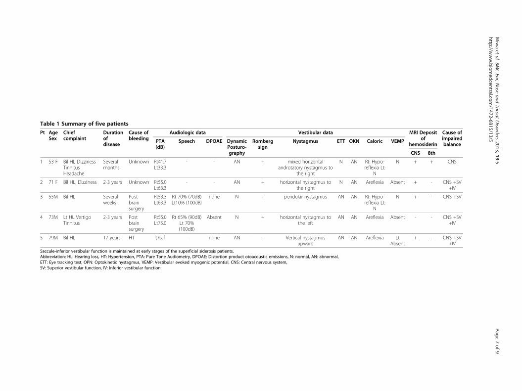
Table 1 Summary of five patients
Pt AgeSex
Chiefcomplaint
Durationofdisease
Cause ofbleeding
Audiologic data Vestibular data MRI Depositof
hemosiderin
Cause ofimpairedbalance
PTA(dB)
Speech DPOAE DynamicPosturo-graphy
Rombergsign
Nystagmus ETT OKN Caloric VEMP
CNS 8th
1 53 F Bil HL DizzinessTinnitusHeadache
Severalmonths
Unknown Rt41.7Lt33.3
- - AN + mixed horizontalandrotatory nystagmus to
the right
N AN Rt: Hypo-reflexia Lt:
N
N + + CNS
2 71 F Bil HL, Dizziness 2-3 years Unknown Rt55.0Lt63.3
- - AN + horizontal nystagmus tothe right
N AN Areflexia Absent + - CNS +SV+IV
3 55M Bil HL Severalweeks
Postbrainsurgery
Rt53.3Lt63.3
Rt 70% (70dB)Lt10% (100dB)
none N + pendular nystagmus AN AN Rt: Hypo-reflexia Lt:
N
N + - CNS +SV
4 73M Lt HL VertigoTinnitus
2-3 years Postbrainsurgery
Rt55.0Lt75.0
Rt 65% (90dB)Lt 70%(100dB)
Absent N + horizontal nystagmus tothe left
AN AN Areflexia Absent - - CNS +SV+IV
5 79M Bil HL 17 years HT Deaf - none AN - Vertical nystagmusupward
AN AN Areflexia LtAbsent
+ - CNS +SV+IV
Saccule-inferior vestibular function is maintained at early stages of the superficial siderosis patients.Abbreviation: HL: Hearing loss, HT: Hypertension, PTA: Pure Tone Audiometry, DPOAE: Distortion product otoacoustic emissions, N: normal, AN: abnormal,ETT: Eye tracking test, OPN: Optokinetic nystagmus, VEMP: Vestibular evoked myogenic potential, CNS: Central nervous system,SV: Superior vestibular function, IV: Inferior vestibular function.
Miwaet
al.BMCEar,N
oseand
ThroatDisorders
2013,13:5Page
7of
9http://w
ww.biom
edcentral.com/1472-6815/13/5

Miwa et al. BMC Ear, Nose and Throat Disorders 2013, 13:5 Page 8 of 9http://www.biomedcentral.com/1472-6815/13/5
develop. Thus, the superior vestibular nerve is more sus-ceptible to damage than the inferior vestibular nerve.However, in our study, the MRI findings revealedhemosiderosis of the 8th nerve in only 1 patient(patient 1). This finding implies that the causes of thebalance impairments are cochlear damage, likely causedby constriction and impaired blood flow, rather thanvestibular nerve damage due to the deposition of hemo-siderin. This result corroborates the report by Fukiyamaet al. indicating that cochlear damage due to SS iscaused by impaired blood flow in the inner ear [5].The treatment of SS involves identifying the cause of
the bleeding and treatment of the underlying cause whenit is apparent. When the cause is not apparent, achelating agent is administered to deplete iron, or ahemostatic agent is administered to stop bleeding. How-ever, such treatment is inadequate, and effective thera-pies have yet to be established [17,26]. Approximately40% of cases of bleeding due to SS are idiopathic [17].Other causes of bleeding, reported as symptomatic, in-clude current or previous CNS tumors, head or backtrauma, arteriovenous malformations/aneurysms, post-surgical changes related to neurosurgeries, brachialplexus injury, amyloid angiopathy (AA), and otherchronic subdural hematomas [26-30]. In patients withAA and/or hypertension, microbleeds into the subarach-noid space continue due to the fragility of the vessels inthe meninges [31,32]. In our study, the cause of bleedingwas apparent in patients 2 and 4, but they had been pre-viously treated when initially examined for this study.We considered that the cause of the bleeding in pa-tient 5 could have been microbleeds due to chronichypertension because he presented no signs of AAand was medicated with an anti-coagulation drug. Inthe other two patients, the causes of the bleedingwere not apparent.The prognosis for SS is relatively good, with some
patients surviving 20 to 30 years after developing thecondition [17,28,33]. Sensorineural deafness often pro-gresses, and quality of life (QOL) is markedly dimin-ished. Recently, many SS patients have received cochlearimplants [18,19,34-40]. Because the impaired balance isdue to central nervous damage and peripheral vestibulardamage, acquiring or achieving balance via the vestibu-loocular reflex often proves difficult. There is no com-pensatory mechanism, and symptoms will persist, oftendiminishing QOL. The course of the current patientscould not be followed, so the diminishing of vestibularfunction could not be ascertained. However, periodicbalance testing could likely facilitate the observation ofthe diminishing of vestibular function.This report revealed that the lesions responsible for
impaired balance due to SS lie in the central nervoussystem and the peripheral vestibular system. Vestibular
function, particularly the saccule-inferior vestibularfunction, diminishes the longer a patient suffers fromthe condition. Nevertheless, the pathology of SS remainsrather unclear, and effective therapies have yet to beestablished. These issues must be examined in the futureby assembling and analyzing additional cases.
Informed consentWritten informed consent documents were obtainedfrom the five patients for publication of this case reportand any accompanying images. The copies of the writtenconsent documents are available for review by theEditor-in-Chief of this journal.
Competing interestsThe authors declare that they have no competing interests.
Authors’ contributionsTM and HM conceived, designed, and carried out the experiments TManalyzed the data and wrote the manuscript. RM conceived of the study,and participated in its coordination. All authors read and approved the finalmanuscript.
AcknowledgementsWe thank Hidetake Matsuyoshi for advice on the manuscript. No sources offunding for the study.
Author details1Department of Otolaryngology and Head and Neck SurgeryKumamoto University, 1-1-1 Honjo, Kumamoto, Japan. 2Matsubase ENT Clinic,Kumamoto, Japan.
Received: 12 July 2012 Accepted: 9 April 2013Published: 23 April 2013
References1. Hamill RC: Report of a case of melanosis of the brain, cord and
meninges. J Nerv Ment Dis 1908, 35(8):594.2. Iwanowski L, Olszewski J: The effects of subarachnoid injections of
iron-containing substances on the central nervous system. J NeuropatholExp Neurol 1960, 19:433–448.
3. Levy M, Turtzo C, Llinas RH: Superficial siderosis: a case report and reviewof the literature. Nat Clin Pract Neurol 2007, 3(1):54–58. quiz 59.
4. Noetzel H: Diffusion of blood pigment in the inner border area and outersurface of the CNS after subarachnoidal hemorrhage [German].Arch Psychol Nervenkr 1940, 111:129–138.
5. Fukiyama M, Matsuura K, Morimitsu T, Kodama T: [A case of superficialsiderosis of the central nervous system with total deafness].Nihon Jibiinkoka Gakkai Kaiho 1993, 96(3):428–434.
6. Colebatch JG, Halmagyi GM, Skuse NF: Myogenic potentials generated bya click-evoked vestibulocollic reflex. J Neurol Neurosurg Psychiatry 1994,57(2):190–197.
7. Colebatch JG, Halmagyi GM: Vestibular evoked potentials in human neckmuscles before and after unilateral vestibular deafferentation. Neurology1992, 42(8):1635–1636.
8. Robertson DD, Ireland DJ: Vestibular evoked myogenic potentials.J Otolaryngol 1995, 24(1):3–8.
9. Bárány R: Some New Methods for Functional Testing of the Vestibular Apparatusand the Cerebellum. Amsterdam: Elsevier Publishing Company; 1967.
10. Vibert D, Hausler R, Lovblad KO, Schroth G: Hearing loss and vertigo insuperficial siderosis of the central nervous system. Am J Otolaryngol 2004,25(2):142–149.
11. Watanabe M, Miyasaka H, Iwata N, Maeda S, Kishida S, Hayashi: [A case ofsuperficial siderosis of the central nervous system with bilateralvestibular dysfunction]. No To Shinkei 1997, 49(10):931–935.

Miwa et al. BMC Ear, Nose and Throat Disorders 2013, 13:5 Page 9 of 9http://www.biomedcentral.com/1472-6815/13/5
12. Ohtsuka H, Ohtsuka C, Yonezawa H, Tateda M, Ishijima K, Sato H: A case ofsuperficial central nervous system siderosis exhibiting vertigo[Japanese].Jibirin 2010, 103(1):9–13.
13. Weekamp HH, Huygen PL, Merx JL, Kremer HP, Cremers CW, Longridge NS:Longitudinal analysis of hearing loss in a case of hemosiderosis of thecentral nervous system. Otol Neurotol 2003, 24(5):738–742.
14. Koeppen AH, Dentinger MP: Brain hemosiderin and superficial siderosis ofthe central nervous system. J Neuropathol Exp Neurol 1988, 47(3):249–270.
15. Koeppen AH, Hurwitz CG, Dearborn RE, Dickson AC, Borke RC, Chu RC:Experimental superficial siderosis of the central nervous system:biochemical correlates. J Neurol Sci 1992, 112(1–2):38–45.
16. Nadol JB Jr, Adams JC, O’Malley JT: Temporal bone histopathology in acase of sensorineural hearing loss caused by superficial siderosis of thecentral nervous system and treated by cochlear implantation. OtolNeurotol 2011, 32(5):748–755.
17. Fearnley JM, Stevens JM, Rudge P: Superficial siderosis of the centralnervous system. Brain 1995, 118(Pt 4):1051–1066.
18. Hathaway B, Hirsch B, Branstetter B: Successful cochlear implantation in apatient with superficial siderosis. Am J Otolaryngol 2006, 27(4):255–258.
19. Irving RM, Graham JM: Cochlear implantation in superficial siderosis.J Laryngol Otol 1996, 110(12):1151–1153.
20. Lai MT, Ohmichi T, Yuen K, Egusa K, Yorizane S, Masuda Y: Superficialsiderosis of the central nervous system: a case with an unrupturedintracranial aneurysm. J Laryngol Otol 1995, 109(6):549–552.
21. Longridge NS, Hashimoto S, Marotta TR, Mezei M: Superficial siderosis–acause of audiovestibular failure. J Otolaryngol 1996, 25(1):41–43.
22. Takasaki K, Tanaka F, Shigeno K, Kanda Y, Kawajiri I, Tashiro T, Kobayashi T:Superficial siderosis of the central nervous system. A case report onexamination by ECoG and DPOAE. ORL J Otorhinolaryngol Relat Spec 2000,62(5):270–273.
23. Ushio M, Iwasaki S, Sugasawa K, Murofushi T: Superficial siderosis causingretrolabyrinthine involvement in both cochlear and vestibular branchesof the eighth cranial nerve. Acta Otolaryngol 2006, 126(9):997–1000.
24. Sydlowski SA, Cevette MJ, Shallop J: Superficial siderosis of the centralnervous system: phenotype and implications for audiology and otology.Otol Neurotol 2011, 32(6):900–908.
25. Goebel JA, O’Mara W, Gianoli G: Anatomic considerations in vestibularneuritis. Otol Neurotol 2001, 22(4):512–518.
26. Anderson NE, Sheffield S, Hope JK: Superficial siderosis of the centralnervous system: a late complication of cerebellar tumors. Neurology 1999,52(1):163–169.
27. Janss AJ, Galetta SL, Freese A, Raps EC, Curtis MT, Grossman RI, Gomori JM,Duhaime AC: Superficial siderosis of the central nervous system:magnetic resonance imaging and pathological correlation. Case report.J Neurosurg 1993, 79(5):756–760.
28. Bonito V, Agostinis C, Ferraresi S, Defanti CA: Superficial siderosis of thecentral nervous system after brachial plexus injury. Case report.J Neurosurg 1994, 80(5):931–934.
29. Tapscott SJ, Eskridge J, Kliot M: Surgical management of superficialsiderosis following cervical nerve root avulsion. Ann Neurol 1996,40(6):936–940.
30. Urban PP, Szegedi A, Muller-Forell W, Hopf HC: Superficial siderosis of theCNS as a rare differential diagnosis of chronic low back pain. J Neurol1999, 246(10):980–981.
31. Katsuragi S, Sakai T, Watanabe K, Shimoji A, Deshimaru M, Kuramoto R,Miyamoto K, Yamashita K, Miyakawa T: An autopsy case of idiopathicsuperficial hemosiderosis of the central nervous system: a microscopicand immunohistochemical study. Clin Neuropathol 1988, 7(2):87–92.
32. Linn J, Halpin A, Demaerel P, Ruhland J, Giese AD, Dichgans M, van BuchemMA, Bruckmann H, Greenberg SM: Prevalence of superficial siderosis inpatients with cerebral amyloid angiopathy. Neurology 2010,74(17):1346–1350.
33. Parnes SM, Weaver SA: Superficial siderosis of the central nervous system:a neglected cause of sensorineural hearing loss. Otolaryngol Head NeckSurg 1992, 107(1):69–77.
34. Bird PA, Monteath P, Healy L: Successful cochlear implantation in apatient with superficial siderosis of the central nervous system.Otol Neurotol 2010, 31(1):177.
35. Dhooge IJ, De Vel E, Urgell H, Gallego S, Vinck B: Cochlear implantation ina patient with superficial siderosis of the central nervous system.Otol Neurotol 2002, 23(4):468–472.
36. Kim CS, Song JJ, Park MH, Kim YH, Koo JW: Cochlear implantation insuperficial siderosis. Acta Otolaryngol 2006, 126(8):892–896.
37. Sugimoto H, Ito M, Hatano M, Yoshizaki T: Cochlear implantation in apatient with superficial siderosis. Auris Nasus Larynx 2011, 39(6):623–626.
38. Sydlowski SA, Cevette MJ, Shallop J, Barrs DM: Cochlear implant patientswith superficial siderosis. J Am Acad Audiol 2009, 20(6):348–352.
39. Wood VH, Bird PA, Giles EC, Baber WJ: Unsuccessful cochlear implantationin two patients with superficial siderosis of the central nervous system.Otol Neurotol 2008, 29(5):622–625.
40. Yoshikawa N, Hirsch BE: Cochlear implantation in a patient withsuperficial siderosis: an update. Am J Otolaryngol 2010, 31(5):390–391.
doi:10.1186/1472-6815-13-5Cite this article as: Miwa et al.: Vestibular function in superficialsiderosis. BMC Ear, Nose and Throat Disorders 2013 13:5.
Submit your next manuscript to BioMed Centraland take full advantage of:
• Convenient online submission
• Thorough peer review
• No space constraints or color figure charges
• Immediate publication on acceptance
• Inclusion in PubMed, CAS, Scopus and Google Scholar
• Research which is freely available for redistribution
Submit your manuscript at www.biomedcentral.com/submit