Very-Long-Chain Fatty Acids Are Involved in Polar … · Very-Long-Chain Fatty Acids Are Involved...
13
Very-Long-Chain Fatty Acids Are Involved in Polar Auxin Transport and Developmental Patterning in Arabidopsis W Franc ¸ ois Roudier, a,1 Lionel Gissot, a Fre ´ de ´ ric Beaudoin, b Richard Haslam, b Louise Michaelson, b Jessica Marion, a,2 Diana Molino, a Amparo Lima, a,3 Lie ˆ n Bach, a Halima Morin, c Fre ´ de ´ rique Tellier, d Jean-Christophe Palauqui, a Yannick Bellec, a Charlotte Renne, a Martine Miquel, a Marco DaCosta, a,4 Julien Vignard, a Christine Rochat, a Jonathan E. Markham, e Patrick Moreau, f Johnathan Napier, b and Jean-Denis Faure a,5 a Institut Jean-Pierre Bourgin, Unite ´ Mixte de Recherche 1318, Institut National de la Recherche Agronomique-AgroParisTech, Centre de Versailles-Grignon, 78026 Versailles Cedex, France b Rothamsted Research, Harpenden, Herts AL5 2JQ, United Kingdom c Plateforme de Cytologie et d’Imagerie Ve ´ ge ´ tale, Institut Jean-Pierre Bourgin, Institut National de la Recherche Agronomique, 78000 Versailles, France d Plateforme de Chimie du Ve ´ ge ´ tale, Institut Jean-Pierre Bourgin, Institut National de la Recherche Agronomique, 78000 Versailles, France e Donald Danforth Plant Science Center, St. Louis, Missouri 63132 f Laboratoire Biogene ` se membranaire, Unite ´ Mixte de Recherche 5200, Centre National de la Recherche Scientifique-Universite ´ Bordeaux 2, BP 33076 Bordeaux Cedex, France Very-long-chain fatty acids (VLCFAs) are essential for many aspects of plant development and necessary for the synthesis of seed storage triacylglycerols, epicuticular waxes, and sphingolipids. Identification of the acetyl-CoA carboxylase PASTICCINO3 and the 3-hydroxy acyl-CoA dehydratase PASTICCINO2 revealed that VLCFAs are important for cell prolifer- ation and tissue patterning. Here, we show that the immunophilin PASTICCINO1 (PAS1) is also required for VLCFA synthesis. Impairment of PAS1 function results in reduction of VLCFA levels that particularly affects the composition of sphingolipids, known to be important for cell polarity in animals. Moreover, PAS1 associates with several enzymes of the VLCFA elongase complex in the endoplasmic reticulum. The pas1 mutants are deficient in lateral root formation and are characterized by an abnormal patterning of the embryo apex, which leads to defective cotyledon organogenesis. Our data indicate that in both tissues, defective organogenesis is associated with the mistargeting of the auxin efflux carrier PIN FORMED1 in specific cells, resulting in local alteration of polar auxin distribution. Furthermore, we show that exogenous VLCFAs rescue lateral root organogenesis and polar auxin distribution, indicating their direct involvement in these processes. Based on these data, we propose that PAS1 acts as a molecular scaffold for the fatty acid elongase complex in the endoplasmic reticulum and that the resulting VLCFAs are required for polar auxin transport and tissue patterning during plant development. INTRODUCTION Very-long-chain fatty acids (VLCFAs) are defined in plants as fatty acids with an acyl chain of at least 20 carbons in length. VLCFAs are components of seed storage triacylglycerols, cutic- ular and epicuticular lipids, and sphingolipids. VLCFA synthesis requires four endoplasmic reticulum (ER)-bound enzymes con- stituting the elongase complex that carry out four sequential reactions: first, the condensation of a substrate acyl-CoA with malonyl-CoA catalyzed by ketoacyl-CoA synthase; second, the reduction of 3-ketoacyl-CoA by a ketoacyl-CoA reductase (KCR) followed by dehydration of the resulting 3-hydroxy acyl-CoA by the 3-hydroxy acyl-CoA dehydratase (PASTICCINO2 [PAS2]). The final enzymatic step is the reduction of the enoyl acyl-CoA by an enoyl-CoA reductase (ECR), resulting in an acyl-CoA that is two carbons longer (Zheng et al., 2005; Joubes et al., 2008). Malonyl-CoA is synthetized from acetyl-CoA by the cytosolic isoform of the acetyl-CoA carboxylase PAS3/GU ¨ RKE (Baud et al., 2003, 2004). Loss of function of the 3-hydroxy acyl-CoA dehydratase PASTICCINO2 leads to embryo lethality, demon- strating that VLCFAs are essential for plant development (Bach et al., 2008). Reduction of VLCFA levels resulting from weak pas3 or pas2 alleles is also associated with abnormal development and ectopic cell proliferation (Faure et al., 1998). Loss of Arabidopsis thaliana ECR CER10 function results in reduced 1 Current address: Epige ´ ne ´ tique et Epige ´ nomique Ve ´ ge ´ tale, Section Ge ´ nomique Environnementale et Evolutive, Institut de Biologie de l’Ecole Normale Supe ´ rieure, Unite ´ Mixte de Recherche 8197, Centre National de la Recherche Scientifique, 46 rue d’Ulm, 75230 Paris cedex 05, France. 2 Institut des Sciences du Ve ´ ge ´ tal, Centre National de la Recherche Scientifique, Avenue de la Terrasse, F-91198 Gif-sur-Yvette Cedex, France. 3 Department of Plant and Microbial Biology, University of California, Berkeley, CA 94720. 4 Laboratoire de Cytologie Expe ´ rimentale et Morphogene ` se Ve ´ ge ´ tale, Paris-VI, 94200 Ivry/Seine, France. 5 Address correspondence to [email protected]. The author responsible for distribution of materials integral to the findings presented in this article in accordance with the policy described in the Instructions for Authors (www.plantcell.org) is: Jean-Denis Faure ([email protected]). W Online version contains Web-only data. www.plantcell.org/cgi/doi/10.1105/tpc.109.071209 The Plant Cell, Vol. 22: 364–375, February 2010, www.plantcell.org ã 2010 American Society of Plant Biologists
Transcript of Very-Long-Chain Fatty Acids Are Involved in Polar … · Very-Long-Chain Fatty Acids Are Involved...

Very-Long-Chain Fatty Acids Are Involved in Polar AuxinTransport and Developmental Patterning in Arabidopsis W
Francois Roudier,a,1 Lionel Gissot,a Frederic Beaudoin,b Richard Haslam,b Louise Michaelson,b Jessica Marion,a,2
Diana Molino,a Amparo Lima,a,3 Lien Bach,a Halima Morin,c Frederique Tellier,d Jean-Christophe Palauqui,a
Yannick Bellec,a Charlotte Renne,a Martine Miquel,a Marco DaCosta,a,4 Julien Vignard,a Christine Rochat,a
Jonathan E. Markham,e Patrick Moreau,f Johnathan Napier,b and Jean-Denis Faurea,5
a Institut Jean-Pierre Bourgin, Unite Mixte de Recherche 1318, Institut National de la Recherche Agronomique-AgroParisTech,
Centre de Versailles-Grignon, 78026 Versailles Cedex, Franceb Rothamsted Research, Harpenden, Herts AL5 2JQ, United Kingdomc Plateforme de Cytologie et d’Imagerie Vegetale, Institut Jean-Pierre Bourgin, Institut National de la Recherche Agronomique,
78000 Versailles, Franced Plateforme de Chimie du Vegetale, Institut Jean-Pierre Bourgin, Institut National de la Recherche Agronomique, 78000 Versailles,
Francee Donald Danforth Plant Science Center, St. Louis, Missouri 63132f Laboratoire Biogenese membranaire, Unite Mixte de Recherche 5200, Centre National de la Recherche Scientifique-Universite
Bordeaux 2, BP 33076 Bordeaux Cedex, France
Very-long-chain fatty acids (VLCFAs) are essential for many aspects of plant development and necessary for the synthesis
of seed storage triacylglycerols, epicuticular waxes, and sphingolipids. Identification of the acetyl-CoA carboxylase
PASTICCINO3 and the 3-hydroxy acyl-CoA dehydratase PASTICCINO2 revealed that VLCFAs are important for cell prolifer-
ation and tissue patterning. Here, we show that the immunophilin PASTICCINO1 (PAS1) is also required for VLCFA synthesis.
Impairment of PAS1 function results in reduction of VLCFA levels that particularly affects the composition of sphingolipids,
known to be important for cell polarity in animals. Moreover, PAS1 associates with several enzymes of the VLCFA elongase
complex in the endoplasmic reticulum. The pas1 mutants are deficient in lateral root formation and are characterized by an
abnormal patterning of the embryo apex, which leads to defective cotyledon organogenesis. Our data indicate that in both
tissues, defective organogenesis is associatedwith themistargeting of the auxin efflux carrier PIN FORMED1 in specific cells,
resulting in local alteration of polar auxin distribution. Furthermore, we show that exogenous VLCFAs rescue lateral root
organogenesis and polar auxin distribution, indicating their direct involvement in these processes. Based on these data, we
propose that PAS1 acts as amolecular scaffold for the fatty acid elongase complex in the endoplasmic reticulum and that the
resulting VLCFAs are required for polar auxin transport and tissue patterning during plant development.
INTRODUCTION
Very-long-chain fatty acids (VLCFAs) are defined in plants as
fatty acids with an acyl chain of at least 20 carbons in length.
VLCFAs are components of seed storage triacylglycerols, cutic-
ular and epicuticular lipids, and sphingolipids. VLCFA synthesis
requires four endoplasmic reticulum (ER)-bound enzymes con-
stituting the elongase complex that carry out four sequential
reactions: first, the condensation of a substrate acyl-CoA with
malonyl-CoA catalyzed by ketoacyl-CoA synthase; second, the
reduction of 3-ketoacyl-CoA by a ketoacyl-CoA reductase (KCR)
followed by dehydration of the resulting 3-hydroxy acyl-CoA by
the 3-hydroxy acyl-CoA dehydratase (PASTICCINO2 [PAS2]).
The final enzymatic step is the reduction of the enoyl acyl-CoA by
an enoyl-CoA reductase (ECR), resulting in an acyl-CoA that is
two carbons longer (Zheng et al., 2005; Joubes et al., 2008).
Malonyl-CoA is synthetized from acetyl-CoA by the cytosolic
isoform of the acetyl-CoA carboxylase PAS3/GURKE (Baud
et al., 2003, 2004). Loss of function of the 3-hydroxy acyl-CoA
dehydratase PASTICCINO2 leads to embryo lethality, demon-
strating that VLCFAs are essential for plant development (Bach
et al., 2008). Reduction of VLCFA levels resulting fromweak pas3
or pas2 alleles is also associated with abnormal development
and ectopic cell proliferation (Faure et al., 1998). Loss of
Arabidopsis thaliana ECR CER10 function results in reduced
1Current address: Epigenetique et Epigenomique Vegetale, SectionGenomique Environnementale et Evolutive, Institut de Biologie de l’EcoleNormale Superieure, Unite Mixte de Recherche 8197, Centre National dela Recherche Scientifique, 46 rue d’Ulm, 75230 Paris cedex 05, France.2 Institut des Sciences du Vegetal, Centre National de la RechercheScientifique, Avenuede laTerrasse,F-91198Gif-sur-YvetteCedex, France.3 Department of Plant and Microbial Biology, University of California,Berkeley, CA 94720.4 Laboratoire de Cytologie Experimentale et Morphogenese Vegetale,Paris-VI, 94200 Ivry/Seine, France.5 Address correspondence to [email protected] author responsible for distribution of materials integral to thefindings presented in this article in accordance with the policy describedin the Instructions for Authors (www.plantcell.org) is: Jean-Denis Faure([email protected]).WOnline version contains Web-only data.www.plantcell.org/cgi/doi/10.1105/tpc.109.071209
The Plant Cell, Vol. 22: 364–375, February 2010, www.plantcell.org ã 2010 American Society of Plant Biologists

cell expansion and eventually in reduced size of aerial organs
(Zheng et al., 2005). Similar observations were recently made
upon RNA interference silencing of Arabidopsis KCR (Beaudoin
et al., 2009). Overexpression of the condensing enzyme FATTY
ELONGASE1 (FAE1) was found to alter chloroplast structure and
cell shape, whereas an increase of the dehydratase activity leads
to cell division and expansion defects (Millar and Kunst, 1997;
Bach et al., 2008). Interestingly, ectopic expression of FAE1with
the epidermal-specific promoter of FIDDLEHEAD results in tri-
chome cell death (Reina-Pinto et al., 2009). Altogether these
studies show that VLCFA homeostasis is essential and limiting
for plant growth and development.
While the analysis of the role of VLCFAs in cell expansion could
be explained by a deficit of essential lipids at the plasma
membrane, the involvement of VLCFAs in cell proliferation and
differentiation remains unclear. The pas mutants were initially
identified as three complementation groups, Pas1, Pas2, and
Pas3, showing ectopic cell proliferation in hypocotyls, cotyle-
dons, and leaves. The pas mutations result in profound devel-
opmental alterations, which are already detectable during
embryogenesis and lead to dwarf seedlings with short and thick
hypocotyls and fused and misshapen leaves (Faure et al., 1998).
The pas seedlings also show an increased competency for cell
division in meristematic and differentiated cells that is enhanced
by exogenous cytokinins and leads to callus-like development
(Haberer et al., 2002; Harrar et al., 2003). Moreover, in these
mutants, the shoot apical meristem is enlarged and shows an
increased expression of meristematic markers (Haberer et al.,
2002; Harrar et al., 2003). This higher proliferative activity is
illustrated by the ability of the pas2 mutation to rescue the
SHOOT MERISTEMLESS mutant that is devoid of meristematic
activity (Harrar et al., 2003). Finally, PAS2was found to interact in
vitro with Cyclin-Dependent Kinase 1, suggesting that VLCFA
synthesis might have a direct link with cell cycle progression (Da
Costa et al., 2006). Cell fate acquisition is also altered in the pas
mutants. The absence of cotyledon development in pas3 (gurke)
embryos is associated with overlapping cell fate territories within
the apex (Kajiwara et al., 2004).
The identification of two PAS genes as essential enzymes of
VLCFA biosynthesis suggest that the pleiotropic pas phenotype
is directly associated with lipids that contain such acyl chains.
However, the role of VLCFAs in cell fate acquisition and tissue
patterning remains unclear, as does the involvement of PAS1 in
VLCFA synthesis. PAS1 encodes a large molecular weight
member of the immunophilin type FK506 binding protein fam-
ily that have been reported in other eukaryotes to regulate
the activity of many proteins involved in signaling pathways
(Vittorioso et al., 1998; Harrar et al., 2001). In previous work, we
have shown that PAS1 interacts in vitro and in vivo through its C
terminuswith amember of theNAC family of transcription factors
and regulates its subcellular localization (Smyczynski et al.,
2006). Here, we show that the pas1 mutant, similar to the two
other pasmutants, has reduced levels of VLCFAs and that PAS1
interacts in the ERwith the core elongase components, suggest-
ing a new role of the immunophilin in scaffolding the fatty acid
elongase complex. We also show that the impairment of PAS1
function results, like in pas3, in defective cotyledon formation
associated with an altered patterning of the embryo apex. We
provide evidence that thismorphological defect and the absence
of lateral root initiation later in development are associated with
alterations in auxin response, resulting from a lack of polar
targeting of the auxin efflux carrier PIN1. In pas1, defective PIN1
polar targeting is correlated with hypersensitivity to the traffick-
ing inhibitor brefeldin A. Fatty acids are directly involved in polar
auxin transport since exogenous application of lipids restore
auxin distribution in lateral roots. Based on these data, we
propose that PAS1 is involved in fatty acid elongation and that
the resulting VLCFAs are essential regulators of cell differentia-
tion during development by regulating polar auxin distribution.
RESULTS
PAS1 Is Required for VLCFA Accumulation in
Triacylglycerols, Free Fatty Acids, and Sphingolipids
The fact that the three pasmutants share similar phenotypes and
that both PAS2 and PAS3 are essential for VLCFA synthesis
(Baud et al., 2003, 2004; Bach et al., 2008) prompted us to
determine whether the pas1-3 mutant was also altered in lipid
metabolism. Quantification of VLCFA content in pas1-3 mature
seeds revealed that the levels of 20:2 and 22:1 fatty acids were
reduced by 50% compared with wild-type levels and that,
conversely, the level of short-chain 16:0 and 18:1 fatty acids
showed a 40% increase (Figure 1A). Similar changes in the levels
of 20:1 and 22:1 but also 16:0 were observed in weak pas2 and
pas3 alleles (Baud et al., 2004; Bach et al., 2008). VLCFA levels in
pas1 roots were also reduced by 38, 42, and 60% for 22:0, 22:1,
and 24:0, respectively (Figure 1B). VLCFAs are also found in
sphingolipids (Dunn et al., 2004). Contrary to seed triacylglyc-
erols, sphingolipids are essential for embryo development (Chen
et al., 2006). Sphingolipids are composed of a long-chain base
(amino alcohol) and an amide linked with a fatty acyl chain (Dunn
et al., 2004). These lipids are characterized by the acyl chain
length and its degree of unsaturation, as well as the nature of the
polar head group, which can be modified by glycosylation and
phosphorylation. Like pas2-1, both pas1-3 and pas3-1 mutants
were affected in sphingolipid content showing a lower amount of
very-long-chain (C24 and C26) sphingolipids, which are the pre-
dominant forms in Arabidopsis (Figure 1C) (Bach et al., 2008).
Specific Sphingolipid Modifications Are Associated with
Altered PAS1 Function
Reduction of C24 and C26 sphingolipids in pas1-3 and pas3-1
was associated with lower levels of D8-unsaturated long-
chain-base moieties 8-sphingenine (d18:18) and 4-hydroxy-8-
sphingenine (t18:18), likely due to modifications of the different
classes of sphingolipids (Borner et al., 2005; Markham et al.,
2006; Chen et al., 2008) (see Supplemental Figure 1A online). To
assess whether PAS1 targets specific classes of sphingolipids,
we performed a complete sphingolipid profiling of both mutants.
The levels of simple sphingolipids like Ceramide (Cer) were
increased by;30 and 20% for pas1-3 and pas3-1, respectively,
whereas hydroxyceramide levels increased (by ;30%) only in
pas1-3 (see Supplemental Figure 1B online). Global increase of
Very-Long-Chain Fatty Acids and Cell Patterning 365

Cer in both mutants was mainly caused by the accumulation of
long-chain 16:0-Cer (see Supplemental Figure 2 online), whereas
the very-long-chain 24:0- and 26:0-Cer levels were substantially
reduced in pas3-1 and decreased only modestly in pas1-3 (see
Supplemental Figure 2 online). We then examined the complex
glycosylated sphingolipids glucosyl inositol phosphoryl cer-
amide (GIPC) and glucosyl ceramide (GluCer). GIPCs levels were
not significantly modified in pas3-1 and were slightly reduced in
pas1-3 (;25%decrease). Contrary toGIPCs,GluCer levelswere
reduced by;50 and 63%, respectively, for pas1-3 and pas3-1
mutants (see Supplemental Figure 1C online). Very-long (C20+)
and long-chain (C18-20) GluCers were specifically reduced (see
Supplemental Figure 2 online). Reduction of GluCer levels could
result from either a decrease in synthesis or a higher turnover rate
(by specific hydrolases). To discriminate between these two
possibilities, GluCer synthesis was measured by 14C-acetate
incorporation followed by extraction of this sphingolipid fraction.
GluCer labeling in pas1-3 and pas3-1 was strongly reduced
compared with the wild type, demonstrating that synthesis of
GluCer was impaired in both mutants (see Supplemental Figure
1D online). Mutations affecting biosynthesis of sterols, which are
associated with sphingolipids in membrane domains, also lead
to strong developmental modifications especially during embryo-
genesis (Schaller, 2004). Contrary to VLCFAs, the levels of sterol
glycosides were not modified and the levels of sterol were even
increased in both pas1-3 and pas3-1mutants compared with the
wild type (see Supplemental Table 1 online). In conclusion, the
pas1-3mutant is characterized by a specific reduction of VLCFAs
like for the otherpasmutants, the 3-hydroxyacyl-CoAdehydratase
pas2, and the cytosolic acetyl CoA carboxylase pas3.
PAS1 Associates with the VLCFA Elongase Complex in
the ER
PAS1 is a member of the immunophilin family known in animals
and plants to target protein complexes and to regulate their
assembly or activities (Barik, 2006; Bouchard et al., 2006; Fu
et al., 2007). An attractive hypothesis to explain the decrease in
VLCFAs would be that PAS1 interacts directly with the VLCFA
elongase complex in the ER. PAS1 was originally described to
accumulate in the cytosolic space but was also found in the
nucleus upon auxin-induced dedifferentiation (Smyczynski et al.,
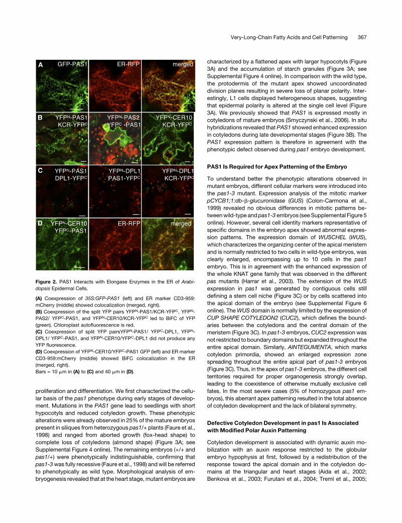
2006). Detailed analysis of the distribution of a green fluorescent
protein (GFP)-PAS1 fusion showed that it was mainly associated
with the ER as confirmed by its colocalization with the CD3-959
marker (Figure 2A). The four enzymes forming the elongase
complex are all localized in the ER (Zheng et al., 2005; Bach et al.,
2008; Joubes et al., 2008). To probe direct interactions between
PAS1 and members of the elongase complex, we used bimolec-
ular fluorescence complementation (BiFC). The different genes
were cloned in frame with the N- or C-terminal half of yellow
fluorescent protein (YFP), and the different combinations were
then tested in transient assays (Desprez et al., 2007;Marion et al.,
2008). YFP fluorescence was detected for all the combinations
involving PAS1 and the elongase complex core members KCR,
PAS2, and ECR (Figures 2B and 2D). In this assay, the interaction
between the elongase complex core members KCR, PAS2, and
CER10 was used as a positive control (Bach et al., 2008) (Figure
2B, right). Conversely, no interaction was detected between
PAS1, KCR, or CER10 and the ER-localized enzymes DPL1 and
SBH2, involved in sphingolipid long chain base metabolism
(Figure 2C; see Supplemental Figure 3C online; Tsegaye et al.,
2007; Chen et al., 2008), demonstrating the specificity of inter-
action between PAS1 and the elongase core enzymes. PAS1
interaction with the core elongase component PAS2 was con-
firmed by pull down of either PAS1-GFP expressed in BY2 cell
culture using a His6-PAS2 affinity column or radiolabeled in vitro–
translated PAS2 using a His6-PAS1 affinity column (see Supple-
mental Figures 3A and 3B online). Interaction of PAS1 with the
elongaseenzymes in theERwasshownbycolocalizing theYFPN-
CER10/YFPC-PAS1 fusion protein with the ER marker CD3-959
(Figure 2D). Altogether, these data demonstrate that PAS1 is
associated in the ERwith the VLCFA elongase complex and thus
is directly linked to VLCFA synthesis, as PAS2 and PAS3 are.
PAS1 Is Involved in Cotyledon Development
during Embryogenesis
The fact that the three pas mutants are severely altered in their
development raises the question of the role of VLCFAs in cell
Figure 1. Lipids with Very Long Acyl Chains Are Altered in pas1-3 and
pas3-1 Mutants.
(A) Triacylglycerol composition of wild-type (Col-0; black bars) and
pas1-3 seeds (white bars).
(B) Total fatty acid composition of wild-type (Col-0; black bars) and
pas1-3 mutant 10-d-old roots (white bars).
(C) Sphingolipid composition of wild-type (Col-0; black bars), pas1-3
(white bars), and pas3-1 (gray bars) seedlings.
Data represents the mean of three independent analyses. Error bar
indicates SE.
366 The Plant Cell
proliferation and differentiation. We first characterized the cellu-
lar basis of the pas1 phenotype during early stages of develop-
ment. Mutations in the PAS1 gene lead to seedlings with short
hypocotyls and reduced cotyledon growth. These phenotypic
alterations were already observed in 25%of themature embryos
present in siliques from heterozygous pas1/+ plants (Faure et al.,
1998) and ranged from aborted growth (fox-head shape) to
complete loss of cotyledons (almond shape) (Figure 3A; see
Supplemental Figure 4 online). The remaining embryos (+/+ and
pas1/+) were phenotypically indistinguishable, confirming that
pas1-3was fully recessive (Faure et al., 1998) andwill be referred
to phenotypically as wild type. Morphological analysis of em-
bryogenesis revealed that at the heart stage,mutant embryos are
characterized by a flattened apex with larger hypocotyls (Figure
3A) and the accumulation of starch granules (Figure 3A; see
Supplemental Figure 4 online). In comparison with the wild type,
the protodermis of the mutant apex showed uncoordinated
division planes resulting in severe loss of planar polarity. Inter-
estingly, L1 cells displayed heterogeneous shapes, suggesting
that epidermal polarity is altered at the single cell level (Figure
3A). We previously showed that PAS1 is expressed mostly in
cotyledons of mature embryos (Smyczynski et al., 2006). In situ
hybridizations revealed that PAS1 showed enhanced expression
in cotyledons during late developmental stages (Figure 3B). The
PAS1 expression pattern is therefore in agreement with the
phenotypic defect observed during pas1 embryo development.
PAS1 Is Required for Apex Patterning of the Embryo
To understand better the phenotypic alterations observed in
mutant embryos, different cellular markers were introduced into
the pas1-3 mutant. Expression analysis of the mitotic marker
pCYCB1;1:db-b-glucuronidase (GUS) (Colon-Carmona et al.,
1999) revealed no obvious differences in mitotic patterns be-
tweenwild-type andpas1-3 embryos (seeSupplemental Figure 5
online). However, several cell identity markers representative of
specific domains in the embryo apex showed abnormal expres-
sion patterns. The expression domain of WUSCHEL (WUS),
which characterizes the organizing center of the apical meristem
and is normally restricted to two cells in wild-type embryos, was
clearly enlarged, encompassing up to 10 cells in the pas1
embryo. This is in agreement with the enhanced expression of
the whole KNAT gene family that was observed in the different
pas mutants (Harrar et al., 2003). The extension of the WUS
expression in pas1 was generated by contiguous cells still
defining a stem cell niche (Figure 3C) or by cells scattered into
the apical domain of the embryo (see Supplemental Figure 6
online). TheWUS domain is normally limited by the expression of
CUP SHAPE COTYLEDON2 (CUC2), which defines the bound-
aries between the cotyledons and the central domain of the
meristem (Figure 3C). In pas1-3 embryos, CUC2 expression was
not restricted to boundary domains but expanded throughout the
entire apical domain. Similarly, AINTEGUMENTA, which marks
cotyledon primordia, showed an enlarged expression zone
spreading throughout the entire apical part of pas1-3 embryos
(Figure 3C). Thus, in the apex of pas1-3 embryos, the different cell
territories required for proper organogenesis strongly overlap,
leading to the coexistence of otherwise mutually exclusive cell
fates. In the most severe cases (5% of homozygous pas1 em-
bryos), this aberrant apex patterning resulted in the total absence
of cotyledon development and the lack of bilateral symmetry.
Defective Cotyledon Development in pas1 Is Associated
with Modified Polar Auxin Patterning
Cotyledon development is associated with dynamic auxin mo-
bilization with an auxin response restricted to the globular
embryo hypophysis at first, followed by a redistribution of the
response toward the apical domain and in the cotyledon do-
mains at the triangular and heart stages (Aida et al., 2002;
Benkova et al., 2003; Furutani et al., 2004; Treml et al., 2005;
Figure 2. PAS1 Interacts with Elongase Enzymes in the ER of Arabi-
dopsis Epidermal Cells.
(A) Coexpression of 35S:GFP-PAS1 (left) and ER marker CD3-959:
mCherry (middle) showed colocalization (merged, right).
(B) Coexpression of the split YFP pairs YFPN-PAS1/KCR-YFPC, YFPN-
PAS2/ YFPC-PAS1, and YFPN-CER10/KCR-YFPC led to BiFC of YFP
(green). Chloroplast autofluorescence is red.
(C) Coexpression of split YFP pairsYFPN-PAS1/ YFPC-DPL1, YFPN-
DPL1/ YFPC-PAS1, and YFPN-CER10/YFPC-DPL1 did not produce any
YFP fluorescence.
(D) Coexpression of YFPN-CER10/YFPC-PAS1 GFP (left) and ER marker
CD3-959:mCherry (middle) showed BiFC colocalization in the ER
(merged, right).
Bars = 10 mm in (A) to (C) and 40 mm in (D).
Very-Long-Chain Fatty Acids and Cell Patterning 367

Izhaki and Bowman, 2007). To monitor auxin response during
cotyledon development, we introduced the synthetic auxin-
responsive promoter DR5 driving the GFP reporter gene in
pas1. As reported previously, wild-type torpedo embryos
showed DR5 expression at the root pole, at the tip of the
cotyledons, and in the vascular tissues (Friml et al., 2003). By
contrast, DR5 expression was detected only at the root pole in
pas1 (Figure 3D). No DR5 expression could be detected in the
apical domain or the vascular tissues of pas1, suggesting that
auxin was not properly redistributed. Auxin distribution in the
embryo involves specific polar influx and efflux carriers (Jenik
and Barton, 2005). In particular, the PIN1 efflux carrier is involved
in downward auxin transport through the vascular tissues to the
root pole and in upward auxin transport through epidermal cells
from the hypophysis to the cotyledon tips. In order to determine
whether the altereddistribution of auxinobserved inpas1-3could
be associated with a defect in polar auxin transport, PIN1
expression and subcellular localization were investigated with a
PIN1-GFP fusion driven by the PIN1 promoter. In pas1-3, ex-
pression of the pPIN1:PIN1-GFP construct was observed in both
epidermal and vascular cells of the embryo, like in the wild type
(Figure 3E). However, whereas PIN1-GFP was localized in the
lateral membranes of epidermal cells in wild-type embryos (Fig-
ure 3F, left), its subcellular distribution was often diffused or
aggregated insidepas1epidermal cells (Figure3F, right). Immuno-
localization analysis confirmed the defective polar distribution of
Figure 3. PAS1 Is Required for Cell Patterning and Polarity in the Embryo Apex.
(A) Development of wild-type (top row) embryo at the dermatogen, globular, heart, torpedo, and late torpedo stage, respectively (from left to right). The
first phenotypic alteration in pas1-3 embryos is visible at the heart stage with the absence of cotyledon formation (bottom row). Mutant embryos
(bottom) were taken at the same respective stage (i.e., from the same silique) as the wild type (top row). Apical cells of pas1 embryos have lost their polar
growth (inset, bottom) compared with the wild type (inset, top). Embryos were fixed and stained with propidium iodide.
(B) In situ hybridization of PAS1 mRNA during embryo development in the wild type. Embryos were taken at globular, young heart, late heart, young
torpedo, and late torpedo stages (left to right).
(C) In situ hybridization of WUS, CUC2, and ANTEGUMENTA (ANT) mRNA in wild-type (left panel of each pair) and pas1-3 (right panel of each pair)
embryos. Bars = 40 mm except for the inset in (A), which is10 mm.
(D) pDR5-GFP distribution in the wild type (left) and pas1-3 mutant (right).
(E) pPIN1:PIN1-GFP distribution in the wild type (left) and pas1-3 mutant (right).
(F) Detail of pPIN1:PIN1-GFP distribution in the tip of a wild-type cotyledon (left) and the apex of pas1-3 embryo (right) at heart stage.
(G) to (J) Immunolocalization of PIN1 in wild-type and pas1-3 embryos.
(G) Detail of PIN1 distribution in the tip of a wild-type cotyledon (left) and in the apex of the pas1-3 mutant where aggregates are visible (right).
(H) and (I) Altered polar distribution of PIN1 in the apex of the pas1-3 mutant with PIN1 localizations facing each other in adjacent cells ([H], arrow) or
facing outward ([I], arrow).
(J) PIN1 polarity is normal in pas1-3 provascular and root pole cells.
Bars = 40 mm in (D) and (E), 20 mm in (F), 10 mm in (G) to (I), and 30 mm in (J).
368 The Plant Cell

PIN1 in pas1-3 apical cells, where it was found aggregated in the
cell and showed no clear polar organization when present at the
plasma membrane (Figure 3G, right). For instance, PIN1 was
observed in contiguous lateral membranes of two adjacent
epidermal cells or in the outward membrane facing the inner
seed cavity (Figures 3H and 3I, arrows). The alteration of PIN1
polar localization in pas1-3 could already be observed at the late
globular stage before any detectable changes in morphology,
suggesting that impaired auxin distribution is likely the cause of
the subsequent patterning defects observed in the pas1-3 em-
bryo (see Supplemental Figure 7 online). In agreement with the
specific expression of PAS1 at the embryo apex, loss of polar
distribution of PIN1was restricted to the apical domain of pas1-3
embryo but was not modified in vascular cells of the hypocotyl
anddidnot affectDR5expression at the root pole (Figures 3Dand
3J). Moreover, similar alterations were observed in embryos of
the VLCFA biosyntheticmutant pas3-1 (see Supplemental Figure
8 online), indicating that pas1-3 auxin transport defectswere also
linked with reduced VLCFA levels. In conclusion, the patterning
defects observed in the apical domain of pas1 embryos were
associated with abnormal polar distribution of PIN1 and com-
promised auxin response due to defective VLCFA synthesis.
PAS1 Is Involved in Lateral Root Development
Since PAS1 is also expressed during postembryonic develop-
ment, we investigated whether other developmental defects
observed in pas1 seedlings could also be related to altered polar
auxin distribution. The pas1-3 mutant was characterized by the
absenceof lateral root development (seeSupplemental Figure 9A
online; Faure et al., 1998; Vittorioso et al., 1998), the initiation of
which is strongly dependent on local auxin accumulation in
pericycle, cortex, and epidermal cells (Benkova et al., 2003;
Tanaka et al., 2006). In primary and lateral roots, PAS1was found
to be specifically expressed in the meristem and the columella
cells but not in the elongation or differentiation zones (see
Supplemental Figure 9B online). PAS1 was expressed at the
initial stage of lateral root formation where periclinal division of
pericycle cells is observed, and its expression was maintained
mostly at the tip of the root primordia until meristem formation
(see Supplemental Figure 9C online). Analysis of DR5:GFP ex-
pression indicated that auxin response in pas1-3 primary roots
andduring theearly stepsof lateral root initiationwassimilar to the
wild type (Figure 4A; see Supplemental Figure 10A online).
However, DR5 expression was rarely detected in the outer
cortical cells and never observed in epidermal cells during lateral
root formation (Figure 4B). In some instances, pas1-3 mutants
produced a few degenerative lateral root primordia with weak or
no DR5 expression (Figures 4C and 4K) that never developed into
lateral roots with proper columella and vascular DR5 expression
(see Supplemental Figure 11 online). Thus, as in embryo apex
patterning, denovoorganogenesis is compromised inpas1 roots.
Defective Lateral Root Development in pas1 Is Associated
with Altered Polar Auxin Transport
Auxin distribution during primary and lateral root develop-
ment is associated with specific expression patterns of PIN1
(Benkova et al., 2003; Tanaka et al., 2006). In the primary root,
PIN1 distribution was similar between the wild type and the
mutant, except that pas1-3 showed a higher occurrence of
cytosolic PIN1:GFP aggregates (Figure 5; see Supplemental
Figure 10B online). During the initial steps of lateral root
Figure 4. VLCFAs Are Involved in Lateral Root Development and Auxin
Polar Distribution.
(A) to (G) pDR5:GFP ([A] to [C]) and pPIN1:PIN1-GFP ([D] to [G])
expression during sequential steps of lateral root development in the wild
type (left) and the pas1-3 mutant (right). In the pas1 mutant, PIN1-GFP
was found accumulated inside primordia cells ([E] to [G], right) often in
aggregates ([G], arrows).
(H) to (J) Exogenous application of VLCFAs restored lateral root devel-
opment in pas1-3mutants. Seedlings were grown in presence ([H], right)
or absence ([H], left) of 200 mM fatty acids (18:0, 20:0, 22:0, and 24:0).
Details of control ([I], green bracket in [H]) or treated roots ([J], red
bracket in [H]) are shown. Arrows point to lateral root outgrowth in the
pas1-3 mutant ([H] and [J]).
(K) to (N) VLCFA application restores polar auxin transport in pas1-3
lateral roots. Normal pDR5:GFP ([K] and [L]) and pPIN1:PIN1-GFP ([M]
and [N]) expression patterns were observed in treated pas1-3 lateral root
tips ([L] and [N]) but not in untreated mutant roots ([K] and [M]).
Bars = 45 mm in (A) to (F), 30 mm in (G) (left), 20 mm in (G) (right), 1 mm in
(H), 300 mm in (I) and (J), and 20 mm in (K) to (N).
Very-Long-Chain Fatty Acids and Cell Patterning 369

initiation, PIN1 expression was clearly visible in the newly
divided pericycle cells for both wild-type and pas1-3 roots
(Figure 4D, right). At the stage of the first periclinal divisions of
the primordia, PIN1:GFP was not sharply localized to the
plasma membrane as in the wild type (Figures 4E and 4F, right)
but showed diffused or aggregated accumulation in the cell
(Figure 4G, right). Interestingly, the influx carrier AUX1, which is
also expressed during the early steps of lateral root develop-
ment, was properly targeted to the plasma membrane in the
pas1-3 mutant (see Supplemental Figures 10C to 10G online).
In conclusion, defective organ formation in pas1 embryos and
roots was associated with specific PIN1mistargeting leading to
altered auxin distribution.
BFA Sensitivity of PIN1 Distribution Is Enhanced in pas1
Polar localization of PIN1 results from a continuous turnover of
the protein from the plasma membrane to endosomal compart-
ments (Geldner et al., 2001, 2003; Grebe et al., 2003; Muday
et al., 2003; Boutte et al., 2006; Jaillais et al., 2006). The lower
VLCFA levels in pas1-3 mutant cells could result in an altered
trafficking of PIN1 and its subsequent accumulation in the
cytosol. In order to test this hypothesis, we used brefeldin A
(BFA), which inhibits anterograde (toward the plasma mem-
brane) trafficking of PIN1 by affecting the activity of GNOM, a
small G protein member of the GTP/GDP exchange factor
family (Steinmann et al., 1999). In the wild type, BFA treatment
results in the accumulation of PIN1 in large endosomal and
Golgi compartments called BFA compartments (Boutte et al.,
2006) and thus provides an interesting tool to monitor PIN1
trafficking. The sensitivity of PIN1 distribution to increasing
concentrations of BFA was investigated in wild-type and
pas1-3 primary roots. BFA was applied to wild-type and
pas1-3 seedlings for 30 min, and the occurrence of PIN1:GFP
in large BFA compartments was monitored in stele cells (Figure
5A). BFA (100 mM) led to the formation of BFA compartments of
PIN1:GFP in most cells in wild-type seedlings, whereas 25 mM
affected;50% of the cells (Figure 5B). On the contrary, pas1-3
mutant cells showed saturating BFA responses already at 25
mM, and >80% cells were BFA responsive at 17.8 mM (com-
pared with 30% in the wild type), indicating that PIN1 localiza-
tion was more sensitive to BFA in the pas1-3 background. This
increased sensitivity of PIN1 localization to BFA suggests that
PAS1, probably via the synthesis of VLCFAs, modifies protein
recycling.
VLCFAs Are Required for Auxin Distribution and for Lateral
Root Development
Finally, to demonstrate the role of VLCFAs in both auxin
distribution and lateral root development, pas1 mutants ex-
pressing pDR5:GFP or pPIN1:PIN1-GFP were directly grown in
the presence or absence of a mixture of VLCFAs (18:0, 20:0,
22:0, and 24:0). After 4 d of treatment, no significant differences
could be observed for wild-type roots (data not shown). How-
ever, VLCFA treatment enhanced lateral root development in
pas1-3 compared with the wild type (Figures 4I and 4J). While
only 2%of pas1 seedlings (n = 74) showed lateral root initiations
in the control condition, the application of VLCFAs led to a
fourfold increase in lateral root outgrowth (9% of pas1 seed-
lings, n = 88). This result was also confirmed by the direct
application of acyl-CoA to pas1 seedlings. Whereas 16:0-CoA
had no effect on the number of seedlings showing lateral root
development, and 18:0-CoA had only a slight effect (13 and
20%, respectively, of seedlings compared with 10% for the
control, n = 30), a mixture of 20:0-, 22:0-, and 24:0-CoA
increased the number of seedlings initiating lateral root devel-
opment (42% of seedlings, n = 30). The effect of VLCFA
treatment on pDR5:GFP and pPIN1:PIN1-GFP distribution
was also analyzed in wild-type and pas1 roots. As for the root
growth assay, the different treatment did not affect DR5 ex-
pression and PIN1-GFP localization in the wild type (data not
shown). However in fatty acid–treated pas1, DR5 was ex-
pressed at the tips of lateral roots (Figures 4K and 4L) and PIN1-
GFP was targeted to the plasma membrane (Figures 4M and
4N), demonstrating that VLCFAs are directly involved in the
distribution of auxin during lateral root development.
Figure 5. BFA-Dependent PIN1-GFP Aggregation Is Enhanced in pas1
Mutant Cells.
(A) BFA induction of PIN1-GFP aggregation in pas1. In contrast with the
wild type (left), PIN1-GFP aggregation can be observed in the pas1-3
mutant (right) even in the absence of BFA (0 mM) and is strongly
enhanced with 25 mM BFA treatment.
(B) The pas1-3mutation enhances sensitivity to BFA. The relative number
of vascular cells showing BFA compartments as illustrated in (A) at 100
mM was monitored in the wild type and pas1 according to the BFA
concentration. Data are the mean of three replicates of 25 roots 6 SE.
370 The Plant Cell

DISCUSSION
PAS1 Is Involved in VLCFA Synthesis
Several lines of evidence suggest that altered lipid metabolism is
directly involved in the pas1 phenotype. First, pas1, like the pas2
and pas3 mutants, is characterized by lower levels of VLCFAs.
Second, most of the developmental defects observed in pas1
were also found in the pas3 mutant, which is deficient in the
synthesis of malonyl-CoA, a precursor of VLCFA synthesis (Baud
et al., 2004). Third, we showed that PAS1 is localized in the ER, at
the site of VLCFA synthesis and directly interacts with at least
three members of the VLCFA elongase complex, KCR, ECR
(CER10), and PAS2. Finally, application of exogenous VLCFAs
was sufficient to rescue lateral root development in pas1.
VLCFAs are specific components of seed storage triacylglyc-
erols, cuticular waxes, suberins, somephospholipids, and sphin-
golipids. Lack of aliphatic suberin in the root led to retarded root
growth but did not change the root architecture and, in particular,
lateral root development (Franke et al., 2009). Defective accu-
mulation of VLCFAs in seed triacylglycerols or cuticle did not
result in embryonic and postembryonic phenotypes equivalent to
those observed in pas1 or pas3mutants. However, sphingolipids
are known to regulate cell polarity and differentiation (Hoekstra
et al., 2003). Polarized epithelial cells showed specific sphingo-
lipid enrichments in apical and basolateral membranes, more-
over perturbing the association of polar protein with sphingolipid
domains and preventing their sorting to the plasma membrane
(Nyasae et al., 2003). Interestingly, sterols and sphingolipids are
both involved in detergent-resistant membrane structure, and it
is noteworthy that the observed GluCer reduction in pas1 is likely
associated with a compensatory increase in sterol and sterol
glycoside levels (Mongrand et al., 2004; Borner et al., 2005; Laloi
et al., 2007). A similar observation was described in yeast where
synthetic lethality resulting from multiple mutations in the sterol
biosynthetic pathway can be suppressed by mutations in the
sphingolipid biosynthesis pathway (Valachovic et al., 2006).
Interestingly, yeast does not seem to have a PAS1 ortholog,
suggesting that the plant elongase complex, which harbors a
large variety of components, such as the FAE1-like ketoacyl-CoA
synthase family and ELO homologs, requires an extra enzymatic
chaperone to carry out its activity.
PAS1-Dependent Synthesis of VLCFAs Is Required for
Polar Auxin Distribution
In animals, part of protein sorting to apical and basolateral
membranes relies on protein partitioning into sphingolipid-
enriched microdomains in the trans-Golgi network and in endo-
somes (Hoekstra et al., 2003). Our work shows that a defect in
VLCFA synthesis results in altered subcellular distribution of the
polar auxin influx carrier PIN1. PIN1 polarity can be modified by
several factors, including the Ser/Thr protein kinase PINOID and
the lipid content of membranes. Indeed, a mutation in the
STEROL METHYL TRANSFERASE1 gene, which caused a
marked decrease in sitosterol and an increase in cholesterol,
leads to the redistribution of PIN1 to lateral instead of basal
membranes of elongated vascular cells (Willemsen et al., 2003).
Involvement of sterols in the polar distribution of PIN proteins is
supported by the fact that the PIN2 efflux carrier colocalizes with
the sterol marker filipin (Grebe et al., 2003). PAS1 is thus not a
general regulator of polar trafficking but instead displays some
specificity toward protein cargo in the anterograde pathway,
which is most likely not limited to PIN1.
Defective Auxin Distribution in pas1 Is Associated with
Abnormal Patterning during Organogenesis
Defective patterning in the apical domain of pas1 embryos was
associated with round shaped cells and random division planes,
confirming that cell dedifferentiation and proliferation were as-
sociated with modified cell polarity. Alteration of cell polarity is
often observed in highly proliferative cells like in mammalian
cancers (Bissell and Radisky, 2001). InDrosophila melanogaster,
loss of cell polarity is associated with Ras-induced tumor pro-
gression and invasiveness (Igaki et al., 2006). In plants, the
involvement of cell polarity in cell differentiation was clearly
demonstrated for polar auxin transport during organogenesis.
The sequential reorientation of epidermal PIN proteins in the late
globular embryo was found to be involved in the generation of
Figure 6. A Model for the Role of PAS Proteins and VLCFAs in Plant Cell
Differentiation.
Fatty acid elongation requires long-chain fatty acyl-CoA (Cn LCFA-CoA)
of n carbons and malonyl-CoA produced by the acetyl-CoA carboxylase
PAS3. Elongation occurs in the ER membrane with four sequential
reactions (boxed enzymes) to eventually produce very-long-chain fatty
acyl-CoA of n+2 carbons (Cn+2 VLCFA-CoA). PAS1, by its association
with elongase enzymes (dashed arrows), is required for fatty acid
elongation. The role of VLCFA-CoA on plant development is most
probably associated with the synthesis of membrane sphingolipids
with long-chain bases (LCBs). Sphingolipids have been described to
be involved in membrane trafficking and cell polarity, which are key
determinants of cell differentiation.
Very-Long-Chain Fatty Acids and Cell Patterning 371

new auxin maxima at the site of cotyledon initiation (Benkova
et al., 2003; Friml et al., 2003). Thus, the defective polarity of PIN1
in the apical region of pas1-3 embryos is involved in the alteration
of polar auxin transport and the lack of local redistribution of
auxin accumulation. The altered auxin gradient at the embryo
apex in pas1-3 would then prevent the establishment of bilateral
symmetry during embryo development as well as the formation
of cotyledons. The absence of cotyledons in pas1 would thus
result from the absence of proper determination of the different
apical territories that are characterized bymutually exclusive cell
fates (Kajiwara et al., 2004). Consistent with this, PAS1 expres-
sion is enhanced in the cotyledons of late developing embryos.
This result confirms the comparative transcriptome analysis of
the different domains of the embryo, in which PAS1 appears to
be one of the most differentially expressed gene in the apical
domains of the embryos at globular and heart stages (Casson
et al., 2005). A similar association between embryo patterning
and PIN1 polarity was observed for the KANADI gene family
during the establishment of embryo bilateral symmetry (Izhaki
and Bowman, 2007). Indeed, embryos of kan1 kan2 kan4 triple
mutants showed ectopic outgrowths on the hypocotyl that were
caused by the reversal of PIN polarity and localized accumulation
of auxin. Similarly, changes of PIN1 polarity are also known to be
essential to relocate auxin into lateral root primordia. As ex-
pected, we found that the absence of lateral root development in
pas1-3 was also associated with the abnormal distribution of
PIN1 but not of AUX1, demonstrating that PAS1 regulates PIN1
polarity in specific cell types throughout plant development, from
early embryonic stages to postgerminative de novo organogen-
esis. Interestingly, the related immunophilin TWISTED DWARF
has been characterized as a regulator of auxin carrier activities of
the PGP1/PGP19 ABCB transporters (Bouchard et al., 2006).
Moreover, PIN1 stability at the plasmamembrane is stabilized by
the presence of PGP1/PGP19 (Titapiwatanakun et al., 2009).
Altogether, these results support the fact that membrane-
localized immunophilins are involved in the regulation of polar
auxin transport.
Like many immunophilins, PAS1 likely interacts with different
substrates (Barik, 2006) andmost probably acts as chaperone or
scaffold factor for several ER proteins. We previously showed
that PAS1 interacts with a transcription factor of the NAC family
and regulates its subcellular localization within the nucleus
(Smyczynski et al., 2006). Overexpression of this transcription
factor in pas1 led to a partial rescue of the shoot apical growth
(Smyczynski et al., 2006), suggesting that PAS1 may interact
with other protein substrates. Here, we showed that PAS1
interacts with several enzymes of the elongase complex in the
ER and is required for VLCFA elongation (Figure 6). Proteins of
the elongase complex likely represent major PAS1 substrates
since mutations in PAS2 and PAS3 genes, which are directly
involved in VLCFA synthesis, are epistatic to pas1 and lead to
similar developmental phenotypes (Faure et al., 1998). We
showed that VLCFAs could rescue lateral root formation in
pas1, demonstrating that PAS1-dependent fatty acid elongation
is necessary for lateral root development. Identification of the
VLCFA-derivedmolecules and their precise cellular functionswill
be essential to understand the role of lipids in the polar targeting
of proteins.
METHODS
Plant Material and Growth Conditions
The pas1-3 and pas3-1mutants are ethyl methanesulfonate alleles in the
Columbia-0 (Col-0) background that were maintained as heterozygous
stocks (Baud et al., 2003, 2004; Smyczynski et al., 2006). Lines express-
ingpDR5:GFP (Friml et al., 2003), pPIN1:PIN1-GFP (Benkova et al., 2003),
pCycB1;1:db-GUS, pWUS:GFP, and pAUX1:AUX1-YFP (Swarup et al.,
2001) were crossed with the pas1-3 or pas3-1 mutants (female donor).
PAS1 promoter sequence corresponding to the 2407-bp sequence
upstream of the ATG was amplified (59-AAAAAAGCAGGCTCCCACAT-
CAGGTGGATATAT-39 and 59-AAGAAAGCTGGGTCCGCCGATCAAAT-
CCAGAGT-39), cloned into pDONOR207 and recombined into pMDC162
to generate the pPAS1:GUS construct (Curtis and Grossniklaus, 2003).
Several independent Col-0 lines expressing pPAS1:GUS were selected,
and a representative line was selected for further studies. All the embryo
analyses were performed on siliques from heterozygous pas1-3/+ or
pas3-1/+ lines homozygous for the GFP/GUSmarker construct to ensure
similar developmental stages andmaternal environment for wild-type and
mutant embryos. Plants were grown in the greenhouse in soil (Tref
Substrates) and watered with Plant-Prod nutritive solution (Fertil). Seed-
lings were germinated on Arabidopsis thaliana agar medium (Estelle and
Somerville, 1987) and grown for 10 d under constant temperature 188C,
16-h/8-h light/dark cycle, and with 60% humidity. VLCFA complemen-
tation was performed by germinating seedlings on horizontal Arabidopsis
medium plates then transferring them to a vertical position for 10 d.
Seedlings were then transferred to horizontal plates supplemented with
200 mM free fatty acids for 4 to 6 d. Free fatty acids and acyl-CoA (18:0,
20:0, 22:0, and 24:0) were prepared as 20 mM stock solutions in toluene
and DMSO, respectively. Control experiments were performed with
toluene or DMSO supplemented plates.
Cytological Analyses
Embryos were fixed, stained with propidium iodide, and finally cleared
with chloral hydrate (Truernit et al., 2008). In situ hybridizations were
performed on serial sections of siliques at different stages as described
previously (Vernoux et al., 2000; Nikovics et al., 2006). For immunolocal-
ization, sampleswere fixed for 1 h in 4% (w/v) paraformaldehyde, embed-
ded in paraplast, and sectioned as previously described (Smyczynski
et al., 2006). Epitope demasking was performed by incubating the slides
in 10 mM citrate buffer, pH 6, in a microwave oven until boiling and then
washing in PBS. Samples were then incubated for 2 h at 378C in primary
antibody diluted 1/250 in PBS containing 2% (w/v) BSA,washedwith PBS
(three changes, 3 min per wash), and then incubated for 2 h at 378C in
secondary antibody, goat anti-rabbit-IgG antibody conjugated to Alexa
Fluor 488 (Molecular Probes, Invitrogen), diluted 1:500 in PBS. After
washing in PBS (three changes, 3 min per wash), the samples were
mounted in antifading agent (Citifluor; Oxford Instruments). PIN1 was
probed with an anti-PIN1 polyclonal serum described previously (Boutte
et al., 2006).
Observations were performed using an inverted Leica TCS SP2-AOBS
spectral confocal laser microscope (Leica Microsystems) using either a
PL APO 20X0.70 NA or 63X1.20 NA water immersion objective. GFP and
Alexa488fluorescencewere recordedafter anexcitation at 488nm (argon
laser) and a selective emission band of 505 to 525 nm. Propidium iodide
fluorescence was recorded with an excitation at 488 nm and emission
band of 600 to 700 nm. GUS staining was performed as described
previously (Harrar et al., 2003). BiFC experiments were performed as
described earlier (Marion et al., 2008). PAS2, KCR, CER10, and DPL1
BiFC constructs were described by Bach et al. (2008), and SBH2 cloning
was reported previously (Chen et al., 2008; Marion et al., 2008). PAS1
372 The Plant Cell

coding sequence was amplified (59-AAAAAAGCAGGCTTCATGGCGG-
TAGGCGATCAGACG-39 and 59-CAAGAAAGCTGGGTCTGTAAATTTG-
GCGCTCACAAA-39) and cloned in pDONOR207. Briefly, all the different
open reading frames were cloned from pDONOR207 by GATEWAY
recombination (Invitrogen) in pBIFC vectors in both N and C configuration
with either N-ter YFP or C-ter YFP and under the control of the 35S
promoter (Desprez et al., 2007; Marion et al., 2008). The four different
constructs expressed under the control of the 35S promoter were sys-
tematically tested leading to eight interaction assays per pair of proteins.
The ER marker CD3-959:mCherry was used as previously described
(Marion et al., 2008).
BFA sensitivity was measured on 7-d-old seedlings (pPIN1:PIN1-GFP
inCol-0 or inpas1-3background) grownonArabidopsis agarmediumand
treated with 10, 17.8, 25, 50, or 100 mM BFA for 30 min. Seedlings were
mounted in the BFA solution, and GFP fluorescence was recorded for 30
min. Percentages of cells carrying BFA compartments were estimated in
an average of 25 roots (700 cells, three replicates) per treatment.
Lipid Analyses
Mass Quantification of Lipid Species
Triacylglycerol analysis was performed as previously described (Bach
et al., 2008). GIPC, GluCer, and Cer from Arabidopsis wild-type and
mutant seedlings were extracted, isolated, and quantified as detailed by
Markham and Jaworski (2007). Long-chain bases of sphingolipids (LCB)
were determined as previously described by Borner et al. (2005). To
isolate neutral lipids, total lipids were loaded onto HPTLC plates devel-
oped in hexane/ethylether/acetic acid (90:15:2, v/v) and separated into
diacylglycerols (RF 0.08), sterols (RF 0.17), fatty alcohols (RF 0.22), and
free fatty acids (RF 0.29). Lipidswere identified by comigrationwith known
standards and quantified by densitometry analysis (Macala et al., 1983)
using a TLC scanner 3 (CAMAG).
De Novo Synthesis of Ceramides and Glucosylceramides
Arabidopsis wild-type and mutant seedlings were placed in small tubes
and incubated with 10mCi of [1-14C]acetate (54 Ci/mol) in 1mL of distilled
water at 248C for 0 to 240 min. After incubations, seedlings were washed
with distilled water three times to recover the remaining acetate. Lipids
were extracted by chloroform:methanol (2:1, v/v) for 60 min at 60 to 708C
and then washed three times with 9% NaCl. The solvent was evaporated
and lipids were dissolved in an appropriate volume of chloroform/
methanol (1:1, v/v). Polar lipids were analyzed by loading total lipids
onto HPTLC plates (60F254; Merck), which were developed in methyl
acetate/n-propanol/chloroform/methanol/0.25% aqueous KCl (25:25:
25:10:9, v/v) (Heape et al., 1985). Under such conditions, glucosylcer-
amides and sterol-glycosides were not separated. To allow their sepa-
ration, the corresponding spot was further applied onto an HPTLC plate
developed with chloroform/methanol (85:15, v/v) (Hillig et al., 2003).
Ceramides were isolated on HPTLC plates as described previously
(Heape et al., 1995). Labeling of the different lipids was determined and
quantified with a phosphor imager (Molecular Dynamics).
Accession Numbers
Sequence data from this article can be found in the Arabidopsis Genome
Initiative or GenBank/EMBL databases under the following accession
numbers: PAS1, At3g54010; PAS2, At5g10480; PAS3, At1g36160; DPL1,
At1g27980;KCR,At1g67730;CER10,At3g55360;SBH2,At1g14290;PIN1,
At1g73590; AUX1, At2g38120; WUS, At2g17950; and ANT, At4g37750.
Supplemental Data
The following materials are available in the online version of this article.
Supplemental Figure 1. VLCFA and Sphingolipid Biosynthesis Is
Altered in pas1-3 and pas3-1 Mutants.
Supplemental Figure 2. Ceramide, Hydroxyceramide, Glucosylcer-
amide, and Glycosylinositolphosphorylceramide Composition of
pas1-3 and pas3-1 Mutants.
Supplemental Figure 3. PAS1 Interacts with Elongase Enzymes.
Supplemental Figure 4. Morphology of pas1-3 Embryos at Late
Torpedo Stage.
Supplemental Figure 5. The Cell Division Pattern Is Not Modified in
the pas1-3 Mutant.
Supplemental Figure 6. The WUS Domain Is Larger in pas1-3.
Supplemental Figure 7. Altered PIN1 Distribution in pas1-3/+ Em-
bryos at Late Globular to Early Heart Stage.
Supplemental Figure 8. Auxin Accumulation and PIN1 Polar Distri-
bution Are Altered in pas3 Embryo.
Supplemental Figure 9. PAS1 Is Involved in Lateral Root Development.
Supplemental Figure 10. Polar Auxin Transport during pas1 Root
Development.
Supplemental Figure 11. pDR5:GFP Expression in Emerging Lateral
Roots of Wild-Type Seedlings.
Supplemental Table 1. Relative Sterol and Sterol Glycoside Levels in
pas1 and pas3 Mutants.
ACKNOWLEDGMENTS
F.R. was funded by a “Haignere” Institut National de la Recherche
Agronomique postdoctoral fellowship. A.L. was funded by the European
Union Marie Curie short-stay training program (Versailles-Evry Research
Training program). J.M. and C.R. were funded by the 6th European
Integrated Project AGRON-OMICS (Grant LSHG-CT-2006-037704). L.B.
was funded by the Bourses Canceropole Ile-de-France. D.M. was funded
by the European Union Versailles-Evry Research Training program and
Agence Nationale pour la Recherche (07-BLAN-202). We thank Jan Traas
(Ecole Normale Superieure, Lyon), Malcolm Bennett (University of Not-
tingham), Patrick Laufs (Institut National de la Recherche Agronomique,
Versailles) for the gifts of pPIN1-PIN1:GFP, pAUX1-AUX1:YFP, and
pWUS:GFP constructs, respectively. We thank Olivier Grandjean and
the Plateforme Cytologie et d’Imagerie du Vegetal de Versailles for their
great help in confocal microscopy as well as Bruno Letarnec for taking
care of the plants. The work was partially funded by the ANR blanc
SphingopolaR (ANR-07-BLAN-0202). Rothamsted Research receives
grant-aided support from the Biotechnology and Biological Sciences
Research Council.
Received September 8, 2009; revised January 21, 2010; accepted
January 27, 2010; published February 9, 2010.
REFERENCES
Aida, M., Vernoux, T., Furutani, M., Traas, J., and Tasaka, M. (2002).
Roles of PIN-FORMED1 and MONOPTEROS in pattern formation of the
apical region of the Arabidopsis embryo. Development 129: 3965–3974.
Bach, L., et al. (2008). The plant very long chain hydroxy fatty Acyl-CoA
dehydratase PASTICCINO2 is essential and limiting for plant devel-
opment. Proc. Natl. Acad. Sci. USA 105: 14727–14731.
Barik, S. (2006). Immunophilins: for the love of proteins. Cell. Mol. Life
Sci. 63: 2889–2900.
Very-Long-Chain Fatty Acids and Cell Patterning 373

Baud, S., Bellec, Y., Miquel, M., Bellini, C., Caboche, M., Lepiniec,
L., Faure, J.D., and Rochat, C. (2004). gurke and pasticcino3
mutants affected in embryo development are impaired in acetyl-
CoA carboxylase. EMBO Rep. 5: 1–6.
Baud, S., Guyon, V., Kronenberger, J., Wuilleme, S., Miquel, M.,
Caboche, M., Lepiniec, L., and Rochat, C. (2003). Multifonctional
acetyl-CoA carboxylase 1 is essential for very long chain fatty acids
elongation and embryo development in Arabidopsis. Plant J. 33: 75–86.
Beaudoin, F., Wu, X., Li, F., Haslam, R.P., Markham, J.E., Zheng, H.,
Napier, J.A., and Kunst, L. (2009). Functional characterization of the
Arabidopsis beta-ketoacyl-coenzyme A reductase candidates of the
fatty acid elongase. Plant Physiol. 150: 1174–1191.
Benkova, E., Michniewicz, M., Sauer, M., Teichmann, T., Seifertova,
D., Jurgens, G., and Friml, J. (2003). Local, efflux-dependent auxin
gradients as a common module for plant organ formation. Cell 115:
591–602.
Bissell, M.J., and Radisky, D. (2001). Putting tumours in context. Nat.
Rev. Cancer 1: 46–54.
Borner, G.H., Sherrier, D.J., Weimar, T., Michaelson, L.V., Hawkins,
N.D., Macaskill, A., Napier, J.A., Beale, M.H., Lilley, K.S., and
Dupree, P. (2005). Analysis of detergent-resistant membranes in
Arabidopsis. Evidence for plasma membrane lipid rafts. Plant Physiol.
137: 104–116.
Bouchard, R., Bailly, A., Blakeslee, J.J., Oehring, S.C., Vincenzetti,
V., Lee, O.R., Paponov, I., Palme, K., Mancuso, S., Murphy, A.S.,
Schulz, B., and Geisler, M. (2006). Immunophilin-like TWISTED
DWARF1 modulates auxin efflux activities of Arabidopsis P-glycopro-
teins. J. Biol. Chem. 281: 30603–30612.
Boutte, Y., Crosnier, M.T., Carraro, N., Traas, J., and Satiat-Jeunemaitre,
B. (2006). The plasma membrane recycling pathway and cell polarity in
plants: studies on PIN proteins. J. Cell Sci. 119: 1255–1265.
Casson, S., Spencer, M., Walker, K., and Lindsey, K. (2005). Laser
capture microdissection for the analysis of gene expression during
embryogenesis of Arabidopsis. Plant J. 42: 111–123.
Chen, M., Han, G., Dietrich, C.R., Dunn, T.M., and Cahoon, E.B. (2006).
The essential nature of sphingolipids in plants as revealed by the
functional identification and characterization of the Arabidopsis LCB1
subunit of serine palmitoyltransferase. Plant Cell 18: 3576–3593.
Chen, M., Markham, J.E., Dietrich, C.R., Jaworski, J.G., and Cahoon,
E.B. (2008). Sphingolipid long-chain base hydroxylation is important for
growth and regulation of sphingolipid content and composition in
Arabidopsis. Plant Cell 20: 1862–1878.
Colon-Carmona, A., You, R., Haimovitch-Gal, T., and Doerner, P.
(1999). Technical advance: Spatio-temporal analysis of mitotic activity
with a labile cyclin-GUS fusion protein. Plant J. 20: 503–508.
Curtis, M.D., and Grossniklaus, U. (2003). A gateway cloning vector
set for high-throughput functional analysis of genes in planta. Plant
Physiol. 133: 462–469.
Da Costa, M., Bach, L., Landrieu, I., Bellec, Y., Catrice, O., Brown,
S., De Veylder, L., Lippens, G., Inze, D., and Faure, J.D. (2006).
Arabidopsis PASTICCINO2 is an antiphosphatase involved in regula-
tion of cyclin-dependent kinase A. Plant Cell 18: 1426–1437.
Desprez, T., Juraniec, M., Crowell, E.F., Jouy, H., Pochylova, Z.,
Parcy, F., Hofte, H., Gonneau, M., and Vernhettes, S. (2007).
Organization of cellulose synthase complexes involved in primary cell
wall synthesis in Arabidopsis thaliana. Proc. Natl. Acad. Sci. USA 104:
15572–15577.
Dunn, T.M., Lynch, D.V., Michaelson, L.V., and Napier, J.A. (2004). A
post-genomic approach to understanding sphingolipid metabolism in
Arabidopsis thaliana. Ann. Bot. (Lond.) 93: 483–497.
Estelle, M., and Somerville, C. (1987). Auxin-resistant mutants of
Arabidopsis thaliana with an altered morphology. Mol. Gen. Genet.
206: 200–206.
Faure, J.D., Vittorioso, P., Santoni, V., Fraisier, V., Prinsen, E.,
Barlier, I., Vanonckelen, H., Caboche, M., and Bellini, C. (1998).
The PASTICCINO genes of Arabidopsis thaliana are involved in the
control of cell division and differentiation. Development 125: 909–918.
Franke, R., Hofer, R., Briesen, I., Emsermann, M., Efremova, N.,
Yephremov, A., and Schreiber, L. (2009). The DAISY gene from
Arabidopsis encodes a fatty acid elongase condensing enzyme
involved in the biosynthesis of aliphatic suberin in roots and the
chalaza-micropyle region of seeds. Plant J. 57: 80–95.
Friml, J., Vieten, A., Sauer, M., Weijers, D., Schwarz, H., Hamann, T.,
Offringa, R., and Jurgens, G. (2003). Efflux-dependent auxin gradi-
ents establish the apical-basal axis of Arabidopsis. Nature 426:
147–153.
Fu, A., He, Z., Cho, H.S., Lima, A., Buchanan, B.B., and Luan, S.
(2007). A chloroplast cyclophilin functions in the assembly and main-
tenance of photosystem II in Arabidopsis thaliana. Proc. Natl. Acad.
Sci. USA 104: 15947–15952.
Furutani, M., Vernoux, T., Traas, J., Kato, T., Tasaka, M., and Aida,
M. (2004). PIN-FORMED1 and PINOID regulate boundary formation
and cotyledon development in Arabidopsis embryogenesis. Develop-
ment 131: 5021–5030.
Geldner, N., Anders, N., Wolters, H., Keicher, J., Kornberger, W.,
Muller, P., Delbarre, A., Ueda, T., Nakano, A., and Jurgens, G.
(2003). The Arabidopsis GNOM ARF-GEF mediates endosomal recy-
cling, auxin transport, and auxin-dependent plant growth. Cell 112:
219–230.
Geldner, N., Friml, J., Stierhof, Y.D., Jurgens, G., and Palme, K.
(2001). Auxin transport inhibitors block PIN1 cycling and vesicle
trafficking. Nature 413: 425–428.
Grebe, M., Xu, J., Mobius, W., Ueda, T., Nakano, A., Geuze, H.J.,
Rook, M.B., and Scheres, B. (2003). Arabidopsis sterol endocytosis
involves actin-mediated trafficking via ARA6-positive early endo-
somes. Curr. Biol. 13: 1378–1387.
Haberer, G., Erschadi, S., and Torres-Ruiz, R.A. (2002). The Arabi-
dopsis gene PEPINO/PASTICCINO2 is required for proliferation con-
trol of meristematic and non-meristematic cells and encodes a
putative anti-phosphatase. Dev. Genes Evol. 212: 542–550.
Harrar, Y., Bellec, Y., Bellini, C., and Faure, J.D. (2003). Hormonal
control of cell proliferation requires PASTICCINO genes. Plant Physiol.
132: 1217–1227.
Harrar, Y., Bellini, C., and Faure, J.D. (2001). FKBPs: At the cross-
roads of folding and transduction. Trends Plant Sci. 6: 426–431.
Heape, A.M., Bessoule, J.J., Boiron-Sargueil, F., Garbay, B., and
Cassagne, C. (1995). Sphingolipid metabolic disorders in Trembler
mouse peripheral nerves in vivo result from an abnormal substrate
supply. J. Neurochem. 65: 1665–1673.
Heape, A.M., Juguelin, H., Boiron, F., and Cassagne, C. (1985).
Improved one-dimensional thin-layer chromatographic technique for
polar lipids. J. Chromatogr. A 322: 391–395.
Hillig, I., Leipelt, M., Ott, C., Zahringer, U., Warnecke, D., and Heinz,
E. (2003). Formation of glucosylceramide and sterol glucoside by a
UDP-glucose-dependent glucosylceramide synthase from cotton ex-
pressed in Pichia pastoris. FEBS Lett. 553: 365–369.
Hoekstra, D., Maier, O., van der Wouden, J.M., Slimane, T.A., and
van IJzendoorn, S.C. (2003). Membrane dynamics and cell polarity:
The role of sphingolipids. J. Lipid Res. 44: 869–877.
Igaki, T., Pagliarini, R.A., and Xu, T. (2006). Loss of cell polarity drives
tumor growth and invasion through JNK activation in Drosophila. Curr.
Biol. 16: 1139–1146.
Izhaki, A., and Bowman, J.L. (2007). KANADI and class III HD-Zip gene
families regulate embryo patterning and modulate auxin flow during
embryogenesis in Arabidopsis. Plant Cell 19: 495–508.
Jaillais, Y., Fobis-Loisy, I., Miege, C., Rollin, C., and Gaude, T. (2006).
374 The Plant Cell

AtSNX1 defines an endosome for auxin-carrier trafficking in Arabi-
dopsis. Nature 443: 106–109.
Jenik, P.D., and Barton, M.K. (2005). Surge and destroy: The role of
auxin in plant embryogenesis. Development 132: 3577–3585.
Joubes, J., Raffaele, S., Bourdenx, B., Garcia, C., Laroche-Traineau,
J., Moreau, P., Domergue, F., and Lessire, R. (2008). The VLCFA
elongase gene family in Arabidopsis thaliana: Phylogenetic analy-
sis, 3D modelling and expression profiling. Plant Mol. Biol. 67:
547–566.
Kajiwara, T., Furutani, M., Hibara, K., and Tasaka, M. (2004). The
GURKE gene encoding an acetyl-CoA carboxylase is required for
partitioning the embryo apex into three subregions in Arabidopsis.
Plant Cell Physiol. 45: 1122–1128.
Laloi, M., et al. (2007). Insights into the role of specific lipids in the
formation and delivery of lipid microdomains to the plasma membrane
of plant cells. Plant Physiol. 143: 461–472.
Macala, L.J., Yu, R.K., and Ando, S. (1983). Analysis of brain lipids
by high performance thin-layer chromatography and densitometry.
J Lipid Res. 24: 1243–1250.
Marion, J., Bach, L., Bellec, Y., Meyer, C., Gissot, L., and Faure, J.D.
(2008). Systematic analysis of protein subcellular localization and
interaction using high-throughput transient transformation of Arabi-
dopsis seedlings. Plant J. 56: 169–179.
Markham, J.E., and Jaworski, J.G. (2007). Rapid measurement
of sphingolipids from Arabidopsis thaliana by reversed-phase high-
performance liquid chromatography coupled to electrospray ioniza-
tion tandem mass spectrometry. Rapid Commun. Mass Spectrom.
21: 1304–1314.
Markham, J.E., Li, J., Cahoon, E.B., and Jaworski, J.G. (2006). Plant
sphingolipids: Separation and identification of major sphingolipid
classes from leaves. J. Biol. Chem. 281: 22684–22694.
Millar, A.A., and Kunst, L. (1997). Very-long-chain fatty acid biosyn-
thesis is controlled through the expression and specificity of the
condensing enzyme. Plant J. 12: 121–131.
Mongrand, S., Morel, J., Laroche, J., Claverol, S., Carde, J.P.,
Hartmann, M.A., Bonneu, M., Simon-Plas, F., Lessire, R., and
Bessoule, J.J. (2004). Lipid rafts in higher plant cells: Purification
and characterization of Triton X-100-insoluble microdomains from
tobacco plasma membrane. J. Biol. Chem. 279: 36277–36286.
Muday, G.K., Peer, W.A., and Murphy, A.S. (2003). Vesicular cycling
mechanisms that control auxin transport polarity. Trends Plant Sci. 8:
301–304.
Nikovics, K., Blein, T., Peaucelle, A., Ishida, T., Morin, H., Aida, M.,
and Laufs, P. (2006). The balance between the MIR164A and CUC2
genes controls leaf margin serration in Arabidopsis. Plant Cell 18:
2929–2945.
Nyasae, L.K., Hubbard, A.L., and Tuma, P.L. (2003). Transcytotic
efflux from early endosomes is dependent on cholesterol and glyco-
sphingolipids in polarized hepatic cells. Mol. Biol. Cell 14: 2689–2705.
Reina-Pinto, J.J., Voisin, D., Kurdyukov, S., Faust, A., Haslam, R.P.,
Michaelson, L.V., Efremova, N., Franke, B., Schreiber, L., Napier,
J.A., and Yephremov, A. (2009). Misexpression of FATTY ACID
ELONGATION1 in the Arabidopsis epidermis induces cell death and
suggests a critical role for phospholipase A2 in this process. Plant Cell
21: 1252–1272.
Schaller, H. (2004). New aspects of sterol biosynthesis in growth and
development of higher plants. Plant Physiol. Biochem. 42: 465–476.
Smyczynski, C., Roudier, F., Gissot, L., Vaillant, E., Grandjean, O.,
Morin, H., Masson, T., Bellec, Y., Geelen, D., and Faure, J.D.
(2006). The C terminus of the immunophilin PASTICCINO1 is required
for plant development and for interaction with a NAC-like transcription
factor. J. Biol. Chem. 281: 25475–25484.
Steinmann, T., Geldner, N., Grebe, M., Mangold, S., Jackson, C.L.,
Paris, S., Galweiler, L., Palme, K., and Jurgens, G. (1999). Coor-
dinated polar localization of auxin efflux carrier PIN1 by GNOM ARF
GEF. Science 286: 316–318.
Swarup, R., Friml, J., Marchant, A., Ljung, K., Sandberg, G., Palme,
K., and Bennett, M. (2001). Localization of the auxin permease AUX1
suggests two functionally distinct hormone transport pathways oper-
ate in the Arabidopsis root apex. Genes Dev. 15: 2648–2653.
Tanaka, H., Dhonukshe, P., Brewer, P.B., and Friml, J. (2006).
Spatiotemporal asymmetric auxin distribution: A means to coordinate
plant development. Cell. Mol. Life Sci. 63: 2738–2754.
Titapiwatanakun, B., et al. (2009). ABCB19/PGP19 stabilises PIN1 in
membrane microdomains in Arabidopsis. Plant J. 57: 27–44.
Treml, B.S., Winderl, S., Radykewicz, R., Herz, M., Schweizer, G.,
Hutzler, P., Glawischnig, E., and Ruiz, R.A. (2005). The gene
ENHANCER OF PINOID controls cotyledon development in the
Arabidopsis embryo. Development 132: 4063–4074.
Truernit, E., Bauby, H., Dubreucq, B., Grandjean, O., Runions, J.,
Barthelemy, J., and Palauqui, J.C. (2008). High-resolution whole-
mount imaging of three-dimensional tissue organization and gene
expression enables the study of phloem development and structure in
Arabidopsis. Plant Cell 20: 1494–1503.
Tsegaye, Y., Richardson, C.G., Bravo, J.E., Mulcahy, B.J., Lynch, D.
V., Markham, J.E., Jaworski, J.G., Chen, M., Cahoon, E.B., and
Dunn, T.M. (2007). Arabidopsis mutants lacking long chain base
phosphate lyase are fumonisin-sensitive and accumulate trihydroxy-
18:1 long chain base phosphate. J. Biol. Chem. 282: 28195–28206.
Valachovic, M., Bareither, B.M., Shah Alam Bhuiyan, M., Eckstein,
J., Barbuch, R., Balderes, D., Wilcox, L., Sturley, S.L., Dickson, R.
C., and Bard, M. (2006). Cumulative mutations affecting sterol bio-
synthesis in the yeast Saccharomyces cerevisiae result in synthetic
lethality that is suppressed by alterations in sphingolipid profiles.
Genetics 173: 1893–1908.
Vernoux, T., Kronenberger, J., Grandjean, O., Laufs, P., and Traas,
J. (2000). PIN-FORMED 1 regulates cell fate at the periphery of the
shoot apical meristem. Development 127: 5157–5165.
Vittorioso, P., Cowling, R., Faure, J.D., Caboche, M., and Bellini, C.
(1998). Mutation in the Arabidopsis PASTICCINO1 gene, which en-
codes a new FK506-binding protein-like protein, has a dramatic effect
on plant development. Mol. Cell. Biol. 18: 3034–3043.
Willemsen, V., Friml, J., Grebe, M., van den Toorn, A., Palme, K., and
Scheres, B. (2003). Cell polarity and PIN protein positioning in
Arabidopsis require STEROL METHYLTRANSFERASE1 function.
Plant Cell 15: 612–625.
Zheng, H., Rowland, O., and Kunst, L. (2005). Disruptions of the
Arabidopsis Enoyl-CoA reductase gene reveal an essential role for
very-long-chain fatty acid synthesis in cell expansion during plant
morphogenesis. Plant Cell 17: 1467–1481.
Very-Long-Chain Fatty Acids and Cell Patterning 375

DOI 10.1105/tpc.109.071209; originally published online February 9, 2010; 2010;22;364-375Plant Cell
Rochat, Jonathan E. Markham, Patrick Moreau, Johnathan Napier and Jean-Denis FaurePalauqui, Yannick Bellec, Charlotte Renne, Martine Miquel, Marco DaCosta, Julien Vignard, ChristineMarion, Diana Molino, Amparo Lima, Liên Bach, Halima Morin, Frédérique Tellier, Jean-Christophe
François Roudier, Lionel Gissot, Frédéric Beaudoin, Richard Haslam, Louise Michaelson, JessicaArabidopsisPatterning in
Very-Long-Chain Fatty Acids Are Involved in Polar Auxin Transport and Developmental
This information is current as of September 13, 2018
Supplemental Data /content/suppl/2010/02/02/tpc.109.071209.DC1.html
References /content/22/2/364.full.html#ref-list-1
This article cites 66 articles, 36 of which can be accessed free at:
Permissions https://www.copyright.com/ccc/openurl.do?sid=pd_hw1532298X&issn=1532298X&WT.mc_id=pd_hw1532298X
eTOCs http://www.plantcell.org/cgi/alerts/ctmain
Sign up for eTOCs at:
CiteTrack Alerts http://www.plantcell.org/cgi/alerts/ctmain
Sign up for CiteTrack Alerts at:
Subscription Information http://www.aspb.org/publications/subscriptions.cfm
is available at:Plant Physiology and The Plant CellSubscription Information for
ADVANCING THE SCIENCE OF PLANT BIOLOGY © American Society of Plant Biologists